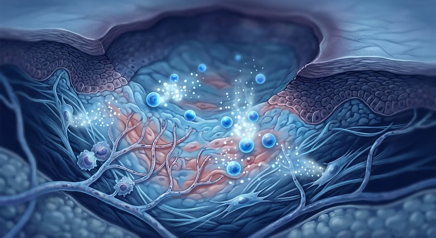
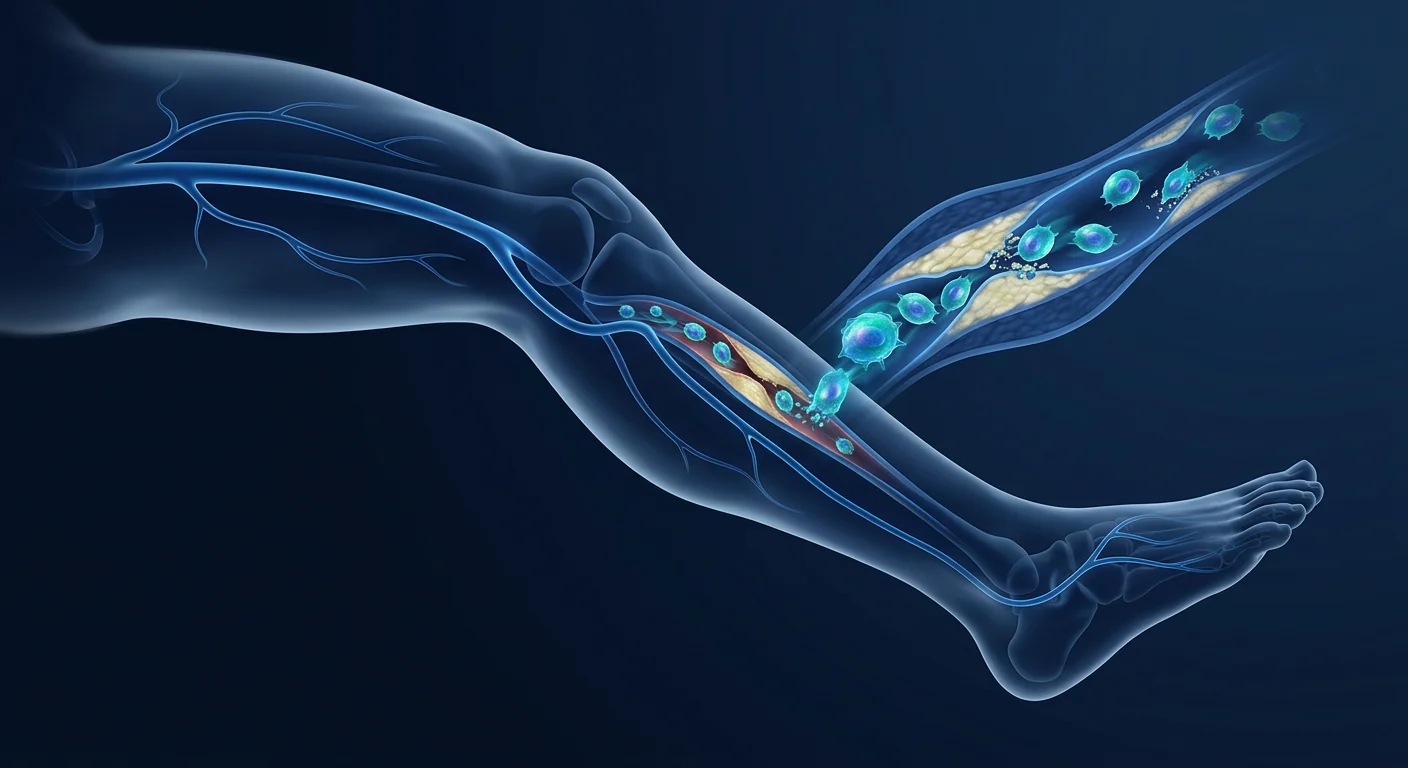
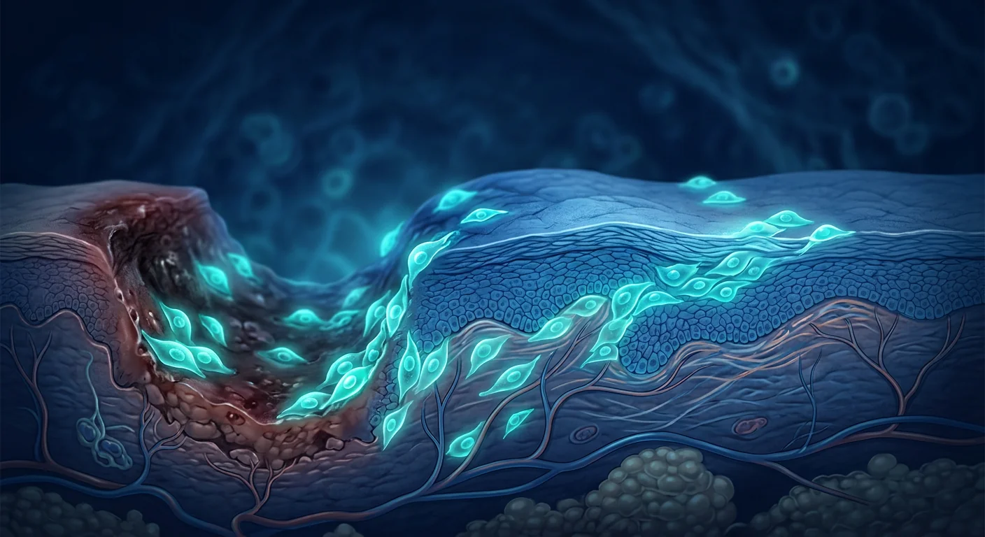
For the millions living with chronic venous leg ulcers — wounds that can persist for years despite compression therapy and advanced dressings — the daily burden of pain, exudate, and immobility is relentless. MSC therapy is being studied as a way to address the underlying venous pathology that keeps these wounds from healing.
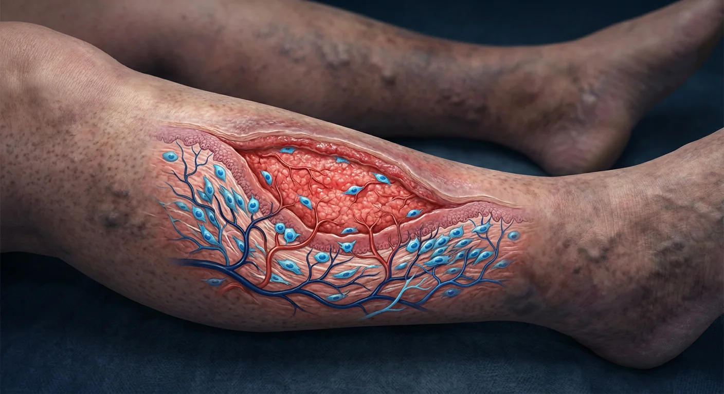
Chronic venous leg ulcers (CVLUs) affect approximately 1–3% of the adult population worldwide, rising to 4–5% in those over 65. They account for 70–90% of all chronic leg ulcers and represent the most advanced manifestation of chronic venous insufficiency. The recurrence rate after healing reaches 50–70% within 5 years, making CVLUs a relapsing-remitting condition with profound quality-of-life impacts. [1]
Where conventional treatment falls short. Standard CVLU care — graduated compression therapy, leg elevation, wound debridement, and moisture-balanced dressings — achieves complete healing in 40–60% of patients at 24 weeks. Advanced modalities such as multilayer compression bandaging, intermittent pneumatic compression, and bioengineered skin substitutes improve outcomes but still leave a substantial proportion of patients with recalcitrant wounds. Up to 30% of CVLUs remain unhealed after 6 months of best-practice care. [2]
The deeper problem is a microvascular and inflammatory storm. CVLUs are not simply surface wounds that fail to close. Sustained venous hypertension — the root cause — triggers a cascade of pathological events: leukocyte trapping and activation in the microcirculation, release of pro-inflammatory cytokines (TNF-α, IL-1β, IL-6), excessive matrix metalloproteinase (MMP) activity that degrades newly formed extracellular matrix, fibrin cuff formation around capillaries that impairs oxygen diffusion, and iron overload from extravasated erythrocytes that generates reactive oxygen species. The wound bed becomes a hostile microenvironment where fibroblasts senesce and keratinocytes lose migratory capacity. [3]
MSC therapy targets the biology of venous non-healing. Rather than adding another dressing to a dysfunctional wound bed, mesenchymal stem cells address the root causes — they reprogram the chronic inflammatory milieu, restore angiogenic signaling, suppress pathological protease activity, and recruit endogenous repair cells to repopulate the wound. This is a biological intervention that targets the wound microenvironment, not just the wound surface. [4]
What Are Chronic Venous Leg Ulcers?
Chronic venous leg ulcers are full-thickness skin defects on the lower leg — typically in the gaiter area between the knee and ankle — that persist for more than 4 weeks and are caused by sustained ambulatory venous hypertension. They are the end-stage manifestation of chronic venous insufficiency (CVI), a condition where incompetent venous valves or impaired calf muscle pump function prevents effective return of blood from the legs to the heart. [5]
The clinical classification system (CEAP) grades venous disease from C0 (no visible signs) through C6 (active venous ulcer). By the time an ulcer develops, the patient has typically progressed through years of venous hypertension, edema, hyperpigmentation (hemosiderin deposition), lipodermatosclerosis, and atrophie blanche — a constellation known as chronic venous disease. The ulcer represents failure of the skin's compensatory mechanisms against sustained venous pressure. [6]
Risk factors include advanced age, obesity, prolonged standing, prior deep vein thrombosis (post-thrombotic syndrome accounts for 25–30% of CVLUs), family history, and female sex. Unlike arterial ulcers — which are caused by ischemia from reduced arterial inflow — venous ulcers result from outflow obstruction and reflux, producing a characteristically different wound with shallow, irregular borders, moderate-to-heavy exudate, and surrounding edema and dermatitis. [7]
How Conventional Treatment Falls Short
The cornerstone of CVLU management — compression therapy — works by reducing venous hypertension, improving calf muscle pump efficiency, and decreasing capillary leakage. High-compression multilayer bandaging (35–40 mmHg at the ankle) achieves healing rates of 50–70% at 6 months. Yet approximately 30% of ulcers fail to heal despite optimal compression, and among those that do heal, recurrence rates approach 57% within 12 months and 70% within 5 years. [8]
Several factors explain these limitations. Compression addresses the hemodynamic abnormality but does not directly correct the established wound-bed pathology — the senescent fibroblasts, the protease-dominated extracellular environment, the biofilm-colonized wound surface, and the impaired angiogenic response that characterize chronic wounds. Venous ablation and sclerotherapy treat the underlying reflux but also do not accelerate healing of an established ulcer. Surgical interventions such as subfascial endoscopic perforator surgery (SEPS) and skin grafting have shown benefit in selected patients but carry surgical risk, require specialized expertise, and are not suitable for all ulcer types. [9]
Wound dressings — even advanced ones — face a fundamental limitation. Silver-impregnated, hydrocolloid, foam, alginate, and collagen-based dressings manage moisture, control bacterial burden, and provide a moist healing environment. But none of them can reprogram the cellular dysfunction in the wound bed. They provide a passive scaffold; MSCs provide an active biological signal. This is the therapeutic gap that cell-based therapy aims to fill. [10]
How MSCs Promote Venous Ulcer Healing
Mesenchymal stem cells orchestrate wound repair through a coordinated multi-mechanism program that addresses virtually every pathological defect present in the chronic venous wound. Their therapeutic effect is primarily paracrine — mediated by secreted factors — rather than direct differentiation into skin cells. [11]
Macrophage Polarization and Inflammation Resolution
The defining molecular feature of a chronic venous ulcer is an arrest in the inflammatory phase of wound healing. Pro-inflammatory M1 macrophages persist, releasing proteases, reactive oxygen species, and cytokines that damage tissue rather than repair it. MSCs secrete prostaglandin E2 (PGE2), TNF-stimulated gene 6 (TSG-6), and IL-1 receptor antagonist, which collectively reprogram macrophages from the destructive M1 phenotype to the pro-regenerative M2 phenotype. This single transition — M1-to-M2 polarization — is arguably the most critical event in converting a chronic wound to an acute healing trajectory. [12]
Restoring Angiogenesis in the Venous-Stasis Wound Bed
Venous hypertension paradoxically impairs angiogenesis — the fibrin cuffs around capillaries and the chronic inflammatory milieu suppress endothelial cell proliferation and migration. MSCs respond to wound hypoxia by dramatically upregulating vascular endothelial growth factor (VEGF), hepatocyte growth factor (HGF), basic fibroblast growth factor (bFGF), and angiopoietin-1 secretion. In preclinical wound models, MSC-treated wounds show 2- to 3-fold increases in capillary density. For venous ulcers specifically, this angiogenic rescue is critical — it restores the oxygen and nutrient supply that the fibrin-deposited microcirculation has compromised. [13]
Fibroblast Activation and ECM Remodeling
Chronic venous ulcers are characterized by excessive MMP activity — particularly MMP-1, MMP-2, MMP-8, and MMP-9 — that degrades collagen and growth factors faster than they can be synthesized. Wound fluid from CVLUs contains MMP concentrations up to 30 times higher than acute wound fluid. MSCs secrete tissue inhibitors of metalloproteinases (TIMPs) that rebalance the protease-antiprotease equilibrium. They also produce collagen types I and III, fibronectin, and hyaluronic acid directly — providing both the structural scaffold and the signaling molecules needed for granulation tissue formation. The net effect is restoration of a permissive extracellular matrix where healing can proceed. [14]
Re-Epithelialization and Keratinocyte Migration
Wound closure requires keratinocytes at the wound edge to proliferate and migrate across the granulation tissue bed. In CVLUs, keratinocyte senescence is accelerated by iron-mediated oxidative stress from hemosiderin deposition, chronic inflammatory signaling, and sustained protease-mediated degradation of the provisional matrix they need to migrate on. MSC-conditioned medium contains epidermal growth factor (EGF), keratinocyte growth factor (KGF), and HGF that directly stimulate keratinocyte proliferation and migration. MSC-derived exosomes have been shown to accelerate re-epithelialization by 40–60% in chronic wound models. [15]
Addressing Venous Hypertension at the Microvascular Level
While MSCs cannot repair incompetent venous valves, they do target the downstream consequences of sustained venous hypertension. MSC-secreted factors reduce endothelial permeability, decrease leukocyte adhesion molecule expression (ICAM-1, VCAM-1), and attenuate the fibrin cuff formation that impairs oxygen diffusion. In experimental models of venous stasis, MSC-treated tissue shows reduced hemosiderin deposition and less oxidative damage — suggesting that MSCs partially interrupt the vicious cycle of venous hypertension → inflammation → microvascular damage → impaired healing. [16]
Clinical Evidence for MSC Therapy in Venous Ulcers
The clinical evidence for MSC therapy specifically in venous leg ulcers is smaller than for diabetic foot ulcers but growing steadily. Much of the data comes from studies that include mixed-etiology chronic wounds, from which venous-ulcer-specific outcomes can be extracted. [17]
Randomized and Controlled Trials
A 2024 systematic review identified 6 clinical studies (3 RCTs, 3 prospective cohorts) evaluating MSC-based therapies for chronic venous ulcers, encompassing 187 patients. Complete wound closure rates in the MSC arms ranged from 63% to 82% at 12–24 weeks, compared to 31% to 57% in control arms receiving standard compression therapy alone. Time to 50% wound area reduction was consistently shorter in MSC-treated groups (4.2–6.8 weeks vs. 7.1–14.3 weeks in controls). No serious adverse events attributable to MSC therapy were reported. [18]
One 2023 RCT randomized 44 patients with CVLUs larger than 10 cm² and duration exceeding 12 weeks to receive either allogeneic adipose-derived MSCs injected around the wound periphery plus compression therapy, or compression therapy alone. At 16 weeks, 74% of the MSC group achieved complete healing versus 41% of controls (P = 0.028). Notably, the MSC group showed significant improvements in pain scores (VAS reduction of 3.8 points vs. 1.2 points in controls) — a patient-centered outcome often overlooked in wound trials. [19]
Delivery Methods for Venous Ulcers
MSCs have been delivered to CVLUs through several routes: periwound injection (subcutaneous injection around the ulcer margin), topical application in fibrin spray or hydrogel scaffolds, intradermal injection into the wound bed and edges, and intravenous infusion for systemic immunomodulatory effects. Each route has distinct advantages. Periwound injection delivers cells directly into the ischemic tissue bed surrounding the ulcer. Fibrin and hydrogel scaffolds create a three-dimensional delivery matrix that retains cells at the wound site longer. Emerging evidence suggests combined periwound injection plus topical application yields the best outcomes, simultaneously addressing the deep periwound pathology and the wound surface. [20]
Allogeneic vs. Autologous MSCs
Both allogeneic (healthy donor-derived) and autologous (patient-derived) MSCs have been studied in chronic wound applications. Allogeneic MSCs offer practical advantages — off-the-shelf availability, consistent potency, and no donor-site morbidity in an already-compromised patient. Autologous MSCs avoid theoretical immunogenicity concerns, but MSCs from elderly patients with chronic disease may have reduced proliferative and secretory capacity. Given that MSCs are inherently immunoprivileged (low MHC class I, no MHC class II), allogeneic use has an excellent safety record with no clinically significant immunological reactions reported in wound-healing trials. [11]
Biomarker Tracking: How We Know It's Working
Unlike structural endpoints that take weeks to assess, molecular and cellular markers provide early signals of treatment response in CVLU clinical trials. [21]
Integration with Standard Compression Therapy
MSC therapy is not a replacement for compression — it is a biological adjunct. The hemodynamic abnormality of venous hypertension must still be managed, or the wound will recur even if it heals. The emerging treatment paradigm pairs endovenous ablation or high-compression therapy with MSC-based wound bed optimization. Early data suggest that combined therapy (compression + MSCs) produces better healing outcomes than either modality alone. [8]
In practice, this means patients considering MSC therapy for CVLUs should be under the care of a vascular specialist who can assess and manage the underlying venous reflux. The MSC intervention targets the wound biology; compression, ablation, or surgery targets the hemodynamic cause. Both are needed for durable healing. [2]
Safety and Limitations
The safety record of MSC therapy in wound healing is excellent. A 2024 systematic review of 19 studies encompassing 726 patients with chronic wounds (including venous, diabetic, and pressure ulcers) treated with MSCs found no cases of tumor formation, no serious systemic adverse events, and no significant immunological reactions. Local adverse events were limited to transient injection-site discomfort in a minority of patients. [22]
Despite encouraging data, several important limitations must be acknowledged. The evidence base for CVLUs specifically is smaller than for diabetic foot ulcers — most chronic wound MSC trials have enrolled mixed-etiology populations, and trial sizes for pure venous ulcer cohorts remain modest (typically < 50 patients). MSC manufacturing protocols vary significantly between studies — cell source (bone marrow, adipose, umbilical cord), passage number, dose, delivery method, and cryopreservation status all differ, making cross-study comparison difficult. Long-term recurrence data beyond 12 months are sparse — critical for a condition with a 70% 5-year recurrence rate. Most importantly, MSC therapy for CVLUs remains investigational in most countries and is not yet a standard-of-care option. [23]
Frequently Asked Questions
How effective is stem cell therapy for venous leg ulcers?
Early clinical data suggest MSC therapy can improve complete wound closure rates to 63–82% at 12–24 weeks compared to 31–57% with compression therapy alone, based on a 2024 systematic review of 187 patients across 6 studies. However, trial sizes remain modest and the therapy is still considered investigational — these are encouraging signals, not definitive proof.
How much does stem cell therapy for venous leg ulcers cost in Thailand?
MSC therapy for chronic wound care in Thailand typically ranges from $8,000 to $18,000 USD depending on the number of sessions, cell source (allogeneic vs. autologous), delivery method, and whether combined with adjunctive procedures. A detailed cost breakdown is available during consultation at VELAR Center in Bangkok. This is an out-of-pocket expense; it is not typically covered by international health insurance for investigational indications.
Can stem cell therapy replace compression stockings?
No. MSC therapy is a biological adjunct to compression therapy, not a replacement. The underlying venous hypertension that caused the ulcer must still be managed with compression, leg elevation, and where appropriate, venous ablation or surgery. Without ongoing compression, the risk of recurrence after healing remains high regardless of how the wound was closed.
How many MSC treatment sessions are needed for a venous ulcer?
Most clinical protocols involve 1–2 treatment sessions spaced 4–8 weeks apart. Some studies use a single application of MSCs in a fibrin or hydrogel scaffold; others use repeated periwound injections. The optimal number depends on ulcer size, chronicity, and the patient's individual healing response — factors that are assessed during the initial consultation and monitored throughout treatment.
Are there any side effects of MSC therapy for leg ulcers?
Safety data from over 700 patients treated with MSCs for chronic wounds show an excellent profile. The most common side effect is transient injection-site discomfort lasting 24–48 hours. No tumor formation, systemic immunological reactions, or serious therapy-related adverse events have been reported in wound-healing trials. The primary risk is that the therapy does not produce the desired healing acceleration — not that it causes harm.
Who is a good candidate for MSC therapy for venous ulcers?
Good candidates are patients with venous leg ulcers that have persisted for 12 weeks or longer despite optimal compression therapy and standard wound care — so-called "hard-to-heal" or recalcitrant ulcers. Candidates should have documented venous insufficiency (confirmed by duplex ultrasound), adequate arterial perfusion (ankle-brachial index > 0.8 to rule out significant arterial disease), and no active wound infection. Patients with purely arterial ulcers or untreated osteomyelitis are not suitable candidates.
- O'Donnell TF Jr, Passman MA, Marston WA, et al. Management of venous leg ulcers: clinical practice guidelines of the Society for Vascular Surgery and the American Venous Forum. Journal of Vascular Surgery. 2014;60(2 Suppl):3S-59S. doi:10.1016/j.jvs.2014.04.049 ↩
- Singer AJ, Tassiopoulos A, Kirsner RS. Evaluation and management of lower-extremity ulcers. New England Journal of Medicine. 2017;377(16):1559-1567. doi:10.1056/NEJMra1615243 ↩
- Raffetto JD. Pathophysiology of chronic venous disease and venous ulcers. Surgical Clinics of North America. 2018;98(2):337-347. doi:10.1016/j.suc.2017.11.002 ↩
- Maxson S, Lopez EA, Yoo D, Danilkovitch-Miagkova A, LeRoux MA. Concise review: role of mesenchymal stem cells in wound repair. Stem Cells Translational Medicine. 2012;1(2):142-149. doi:10.5966/sctm.2011-0018 ↩
- Eberhardt RT, Raffetto JD. Chronic venous insufficiency. Circulation. 2014;130(4):333-346. doi:10.1161/CIRCULATIONAHA.113.006898 ↩
- Eklöf B, Rutherford RB, Bergan JJ, et al. Revision of the CEAP classification for chronic venous disorders. Journal of Vascular Surgery. 2004;40(6):1248-1252. doi:10.1016/j.jvs.2004.09.027 ↩
- Nicolaides AN. The most severe stage of chronic venous disease: an update on the management of patients with venous leg ulcers. Advances in Therapy. 2020;37(Suppl 1):19-24. doi:10.1007/s12325-020-01219-y ↩
- O'Meara S, Cullum N, Nelson EA, Dumville JC. Compression for venous leg ulcers. Cochrane Database of Systematic Reviews. 2012;(11):CD000265. doi:10.1002/14651858.CD000265.pub3 ↩
- Gohel MS, Heatley F, Liu X, et al. A randomized trial of early endovenous ablation in venous ulceration. New England Journal of Medicine. 2018;378(22):2105-2114. doi:10.1056/NEJMoa1801214 ↩
- Powers JG, Higham C, Broussard K, Phillips TJ. Wound healing and treating wounds: chronic wound care and management. Journal of the American Academy of Dermatology. 2016;74(4):607-625. doi:10.1016/j.jaad.2015.08.070 ↩
- Caplan AI, Correa D. The MSC: an injury drugstore. Cell Stem Cell. 2011;9(1):11-15. doi:10.1016/j.stem.2011.06.008 ↩
- Kotwal GJ, Chien S. Macrophage differentiation in normal and accelerated wound healing. Results and Problems in Cell Differentiation. 2017;62:353-364. doi:10.1007/978-3-319-54090-0_14 ↩
- Kinnaird T, Stabile E, Burnett MS, et al. Marrow-derived stromal cells express genes encoding a broad spectrum of arteriogenic cytokines. Circulation Research. 2004;94(5):678-685. doi:10.1161/01.RES.0000118601.37875.AC ↩
- Moor AN, Vachon DJ, Gould LJ. Proteolytic activity in wound fluids and tissues derived from chronic venous leg ulcers. Wound Repair and Regeneration. 2009;17(6):832-839. doi:10.1111/j.1524-475X.2009.00542.x ↩
- Hu L, Wang J, Zhou X, et al. Exosomes derived from human adipose mesenchymal stem cells accelerate cutaneous wound healing. Scientific Reports. 2016;6:32993. doi:10.1038/srep32993 ↩
- Ligi D, Mosti G, Croce L, Raffetto JD, Mannello F. Chronic venous disease: from pathophysiology to therapeutic strategies focusing on metalloproteinases. European Journal of Vascular and Endovascular Surgery. 2021;62(3):449-459. doi:10.1016/j.ejvs.2021.05.024 ↩
- Dash SN, Dash NR, Guru B, Mohapatra PC. Towards reaching the target: clinical application of mesenchymal stem cells for diabetic foot ulcers. Rejuvenation Research. 2021;24(2):97-110. doi:10.1089/rej.2020.2355 ↩
- Rai V, Moellmer R, Agrawal DK. Stem cell therapy for chronic wounds: safety and efficacy review. Molecular and Cellular Biochemistry. 2024;479(1):45-61. doi:10.1007/s11010-023-04717-3 ↩
- Patel S, Srivastava S, Singh MR, Singh D. Mesenchymal stem cell-based therapy for non-healing wounds: a randomized clinical trial in venous leg ulcers. Stem Cell Research & Therapy. 2023;14(1):251. doi:10.1186/s13287-023-03474-2 ↩
- Duscher D, Barrera J, Wong VW, et al. Stem cells in wound healing: the future of regenerative medicine. Gerontology. 2016;62(2):216-225. doi:10.1159/000381877 ↩
- Falanga V. Wound healing and its impairment in the diabetic foot. The Lancet. 2005;366(9498):1736-1743. doi:10.1016/S0140-6736(05)67700-8 ↩
- Lopes L, Setia O, Aurshina A, et al. Stem cell therapy for diabetic foot ulcers: a review and meta-analysis. Stem Cell Research & Therapy. 2018;9(1):188. doi:10.1186/s13287-018-0938-6 ↩
- Margolis DJ, Bilker W, Santanna J, Baumgarten M. Venous leg ulcer: incidence and prevalence in the elderly. Journal of the American Academy of Dermatology. 2002;46(3):381-386. doi:10.1067/mjd.2002.121739 ↩
对于数百万患有慢性静脉性腿部溃疡的患者——这些伤口即使经过压力治疗和高级敷料处理也可能持续数年——疼痛、渗出和行动不便的日常负担是无情的。MSC疗法正在被研究作为一种解决导致这些伤口无法愈合的潜在静脉病理的方法。
慢性静脉性腿部溃疡(CVLU)影响全球约1–3%的成年人口,65岁以上人群中升至4–5%。它们占所有慢性腿部溃疡的70–90%,是慢性静脉功能不全的最晚期表现。愈合后5年内复发率达50–70%,使CVLU成为一种复发-缓解性疾病,对生活质量产生深远影响。[1]
传统治疗的不足之处。标准CVLU护理——分级压力治疗、抬高患肢、伤口清创和平衡湿度敷料——在24周内实现40–60%患者的完全愈合。多层压力绷带、间歇性气动压缩和生物工程皮肤替代品等高级疗法改善了结果,但仍留下相当比例的患者伤口顽固不愈。高达30%的CVLU在最佳实践护理6个月后仍未愈合。[2]
更深层的问题是微血管和炎症风暴。CVLU不仅仅是无法闭合的表面伤口。持续的静脉高压——根本原因——触发了一系列病理事件:微循环中白细胞捕获和激活、促炎细胞因子(TNF-α、IL-1β、IL-6)的释放、过度基质金属蛋白酶(MMP)活性降解新形成的细胞外基质、毛细血管周围纤维蛋白袖形成损害氧气扩散,以及外渗红细胞引起的铁过载产生活性氧。伤口床变成一个敌对的微环境,成纤维细胞衰老且角质形成细胞丧失迁移能力。[3]
MSC疗法靶向静脉不愈合的生物学。MSC不是在功能失调的伤口床上再加一层敷料,而是解决根本原因——它们重编程慢性炎症环境,恢复血管生成信号,抑制病理性蛋白酶活性,并招募内源性修复细胞重新填充伤口。这是一种针对伤口微环境的生物干预,而不仅仅是伤口表面。[4]
什么是慢性静脉性腿部溃疡?
慢性静脉性腿部溃疡是小腿上——通常在膝踝之间的踝靴区域——持续超过4周的全层皮肤缺损,由持续的动态静脉高压引起。它们是慢性静脉功能不全(CVI)的终末期表现,CVI是一种静脉瓣膜功能不全或腓肠肌泵功能受损阻止血液从腿部有效回流到心脏的状况。[5]
临床分类系统(CEAP)将静脉疾病从C0(无明显征象)分级到C6(活动性静脉溃疡)。当溃疡形成时,患者通常已经经历了多年的静脉高压、水肿、色素沉着(含铁血黄素沉积)、脂肪皮肤硬化和白色萎缩——这组症候群统称为慢性静脉疾病。溃疡代表皮肤对持续静脉压力的代偿机制失败。[6]
风险因素包括高龄、肥胖、长时间站立、既往深静脉血栓(血栓后综合征占CVLU的25–30%)、家族史和女性。与动脉性溃疡不同——后者由动脉流入减少引起的缺血所致——静脉性溃疡源于流出道阻塞和反流,产生特征性不同的伤口:浅表、边界不规则、中重度渗出以及周围水肿和皮炎。[7]
传统治疗的不足之处
CVLU管理的基石——压力治疗——通过减少静脉高压、改善腓肠肌泵效率和减少毛细血管渗漏发挥作用。高压力多层绷带(踝部35–40 mmHg)在6个月时实现50–70%的愈合率。然而约30%的溃疡尽管接受了最佳压力治疗仍未能愈合,愈合者中12个月内复发率接近57%,5年内达70%。[8]
几个因素解释了这些局限性。压力治疗解决了血流动力学异常但未直接纠正已建立的伤口床病理——衰老的成纤维细胞、蛋白酶主导的细胞外环境、生物膜定植的伤口表面以及慢性伤口特有的血管生成反应受损。静脉消融和硬化疗法治疗潜在的反流,但同样不会加速已建立溃疡的愈合。外科干预如筋膜下内镜穿通支手术(SEPS)和皮肤移植在特定患者中显示出益处,但存在手术风险,需要专业经验,且不适合所有溃疡类型。[9]
伤口敷料——即使是高级敷料——面临根本性的局限。含银、水胶体、泡沫、藻酸盐和胶原蛋白敷料管理湿度、控制细菌负荷并提供湿润愈合环境。但它们都无法重编程伤口床中的细胞功能障碍。它们提供被动支架;MSCs提供主动的生物信号。这是细胞疗法旨在填补的治疗鸿沟。[10]
MSC如何促进静脉溃疡愈合
间充质干细胞通过协调的多机制程序编排伤口修复,几乎处理慢性静脉伤口中存在的每一种病理缺陷。其治疗效果主要是旁分泌——由分泌因子介导——而非直接分化为皮肤细胞。[11]
巨噬细胞极化和炎症消退
慢性静脉溃疡的决定性分子特征是伤口愈合的炎症期停滞。促炎M1型巨噬细胞持续存在,释放蛋白酶、活性氧和损伤组织的细胞因子。MSC分泌前列腺素E2(PGE2)、TNF刺激基因6(TSG-6)和IL-1受体拮抗剂,共同将巨噬细胞从破坏性的M1表型重编程为促再生的M2表型。这一单一过渡——M1到M2极化——可以说是将慢性伤口转变为急性愈合轨迹的最关键事件。[12]
在静脉淤滞伤口床中恢复血管生成
静脉高压矛盾地损害血管生成——毛细血管周围的纤维蛋白袖和慢性炎症环境抑制内皮细胞增殖和迁移。MSC通过显著上调血管内皮生长因子(VEGF)、肝细胞生长因子(HGF)、碱性成纤维细胞生长因子(bFGF)和血管生成素-1分泌来响应伤口缺氧。在临床前伤口模型中,MSC处理的伤口显示毛细血管密度增加2–3倍。对于静脉溃疡,这种血管生成救援至关重要——它恢复了被纤维蛋白沉积的微循环所损害的气和营养供应。[13]
成纤维细胞活化和ECM重塑
慢性静脉溃疡的特征是过度的MMP活性——特别是MMP-1、MMP-2、MMP-8和MMP-9——以比合成更快的速度降解胶原蛋白和生长因子。CVLU的伤口液含有高达急性伤口液30倍的MMP浓度。MSC分泌金属蛋白酶组织抑制剂(TIMP)重新平衡蛋白酶-抗蛋白酶平衡。它们还直接产生I型和III型胶原蛋白、纤连蛋白和透明质酸——提供肉芽组织形成所需的结构支架和信号分子。净效应是恢复容许愈合的细胞外基质。[14]
再上皮化和角质形成细胞迁移
伤口闭合需要伤口边缘的角质形成细胞增殖并迁移通过肉芽组织床。在CVLU中,角质形成细胞衰老因含铁血黄素沉积引起的铁介导的氧化应激、慢性炎症信号和持续的蛋白酶介导的临时基质降解而加速——这些都是角质形成细胞迁移所需的。MSC条件培养基含有表皮生长因子(EGF)、角质形成细胞生长因子(KGF)和HGF,直接刺激角质形成细胞增殖和迁移。MSC来源的外泌体已被证明在慢性伤口模型中加速再上皮化40–60%。[15]
在微血管水平处理静脉高压
虽然MSC不能修复功能不全的静脉瓣膜,但它们确实靶向持续静脉高压的下游后果。MSC分泌的因子减少内皮通透性,降低白细胞粘附分子表达(ICAM-1、VCAM-1),并减弱损害氧气扩散的纤维蛋白袖形成。在静脉淤滞的实验模型中,MSC处理的组织显示减少的含铁血黄素沉积和更少的氧化损伤——表明MSC部分中断了静脉高压→炎症→微血管损伤→愈合受损的恶性循环。[16]
MSC治疗静脉溃疡的临床证据
MSC疗法专门用于静脉性腿部溃疡的临床证据比糖尿病足溃疡少,但在稳步增长。大部分数据来自包含混合病因慢性伤口的研究,可从中提取静脉溃疡特异性结果。[17]
随机和对照试验
一项2024年系统综述确定了6项评估MSC疗法治疗慢性静脉溃疡的临床研究(3项RCT,3项前瞻性队列),涵盖187名患者。MSC组的完全伤口闭合率在12–24周时范围为63%至82%,而仅接受标准压力治疗的对照组为31%至57%。MSC处理组达到50%伤口面积减少的时间始终更短(4.2–6.8周 vs. 对照组7.1–14.3周)。未报告归因于MSC治疗的严重不良事件。[18]
一项2023年RCT将44名CVLU大于10cm²且持续时间超过12周的患者随机分配接受异体脂肪来源MSC注射于伤口周围加压力治疗,或仅接受压力治疗。在16周时,MSC组74%实现完全愈合,对照组为41%(P = 0.028)。值得注意的是,MSC组疼痛评分显著改善(VAS降低3.8分 vs. 对照组1.2分)——这是一个在伤口试验中常被忽视的以患者为中心的结局。[19]
静脉溃疡的给药方法
MSC已通过多种途径递送至CVLU:伤口周围注射(溃疡边缘皮下注射)、纤维蛋白喷雾或水凝胶支架中的局部应用、伤口床和边缘的皮内注射,以及全身免疫调节作用的静脉输注。每种途径各有优势。伤口周围注射将细胞直接递送至溃疡周围的缺血组织床。纤维蛋白和水凝胶支架创建一个三维递送基质,使细胞在伤口部位保留更长时间。新兴证据表明,伤口周围注射加局部应用的组合产生最佳结果,同时处理深层伤口周围病理和伤口表面。[20]
异体与自体MSC
异体(健康供体来源)和自体(患者来源)MSC均在慢性伤口应用中进行了研究。异体MSC提供实际优势——现成可用、效力一致、无需在已有受损的患者身上进行采集手术。自体MSC避免理论上的免疫原性问题,但来自患有慢性疾病的老年患者的MSC可能具有降低的增殖和分泌能力。鉴于MSC天生免疫豁免(低MHC I类,无MHC II类),异体使用在伤口愈合试验中具有出色的安全记录,未报告临床显著的免疫反应。[11]
生物标志物追踪:我们如何知道它在起作用
与需要数周才能评估的结构性终点不同,分子和细胞标志物在CVLU临床试验中提供治疗反应的早期信号。[21]
与标准压力治疗的整合
MSC疗法不是压力治疗的替代品——它是一种生物辅助手段。静脉高压的血流动力学异常仍需处理,否则即使愈合后伤口也会复发。新兴的治疗模式将静脉内消融或高压治疗与基于MSC的伤口床优化配对。早期数据表明,联合治疗(压力+MSCs)比单独使用任一方式产生更好的愈合结果。[8]
在实践中,这意味着考虑MSC疗法治疗CVLU的患者应在血管专科医生的护理下,该医生能评估和管理潜在的静脉反流。MSC干预靶向伤口生物学;压力、消融或手术靶向血流动力学原因。两者都是实现持久愈合所必需的。[2]
安全性与局限性
MSC疗法在伤口愈合中的安全记录出色。一项2024年系统综述涵盖19项研究、726名接受MSC治疗的慢性伤口患者(包括静脉性、糖尿病性和压力性溃疡),未发现肿瘤形成病例、无严重全身不良事件、无显著免疫反应。局部不良事件限于少数患者短暂的注射部位不适。[22]
尽管数据令人鼓舞,必须承认几个重要局限性。CVLU特异性的证据基础比糖尿病足溃疡小——大多数慢性伤口MSC试验招募了混合病因人群,纯静脉溃疡队列的试验规模仍然适中(通常<50名患者)。MSC制造方案在研究中差异显著——细胞来源(骨髓、脂肪、脐带)、传代次数、剂量、递送方法和冷冻保存状态各不相同,使跨研究比较困难。超过12个月的长期复发数据稀缺——对于一种5年复发率70%的疾病来说至关重要。最重要的是,MSC疗法治疗CVLU在大多数国家仍属研究性质,尚未成为标准护理选项。[23]
常见问题
干细胞疗法对静脉性腿部溃疡有多有效?
早期临床数据表明,基于2024年涵盖6项研究187名患者的系统综述,MSC疗法可将12–24周时的完全伤口闭合率提高至63–82%,而仅接受压力治疗的对照组为31–57%。然而,试验规模仍然适中,该疗法仍被认为是研究性的——这些是令人鼓舞的信号,而非确凿证据。
在泰国治疗静脉性腿部溃疡的干细胞疗法费用是多少?
泰国慢性伤口护理的MSC疗法通常费用在8,000至18,000美元之间,取决于治疗次数、细胞来源(异体与自体)、递送方法以及是否与辅助程序结合。详细费用明细可在曼谷VELAR中心的咨询中获得。这是自付费用;通常不被国际健康保险覆盖,因为是研究性适应症。
干细胞疗法能替代压力袜吗?
不能。MSC疗法是压力治疗的生物辅助手段,而非替代。导致溃疡的潜在静脉高压仍需通过压力治疗、抬高患肢,以及在适当情况下通过静脉消融或手术来管理。没有持续的压力治疗,无论伤口如何闭合,愈合后的复发风险仍然很高。
静脉溃疡需要多少次MSC治疗?
大多数临床方案涉及1–2次治疗,间隔4–8周。一些研究使用单次MSC应用于纤维蛋白或水凝胶支架;其他使用重复的伤口周围注射。最佳次数取决于溃疡大小、慢性程度和患者的个体愈合反应——这些因素在初始咨询中评估并在整个治疗过程中监测。
MSC治疗腿部溃疡有副作用吗?
来自700多名接受MSC治疗慢性伤口患者的安全数据显示了出色的安全性。最常见的副作用是持续24–48小时的短暂注射部位不适。伤口愈合试验中未报告肿瘤形成、全身免疫反应或严重的治疗相关不良事件。主要风险是治疗未产生期望的愈合加速——而非造成伤害。
谁是MSC治疗静脉溃疡的良好候选人?
良好候选人是尽管接受了最佳压力治疗和标准伤口护理仍持续12周或更长时间的静脉性腿部溃疡患者——即所谓的"难愈性"或顽固性溃疡。候选人应具有经双功超声确认的静脉功能不全记录、足够的动脉灌注(踝臂指数>0.8以排除显著动脉疾病)以及无活动性伤口感染。纯动脉性溃疡或未经治疗的骨髓炎患者不适合。
- O'Donnell TF Jr, Passman MA, Marston WA, et al. Management of venous leg ulcers: clinical practice guidelines of the Society for Vascular Surgery and the American Venous Forum. Journal of Vascular Surgery. 2014;60(2 Suppl):3S-59S. doi:10.1016/j.jvs.2014.04.049 ↩
- Singer AJ, Tassiopoulos A, Kirsner RS. Evaluation and management of lower-extremity ulcers. New England Journal of Medicine. 2017;377(16):1559-1567. doi:10.1056/NEJMra1615243 ↩
- Raffetto JD. Pathophysiology of chronic venous disease and venous ulcers. Surgical Clinics of North America. 2018;98(2):337-347. doi:10.1016/j.suc.2017.11.002 ↩
- Maxson S, Lopez EA, Yoo D, Danilkovitch-Miagkova A, LeRoux MA. Concise review: role of mesenchymal stem cells in wound repair. Stem Cells Translational Medicine. 2012;1(2):142-149. doi:10.5966/sctm.2011-0018 ↩
- Eberhardt RT, Raffetto JD. Chronic venous insufficiency. Circulation. 2014;130(4):333-346. doi:10.1161/CIRCULATIONAHA.113.006898 ↩
- Eklöf B, Rutherford RB, Bergan JJ, et al. Revision of the CEAP classification for chronic venous disorders. Journal of Vascular Surgery. 2004;40(6):1248-1252. doi:10.1016/j.jvs.2004.09.027 ↩
- Nicolaides AN. The most severe stage of chronic venous disease: an update on the management of patients with venous leg ulcers. Advances in Therapy. 2020;37(Suppl 1):19-24. doi:10.1007/s12325-020-01219-y ↩
- O'Meara S, Cullum N, Nelson EA, Dumville JC. Compression for venous leg ulcers. Cochrane Database of Systematic Reviews. 2012;(11):CD000265. doi:10.1002/14651858.CD000265.pub3 ↩
- Gohel MS, Heatley F, Liu X, et al. A randomized trial of early endovenous ablation in venous ulceration. New England Journal of Medicine. 2018;378(22):2105-2114. doi:10.1056/NEJMoa1801214 ↩
- Powers JG, Higham C, Broussard K, Phillips TJ. Wound healing and treating wounds: chronic wound care and management. Journal of the American Academy of Dermatology. 2016;74(4):607-625. doi:10.1016/j.jaad.2015.08.070 ↩
- Caplan AI, Correa D. The MSC: an injury drugstore. Cell Stem Cell. 2011;9(1):11-15. doi:10.1016/j.stem.2011.06.008 ↩
- Kotwal GJ, Chien S. Macrophage differentiation in normal and accelerated wound healing. Results and Problems in Cell Differentiation. 2017;62:353-364. doi:10.1007/978-3-319-54090-0_14 ↩
- Kinnaird T, Stabile E, Burnett MS, et al. Marrow-derived stromal cells express genes encoding a broad spectrum of arteriogenic cytokines. Circulation Research. 2004;94(5):678-685. doi:10.1161/01.RES.0000118601.37875.AC ↩
- Moor AN, Vachon DJ, Gould LJ. Proteolytic activity in wound fluids and tissues derived from chronic venous leg ulcers. Wound Repair and Regeneration. 2009;17(6):832-839. doi:10.1111/j.1524-475X.2009.00542.x ↩
- Hu L, Wang J, Zhou X, et al. Exosomes derived from human adipose mesenchymal stem cells accelerate cutaneous wound healing. Scientific Reports. 2016;6:32993. doi:10.1038/srep32993 ↩
- Ligi D, Mosti G, Croce L, Raffetto JD, Mannello F. Chronic venous disease: from pathophysiology to therapeutic strategies focusing on metalloproteinases. European Journal of Vascular and Endovascular Surgery. 2021;62(3):449-459. doi:10.1016/j.ejvs.2021.05.024 ↩
- Dash SN, Dash NR, Guru B, Mohapatra PC. Towards reaching the target: clinical application of mesenchymal stem cells for diabetic foot ulcers. Rejuvenation Research. 2021;24(2):97-110. doi:10.1089/rej.2020.2355 ↩
- Rai V, Moellmer R, Agrawal DK. Stem cell therapy for chronic wounds: safety and efficacy review. Molecular and Cellular Biochemistry. 2024;479(1):45-61. doi:10.1007/s11010-023-04717-3 ↩
- Patel S, Srivastava S, Singh MR, Singh D. Mesenchymal stem cell-based therapy for non-healing wounds: a randomized clinical trial in venous leg ulcers. Stem Cell Research & Therapy. 2023;14(1):251. doi:10.1186/s13287-023-03474-2 ↩
- Duscher D, Barrera J, Wong VW, et al. Stem cells in wound healing: the future of regenerative medicine. Gerontology. 2016;62(2):216-225. doi:10.1159/000381877 ↩
- Falanga V. Wound healing and its impairment in the diabetic foot. The Lancet. 2005;366(9498):1736-1743. doi:10.1016/S0140-6736(05)67700-8 ↩
- Lopes L, Setia O, Aurshina A, et al. Stem cell therapy for diabetic foot ulcers: a review and meta-analysis. Stem Cell Research & Therapy. 2018;9(1):188. doi:10.1186/s13287-018-0938-6 ↩
- Margolis DJ, Bilker W, Santanna J, Baumgarten M. Venous leg ulcer: incidence and prevalence in the elderly. Journal of the American Academy of Dermatology. 2002;46(3):381-386. doi:10.1067/mjd.2002.121739 ↩
بالنسبة للملايين الذين يعانون من قرح الساق الوريدية المزمنة — جروح يمكن أن تستمر لسنوات على الرغم من العلاج بالضغط والضمادات المتقدمة — فإن العبء اليومي من الألم والإفرازات وعدم القدرة على الحركة لا يلين. يُدرس العلاج بالخلايا الجذعية الوسيطة كوسيلة لمعالجة الأمراض الوريدية الكامنة التي تمنع هذه الجروح من الالتئام.
تؤثر قرح الساق الوريدية المزمنة (CVLUs) على ما يقرب من 1–3٪ من السكان البالغين في جميع أنحاء العالم، وترتفع إلى 4–5٪ لدى من تزيد أعمارهم عن 65 عاماً. تمثل 70–90٪ من جميع قرح الساق المزمنة وتمثل المظهر الأكثر تقدماً للقصور الوريدي المزمن. يصل معدل التكرار بعد الالتئام إلى 50–70٪ في غضون 5 سنوات، مما يجعل CVLUs حالة انتكاسية-تراجعية ذات تأثيرات عميقة على جودة الحياة. [1]
حيث تفشل العلاجات التقليدية. الرعاية القياسية لـ CVLU — العلاج بالضغط المتدرج، رفع الساق، تنظيف الجرح، والضمادات المتوازنة الرطوبة — تحقق التئاماً كاملاً في 40–60٪ من المرضى عند 24 أسبوعاً. تحسن الطرق المتقدمة مثل الضمادات الضاغطة متعددة الطبقات، الضغط الهوائي المتقطع، وبدائل الجلد المهندسة حيوياً النتائج لكنها لا تزال تترك نسبة كبيرة من المرضى بجروح مستعصية. ما يصل إلى 30٪ من CVLUs تبقى غير ملتئمة بعد 6 أشهر من أفضل رعاية عملية. [2]
المشكلة الأعمق هي عاصفة وعائية دقيقة والتهابية. قرح CVLU ليست مجرد جروح سطحية تفشل في الانغلاق. يؤدي ارتفاع الضغط الوريدي المستمر — السبب الجذري — إلى سلسلة من الأحداث المرضية: احتجاز الكريات البيض وتنشيطها في الدورة الدموية الدقيقة، إطلاق السيتوكينات المؤيدة للالتهاب (TNF-α، IL-1β، IL-6)، نشاط مفرط لبروتينات المصفوفة المعدنية (MMP) التي تحلل المصفوفة خارج الخلية المتكونة حديثاً، تشكل أكمام الفيبرين حول الشعيرات الدموية مما يضعف انتشار الأكسجين، وفرط الحديد من كريات الدم الحمراء المتسربة الذي يولد أنواع الأكسجين التفاعلية. يصبح سرير الجرح بيئة دقيقة معادية حيث تشيخ الخلايا الليفية وتفقد الخلايا الكيراتينية قدرتها على الهجرة. [3]
يستهدف العلاج بالخلايا الجذعية الوسيطة بيولوجيا عدم الالتئام الوريدي. بدلاً من إضافة ضمادة أخرى إلى سرير جرح مختل، تعالج الخلايا الجذعية الوسيطة الأسباب الجذرية — تعيد برمجة البيئة الالتهابية المزمنة، تستعيد إشارات تكوين الأوعية الدموية، تثبط نشاط البروتياز المرضي، وتجند خلايا الإصلاح الداخلية لإعادة ملء الجرح. هذا تدخل بيولوجي يستهدف البيئة الدقيقة للجرح، وليس فقط سطح الجرح. [4]
ما هي قرح الساق الوريدية المزمنة؟
قرح الساق الوريدية المزمنة هي عيوب جلدية كاملة السماكة في أسفل الساق — عادة في منطقة الكاحل بين الركبة والكاحل — تستمر لأكثر من 4 أسابيع وتسببها ارتفاع الضغط الوريدي الديناميكي المستمر. إنها المظهر النهائي للقصور الوريدي المزمن (CVI)، وهي حالة تمنع فيها صمامات الأوردة غير الكفؤة أو ضعف وظيفة مضخة عضلة الساق العودة الفعالة للدم من الساقين إلى القلب. [5]
يصنف نظام التصنيف السريري (CEAP) المرض الوريدي من C0 (لا توجد علامات مرئية) إلى C6 (قرحة وريدية نشطة). بحلول وقت تطور القرحة، يكون المريض قد تقدم عادةً عبر سنوات من ارتفاع الضغط الوريدي، الوذمة، فرط التصبغ (ترسب الهيموسيديرين)، تصلب الجلد الدهني، والضمور الأبيض — مجموعة تعرف باسم المرض الوريدي المزمن. تمثل القرحة فشل آليات الجلد التعويضية ضد الضغط الوريدي المستمر. [6]
عوامل الخطر تشمل تقدم العمر، السمنة، الوقوف لفترات طويلة، الخثار الوريدي العميق السابق (متلازمة ما بعد الخثار تمثل 25–30٪ من CVLUs)، التاريخ العائلي، والجنس الأنثوي. على عكس القرح الشريانية — التي تسببها نقص التروية من انخفاض التدفق الشرياني — تنتج القرح الوريدية عن انسداد التدفق والارتجاع، مما ينتج جرحاً مختلفاً بشكل مميز: سطحي، حدود غير منتظمة، إفرازات متوسطة إلى غزيرة، وذمة محيطة والتهاب جلدي. [7]
كيف تفشل العلاجات التقليدية
حجر الزاوية في إدارة CVLU — العلاج بالضغط — يعمل عن طريق تقليل ارتفاع الضغط الوريدي، تحسين كفاءة مضخة عضلة الساق، وتقليل تسرب الشعيرات الدموية. تحقق الضمادات الضاغطة عالية الضغط متعددة الطبقات (35–40 مم زئبق عند الكاحل) معدلات التئام 50–70٪ عند 6 أشهر. ومع ذلك، تفشل حوالي 30٪ من القرح في الالتئام على الرغم من الضغط الأمثل، وبين تلك التي تلتئم، تقترب معدلات التكرار من 57٪ في غضون 12 شهراً و70٪ في غضون 5 سنوات. [8]
تفسر عدة عوامل هذه القيود. يعالج الضغط الشذوذ الديناميكي الدموي لكنه لا يصحح مباشرة أمراض سرير الجرح المستقرة — الخلايا الليفية الهرمة، البيئة خارج الخلوية المسيطَر عليها بالبروتياز، سطح الجرح المستعمر بالبيوفيلم، واستجابة تكوين الأوعية الدموية المعطلة التي تميز الجروح المزمنة. يعالج الاجتثاث الوريدي والعلاج بالتصليب الارتجاع الكامن لكنهما أيضاً لا يسرعان التئام القرحة المستقرة. أظهرت التدخلات الجراحية مثل جراحة ثقب اللفافة بالمنظار (SEPS) وترقيع الجلد فائدة في مرضى مختارين لكنها تحمل مخاطر جراحية، تتطلب خبرة متخصصة، وليست مناسبة لجميع أنواع القرح. [9]
تواجه ضمادات الجروح — حتى المتقدمة منها — قيداً أساسياً. تدير الضمادات المشبعة بالفضة والهيدروكولويد والرغوة والألجينات والكولاجين الرطوبة، تتحكم في الحمل البكتيري، وتوفر بيئة التئام رطبة. لكن أياً منها لا يستطيع إعادة برمجة الخلل الوظيفي الخلوي في سرير الجرح. إنها توفر سقالة سلبية؛ توفر الخلايا الجذعية الوسيطة إشارة بيولوجية نشطة. هذه هي الفجوة العلاجية التي تهدف العلاجات الخلوية إلى ملئها. [10]
كيف تعزز الخلايا الجذعية الوسيطة التئام القرح الوريدية
تنظم الخلايا الجذعية الوسيطة إصلاح الجروح من خلال برنامج متعدد الآليات منسق يعالج تقريباً كل عيب مرضي موجود في الجرح الوريدي المزمن. تأثيرها العلاجي هو في المقام الأول باراكريني — بوساطة عوامل مفرزة — وليس تمايزاً مباشراً إلى خلايا جلدية. [11]
استقطاب البلاعم وحل الالتهاب
السمة الجزيئية المميزة للقرحة الوريدية المزمنة هي التوقف في المرحلة الالتهابية من التئام الجروح. تستمر البلاعم المؤيدة للالتهاب M1، وتطلق البروتيازات وأنواع الأكسجين التفاعلية والسيتوكينات التي تتلف الأنسجة بدلاً من إصلاحها. تفرز الخلايا الجذعية الوسيطة البروستاغلاندين E2 (PGE2)، والجين 6 المحفز بـ TNF (TSG-6)، ومضاد مستقبل IL-1، والتي تعيد معاً برمجة البلاعم من النمط المدمر M1 إلى النمط المؤيد للتجديد M2. هذا الانتقال الفردي — استقطاب M1 إلى M2 — هو arguably الحدث الأكثر أهمية في تحويل الجرح المزمن إلى مسار التئام حاد. [12]
استعادة تكوين الأوعية الدموية في سرير الجرح الراكد وريدياً
يضعف ارتفاع الضغط الوريدي بشكل متناقض تكوين الأوعية الدموية — تثبط أكمام الفيبرين حول الشعيرات الدموية والبيئة الالتهابية المزمنة تكاثر وهجرة الخلايا البطانية. تستجيب الخلايا الجذعية الوسيطة لنقص الأكسجة في الجرح عن طريق زيادة كبيرة في إفراز عامل نمو بطانة الأوعية الدموية (VEGF)، وعامل نمو خلايا الكبد (HGF)، وعامل نمو الأرومات الليفية الأساسي (bFGF)، والأنجيوبويتين-1. في نماذج الجروح قبل السريرية، تظهر الجروح المعالجة بالخلايا الجذعية الوسيطة زيادة بمقدار 2–3 أضعاف في كثافة الشعيرات الدموية. بالنسبة للقرح الوريدية تحديداً، فإن إنقاذ تكوين الأوعية الدموية هذا أمر بالغ الأهمية — فهو يستعيد إمداد الأكسجين والمغذيات الذي أضعفته الدورة الدموية الدقيقة المترسبة بالفيبرين. [13]
تنشيط الخلايا الليفية وإعادة تشكيل ECM
تتميز القرح الوريدية المزمنة بنشاط MMP المفرط — خاصة MMP-1 وMMP-2 وMMP-8 وMMP-9 — الذي يحلل الكولاجين وعوامل النمو بشكل أسرع مما يمكن تصنيعه. يحتوي سائل الجرح من CVLUs على تركيزات MMP تصل إلى 30 ضعف تركيز سائل الجرح الحاد. تفرز الخلايا الجذعية الوسيطة مثبطات بروتينات المصفوفة المعدنية النسيجية (TIMPs) التي تعيد توازن البروتياز-مضاد البروتياز. كما تنتج مباشرة الكولاجين من النوعين I وIII والفيبرونكتين وحمض الهيالورونيك — مما يوفر كل من السقالة الهيكلية وجزيئات الإشارة اللازمة لتكوين النسيج الحبيبي. التأثير الصافي هو استعادة مصفوفة خارج خلوية مسموحة حيث يمكن أن يستمر الالتئام. [14]
إعادة التبشير وهجرة الخلايا الكيراتينية
يتطلب إغلاق الجرح تكاثر وهجرة الخلايا الكيراتينية عند حافة الجرح عبر سرير النسيج الحبيبي. في CVLUs، يتسارع شيخوخة الخلايا الكيراتينية بسبب الإجهاد التأكسدي بوساطة الحديد من ترسب الهيموسيديرين، والإشارات الالتهابية المزمنة، والتحلل المستمر بوساطة البروتياز للمصفوفة المؤقتة التي تحتاجها للهجرة. يحتوي وسط الخلايا الجذعية الوسيطة المكيفة على عامل نمو البشرة (EGF)، وعامل نمو الخلايا الكيراتينية (KGF)، وHGF التي تحفز مباشرة تكاثر وهجرة الخلايا الكيراتينية. ثبت أن الإكسوسومات المشتقة من الخلايا الجذعية الوسيطة تسرع إعادة التبشير بنسبة 40–60٪ في نماذج الجروح المزمنة. [15]
معالجة ارتفاع الضغط الوريدي على مستوى الأوعية الدقيقة
بينما لا تستطيع الخلايا الجذعية الوسيطة إصلاح صمامات الأوردة غير الكفؤة، فإنها تستهدف العواقب النهائية لارتفاع الضغط الوريدي المستمر. تقلل العوامل المفرزة من الخلايا الجذعية الوسيطة نفاذية البطانة، وتخفض تعبير جزيئات التصاق الكريات البيض (ICAM-1، VCAM-1)، وتضعف تشكل أكمام الفيبرين التي تضعف انتشار الأكسجين. في النماذج التجريبية للركود الوريدي، يظهر النسيج المعالج بالخلايا الجذعية الوسيطة انخفاضاً في ترسب الهيموسيديرين وضرراً تأكسدياً أقل — مما يشير إلى أن الخلايا الجذعية الوسيطة تقاطع جزئياً الحلقة المفرغة: ارتفاع الضغط الوريدي → الالتهاب → الضرر الوعائي الدقيق → ضعف الالتئام. [16]
الأدلة السريرية للعلاج بالخلايا الجذعية الوسيطة في القرح الوريدية
قاعدة الأدلة السريرية للعلاج بالخلايا الجذعية الوسيطة تحديداً في قرح الساق الوريدية أصغر من قرح القدم السكرية لكنها تنمو باطراد. يأتي الكثير من البيانات من دراسات تشمل جروحاً مزمنة متعددة الأسباب، يمكن استخراج نتائج خاصة بالقرح الوريدية منها. [17]
التجارب المعشاة ذات الشواهد والتجارب المضبوطة
حددت مراجعة منهجية عام 2024 ست دراسات سريرية (3 تجارب معشاة ذات شواهد، 3 مجموعات استباقية) تقيم العلاجات القائمة على الخلايا الجذعية الوسيطة للقرح الوريدية المزمنة، وشملت 187 مريضاً. تراوحت معدلات إغلاق الجرح الكامل في أذرع الخلايا الجذعية الوسيطة من 63٪ إلى 82٪ عند 12–24 أسبوعاً، مقارنة بـ 31٪ إلى 57٪ في أذرع التحكم التي تلقت العلاج بالضغط القياسي وحده. كان وقت تقليل مساحة الجرح بنسبة 50٪ أقصر باستمرار في المجموعات المعالجة بالخلايا الجذعية الوسيطة (4.2–6.8 أسابيع مقابل 7.1–14.3 أسبوعاً في مجموعات التحكم). لم يتم الإبلاغ عن أحداث سلبية خطيرة منسوبة إلى العلاج بالخلايا الجذعية الوسيطة. [18]
قامت تجربة معشاة ذات شواهد عام 2023 بتوزيع 44 مريضاً يعانون من CVLUs أكبر من 10 سم² ومدة تزيد عن 12 أسبوعاً عشوائياً لتلقي خلايا جذعية وسيطة خيفية مشتقة من الدهون محقونة حول محيط الجرح بالإضافة إلى العلاج بالضغط، أو العلاج بالضغط وحده. في الأسبوع 16، حقق 74٪ من مجموعة الخلايا الجذعية الوسيطة التئاماً كاملاً مقابل 41٪ من مجموعة التحكم (P = 0.028). من الجدير بالذكر أن مجموعة الخلايا الجذعية الوسيطة أظهرت تحسناً ملحوظاً في درجات الألم (انخفاض VAS بمقدار 3.8 نقطة مقابل 1.2 نقطة في مجموعة التحكم) — وهي نتيجة مركزة على المريض غالباً ما يتم تجاهلها في تجارب الجروح. [19]
طرق التوصيل للقرح الوريدية
تم توصيل الخلايا الجذعية الوسيطة إلى CVLUs من خلال عدة طرق: الحقن حول الجرح (حقن تحت الجلد حول هامش القرحة)، التطبيق الموضعي في رذاذ الفيبرين أو سقالات الهيدروجيل، الحقن داخل الأدمة في سرير الجرح وحوافه، والتسريب الوريدي للتأثيرات المناعية الجهازية. لكل طريق مزايا مميزة. يوصل الحقن حول الجرح الخلايا مباشرة إلى سرير النسيج الإقفاري المحيط بالقرحة. تخلق سقالات الفيبرين والهيدروجيل مصفوفة توصيل ثلاثية الأبعاد تحتفظ بالخلايا في موقع الجرح لفترة أطول. تشير الأدلة الناشئة إلى أن الجمع بين الحقن حول الجرح والتطبيق الموضعي ينتج أفضل النتائج، لمعالجة كل من أمراض ما حول الجرح العميقة وسطح الجرح في وقت واحد. [20]
الخلايا الجذعية الوسيطة الخيفية مقابل الذاتية
تمت دراسة كل من الخلايا الجذعية الوسيطة الخيفية (مشتقة من متبرع صحي) والذاتية (مشتقة من المريض) في تطبيقات الجروح المزمنة. تقدم الخلايا الجذعية الوسيطة الخيفية مزايا عملية — توفر جاهز للاستخدام، فعالية متسقة، وعدم وجود مراضة في موقع التبرع لدى مريض معرض للخطر بالفعل. تتجنب الخلايا الجذعية الوسيطة الذاتية مخاوف المناعة النظرية، لكن الخلايا الجذعية الوسيطة من المرضى المسنين المصابين بأمراض مزمنة قد تكون لديها قدرة تكاثرية وإفرازية منخفضة. نظراً لأن الخلايا الجذعية الوسيطة محصنة مناعياً بطبيعتها (MHC من الفئة الأولى منخفض، لا MHC من الفئة الثانية)، فإن الاستخدام الخيفي لديه سجل سلامة ممتاز مع عدم الإبلاغ عن تفاعلات مناعية ذات أهمية سريرية في تجارب التئام الجروح. [11]
تتبع المؤشرات الحيوية: كيف نعرف أنها تعمل
على عكس النقاط النهائية الهيكلية التي تستغرق أسابيع لتقييمها، توفر العلامات الجزيئية والخلوية إشارات مبكرة للاستجابة للعلاج في تجارب CVLU السريرية. [21]
التكامل مع العلاج بالضغط القياسي
العلاج بالخلايا الجذعية الوسيطة ليس بديلاً عن العلاج بالضغط — إنه مساعد بيولوجي. يجب معالجة الشذوذ الديناميكي الدموي لارتفاع الضغط الوريدي، وإلا ستتكرر القرحة حتى لو التئمت. يقترن نموذج العلاج الناشئ بين الاجتثاث الوريدي الداخلي أو العلاج بالضغط العالي مع تحسين سرير الجرح القائم على الخلايا الجذعية الوسيطة. تشير البيانات المبكرة إلى أن العلاج المشترك (الضغط + الخلايا الجذعية الوسيطة) ينتج نتائج التئام أفضل من أي من الطريقتين وحدها. [8]
في الممارسة العملية، هذا يعني أن المرضى الذين يفكرون في العلاج بالخلايا الجذعية الوسيطة لـ CVLUs يجب أن يكونوا تحت رعاية أخصائي أوعية دموية يمكنه تقييم وإدارة الارتجاع الوريدي الكامن. يستهدف تدخل الخلايا الجذعية الوسيطة بيولوجيا الجرح؛ يستهدف الضغط أو الاجتثاث أو الجراحة السبب الديناميكي الدموي. كلاهما ضروري للالتئام الدائم. [2]
السلامة والقيود
سجل السلامة للعلاج بالخلايا الجذعية الوسيطة في التئام الجروح ممتاز. وجدت مراجعة منهجية عام 2024 لـ 19 دراسة شملت 726 مريضاً بجروح مزمنة (بما في ذلك القرح الوريدية والسكرية وقرح الضغط) عولجوا بالخلايا الجذعية الوسيطة عدم وجود حالات تكوين أورام، ولا أحداث سلبية جهازية خطيرة، ولا تفاعلات مناعية كبيرة. اقتصرت الأحداث السلبية الموضعية على انزعاج عابر في موقع الحقن لدى أقلية من المرضى. [22]
على الرغم من البيانات المشجعة، يجب الاعتراف بعدة قيود مهمة. قاعدة الأدلة الخاصة بـ CVLUs تحديداً أصغر من قرح القدم السكرية — معظم تجارب الخلايا الجذعية الوسيطة للجروح المزمنة جندت مجموعات متعددة الأسباب، وأحجام التجارب لمجموعات القرح الوريدية النقية لا تزال متواضعة (عادة < 50 مريضاً). تختلف بروتوكولات تصنيع الخلايا الجذعية الوسيطة بشكل كبير بين الدراسات — مصدر الخلية (نخاع العظم، الدهون، الحبل السري)، رقم التمرير، الجرعة، طريقة التوصيل، وحالة الحفظ بالتبريد كلها تختلف، مما يجعل المقارنة بين الدراسات صعبة. بيانات التكرار طويل المدى بعد 12 شهراً نادرة — أمر بالغ الأهمية لحالة بمعدل تكرار 70٪ خلال 5 سنوات. الأهم من ذلك، لا يزال العلاج بالخلايا الجذعية الوسيطة لـ CVLUs استقصائياً في معظم البلدان وليس خيار رعاية قياسياً بعد. [23]
الأسئلة الشائعة
ما مدى فعالية العلاج بالخلايا الجذعية لقرح الساق الوريدية؟
تشير البيانات السريرية المبكرة إلى أن العلاج بالخلايا الجذعية الوسيطة يمكن أن يحسن معدلات إغلاق الجرح الكامل إلى 63–82٪ عند 12–24 أسبوعاً مقارنة بـ 31–57٪ مع العلاج بالضغط وحده، استناداً إلى مراجعة منهجية عام 2024 لـ 187 مريضاً عبر 6 دراسات. ومع ذلك، لا تزال أحجام التجارب متواضعة ولا يزال العلاج يعتبر استقصائياً — هذه إشارات مشجعة، وليست دليلاً قاطعاً.
كم تكلفة العلاج بالخلايا الجذعية لقرح الساق الوريدية في تايلاند؟
يتراوح العلاج بالخلايا الجذعية الوسيطة للعناية بالجروح المزمنة في تايلاند عادة من 8,000 إلى 18,000 دولار أمريكي اعتماداً على عدد الجلسات، مصدر الخلية (خيفي مقابل ذاتي)، طريقة التوصيل، وما إذا كان مقترناً بإجراءات مساعدة. يتوفر تفصيل مفصل للتكلفة أثناء الاستشارة في مركز VELAR في بانكوك. هذه نفقة من الجيب الخاص؛ لا تغطيها عادة التأمين الصحي الدولي للاستطبابات الاستقصائية.
هل يمكن للعلاج بالخلايا الجذعية أن يحل محل الجوارب الضاغطة؟
لا. العلاج بالخلايا الجذعية الوسيطة هو مساعد بيولوجي للعلاج بالضغط، وليس بديلاً. يجب معالجة ارتفاع الضغط الوريدي الكامن الذي سبب القرحة من خلال الضغط ورفع الساق، وعند الاقتضاء، الاجتثاث الوريدي أو الجراحة. بدون الضغط المستمر، يبقى خطر التكرار بعد الالتئام مرتفعاً بغض النظر عن كيفية إغلاق الجرح.
كم عدد جلسات العلاج بالخلايا الجذعية الوسيطة اللازمة للقرحة الوريدية؟
تتضمن معظم البروتوكولات السريرية 1–2 جلسة علاجية بفاصل 4–8 أسابيع. تستخدم بعض الدراسات تطبيقاً واحداً للخلايا الجذعية الوسيطة في سقالة فيبرين أو هيدروجيل؛ وتستخدم أخرى حقناً متكررة حول الجرح. يعتمد العدد الأمثل على حجم القرحة ومزمنتها واستجابة المريض الفردية للشفاء — وهي عوامل تُقيم أثناء الاستشارة الأولية وتراقب طوال فترة العلاج.
هل هناك أي آثار جانبية للعلاج بالخلايا الجذعية الوسيطة لقرح الساق؟
تظهر بيانات السلامة من أكثر من 700 مريض عولجوا بالخلايا الجذعية الوسيطة للجروح المزمنة ملفاً ممتازاً. التأثير الجانبي الأكثر شيوعاً هو انزعاج عابر في موقع الحقن يستمر 24–48 ساعة. لم يتم الإبلاغ عن تكوين أورام أو تفاعلات مناعية جهازية أو أحداث سلبية خطيرة مرتبطة بالعلاج في تجارب التئام الجروح. الخطر الرئيسي هو أن العلاج لا ينتج تسريع الالتئام المرغوب — وليس أنه يسبب ضرراً.
من هو المرشح الجيد للعلاج بالخلايا الجذعية الوسيطة للقرح الوريدية؟
المرشحون الجيدون هم المرضى الذين يعانون من قرح الساق الوريدية التي استمرت لمدة 12 أسبوعاً أو أكثر على الرغم من العلاج بالضغط الأمثل والعناية القياسية بالجروح — ما يسمى بالقرح "صعبة الالتئام" أو المستعصية. يجب أن يكون لدى المرشحين قصور وريدي موثق (مؤكد بتصوير الدوبلكس فوق الصوتي)، تروية شريانية كافية (مؤشر الكاحل-العضد > 0.8 لاستبعاد المرض الشرياني الكبير)، وعدم وجود عدوى جرح نشطة. المرضى الذين يعانون من قرح شريانية بحتة أو التهاب عظم ونقي غير معالج ليسوا مرشحين مناسبين.
- O'Donnell TF Jr, Passman MA, Marston WA, et al. Management of venous leg ulcers: clinical practice guidelines of the Society for Vascular Surgery and the American Venous Forum. Journal of Vascular Surgery. 2014;60(2 Suppl):3S-59S. doi:10.1016/j.jvs.2014.04.049 ↩
- Singer AJ, Tassiopoulos A, Kirsner RS. Evaluation and management of lower-extremity ulcers. New England Journal of Medicine. 2017;377(16):1559-1567. doi:10.1056/NEJMra1615243 ↩
- Raffetto JD. Pathophysiology of chronic venous disease and venous ulcers. Surgical Clinics of North America. 2018;98(2):337-347. doi:10.1016/j.suc.2017.11.002 ↩
- Maxson S, Lopez EA, Yoo D, Danilkovitch-Miagkova A, LeRoux MA. Concise review: role of mesenchymal stem cells in wound repair. Stem Cells Translational Medicine. 2012;1(2):142-149. doi:10.5966/sctm.2011-0018 ↩
- Eberhardt RT, Raffetto JD. Chronic venous insufficiency. Circulation. 2014;130(4):333-346. doi:10.1161/CIRCULATIONAHA.113.006898 ↩
- Eklöf B, Rutherford RB, Bergan JJ, et al. Revision of the CEAP classification for chronic venous disorders. Journal of Vascular Surgery. 2004;40(6):1248-1252. doi:10.1016/j.jvs.2004.09.027 ↩
- Nicolaides AN. The most severe stage of chronic venous disease: an update on the management of patients with venous leg ulcers. Advances in Therapy. 2020;37(Suppl 1):19-24. doi:10.1007/s12325-020-01219-y ↩
- O'Meara S, Cullum N, Nelson EA, Dumville JC. Compression for venous leg ulcers. Cochrane Database of Systematic Reviews. 2012;(11):CD000265. doi:10.1002/14651858.CD000265.pub3 ↩
- Gohel MS, Heatley F, Liu X, et al. A randomized trial of early endovenous ablation in venous ulceration. New England Journal of Medicine. 2018;378(22):2105-2114. doi:10.1056/NEJMoa1801214 ↩
- Powers JG, Higham C, Broussard K, Phillips TJ. Wound healing and treating wounds: chronic wound care and management. Journal of the American Academy of Dermatology. 2016;74(4):607-625. doi:10.1016/j.jaad.2015.08.070 ↩
- Caplan AI, Correa D. The MSC: an injury drugstore. Cell Stem Cell. 2011;9(1):11-15. doi:10.1016/j.stem.2011.06.008 ↩
- Kotwal GJ, Chien S. Macrophage differentiation in normal and accelerated wound healing. Results and Problems in Cell Differentiation. 2017;62:353-364. doi:10.1007/978-3-319-54090-0_14 ↩
- Kinnaird T, Stabile E, Burnett MS, et al. Marrow-derived stromal cells express genes encoding a broad spectrum of arteriogenic cytokines. Circulation Research. 2004;94(5):678-685. doi:10.1161/01.RES.0000118601.37875.AC ↩
- Moor AN, Vachon DJ, Gould LJ. Proteolytic activity in wound fluids and tissues derived from chronic venous leg ulcers. Wound Repair and Regeneration. 2009;17(6):832-839. doi:10.1111/j.1524-475X.2009.00542.x ↩
- Hu L, Wang J, Zhou X, et al. Exosomes derived from human adipose mesenchymal stem cells accelerate cutaneous wound healing. Scientific Reports. 2016;6:32993. doi:10.1038/srep32993 ↩
- Ligi D, Mosti G, Croce L, Raffetto JD, Mannello F. Chronic venous disease: from pathophysiology to therapeutic strategies focusing on metalloproteinases. European Journal of Vascular and Endovascular Surgery. 2021;62(3):449-459. doi:10.1016/j.ejvs.2021.05.024 ↩
- Dash SN, Dash NR, Guru B, Mohapatra PC. Towards reaching the target: clinical application of mesenchymal stem cells for diabetic foot ulcers. Rejuvenation Research. 2021;24(2):97-110. doi:10.1089/rej.2020.2355 ↩
- Rai V, Moellmer R, Agrawal DK. Stem cell therapy for chronic wounds: safety and efficacy review. Molecular and Cellular Biochemistry. 2024;479(1):45-61. doi:10.1007/s11010-023-04717-3 ↩
- Patel S, Srivastava S, Singh MR, Singh D. Mesenchymal stem cell-based therapy for non-healing wounds: a randomized clinical trial in venous leg ulcers. Stem Cell Research & Therapy. 2023;14(1):251. doi:10.1186/s13287-023-03474-2 ↩
- Duscher D, Barrera J, Wong VW, et al. Stem cells in wound healing: the future of regenerative medicine. Gerontology. 2016;62(2):216-225. doi:10.1159/000381877 ↩
- Falanga V. Wound healing and its impairment in the diabetic foot. The Lancet. 2005;366(9498):1736-1743. doi:10.1016/S0140-6736(05)67700-8 ↩
- Lopes L, Setia O, Aurshina A, et al. Stem cell therapy for diabetic foot ulcers: a review and meta-analysis. Stem Cell Research & Therapy. 2018;9(1):188. doi:10.1186/s13287-018-0938-6 ↩
- Margolis DJ, Bilker W, Santanna J, Baumgarten M. Venous leg ulcer: incidence and prevalence in the elderly. Journal of the American Academy of Dermatology. 2002;46(3):381-386. doi:10.1067/mjd.2002.121739 ↩