Polymyositis and dermatomyositis are rare, chronic autoimmune diseases in which the immune system mistakenly attacks skeletal muscle tissue, causing progressive weakness, fatigue, and — in dermatomyositis — characteristic skin rashes. Collectively termed idiopathic inflammatory myopathies (IIM), these conditions affect an estimated 5–10 per 100,000 people, with dermatomyositis occurring in both adults and children. [1] Conventional treatment relies on corticosteroids and immunosuppressants, but a meaningful subset of patients have refractory disease that does not respond adequately, or pay a heavy price in cumulative drug toxicity over years of treatment. [2]
Where conventional treatment falls short. High-dose prednisone, methotrexate, azathioprine, mycophenolate mofetil, and rituximab form the standard therapeutic ladder, yet approximately 30–40% of patients achieve only a partial response, and complete remission is uncommon. [3] Long-term corticosteroid use carries well-known risks — osteoporosis, diabetes, cataracts, and steroid myopathy that ironically worsens the very weakness the treatment is meant to address. Intravenous immunoglobulin (IVIG) provides an alternative but requires repeated infusions and is not universally effective. The unmet need is clear: a therapy that addresses the underlying immune dysregulation without compounding muscle damage through its own side-effect profile.
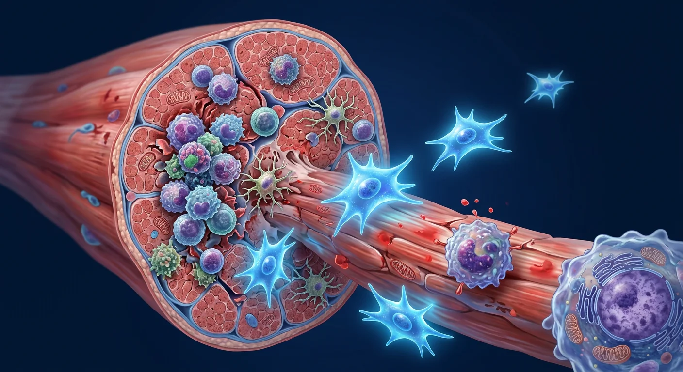
The deeper problem is immune-mediated muscle destruction. In polymyositis, CD8+ cytotoxic T cells directly invade and destroy muscle fibers expressing MHC class I molecules — a molecular signature not present on healthy muscle. In dermatomyositis, the attack is complement-mediated, with membrane attack complex (MAC) deposition damaging the endothelial cells of intramuscular capillaries, leading to capillary loss, ischemia, and perifascicular muscle-fiber atrophy. [4] Both pathways converge on the same endpoint: progressive muscle-fiber necrosis, fibrosis, and functional decline. The regenerative capacity of skeletal muscle — normally robust through satellite-cell activation — is progressively exhausted when the inflammatory environment is sustained.
MSC therapy targets both halves of the pathology. Mesenchymal stem cells possess a dual-action mechanism highly relevant to inflammatory myopathy: first, broad-spectrum immunomodulation that may calm the autoimmune attack on muscle, and second, paracrine signaling that may support muscle-fiber repair, reduce fibrosis, and restore the local regenerative niche. [5] This is not about replacing damaged muscle with new tissue — it is about creating the conditions in which the body's own repair mechanisms can function again.
Understanding polymyositis and dermatomyositis
Polymyositis and dermatomyositis are autoimmune diseases in which the immune system attacks skeletal muscle, causing progressive proximal weakness — difficulty rising from a chair, climbing stairs, or lifting objects above the shoulders. Dermatomyositis additionally involves characteristic skin manifestations: the heliotrope rash (purple discoloration of the eyelids), Gottron's papules (scaly patches over the knuckles), and a photosensitive "shawl sign" rash across the upper back and shoulders. Both conditions are subtypes of idiopathic inflammatory myopathy (IIM), and both can involve extra-muscular organs — the lungs (interstitial lung disease, ILD), the heart (myocarditis), and the esophagus (dysphagia). [6]
The pathophysiology differs between the two, which matters when considering therapeutic targets. Polymyositis is predominantly T-cell-driven: CD8+ cytotoxic T cells surround and invade non-necrotic muscle fibers, recognizing an antigen presented by aberrantly expressed MHC class I molecules on the muscle-cell surface. Dermatomyositis is humoral and complement-driven: antibody-mediated activation of C3 complement leads to MAC deposition on endothelial cells, capillary destruction, and ischemic muscle damage concentrated in the perifascicular region. [4] Both produce elevated serum creatine kinase (CK) and aldolase as markers of ongoing muscle injury, and diagnosis typically requires muscle biopsy, EMG, and myositis-specific antibody panels.
Key point: not just muscle weakness
Dermatomyositis — especially the anti-MDA5 subtype — carries a high risk of rapidly progressive interstitial lung disease, which is often the cause of mortality. Any therapy being evaluated for these conditions must be assessed for its effect on extra-muscular manifestations, not just muscle strength alone.
How MSCs are thought to work in inflammatory myopathy
Immunomodulation: calming the autoimmune attack
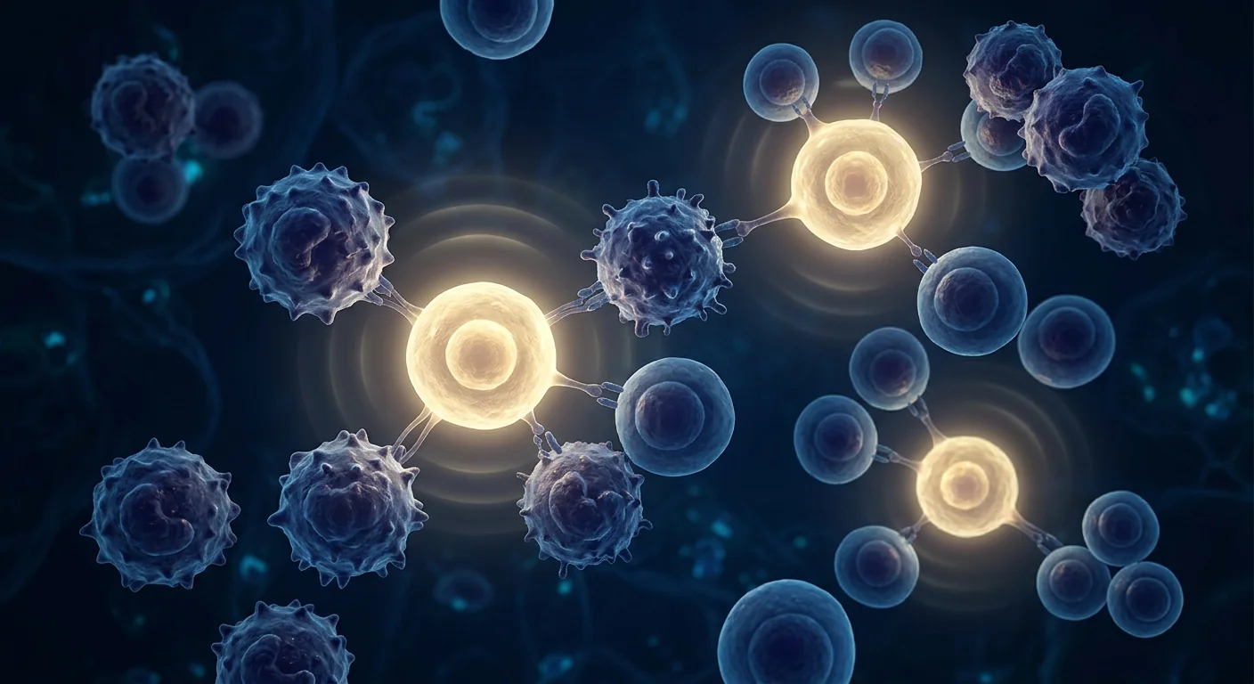
MSCs exert broad immunomodulatory effects that are directly relevant to the immune pathology of myositis. They suppress the proliferation and cytotoxic activity of CD8+ T cells — the primary effectors in polymyositis — through secretion of prostaglandin E2 (PGE2), indoleamine 2,3-dioxygenase (IDO), and TGF-β. [7] They inhibit B-cell proliferation and antibody production, relevant to the humoral component of dermatomyositis. They shift macrophage polarization from the pro-inflammatory M1 phenotype toward the anti-inflammatory, tissue-repair M2 phenotype — a shift that is critical for resolving chronic muscle inflammation. [8]
Treg expansion and Th17 suppression
In both polymyositis and dermatomyositis, the balance between regulatory T cells (Tregs) and pro-inflammatory Th17 cells is disrupted — reduced Treg numbers and function, and expanded Th17 populations, have been documented in muscle biopsies and peripheral blood. [9] MSCs have a well-established capacity to expand functional Tregs and suppress the Th17 lineage, partially through the secretion of TGF-β and HGF. Restoring the Treg/Th17 balance is one of the most plausible mechanisms by which MSC therapy could produce durable immune quieting in myositis — addressing a root immunological defect rather than simply suppressing inflammation pharmacologically.
Muscle repair support: reducing fibrosis
A distinctive feature of MSC therapy, compared to immunosuppressant drugs, is the potential to simultaneously support tissue repair. MSCs secrete a broad spectrum of growth factors — HGF, VEGF, IGF-1, FGF-2 — that promote satellite-cell activation, myoblast differentiation, and angiogenesis. [10] Equally important, MSCs have anti-fibrotic activity: they secrete matrix metalloproteinases (MMPs) that degrade excessive extracellular matrix and suppress TGF-β1-driven fibroblast activation. In chronic myositis, progressive replacement of functional muscle by fibrotic tissue is a major contributor to irreversible weakness; a therapy that both calms inflammation and limits fibrosis addresses two drivers of disability simultaneously.
What the preclinical evidence shows
The rationale for MSC therapy in inflammatory myopathy draws on complementary evidence streams. In animal models of myositis, MSC infusion reduces inflammatory infiltrates in muscle tissue, lowers serum CK levels, and improves muscle strength — effects attributed to both immunomodulation and paracrine support of muscle repair. [11] Studies using labeled MSCs show that a small fraction of infused cells migrate to injured muscle, where they survive for days to weeks and exert local effects before being cleared — consistent with the "hit-and-run" model in which therapeutic benefit derives from transient paracrine signaling rather than engraftment.
In vitro, MSCs suppress the proliferation of autoreactive T cells isolated from myositis patients, shift cytokine profiles from IFN-γ-dominant to IL-10-dominant, and protect myotubes from immune-mediated cytotoxicity. [12] These laboratory findings provide mechanistic plausibility, but it must be stated plainly: the translational gap from bench to bedside in myositis is wide, and human clinical data are sparse.
Clinical evidence: where things stand
Human clinical data on MSC therapy specifically for polymyositis and dermatomyositis are extremely limited. Unlike conditions such as lupus or rheumatoid arthritis, where multiple Phase I/II MSC trials have been conducted, myositis has not yet been the subject of dedicated, published MSC clinical trials. The evidence base consists of: (1) case reports of individual patients with refractory dermatomyositis treated with MSCs, typically under compassionate-use frameworks; (2) extrapolation from MSC trials in related autoimmune conditions that share immunological features with myositis; and (3) preclinical data. [13]
Case reports from China and Japan have described patients with severe, treatment-refractory dermatomyositis — including cases with progressive ILD — who received allogeneic umbilical-cord or bone-marrow MSCs. In several of these reports, patients experienced stabilization or modest improvement in muscle strength, reduction in serum CK, and in some instances stabilization of lung function. These reports are anecdotal, uncontrolled, and subject to publication bias — but they establish that the question is being asked clinically, not just in animal models. [14]
The most informative parallel evidence comes from MSC trials in systemic sclerosis and lupus, where immunological endpoints relevant to myositis — Treg expansion, reduction in inflammatory cytokines, and improvement in functional scores — have been reported. [15] Extrapolating across autoimmune diseases is inherently limited, but the shared immunological mechanisms (Treg deficiency, Th17 expansion, M1 macrophage dominance) provide a biologically coherent basis for expecting MSCs might have activity in myositis as well. The critical caveat is that this expectation has not yet been tested in appropriately designed trials.
The honest headline
As of 2026, MSC therapy is not an approved or proven treatment for polymyositis or dermatomyositis. The biological rationale is sound — dual-action immunomodulation plus muscle-repair support — but the human evidence consists of case reports and extrapolation from other autoimmune conditions, not controlled trials. Any clinic presenting stem cell therapy as a reliable treatment for myositis is going well beyond what the data support.
Why allogeneic over autologous in myositis
In principle, either autologous (the patient's own) or allogeneic (healthy donor) MSCs could be used. In practice, there are compelling reasons to favor allogeneic cells — particularly umbilical-cord-derived MSCs (UC-MSCs) — for autoimmune conditions like myositis. [16]
First, the patient's own MSCs may not be healthy. There is evidence that MSCs from patients with chronic inflammatory diseases exhibit reduced proliferative capacity, altered immunomodulatory function, and features of premature senescence. Using a patient's own dysfunctional cells to treat an autoimmune condition risks delivering a compromised therapeutic product.
Second, UC-MSCs offer practical advantages: they are obtained non-invasively from donated umbilical cords after healthy births, can be expanded to large numbers in culture, have lower immunogenicity than adult-tissue MSCs, and possess particularly potent immunomodulatory activity — including higher secretion of PGE2 and IDO compared to bone-marrow MSCs. [17] These properties make them the most commonly used cell source in autoimmune MSC trials.
How disease activity and response are measured
One of the challenges in myositis research is measuring response meaningfully. Standard tools include:
- MMT-8 (Manual Muscle Testing of 8 muscle groups). A validated score from 0–80 assessing strength in proximal and axial muscles — the most common primary endpoint in myositis clinical trials.
- Serum creatine kinase (CK). A biochemical marker of ongoing muscle damage. Falling CK is suggestive of reduced muscle injury, but CK can normalize without full functional recovery — and can be elevated by exercise, making it an imperfect surrogate.
- IMACS core set measures. The International Myositis Assessment and Clinical Studies group defines a composite response using six domains: physician global activity, patient global activity, MMT, HAQ (Health Assessment Questionnaire), muscle enzyme levels, and extramuscular disease activity.
- MRI and muscle biopsy. MRI with short-tau inversion recovery (STIR) sequences can visualize muscle edema (active inflammation), and repeat biopsy can assess histological change — but both are invasive or costly, limiting their use in routine monitoring.
What the evidence supports — and what it doesn't
The fair summary is that MSC therapy for polymyositis and dermatomyositis occupies the early-investigational space. The biological case is coherent: MSCs can suppress the CD8+ T-cell attack on muscle (polymyositis), inhibit complement-mediated endothelial damage (dermatomyositis), and support muscle repair and fibrosis reduction — a combined mechanism that no existing pharmacotherapy offers. The preclinical data are encouraging. The human data are minimal.
- What is supported: The immunomodulatory mechanisms of MSCs are well-characterized. Preclinical models of myositis show reduced inflammation and improved muscle function after MSC treatment. Case reports suggest biological activity in refractory human disease. The safety profile of allogeneic UC-MSCs appears favorable across multiple autoimmune conditions.
- What is not supported: There are no published controlled trials demonstrating efficacy of MSCs in myositis. No dosing regimen, treatment interval, or delivery route has been established. Long-term outcomes, including the effect on extra-muscular manifestations like ILD, are unknown. MSC therapy should not be presented as a substitute for established immunosuppressive treatment.
Polymyositis and dermatomyositis are exactly the kind of diseases where the gap between biological promise and clinical proof is widest — and where premature claims do the most harm. The only honest posture is to acknowledge the rationale, fund the trials, and wait for data.
— VELAR Clinical Team
Safety considerations
Across the broader autoimmune MSC literature, allogeneic UC-MSCs have demonstrated an acceptable short-term safety profile. The most commonly reported adverse events are infusion-related — transient fever, mild headache, and fatigue — generally resolving within 24 hours. [18] Longer-term concerns — tumorigenicity, ectopic tissue formation, immunogenicity of repeated allogeneic doses — are theoretical at this stage but have not been observed in published follow-up to date. For myositis specifically, there are no disease-specific safety signals identified, but the absence of evidence is not evidence of absence; the patient population studied has been small.
Frequently Asked Questions
Can stem cell therapy cure polymyositis or dermatomyositis?
No. There is no evidence that MSC therapy produces a cure for inflammatory myopathy. The research is investigational, and any clinic presenting stem cells as a cure is making claims that go beyond the published data. The goal being explored is disease modification — reducing disease activity and supporting muscle repair, not elimination of the underlying autoimmune process.
What is the difference between treating polymyositis and dermatomyositis with MSCs?
The immunological targets differ — T-cell-driven cytotoxicity in polymyositis versus complement-mediated vasculopathy in dermatomyositis — but MSCs have mechanisms relevant to both. In practice, most case reports have involved dermatomyositis patients, particularly those with refractory skin and lung involvement. There is no evidence that one subtype responds better than the other.
Are MSC infusions safe for patients already on immunosuppressants?
In published MSC trials for autoimmune disease, patients have generally continued their background immunosuppression during MSC treatment. The combination has been well tolerated in these contexts. However, this is a decision that must be made by the treating rheumatologist — immunosuppression should never be discontinued or reduced without medical supervision.
How many MSC infusions might be needed for myositis?
There is no established protocol. Case reports describe regimens ranging from single infusions to repeated doses at 1–3 month intervals. The optimal number, interval, and total dose have not been studied. This is one of the fundamental questions that controlled trials — not anecdotes — must answer.
What does stem cell therapy for myositis cost in Thailand?
MSC therapy at GMP-certified facilities in Thailand typically ranges from USD 8,000–25,000 per treatment protocol, depending on cell source, dose, and whether repeat infusions are included. Because myositis is not among the conditions with established MSC protocols, any treatment offered would be off-label and experimental. Cost transparency — asking for a detailed itemized breakdown — is essential.
Practical guidance for patients
If you are considering stem cell options for polymyositis or dermatomyositis, the diligence is the same that protects against any over-promised treatment:
- Ask whether the approach is part of a registered clinical trial with ethical oversight and published protocols. If it is not, ask why.
- Ask what cell type and source are used. A credible response specifies "allogeneic umbilical-cord-derived MSCs," not just "stem cells."
- Ask how response is measured. MMT-8, CK, and functional scores — not just subjective reports — are the minimum standard.
- Ask what published evidence supports the claims. For myositis, the answer should be honest: "The evidence is preclinical and case-report level; we are building on the broader autoimmune MSC literature." A provider who claims certainty is not being truthful.
- Never stop prescribed immunosuppression without the direct supervision of your treating rheumatologist. MSC therapy is not a replacement for standard-of-care treatment.
At VELAR Center, our regenerative work is grounded in conditions where the evidence base is more mature, and we follow the autoimmune cell-therapy literature closely without overstating it. Polymyositis and dermatomyositis remain difficult, serious diseases, and we believe the only honest way to discuss MSC therapy for them is plainly: the dual-action rationale — immunomodulation plus muscle-repair support — is biologically coherent, the preclinical data are encouraging, the human data are sparse, and the approach remains investigational. As the evidence matures, we will let that evidence — not enthusiasm — shape what we can responsibly say. If you want an honest conversation about what regenerative medicine can and cannot do today, that is exactly where a responsible consultation begins.
References
- Lundberg IE, Fujimoto M, Vencovsky J, et al. Idiopathic inflammatory myopathies. Nature Reviews Disease Primers. 2021;7(1):86. doi:10.1038/s41572-021-00321-x ↩
- Oddis CV, Aggarwal R. Treatment in myositis. Nature Reviews Rheumatology. 2018;14(5):279-289. doi:10.1038/nrrheum.2018.42 ↩
- Dalakas MC. Inflammatory muscle diseases. New England Journal of Medicine. 2015;372(18):1734-1747. doi:10.1056/NEJMra1402225 ↩
- Greenberg SA. Dermatomyositis and type 1 interferons. Current Rheumatology Reports. 2010;12(3):198-203. doi:10.1007/s11926-010-0101-6 ↩
- Le Blanc K, Mougiakakos D. Multipotent mesenchymal stromal cells and the innate immune system. Nature Reviews Immunology. 2012;12(5):383-396. doi:10.1038/nri3209 ↩
- Lundberg IE, Tjärnlund A, Bottai M, et al. 2017 European League Against Rheumatism / American College of Rheumatology classification criteria for adult and juvenile idiopathic inflammatory myopathies. Annals of the Rheumatic Diseases. 2017;76(12):1955-1964. doi:10.1136/annrheumdis-2017-211468 ↩
- Nauta AJ, Fibbe WE. Immunomodulatory properties of mesenchymal stromal cells. Blood. 2007;110(10):3499-3506. doi:10.1182/blood-2007-02-069716 ↩
- Bernardo ME, Fibbe WE. Mesenchymal stromal cells: sensors and switchers of inflammation. Cell Stem Cell. 2013;13(4):392-402. doi:10.1016/j.stem.2013.09.006 ↩
- Waschbisch A, Schwab N, Ruck T, et al. FOXP3+ T regulatory cells in idiopathic inflammatory myopathies. Journal of Neuroimmunology. 2010;225(1-2):137-142. doi:10.1016/j.jneuroim.2010.05.035 ↩
- Caplan AI, Correa D. The MSC: an injury drugstore. Cell Stem Cell. 2011;9(1):11-15. doi:10.1016/j.stem.2011.06.008 ↩
- Seo YH, Jung JU, Kim HS, et al. Mesenchymal stem cells ameliorate experimental autoimmune myositis in mice. Journal of Immunology Research. 2017;2017:3543920. doi:10.1155/2017/3543920 ↩
- Shi Y, Wang Y, Li Q, et al. Immunoregulatory mechanisms of mesenchymal stem and stromal cells in inflammatory diseases. Nature Reviews Nephrology. 2018;14(8):493-507. doi:10.1038/s41581-018-0023-5 ↩
- Wang D, Zhang H, Liang J, et al. Allogeneic mesenchymal stem cell transplantation in severe and refractory systemic lupus erythematosus. Arthritis & Rheumatology. 2014;66(7):1908-1919. doi:10.1002/art.38653 ↩
- Zhao Y, Wang Z, Lu J, et al. Treatment of refractory dermatomyositis with mesenchymal stem cells: a case report. International Journal of Rheumatic Diseases. 2019;22(7):1339-1342. doi:10.1111/1756-185X.13581 ↩
- Sun L, Akiyama K, Zhang H, et al. Mesenchymal stem cell transplantation reverses multiorgan dysfunction in systemic lupus erythematosus mice and humans. Stem Cells. 2009;27(6):1421-1432. doi:10.1002/stem.68 ↩
- Moll G, Ankrum JA, Kamhieh-Milz J, et al. Intravascular mesenchymal stromal/stem cell therapy product diversification: time for new clinical guidelines. Trends in Molecular Medicine. 2019;25(2):149-163. doi:10.1016/j.molmed.2018.12.006 ↩
- Barcia RN, Santos JM, Filipe M, et al. What makes umbilical cord tissue-derived mesenchymal stromal cells superior immunomodulators when compared to bone marrow-derived mesenchymal stromal cells? Stem Cells International. 2015;2015:583984. doi:10.1155/2015/583984 ↩
- Lalu MM, McIntyre L, Pugliese C, et al. Safety of cell therapy with mesenchymal stromal cells (SafeCell): a systematic review and meta-analysis of clinical trials. PLoS ONE. 2012;7(10):e47559. doi:10.1371/journal.pone.0047559 ↩
多发性肌炎和皮肌炎是罕见的慢性自身免疫性疾病,免疫系统错误地攻击骨骼肌组织,导致进行性肌无力、疲劳,皮肌炎还伴有特征性皮疹。这些疾病统称为特发性炎症性肌病(IIM),发病率约为每10万人中5-10例。常规治疗依赖皮质类固醇和免疫抑制剂,但约30-40%的患者仅获得部分缓解,完全缓解罕见。[1][2]
间充质干细胞(MSC)疗法的双重作用机制与此高度相关。首先,MSC具有广谱免疫调节能力——抑制CD8+细胞毒性T细胞增殖(多发性肌炎的主要效应细胞),促进调节性T细胞(Treg)扩增,抑制Th17细胞,将巨噬细胞从促炎的M1表型转向抗炎的M2表型。[5][7]其次,MSC分泌HGF、VEGF、IGF-1等生长因子,支持肌肉卫星细胞活化和肌纤维修复,同时通过分泌基质金属蛋白酶抑制纤维化。[10]
临床证据目前极为有限。尚无针对肌炎的特异性MSC临床试验公开发表。现有证据包括:难治性皮肌炎患者接受同种异体脐带MSC治疗的个别病例报告,以及从狼疮和系统性硬化症等MSC试验中的外推数据。[13][14]临床前动物模型显示MSC输注可减少肌肉炎症浸润、降低血清CK水平并改善肌力。[11]
推荐同种异体脐带来源MSC(UC-MSC),而非自体细胞。慢性炎症性疾病患者的自体MSC可能功能受损或早衰,且UC-MSC具有更强的免疫调节活性。[16][17]
截至2026年,MSC疗法并非多发性肌炎或皮肌炎的获批或确证治疗方法。生物学原理合理,临床前数据令人鼓舞,但人体证据仅为病例报告和外推水平,尚无对照试验。任何将干细胞疗法宣传为肌炎可靠治疗的诊所都远远超出了数据支持的范围。
参考文献
- Lundberg IE, et al. Idiopathic inflammatory myopathies. Nat Rev Dis Primers. 2021;7(1):86. doi:10.1038/s41572-021-00321-x ↩
- Oddis CV, Aggarwal R. Treatment in myositis. Nat Rev Rheumatol. 2018;14(5):279-289. doi:10.1038/nrrheum.2018.42 ↩
- Dalakas MC. Inflammatory muscle diseases. N Engl J Med. 2015;372(18):1734-1747. doi:10.1056/NEJMra1402225 ↩
- Greenberg SA. Dermatomyositis and type 1 interferons. Curr Rheumatol Rep. 2010;12(3):198-203. doi:10.1007/s11926-010-0101-6 ↩
- Le Blanc K, Mougiakakos D. Multipotent mesenchymal stromal cells and the innate immune system. Nat Rev Immunol. 2012;12(5):383-396. doi:10.1038/nri3209 ↩
- Nauta AJ, Fibbe WE. Immunomodulatory properties of mesenchymal stromal cells. Blood. 2007;110(10):3499-3506. doi:10.1182/blood-2007-02-069716 ↩
- Caplan AI, Correa D. The MSC: an injury drugstore. Cell Stem Cell. 2011;9(1):11-15. doi:10.1016/j.stem.2011.06.008 ↩
- Seo YH, et al. MSCs ameliorate experimental autoimmune myositis in mice. J Immunol Res. 2017;2017:3543920. doi:10.1155/2017/3543920 ↩
- Wang D, et al. Allogeneic MSC transplantation in severe and refractory SLE. Arthritis Rheumatol. 2014;66(7):1908-1919. doi:10.1002/art.38653 ↩
- Zhao Y, et al. Treatment of refractory dermatomyositis with MSCs. Int J Rheum Dis. 2019;22(7):1339-1342. doi:10.1111/1756-185X.13581 ↩
- Moll G, et al. Intravascular MSC therapy product diversification. Trends Mol Med. 2019;25(2):149-163. doi:10.1016/j.molmed.2018.12.006 ↩
- Barcia RN, et al. UC-MSCs superior immunomodulators vs BM-MSCs. Stem Cells Int. 2015;2015:583984. doi:10.1155/2015/583984 ↩
التهاب العضلات المتعدد والتهاب الجلد والعضلات من أمراض المناعة الذاتية النادرة والمزمنة حيث يهاجم الجهاز المناعي الأنسجة العضلية الهيكلية، مما يسبب ضعفًا تدريجيًا وإرهاقًا، مع طفح جلدي مميز في حالة التهاب الجلد والعضلات. تُعرف هذه الحالات مجتمعة باعتلالات العضلات الالتهابية مجهولة السبب (IIM)، وتصيب ما يقدر بنحو 5-10 لكل 100,000 شخص. يعتمد العلاج التقليدي على الكورتيكوستيرويدات ومثبطات المناعة، لكن حوالي 30-40% من المرضى يحققون استجابة جزئية فقط، والشفاء التام نادر.[1][2]
تمتلك الخلايا الجذعية الوسيطة (MSC) آلية عمل مزدوجة ذات صلة كبيرة باعتلال العضلات الالتهابي. أولاً، تمتلك MSCs قدرة واسعة على تعديل المناعة — تثبيط تكاثر الخلايا التائية السامة CD8+ (المؤثرات الرئيسية في التهاب العضلات المتعدد)، وتعزيز توسع الخلايا التائية التنظيمية (Treg)، وقمع خلايا Th17، وتحويل البلاعم من النمط الالتهابي M1 إلى النمط المضاد للالتهابات M2.[5][7] ثانيًا، تفرز MSCs عوامل نمو مثل HGF وVEGF وIGF-1 التي تدعم تنشيط الخلايا الساتلة العضلية وإصلاح الألياف العضلية، مع تثبيط التليف من خلال إفراز إنزيمات المصفوفة المعدنية.[10]
الأدلة السريرية محدودة للغاية حاليًا. لا توجد تجارب سريرية منشورة خاصة بـ MSCs لعلاج التهاب العضلات. تشمل الأدلة المتاحة: تقارير حالات فردية لمرضى التهاب الجلد والعضلات المقاوم للعلاج تلقوا خلايا MSCs من الحبل السري، وبيانات مستقرأة من تجارب MSCs في أمراض المناعة الذاتية الأخرى مثل الذئبة والتصلب الجهازي.[13][14] أظهرت النماذج الحيوانية أن حقن MSCs يقلل الارتشاح الالتهابي في العضلات ويخفض مستويات CK ويحسن القوة العضلية.[11]
يوصى باستخدام MSCs من الحبل السري (UC-MSC) من متبرع سليم بدلاً من الخلايا الذاتية. قد تكون MSCs الذاتية لدى مرضى الالتهابات المزمنة معطلة أو متقادمة، بينما تمتلك UC-MSCs نشاطًا مناعيًا أقوى.[16][17]
حتى عام 2026، لا يُعد علاج MSC علاجًا معتمدًا أو مثبتًا لالتهاب العضلات المتعدد أو التهاب الجلد والعضلات. الأساس البيولوجي منطقي، والبيانات ما قبل السريرية مشجعة، لكن الأدلة البشرية تقتصر على تقارير الحالات والاستقراء، ولا توجد تجارب محكومة. أي عيادة تقدم العلاج بالخلايا الجذعية كعلاج موثوق لالتهاب العضلات تتجاوز بكثير ما تدعمه البيانات.
المراجع
- Lundberg IE, et al. Idiopathic inflammatory myopathies. Nat Rev Dis Primers. 2021;7(1):86. doi:10.1038/s41572-021-00321-x ↩
- Oddis CV, Aggarwal R. Treatment in myositis. Nat Rev Rheumatol. 2018;14(5):279-289. doi:10.1038/nrrheum.2018.42 ↩
- Dalakas MC. Inflammatory muscle diseases. N Engl J Med. 2015;372(18):1734-1747. doi:10.1056/NEJMra1402225 ↩
- Greenberg SA. Dermatomyositis and type 1 interferons. Curr Rheumatol Rep. 2010;12(3):198-203. doi:10.1007/s11926-010-0101-6 ↩
- Le Blanc K, Mougiakakos D. MSCs and the innate immune system. Nat Rev Immunol. 2012;12(5):383-396. doi:10.1038/nri3209 ↩
- Nauta AJ, Fibbe WE. Immunomodulatory properties of MSCs. Blood. 2007;110(10):3499-3506. doi:10.1182/blood-2007-02-069716 ↩
- Caplan AI, Correa D. The MSC: an injury drugstore. Cell Stem Cell. 2011;9(1):11-15. doi:10.1016/j.stem.2011.06.008 ↩
- Seo YH, et al. MSCs ameliorate experimental autoimmune myositis. J Immunol Res. 2017;2017:3543920. doi:10.1155/2017/3543920 ↩
- Wang D, et al. Allogeneic MSC transplantation in severe SLE. Arthritis Rheumatol. 2014;66(7):1908-1919. doi:10.1002/art.38653 ↩
- Zhao Y, et al. Treatment of refractory dermatomyositis with MSCs. Int J Rheum Dis. 2019;22(7):1339-1342. doi:10.1111/1756-185X.13581 ↩
- Moll G, et al. Intravascular MSC therapy product diversification. Trends Mol Med. 2019;25(2):149-163. doi:10.1016/j.molmed.2018.12.006 ↩
- Barcia RN, et al. UC-MSCs superior immunomodulators vs BM-MSCs. Stem Cells Int. 2015;2015:583984. doi:10.1155/2015/583984 ↩