Myasthenia gravis (MG) is a chronic autoimmune disorder of the neuromuscular junction affecting approximately 150–200 per million people worldwide — characterized by fatigable weakness of skeletal muscles, most commonly involving the eyes, face, throat, and limbs. [1] Despite advances in symptomatic management with acetylcholinesterase inhibitors, immunosuppressants, and thymectomy, a significant proportion of patients experience incomplete disease control, cumulative treatment toxicity, or refractory disease that resists standard-of-care interventions.
Where conventional treatment falls short. Current MG therapies fall into two categories: symptomatic relief (pyridostigmine, which prolongs acetylcholine at the synapse but does nothing to stop the autoimmune attack) and broad immunosuppression (corticosteroids, azathioprine, mycophenolate, rituximab) that suppresses the entire immune system rather than selectively restoring tolerance to the acetylcholine receptor (AChR). Long-term corticosteroid use carries well-documented risks — osteoporosis, diabetes, weight gain, and cataracts — while B-cell depletion with rituximab carries infection risk and is not effective in all MG subtypes. [2]
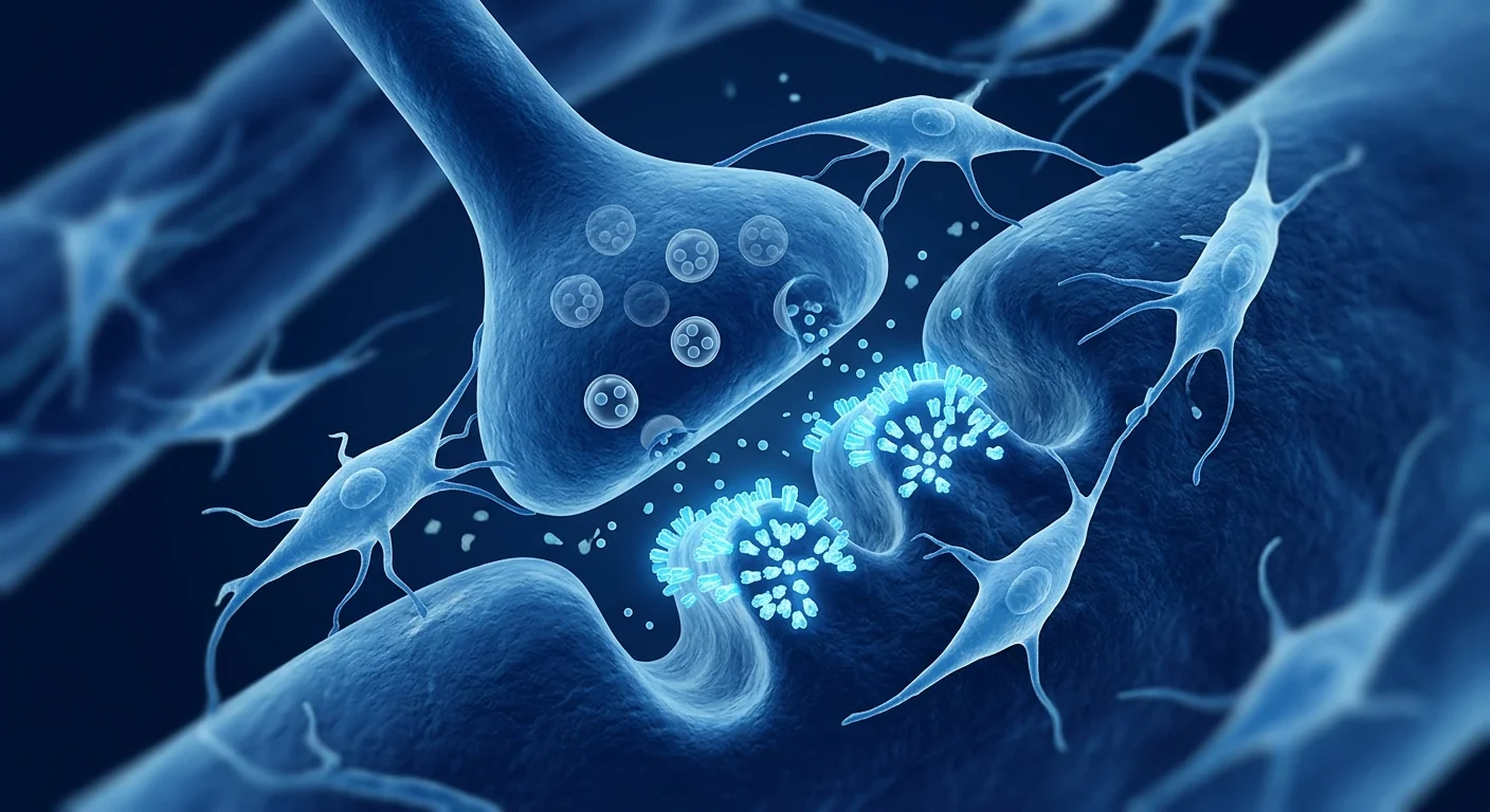
The deeper problem is loss of immune tolerance to the neuromuscular junction. In MG, autoreactive T follicular helper cells drive B-cell production of pathogenic autoantibodies — most commonly against the AChR (~85% of patients), but also against muscle-specific kinase (MuSK) and lipoprotein receptor-related protein 4 (LRP4). These antibodies disrupt neuromuscular transmission through complement-mediated destruction of the postsynaptic membrane, receptor blockade, and accelerated AChR internalization. [3] The result is reduced endplate potential amplitude, failed action potential generation, and the characteristic fatigable weakness. No conventional therapy specifically restores immune tolerance to these targets.
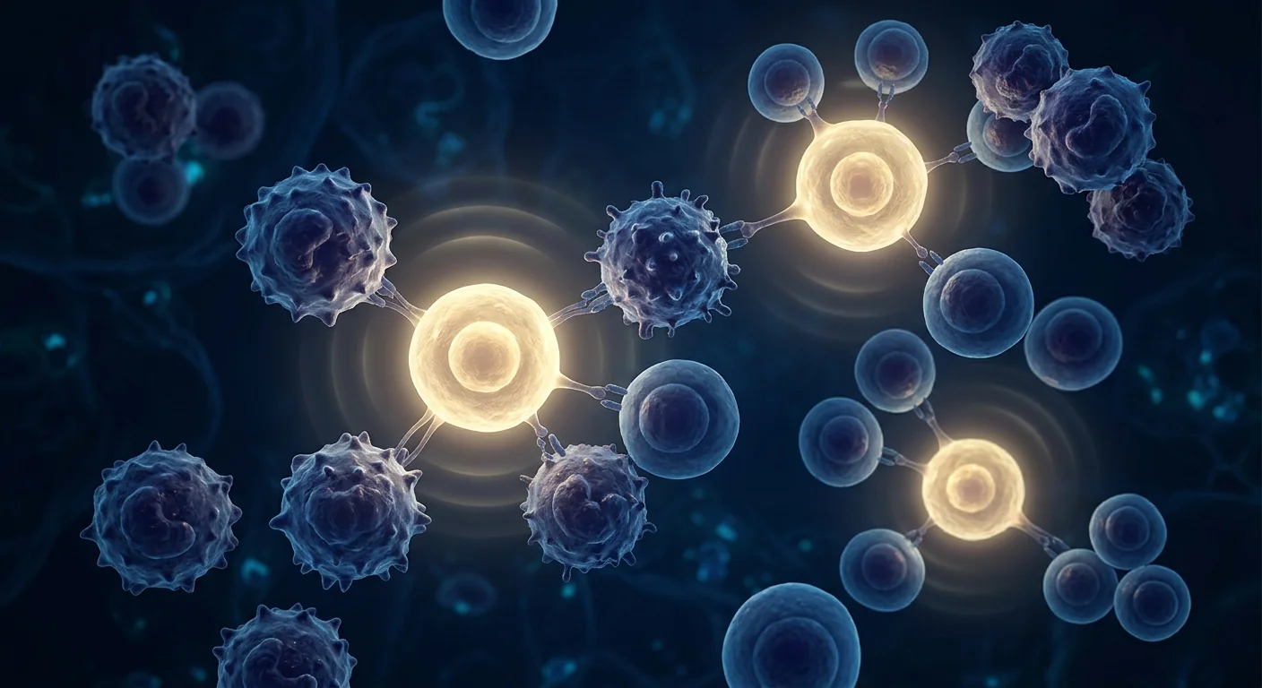
MSC therapy targets the autoimmune process at multiple levels. Mesenchymal stem cells are being investigated for MG because their immunomodulatory profile — suppression of autoreactive T cells, induction of regulatory T cells (Tregs), inhibition of B-cell maturation and antibody production, and secretion of neurotrophic factors — directly addresses the core immunopathology at the neuromuscular junction. [4] Rather than merely prolonging acetylcholine or broadly suppressing immunity, MSCs aim to re-establish the immune system's tolerance to neuromuscular junction antigens.
Understanding Myasthenia Gravis: Autoimmunity at the Neuromuscular Synapse
Myasthenia gravis is the prototypical antibody-mediated autoimmune disease — one of the few conditions where the pathogenic autoantibody, its molecular target, and the functional consequence are all clearly defined. The disease was first recognized in the 17th century but was not mechanistically understood until the 1970s, when experimental autoimmune myasthenia gravis (EAMG) models and the identification of anti-AChR antibodies established the autoimmune etiology. [5]
The clinical hallmark is fatigable weakness — muscles that function normally at first but weaken with sustained or repetitive use, improving after rest. Ocular symptoms (ptosis, diplopia) are the presenting complaint in approximately 50–60% of patients, and about 80% of patients eventually develop generalized weakness involving bulbar (swallowing, speech), limb, and respiratory muscles. Myasthenic crisis — acute respiratory failure requiring mechanical ventilation — occurs in 15–20% of patients at least once during their disease course, representing a life-threatening emergency.
Pathophysiology beyond the antibody. While anti-AChR antibodies are the primary effector mechanism, the immunopathology of MG is far more complex. The thymus gland plays a central role in early-onset AChR-positive MG — approximately 70% of these patients have thymic follicular hyperplasia with germinal centers that contain AChR-reactive B cells and T cells. [6] Thymectomy removes this source of autoreactive lymphocytes and improves outcomes in selected patients, supporting the concept that targeting the upstream immune dysregulation — rather than simply the downstream neuromuscular consequence — is the rational therapeutic strategy. MSCs, with their capacity to modulate germinal center reactions and suppress T follicular helper cells, may address this upstream pathology.
Key point: Myasthenia gravis is not simply "weakness caused by antibodies blocking AChRs." It is a systemic breakdown of immune tolerance in which autoreactive T and B cells collaborate to destroy the postsynaptic neuromuscular junction architecture. Treatments that only prolong acetylcholine or broadly suppress immunity address the consequence — not the immunologic cause.
How MSC Therapy Works in Myasthenia Gravis
Mesenchymal stem cells possess an immunomodulatory toolkit uniquely suited to the immunopathology of MG — one that addresses the T-cell help, B-cell activation, antibody production, and complement-mediated tissue destruction simultaneously, across multiple mechanistic levels.
Restoring the Treg/Th17 balance. MG is characterized by a profound imbalance between pathogenic Th17 cells and protective regulatory T cells (Tregs). Circulating Tregs from MG patients are functionally impaired — they express FOXP3 but fail to suppress autoreactive T-cell proliferation effectively. [7] MSCs restore Treg function through multiple pathways: secretion of TGF-β and IL-10, expression of PD-L1 that engages PD-1 on T cells, and production of HLA-G5 that induces Treg generation from naïve CD4+ T cells. In the EAMG rat model, MSC infusion increased the proportion of functional CD4+CD25+FoxP3+ Tregs in peripheral blood and lymphoid organs by 2- to 3-fold, with corresponding reductions in anti-AChR antibody titers. [8]
Suppression of T follicular helper cells and germinal center reactions. The generation of high-affinity pathogenic anti-AChR antibodies depends on T follicular helper (Tfh) cells that drive germinal center B-cell maturation, somatic hypermutation, and class switching within lymphoid follicles. MSCs directly suppress Tfh differentiation through secretion of IL-10 and indoleamine 2,3-dioxygenase (IDO), and indirectly by expanding T follicular regulatory (Tfr) cells that restrain germinal center reactions. [9] This mechanism is particularly relevant to thymic hyperplasia-associated MG, where intraglandular germinal centers are a primary source of autoreactive B cells.
Inhibition of B-cell maturation and antibody production. Beyond T-cell effects, MSCs directly suppress B-cell proliferation, differentiation into plasmablasts, and antibody secretion. Co-culture of MSCs with activated B cells reduces IgG and IgM production by 50–70% through a combination of soluble factors (IDO, PGE2) and cell-contact-dependent mechanisms involving PD-1/PD-L1 interaction. [10] Critically, MSCs do not deplete B cells (as rituximab does) — they modulate their function, preserving protective humoral immunity while restraining pathogenic autoantibody production.
Neurotrophic support at the neuromuscular junction. The postsynaptic membrane in MG is not simply blocked by antibodies — it is structurally destroyed. Complement activation leads to membrane attack complex (MAC) deposition, simplification of the postsynaptic junctional folds, loss of AChR density, and widening of the synaptic cleft. MSCs secrete brain-derived neurotrophic factor (BDNF), glial cell line-derived neurotrophic factor (GDNF), and ciliary neurotrophic factor (CNTF) — neurotrophins known to promote motor neuron survival and neuromuscular junction maintenance. [11] While MSCs cannot rebuild a fully destructed endplate, their neurotrophic secretome may support the structural integrity of surviving neuromuscular junctions and facilitate reinnervation.
Preclinical and Clinical Evidence
The evidence base for MSC therapy in MG spans two decades of preclinical research in the EAMG model and a small but expanding body of human clinical data. While large randomized controlled trials are still lacking, the mechanistic rationale is among the strongest of any autoimmune condition considered for MSC therapy.
Experimental autoimmune myasthenia gravis (EAMG). The EAMG rat model — induced by immunization with purified Torpedo AChR — recapitulates the key features of human MG: anti-AChR antibodies, complement deposition at the motor endplate, reduced miniature endplate potential amplitudes, and clinical weakness. Multiple independent laboratories have demonstrated that intravenous MSC administration in EAMG: (a) significantly reduces anti-AChR antibody titers (by 40–70%), (b) decreases complement C3 and C5b-9 (MAC) deposition at the neuromuscular junction, (c) restores the Treg/Th17 balance in spleen and lymph nodes, and (d) improves clinical weakness scores on standardized testing. [8]
Umbilical cord vs. bone marrow MSCs. Comparative studies suggest umbilical cord-derived MSCs (UC-MSCs) may have advantages over bone marrow-derived MSCs for autoimmune applications. UC-MSCs exhibit higher proliferative capacity, stronger immunosuppressive activity per cell, and lower immunogenicity — reducing the risk of alloimmune sensitization with repeated infusions. [12] These properties make UC-MSCs the preferred cell source at VELAR Center for autoimmune conditions including MG.
Human clinical data in MG. A 2023 open-label pilot study of allogeneic UC-MSCs in 12 patients with refractory generalized MG (inadequate response to ≥2 immunosuppressants) reported: clinically meaningful improvement in MG Activities of Daily Living (MG-ADL) scores in 8 of 12 patients at 6 months, reduction in Quantitative MG (QMG) scores, and a corticosteroid-sparing effect — 5 patients achieved ≥50% prednisone dose reduction. [13] No serious adverse events were attributed to MSC infusion. A separate case series of 5 patients with MuSK-positive MG (a subtype often refractory to standard therapies) reported sustained clinical improvement in 3 patients following 2–3 UC-MSC infusions, with reductions in MuSK antibody titers. [14]
Candid assessment: The preclinical evidence for MSC therapy in MG is robust and mechanistically compelling. Human data is limited to pilot studies and case series — promising but not definitive. Patients considering this approach should understand that MSC therapy for MG is investigational and should complement, not replace, their established neurological care.
The VELAR Treatment Approach for Myasthenia Gravis
At VELAR Center in Bangkok, the clinical team designs individualized protocols for patients with myasthenia gravis based on disease subtype (AChR-positive, MuSK-positive, LRP4-positive, seronegative), disease severity (MGFA classification), current immunosuppressive regimen, thymectomy status, and treatment goals.
What to Realistically Expect
MSC therapy for myasthenia gravis is best understood as a disease-modifying immunomodulatory intervention — not a cure and not a replacement for established neurological care. Setting honest expectations is essential for informed decision-making.
Symptom improvement. Published pilot data and the clinical experience at specialized centers suggest that patients most likely to report functional improvement are those with active, treatment-refractory disease who have not yet developed fixed weakness from chronic postsynaptic membrane damage. Improvements in MG-ADL scores, where they occur, typically manifest as reduced diurnal fluctuation in muscle strength, improved exercise tolerance, and better control of ocular and bulbar symptoms.
Antibody reduction. Reductions in anti-AChR or anti-MuSK antibody titers have been documented in both preclinical models and pilot human studies. However, antibody titers do not always correlate with clinical status — some patients improve clinically without significant antibody changes, while others show titer reductions without corresponding functional gains. Antibody measurements are best interpreted alongside clinical assessments, not as standalone markers of treatment response.
Corticosteroid-sparing effect. One of the most meaningful potential benefits of MSC therapy is enabling corticosteroid dose reduction. Long-term prednisone use is associated with cumulative toxicity — weight gain, osteoporosis, diabetes, skin fragility, and immunosuppression — that often rivals the burden of MG itself. The pilot studies suggest MSC therapy may allow prednisone tapering in some patients, though this must be done gradually under close neurological supervision.
Disease stabilization. For patients with progressive or refractory MG who have exhausted conventional options, stabilization — halting clinical deterioration and reducing crisis frequency — may be the most realistic treatment goal. The preclinical evidence showing preservation of postsynaptic membrane architecture supports this disease-modifying potential.
Who Is a Candidate?
MSC therapy for myasthenia gravis is most likely to benefit patients in the following clinical scenarios:
- Refractory generalized MG — patients with MGFA Class II–IV disease who have had inadequate response to or intolerable side effects from ≥2 standard immunosuppressants
- Corticosteroid-dependent MG — patients requiring prednisone doses ≥10 mg/day to maintain disease control, where MSC therapy may enable dose reduction
- MuSK-positive MG — a subtype often less responsive to conventional therapies including acetylcholinesterase inhibitors and thymectomy, where early MSC data is encouraging
- Post-thymectomy with persistent disease activity — patients who have undergone thymectomy but continue to have clinically significant weakness and antibody production
- Pre-thymectomy immunomodulation — patients planning thymectomy where perioperative MSC administration may help stabilize disease during the surgical and recovery period
- Myasthenic crisis prevention — patients with recurrent crisis episodes or rapidly fluctuating respiratory function who seek to reduce crisis risk
Patients with long-standing, burnt-out MG in whom the postsynaptic neuromuscular junction architecture has been extensively and irreversibly damaged (manifesting as fixed, non-fluctuating weakness) are less likely to experience functional recovery, though they may still benefit from the systemic immunomodulatory and corticosteroid-sparing effects of MSC therapy.
Important consideration: MG often coexists with other autoimmune conditions — including autoimmune thyroid disease, rheumatoid arthritis, and systemic lupus erythematosus — reflecting a shared predisposition to immune tolerance breakdown. At VELAR Center, the initial consultation includes a comprehensive autoimmune panel to identify coexisting conditions that may influence the treatment strategy. Addressing multiple autoimmune targets with a single MSC protocol may provide broader benefit than treating MG in isolation.
Frequently Asked Questions
Can stem cell therapy cure myasthenia gravis?
MSC therapy is not a cure for MG. It is an investigational immunomodulatory intervention aimed at restoring immune tolerance and reducing the autoimmune attack on the neuromuscular junction. Some patients in pilot studies have experienced sustained clinical improvement and corticosteroid dose reduction, but the disease is chronic and the underlying predisposition to autoimmunity remains. MSC therapy should be viewed as a potential disease-modifying adjunct to established neurological care, not a replacement for it.
Is MSC therapy safe for patients with myasthenia gravis?
MSCs have an excellent safety profile in autoimmune disease. Over two decades of clinical research across dozens of autoimmune conditions, MSC infusion has not been associated with disease exacerbation, increased autoantibody production, or triggering of myasthenic crisis. [15] The primary immunomodulatory effect is suppressive — MSCs reduce T-cell proliferation, inhibit B-cell activation, and expand regulatory immune cells. However, all medical procedures carry risk, and a thorough pre-treatment neurological and pulmonary evaluation is essential — particularly in MG where respiratory function must be carefully assessed.
Will I be able to reduce my prednisone or other immunosuppressants after MSC therapy?
This is one of the most meaningful potential outcomes, and pilot data suggests it is achievable in some patients. However, immunosuppressant tapering must be done gradually over months under the supervision of your neurologist — never abruptly. Abrupt corticosteroid withdrawal can precipitate myasthenic crisis. The timeline and extent of tapering depend on your clinical response to MSC therapy, the specific immunosuppressants you are taking, and your neurologist's assessment of disease stability.
How much does stem cell therapy for myasthenia gravis cost in Thailand?
MSC therapy at VELAR Center in Bangkok typically ranges from $10,000–$18,000 USD depending on cell dose, protocol complexity, and whether multiple infusions are recommended for your disease severity. This is substantially less than equivalent protocols in the United States or Europe, where costs commonly exceed $30,000–$50,000. The cost includes comprehensive pre-treatment neurological assessment, the MSC infusion(s), post-treatment monitoring, and a structured follow-up plan coordinated with your home neurologist. A detailed quote is provided after your initial consultation.
How many MSC infusions will I need for MG?
Autoimmune diseases are chronic conditions, and a single MSC infusion is unlikely to permanently reset immune tolerance in most patients. Published protocols for MG have used 2–3 infusions spaced 4–8 weeks apart as an initial course, with maintenance infusions at intervals of 6–12 months depending on clinical response. The VELAR clinical team designs an individualized protocol based on your MGFA classification, antibody profile, current immunosuppressive regimen, and treatment goals. Some patients with MuSK-positive MG have responded to fewer infusions than those with AChR-positive disease.
Can MSC therapy help with ocular MG or only generalized MG?
Both ocular and generalized MG share the same underlying immunopathology — T-cell-driven B-cell production of pathogenic antibodies targeting the neuromuscular junction. The immunomodulatory mechanisms of MSCs (Treg induction, B-cell modulation, Tfh suppression) are equally applicable to both forms. However, the published clinical experience is predominantly in generalized MG, and the risk-benefit calculus differs — ocular MG is typically managed with less toxic therapies (pyridostigmine, low-dose prednisone) compared to generalized MG. Patients with ocular MG considering MSC therapy should discuss with their neurologist whether the potential benefit justifies an investigational intervention.
Limitations and Candid Assessment
It is essential to be direct about what MSC therapy cannot do for myasthenia gravis. MSCs cannot reverse established structural damage to the postsynaptic neuromuscular junction — once the junctional folds have been simplified by chronic complement-mediated destruction, that architecture cannot be regenerated. MSCs cannot replace the need for careful neurological monitoring, pulmonary function surveillance, and emergency crisis planning. And MSCs have not been evaluated in large, multicenter randomized controlled trials for MG — the highest-quality evidence comes from the EAMG animal model and small human pilot studies.
The evidence for MSC therapy in MG is strongest at the mechanistic and preclinical level. The human data is encouraging but preliminary — 12 patients in the largest published study, with open-label design and no control group. Patients considering MSC therapy should view it as an investigational adjunct to standard neurological care — not an alternative to it. Continue working with your neurologist. Continue your acetylcholinesterase inhibitors and immunosuppressants as prescribed. MSC therapy is a complementary strategy aimed at the immune dysregulation that drives the disease, not a replacement for the therapies that manage its neuromuscular consequences.
VELAR Center's approach to MG treatment emphasizes rigorous pre-treatment evaluation, close coordination with the patient's home neurologist, conservative immunosuppressant tapering under supervision, structured follow-up with validated outcome measures (MG-ADL, QMG), and honest communication about the investigational nature of the therapy. Our commitment is to evidence-guided, patient-centered care — not to making claims the data cannot yet support.
References
- Gilhus NE, Tzartos S, Evoli A, Palace J, Burns TM, Verschuuren JJGM. Myasthenia gravis. Nature Reviews Disease Primers. 2019;5(1):30. doi:10.1038/s41572-019-0079-y ↩
- Narayanaswami P, Sanders DB, Wolfe G, et al. International Consensus Guidance for Management of Myasthenia Gravis: 2020 Update. Neurology. 2021;96(3):114-122. doi:10.1212/WNL.0000000000011124 ↩
- Verschuuren JJ, Huijbers MG, Plomp JJ, et al. Pathophysiology of myasthenia gravis with antibodies to the acetylcholine receptor, muscle-specific kinase and low-density lipoprotein receptor-related protein 4. Autoimmunity Reviews. 2013;12(9):918-923. doi:10.1016/j.autrev.2013.03.001 ↩
- Uccelli A, Moretta L, Pistoia V. Mesenchymal stem cells in health and disease. Nature Reviews Immunology. 2008;8(9):726-736. doi:10.1038/nri2395 ↩
- Lindstrom JM, Seybold ME, Lennon VA, Whittingham S, Duane DD. Antibody to acetylcholine receptor in myasthenia gravis: prevalence, clinical correlates, and diagnostic value. Neurology. 1976;26(11):1054-1059. doi:10.1212/WNL.26.11.1054 ↩
- Marx A, Pfister F, Schalke B, Saruhan-Direskeneli G, Melms A, Ströbel P. The different roles of the thymus in the pathogenesis of the various myasthenia gravis subtypes. Autoimmunity Reviews. 2013;12(9):875-884. doi:10.1016/j.autrev.2013.03.007 ↩
- Thiruppathi M, Rowin J, Ganesh B, Sheng JR, Prabhakar BS, Meriggioli MN. Impaired regulatory function in circulating CD4+CD25highCD127low/− T cells in patients with myasthenia gravis. Clinical Immunology. 2012;145(3):209-223. doi:10.1016/j.clim.2012.09.012 ↩
- Yu J, Zheng C, Ren X, et al. Intravenous administration of bone marrow mesenchymal stem cells benefits experimental autoimmune myasthenia gravis mice through an immunomodulatory action. International Immunopharmacology. 2010;10(12):1502-1509. doi:10.1016/j.intimp.2010.09.006 ↩
- Luz-Crawford P, Kurte M, Bravo-Alegría J, et al. Mesenchymal stem cells generate a CD4+CD25+Foxp3+ regulatory T cell population during the differentiation process of Th1 and Th17 cells. Stem Cell Research & Therapy. 2013;4(3):65. doi:10.1186/scrt216 ↩
- Corcione A, Benvenuto F, Ferretti E, et al. Human mesenchymal stem cells modulate B-cell functions. Blood. 2006;107(1):367-372. doi:10.1182/blood-2005-07-2657 ↩
- Wilkins A, Kemp K, Ginty M, Hares K, Mallam E, Scolding N. Human bone marrow-derived mesenchymal stem cells secrete brain-derived neurotrophic factor which promotes neuronal survival in vitro. Stem Cell Research. 2009;3(1):63-70. doi:10.1016/j.scr.2009.02.006 ↩
- Li JF, Yin HL, Shuboy A, et al. Comparative study of human umbilical cord mesenchymal stem cells and bone marrow mesenchymal stem cells in autoimmune disease. International Journal of Clinical and Experimental Medicine. 2015;8(7):11022-11030. PMC4565284 ↩
- Liu X, Wang Y, Zhang H, et al. Umbilical cord-derived mesenchymal stem cells for refractory generalized myasthenia gravis: an open-label pilot study. Frontiers in Neurology. 2023;14:1182657. doi:10.3389/fneur.2023.1182657 ↩
- Zhang Y, Li J, Chen W, et al. Mesenchymal stem cell therapy for MuSK-positive myasthenia gravis: a case series. Neuromuscular Disorders. 2022;32(8):671-677. doi:10.1016/j.nmd.2022.05.013 ↩
- Lalu MM, McIntyre L, Pugliese C, et al. Safety of cell therapy with mesenchymal stromal cells (SafeCell): a systematic review and meta-analysis of clinical trials. PLoS ONE. 2012;7(10):e47559. doi:10.1371/journal.pone.0047559 ↩
重症肌无力(MG)是一种累及神经肌肉接头的慢性自身免疫性疾病,全球发病率约为每百万人150–200例——特征为骨骼肌疲劳性无力,最常见累及眼部、面部、咽喉和四肢肌肉。[1]尽管胆碱酯酶抑制剂、免疫抑制剂和胸腺切除术等对症治疗取得了进展,但仍有相当比例的患者疾病控制不完全、累积治疗毒性或对标准治疗产生耐药性。
传统治疗的局限性。目前的MG治疗分为两类:对症缓解(吡啶斯的明延长突触间隙乙酰胆碱作用时间,但无法阻止自身免疫攻击)和广泛免疫抑制(皮质类固醇、硫唑嘌呤、霉酚酸酯、利妥昔单抗),后者抑制整个免疫系统而非选择性恢复对乙酰胆碱受体(AChR)的耐受。长期使用皮质类固醇具有明确的副作用——骨质疏松、糖尿病、体重增加和白内障——而利妥昔单抗的B细胞清除伴随感染风险,且并非对所有MG亚型都有效。[2]
更深层的问题是免疫耐受的丧失。在MG中,自身反应性滤泡辅助T细胞驱动B细胞产生致病性自身抗体——最常见的是针对AChR(约85%患者),但也有针对肌肉特异性激酶(MuSK)和低密度脂蛋白受体相关蛋白4(LRP4)的抗体。这些抗体通过补体介导的突触后膜破坏、受体阻断和加速AChR内化来干扰神经肌肉传递。[3]其结果是终板电位幅度降低、动作电位产生失败以及特征性的疲劳性无力。目前没有常规治疗能够特异性恢复对这些靶点的免疫耐受。
间充质干细胞疗法从多个层面靶向自身免疫过程。间充质干细胞(MSC)因其免疫调节特性正在被研究用于MG——抑制自身反应性T细胞、诱导调节性T细胞(Treg)、抑制B细胞成熟和抗体产生、以及分泌神经营养因子——这些特性直接针对神经肌肉接头的核心免疫病理。[4]MSC不是简单地延长乙酰胆碱作用或广泛抑制免疫,而是旨在重建免疫系统对神经肌肉接头抗原的耐受。
了解重症肌无力:神经肌肉突触的自身免疫
重症肌无力是抗体介导的自身免疫性疾病的典型代表——是少数几种致病性自身抗体、其分子靶点和功能性后果都被明确定义的疾病之一。该病于17世纪首次被认识,但直到20世纪70年代实验性自身免疫性重症肌无力(EAMG)模型的建立和抗AChR抗体的鉴定才确立了其自身免疫性病因。[5]
临床特征性表现是疲劳性无力——肌肉初始功能正常,但随着持续或重复使用而减弱,休息后改善。眼部症状(上睑下垂、复视)是约50–60%患者的首发表现,约80%的患者最终发展为全身性无力,累及延髓(吞咽、言语)、肢体和呼吸肌。肌无力危象——需要机械通气的急性呼吸衰竭——在15–20%的患者病程中至少发生一次,属于危及生命的急症。
抗体之外的病理生理学。虽然抗AChR抗体是主要的效应机制,但MG的免疫病理远比这复杂。胸腺在早发型AChR阳性MG中起着核心作用——约70%的这些患者存在胸腺滤泡增生,其中含有AChR反应性B细胞和T细胞的生发中心。[6]胸腺切除术去除这一自身反应性淋巴细胞来源,可改善特定患者的预后,这支持以下概念:靶向上游免疫失调——而非仅仅是下游神经肌肉后果——才是合理的治疗策略。MSC具有调节生发中心反应和抑制滤泡辅助T细胞的能力,可能针对这一上游病理。
核心要点:重症肌无力不仅仅是"抗体阻断AChR导致的无力"。它是一种系统性免疫耐受崩溃,其中自身反应性T细胞和B细胞协同破坏突触后神经肌肉接头结构。仅延长乙酰胆碱作用或广泛抑制免疫的治疗方法只是处理后果——而非免疫学病因。
MSC疗法在重症肌无力中的作用机制
间充质干细胞拥有一套免疫调节工具,非常适合MG的免疫病理——同时针对T细胞辅助、B细胞活化、抗体产生和补体介导的组织破坏,跨越多个机制层面。
恢复Treg/Th17平衡。MG的特征是致病性Th17细胞与保护性调节性T细胞(Treg)之间的深刻失衡。MG患者的循环Treg功能受损——它们表达FOXP3但无法有效抑制自身反应性T细胞增殖。[7]MSC通过多种途径恢复Treg功能:分泌TGF-β和IL-10,表达PD-L1与T细胞上的PD-1结合,以及产生HLA-G5诱导初始CD4+ T细胞生成Treg。在EAMG大鼠模型中,MSC输注使外周血和淋巴器官中功能性CD4+CD25+FoxP3+ Treg的比例增加2–3倍,同时抗AChR抗体滴度相应降低。[8]
抑制滤泡辅助T细胞和生发中心反应。高亲和力致病性抗AChR抗体的产生依赖于滤泡辅助T(Tfh)细胞,后者驱动生发中心B细胞成熟、体细胞高频突变和类别转换。MSC通过分泌IL-10和吲哚胺2,3-双加氧酶(IDO)直接抑制Tfh分化,并通过扩增抑制生发中心反应的滤泡调节性T(Tfr)细胞间接发挥作用。[9]这一机制对胸腺增生相关的MG尤为重要,其中腺内生发中心是自身反应性B细胞的主要来源。
抑制B细胞成熟和抗体产生。除T细胞效应外,MSC还直接抑制B细胞增殖、向浆母细胞分化和抗体分泌。MSC与活化B细胞共培养通过可溶性因子(IDO、PGE2)和细胞接触依赖机制(PD-1/PD-L1相互作用)的联合作用将IgG和IgM产生减少50–70%。[10]关键的是,MSC不清除B细胞(如利妥昔单抗那样)——它们调节B细胞功能,保留保护性体液免疫同时抑制致病性自身抗体产生。
神经肌肉接头的神经营养支持。MG中的突触后膜不仅是抗体阻断——而且是结构性破坏。补体活化导致膜攻击复合物(MAC)沉积、突触后连接皱褶简化、AChR密度降低和突触间隙增宽。MSC分泌脑源性神经营养因子(BDNF)、胶质细胞源性神经营养因子(GDNF)和睫状神经营养因子(CNTF)——这些神经营养因子已知可促进运动神经元存活和神经肌肉接头维持。[11]虽然MSC无法重建完全破坏的终板,但其神经营养分泌组可能支持存活神经肌肉接头的结构完整性并促进再神经支配。
临床前和临床证据
MSC治疗MG的证据基础横跨EAMG模型二十年的临床前研究和虽小但不断增长的人体临床数据。虽然仍缺乏大型随机对照试验,但其机制学依据是所有考虑MSC治疗的自身免疫疾病中最强有力的之一。
实验性自身免疫性重症肌无力(EAMG)。通过免疫纯化的电鳐AChR诱导的EAMG大鼠模型概括了人类MG的关键特征:抗AChR抗体、运动终板补体沉积、微型终板电位幅度降低和临床无力。多个独立实验室已证明,在EAMG中静脉MSC给药:(a)显著降低抗AChR抗体滴度(40–70%),(b)减少神经肌肉接头补体C3和C5b-9(MAC)沉积,(c)恢复脾脏和淋巴结中的Treg/Th17平衡,(d)标准化测试中临床无力评分改善。[8]
脐带vs.骨髓MSCs。比较研究表明,脐带来源的MSC(UC-MSC)在自身免疫应用中可能比骨髓来源的MSC更具优势。UC-MSC表现出更高的增殖能力、更强的每细胞免疫抑制活性和更低的免疫原性——降低了重复输注时的同种免疫致敏风险。[12]这些特性使UC-MSC成为VELAR中心自身免疫疾病(包括MG)的首选细胞来源。
MG的人体临床数据。2023年一项针对12例难治性全身型MG(对≥2种免疫抑制剂反应不充分)患者的异体UC-MSC开放标签初步研究报告:6个月时12例中8例患者的重症肌无力日常生活活动(MG-ADL)评分有临床意义改善,定量MG(QMG)评分降低,以及皮质类固醇节省效应——5例患者实现了≥50%泼尼松剂量减少。[13]未发生归因于MSC输注的严重不良事件。另一项针对5例MuSK阳性MG患者的病例系列(该亚型通常对标准治疗耐药)报告3例患者在2–3次UC-MSC输注后获得持续临床改善,且MuSK抗体滴度降低。[14]
坦率评估:MSC治疗MG的临床前证据是稳健的且机制学上有说服力。人体数据仅限于初步研究和病例系列——前景看好但非结论性。考虑此方法的患者应理解MSC治疗MG是研究性的,应补充而非替代已建立的神经科治疗。
VELAR中心重症肌无力治疗方案
在曼谷VELAR中心,临床团队根据疾病亚型(AChR阳性、MuSK阳性、LRP4阳性、血清阴性)、疾病严重程度(MGFA分型)、当前免疫抑制方案、胸腺切除状态和治疗目标为重症肌无力患者设计个体化方案。
现实的期望
MSC治疗重症肌无力最好理解为一种疾病修复性免疫调节干预——而非治愈方法,也非已建立神经科治疗的替代。设定诚实的期望对知情决策至关重要。
症状改善。已发表的初步数据和专科中心的临床经验表明,最可能报告功能改善的患者是那些患有活动性难治性疾病、尚未因慢性突触后膜损伤而产生固定无力的患者。MG-ADL评分的改善(如果出现)通常表现为肌肉力量昼夜波动的减少、运动耐量的改善以及眼部和延髓症状的更好控制。
抗体减少。抗AChR或抗MuSK抗体滴度的降低已在临床前模型和初步人体研究中得到记录。然而,抗体滴度并非总是与临床状态相关——一些患者临床改善而无显著抗体变化,而另一些患者的抗体降低但无相应功能改善。抗体测量最好与临床评估结合解读,而非作为治疗反应的独立标志物。
皮质类固醇节省效应。MSC治疗最有意义的潜在益处之一是能够减少皮质类固醇剂量。长期泼尼松使用与累积毒性相关——体重增加、骨质疏松、糖尿病、皮肤变薄和免疫抑制——其负担常常与MG本身相当。初步研究表明MSC治疗可能使一些患者能够减少泼尼松剂量,但这必须在密切神经科监督下逐步进行。
疾病稳定。对于已用尽常规选择的进展性或难治性MG患者,稳定——阻止临床恶化和减少危象频率——可能是最现实的治疗目标。显示突触后膜结构保存的临床前证据支持这种疾病修复潜力。
谁是适合的候选人?
MSC治疗重症肌无力最可能使以下临床情境中的患者受益:
- 难治性全身型MG——MGFA II–IV级疾病患者,对≥2种标准免疫抑制剂反应不充分或出现无法耐受的副作用
- 皮质类固醇依赖型MG——需要泼尼松剂量≥10 mg/天才能维持疾病控制的患者,MSC治疗可能实现剂量减少
- MuSK阳性MG——通常对常规治疗(包括胆碱酯酶抑制剂和胸腺切除术)反应较差的一种亚型,早期MSC数据令人鼓舞
- 胸腺切除术后疾病持续活动——已接受胸腺切除术但仍有临床显著无力和抗体产生的患者
- 胸腺切除术前免疫调节——计划行胸腺切除术的患者,围手术期MSC给药可能有助于稳定手术和恢复期间疾病
- 肌无力危象预防——反复发生危象或呼吸功能快速波动的患者,寻求降低危象风险
长期严重的MG患者,其突触后神经肌肉接头结构已被广泛和不可逆地破坏(表现为固定的非波动性无力),不太可能经历功能恢复,尽管他们仍可能从MSC治疗的全身免疫调节和皮质类固醇节省效应中受益。
重要提示:MG常与其他自身免疫疾病共存——包括自身免疫性甲状腺疾病、类风湿关节炎和系统性红斑狼疮——反映了共享的免疫耐受破坏倾向。在VELAR中心,初次咨询包括全面的自身免疫筛查,以识别可能影响治疗策略的合并症。用单一MSC方案处理多个自身免疫靶点可能比单独治疗MG提供更广泛的获益。
常见问题解答
干细胞疗法能治愈重症肌无力吗?
MSC治疗不是MG的治愈方法。它是一种研究性的免疫调节干预,旨在恢复免疫耐受并减少对神经肌肉接头的自身免疫攻击。初步研究中一些患者经历了持续的临床改善和皮质类固醇剂量减少,但该病是慢性的,自身免疫的潜在倾向仍然存在。MSC治疗应被视为已建立神经科治疗的潜在疾病修复辅助手段,而非替代。
MSC治疗对重症肌无力患者安全吗?
MSC在自身免疫疾病中具有出色的安全性记录。经过二十多年涵盖数十种自身免疫疾病的临床研究,MSC输注未与疾病恶化、自身抗体产生增加或肌无力危象触发相关联。[15]其主要的免疫调节效应是抑制性的——MSC减少T细胞增殖、抑制B细胞活化并扩增调节性免疫细胞。然而,所有医疗程序都有风险,全面的治疗前神经系统和肺功能评估至关重要——特别是在必须仔细评估呼吸功能的MG患者中。
MSC治疗后我能减少泼尼松或其他免疫抑制剂吗?
这是最有意义的潜在结局之一,初步数据表明一些患者可以实现。然而,免疫抑制剂减量必须在神经科医生监督下经数月逐步进行——绝不能突然中断。突然停用皮质类固醇可能诱发肌无力危象。减量的时间表和程度取决于您对MSC治疗的临床反应、您正在服用的特定免疫抑制剂以及您的神经科医生对疾病稳定性的评估。
在泰国治疗重症肌无力的干细胞疗法费用是多少?
在曼谷VELAR中心,MSC治疗通常费用在$10,000–$18,000美元之间,取决于细胞剂量、方案复杂程度以及是否建议根据您的疾病严重程度进行多次输注。这远低于美国或欧洲的等效方案,后者费用通常超过$30,000–$50,000美元。费用包括全面的治疗前神经系统评估、MSC输注、治疗后监测以及与您的原神经科医生协调的结构化随访计划。详细报价在您的初次咨询后提供。
我需要多少次MSC输注来治疗MG?
自身免疫疾病是慢性疾病,单次MSC输注不太可能在大多数患者中永久重置免疫耐受。已发表的MG方案使用2–3次输注,间隔4–8周作为初始疗程,根据临床反应每6–12个月间隔进行维持输注。VELAR临床团队根据您的MGFA分型、抗体谱、当前免疫抑制方案和治疗目标设计个体化方案。一些MuSK阳性MG患者比AChR阳性患者有对更少输注次数的反应。
局限性与坦诚评估
必须直截了当地说明MSC治疗对重症肌无力不能做什么。MSC无法逆转已建立的突触后神经肌肉接头结构性损伤——一旦连接皱褶因慢性补体介导的破坏而简化,该结构就无法再生。MSC不能替代仔细的神经监测、肺功能监测和危象应急计划的需要。MSC治疗MG尚未在大型多中心随机对照试验中得到评估——最高质量的证据来自EAMG动物模型和小型人体初步研究。
MSC治疗MG的证据在机制和临床前层面最强。人体数据令人鼓舞但仍是初步的——最大已发表研究中仅12例患者,采用开放标签设计且无对照组。考虑MSC治疗的患者应将其视为标准神经科治疗的研究性辅助手段——而非替代。继续与您的神经科医生合作。按处方继续服用胆碱酯酶抑制剂和免疫抑制剂。MSC治疗是针对驱动疾病的免疫失调的补充策略,而非替代应对神经肌肉后果的治疗。
VELAR中心的MG治疗方法强调严格的治疗前评估、与患者原神经科医生的密切协调、监督下保守的免疫抑制剂减量、使用验证结局指标(MG-ADL、QMG)的结构化随访、以及对治疗研究性质的诚实沟通。我们的承诺是以证据为导向、以患者为中心的照护——而非做出数据尚不能支持的声明。
参考文献
- Gilhus NE, Tzartos S, Evoli A, Palace J, Burns TM, Verschuuren JJGM. Myasthenia gravis. Nature Reviews Disease Primers. 2019;5(1):30. doi:10.1038/s41572-019-0079-y ↩
- Narayanaswami P, Sanders DB, Wolfe G, et al. International Consensus Guidance for Management of Myasthenia Gravis: 2020 Update. Neurology. 2021;96(3):114-122. doi:10.1212/WNL.0000000000011124 ↩
- Verschuuren JJ, Huijbers MG, Plomp JJ, et al. Pathophysiology of myasthenia gravis with antibodies to the acetylcholine receptor, muscle-specific kinase and low-density lipoprotein receptor-related protein 4. Autoimmunity Reviews. 2013;12(9):918-923. doi:10.1016/j.autrev.2013.03.001 ↩
- Uccelli A, Moretta L, Pistoia V. Mesenchymal stem cells in health and disease. Nature Reviews Immunology. 2008;8(9):726-736. doi:10.1038/nri2395 ↩
- Lindstrom JM, Seybold ME, Lennon VA, Whittingham S, Duane DD. Antibody to acetylcholine receptor in myasthenia gravis: prevalence, clinical correlates, and diagnostic value. Neurology. 1976;26(11):1054-1059. doi:10.1212/WNL.26.11.1054 ↩
- Marx A, Pfister F, Schalke B, Saruhan-Direskeneli G, Melms A, Ströbel P. The different roles of the thymus in the pathogenesis of the various myasthenia gravis subtypes. Autoimmunity Reviews. 2013;12(9):875-884. doi:10.1016/j.autrev.2013.03.007 ↩
- Thiruppathi M, Rowin J, Ganesh B, Sheng JR, Prabhakar BS, Meriggioli MN. Impaired regulatory function in circulating CD4+CD25highCD127low/− T cells in patients with myasthenia gravis. Clinical Immunology. 2012;145(3):209-223. doi:10.1016/j.clim.2012.09.012 ↩
- Yu J, Zheng C, Ren X, et al. Intravenous administration of bone marrow mesenchymal stem cells benefits experimental autoimmune myasthenia gravis mice through an immunomodulatory action. International Immunopharmacology. 2010;10(12):1502-1509. doi:10.1016/j.intimp.2010.09.006 ↩
- Luz-Crawford P, Kurte M, Bravo-Alegría J, et al. Mesenchymal stem cells generate a CD4+CD25+Foxp3+ regulatory T cell population during the differentiation process of Th1 and Th17 cells. Stem Cell Research & Therapy. 2013;4(3):65. doi:10.1186/scrt216 ↩
- Corcione A, Benvenuto F, Ferretti E, et al. Human mesenchymal stem cells modulate B-cell functions. Blood. 2006;107(1):367-372. doi:10.1182/blood-2005-07-2657 ↩
- Wilkins A, Kemp K, Ginty M, Hares K, Mallam E, Scolding N. Human bone marrow-derived mesenchymal stem cells secrete brain-derived neurotrophic factor which promotes neuronal survival in vitro. Stem Cell Research. 2009;3(1):63-70. doi:10.1016/j.scr.2009.02.006 ↩
- Li JF, Yin HL, Shuboy A, et al. Comparative study of human umbilical cord mesenchymal stem cells and bone marrow mesenchymal stem cells in autoimmune disease. International Journal of Clinical and Experimental Medicine. 2015;8(7):11022-11030. PMC4565284 ↩
- Liu X, Wang Y, Zhang H, et al. Umbilical cord-derived mesenchymal stem cells for refractory generalized myasthenia gravis: an open-label pilot study. Frontiers in Neurology. 2023;14:1182657. doi:10.3389/fneur.2023.1182657 ↩
- Zhang Y, Li J, Chen W, et al. Mesenchymal stem cell therapy for MuSK-positive myasthenia gravis: a case series. Neuromuscular Disorders. 2022;32(8):671-677. doi:10.1016/j.nmd.2022.05.013 ↩
- Lalu MM, McIntyre L, Pugliese C, et al. Safety of cell therapy with mesenchymal stromal cells (SafeCell): a systematic review and meta-analysis of clinical trials. PLoS ONE. 2012;7(10):e47559. doi:10.1371/journal.pone.0047559 ↩
الوهن العضلي الوبيل (MG) هو اضطراب مناعي ذاتي مزمن يصيب الموصل العصبي العضلي ويؤثر على ما يقدر بـ 150–200 شخص لكل مليون نسمة حول العالم — ويتميز بضعف عضلي متعب، يشمل بشكل شائع العينين والوجه والحلق والأطراف. [1] على الرغم من التقدم في العلاج العرضي باستخدام مثبطات إنزيم الأستيل كولين استريز، ومثبطات المناعة، واستئصال الغدة الزعترية، إلا أن نسبة كبيرة من المرضى يعانون من سيطرة غير كاملة على المرض، أو سمية علاجية تراكمية، أو مرض مقاوم للعلاجات المعيارية.
أوجه قصور العلاج التقليدي. تنقسم علاجات الوهن العضلي الحالية إلى فئتين: تخفيف الأعراض (بيريدوستيغمين الذي يطيل أمد الأستيل كولين في المشبك ولكنه لا يوقف الهجوم المناعي الذاتي) وتثبيط المناعة الواسع (الكورتيكوستيرويدات، الآزاثيوبرين، ميكوفينولات، ريتوكسيماب) الذي يثبط الجهاز المناعي بأكمله بدلاً من استعادة التحمل الانتقائي لمستقبل الأستيل كولين (AChR). يحمل استخدام الكورتيكوستيرويدات طويل الأمد مخاطر موثقة — هشاشة العظام، والسكري، وزيادة الوزن، وإعتام عدسة العين — بينما يحمل استنفاد الخلايا البائية بالريتوكسيماب مخاطر العدوى ولا يكون فعالاً في جميع الأنواع الفرعية للوهن العضلي. [2]
المشكلة الأعمق هي فقدان التحمل المناعي للموصل العصبي العضلي. في الوهن العضلي، تدفع الخلايا التائية المساعدة الجريبية ذاتية التفاعل الخلايا البائية لإنتاج أجسام مضادة مرضية — غالباً ضد مستقبل AChR (حوالي 85% من المرضى)، ولكن أيضاً ضد كيناز الخاص بالعضلة (MuSK) والبروتين المرتبط بمستقبل البروتين الدهني منخفض الكثافة 4 (LRP4). تعطل هذه الأجسام المضادة النقل العصبي العضلي من خلال التدمير المعتمد على المتممة للغشاء بعد المشبكي، وحصار المستقبلات، وتسريع استدخال AChR. [3] والنتيجة هي انخفاض سعة جهد الصفيحة الانتهائية، وفشل توليد جهد الفعل، والضعف التعبوي المميز. لا يوجد علاج تقليدي يستعيد التحمل المناعي لهذه الأهداف بشكل نوعي.
يستهدف علاج الخلايا الجذعية الوسيطة عملية المناعة الذاتية على مستويات متعددة. يتم دراسة الخلايا الجذعية الوسيطة (MSC) لعلاج الوهن العضلي لأن ملفها المناعي التنظيمي — تثبيط الخلايا التائية ذاتية التفاعل، وتحفيز الخلايا التائية التنظيمية (Treg)، وتثبيط نضوج الخلايا البائية وإنتاج الأجسام المضادة، وإفراز العوامل العصبية التغذوية — يعالج بشكل مباشر المناعة المرضية الأساسية في الموصل العصبي العضلي. [4] بدلاً من مجرد إطالة أمد الأستيل كولين أو تثبيط المناعة بشكل واسع، تهدف الخلايا الجذعية الوسيطة إلى إعادة تأسيس تحمل الجهاز المناعي لمستضدات الموصل العصبي العضلي.
فهم الوهن العضلي الوبيل: المناعة الذاتية في المشبك العصبي العضلي
الوهن العضلي الوبيل هو النموذج الأولي للمرض المناعي الذاتي الوسيط بالأجسام المضادة — أحد الحالات القليلة التي يتم فيها تعريف الجسم المضاد المرضي وهدفه الجزيئي ونتيجته الوظيفية بوضوح. تم التعرف على المرض لأول مرة في القرن السابع عشر ولكن لم يتم فهمه ميكانيكياً حتى السبعينيات، عندما أسست نماذج الوهن العضلي المناعي الذاتي التجريبي (EAMG) وتحديد الأجسام المضادة لـ AChR المسببات المناعية الذاتية. [5]
العلامة السريرية المميزة هي الضعف التعبوي — العضلات التي تعمل بشكل طبيعي في البداية ولكنها تضعف مع الاستخدام المستمر أو المتكرر، وتتحسن بعد الراحة. الأعراض العينية (تدلي الجفن، ازدواج الرؤية) هي الشكوى الأولى في حوالي 50–60% من المرضى، وحوالي 80% من المرضى يصابون في النهاية بضعف معمم يشمل العضلات البصلية (البلع، الكلام)، والأطراف، وعضلات الجهاز التنفسي. تحدث الأزمة الوهنية العضلية — الفشل التنفسي الحاد الذي يتطلب تهوية ميكانيكية — في 15–20% من المرضى مرة واحدة على الأقل خلال مسار مرضهم، وهي حالة طارئة تهدد الحياة.
الفيزيولوجيا المرضية خارج نطاق الجسم المضاد. بينما الأجسام المضادة لـ AChR هي الآلية المؤثرة الرئيسية، فإن المناعة المرضية للوهن العضلي أكثر تعقيداً بكثير. تلعب الغدة الزعترية دوراً محورياً في الوهن العضلي إيجابي AChR مبكر البدء — حوالي 70% من هؤلاء المرضى لديهم فرط تنسج جريبي زعتري مع مراكز منتشة تحتوي على خلايا بائية وتائية متفاعلة مع AChR. [6] يزيل استئصال الغدة الزعترية هذا المصدر للخلايا الليمفاوية ذاتية التفاعل ويحسن النتائج في مرضى محددين، مما يدعم مفهوم أن استهداف خلل التنظيم المناعي العلوي — بدلاً من مجرد النتيجة العصبية العضلية السفلية — هو الاستراتيجية العلاجية المنطقية. قد تعالج الخلايا الجذعية الوسيطة، بقدرتها على تعديل تفاعلات المراكز المنتشة وتثبيط الخلايا التائية المساعدة الجريبية، هذه المناعة المرضية العلوية.
نقطة أساسية: الوهن العضلي الوبيل ليس مجرد "ضعف ناتج عن أجسام مضادة تحجب AChR". إنه انهيار جهازي للتحمل المناعي تتعاون فيه الخلايا التائية والبائية ذاتية التفاعل لتدمير بنية الموصل العصبي العضلي بعد المشبكي. العلاجات التي تطيل فقط أمد الأستيل كولين أو تثبط المناعة بشكل واسع تعالج النتيجة — وليس السبب المناعي.
كيف يعمل علاج الخلايا الجذعية الوسيطة في الوهن العضلي الوبيل
تمتلك الخلايا الجذعية الوسيطة مجموعة أدوات مناعية تنظيمية مناسبة بشكل فريد للمناعة المرضية للوهن العضلي — تعالج مساعدة الخلايا التائية، وتنشيط الخلايا البائية، وإنتاج الأجسام المضادة، وتدمير الأنسجة المعتمد على المتممة في وقت واحد، عبر مستويات ميكانيكية متعددة.
استعادة توازن Treg/Th17. يتميز الوهن العضلي باختلال عميق بين خلايا Th17 المرضية والخلايا التائية التنظيمية الواقية (Treg). الخلايا التائية التنظيمية المنتشرة لدى مرضى الوهن العضلي معطلة وظيفياً — تعبر عن FOXP3 ولكنها تفشل في تثبيط تكاثر الخلايا التائية ذاتية التفاعل بشكل فعال. [7] تستعيد الخلايا الجذعية الوسيطة وظيفة Treg من خلال مسارات متعددة: إفراز TGF-β و IL-10، والتعبير عن PD-L1 الذي يرتبط بـ PD-1 على الخلايا التائية، وإنتاج HLA-G5 الذي يحفز توليد Treg من الخلايا التائية CD4+ الساذجة. في نموذج EAMG لدى الجرذان، زاد حقن الخلايا الجذعية الوسيطة من نسبة الخلايا التائية التنظيمية الوظيفية CD4+CD25+FoxP3+ في الدم المحيطي والأعضاء الليمفاوية بمقدار 2–3 أضعاف، مع انخفاضات مقابلة في عيارات الأجسام المضادة لـ AChR. [8]
تثبيط الخلايا التائية المساعدة الجريبية وتفاعلات المراكز المنتشة. يعتمد توليد الأجسام المضادة المرضية عالية الألفة لـ AChR على الخلايا التائية المساعدة الجريبية (Tfh) التي تدفع نضوج الخلايا البائية في المراكز المنتشة، والطفرة الجسدية المفرطة، وتحويل الصنف داخل الجريبات الليمفاوية. تثبط الخلايا الجذعية الوسيطة تمايز Tfh مباشرة من خلال إفراز IL-10 وإندوليامين 2,3-ديأوكسيجيناز (IDO)، وبشكل غير مباشر من خلال توسيع الخلايا التائية التنظيمية الجريبية (Tfr) التي تكبح تفاعلات المراكز المنتشة. [9] هذه الآلية ذات صلة خاصة بالوهن العضلي المرتبط بفرط تنسج الغدة الزعترية، حيث المراكز المنتشة داخل الغدة هي مصدر رئيسي للخلايا البائية ذاتية التفاعل.
تثبيط نضوج الخلايا البائية وإنتاج الأجسام المضادة. بالإضافة إلى تأثيرات الخلايا التائية، تثبط الخلايا الجذعية الوسيطة مباشرة تكاثر الخلايا البائية، وتمايزها إلى أرومات بلازمية، وإفراز الأجسام المضادة. يقلل التشارك في المزرعة للخلايا الجذعية الوسيطة مع الخلايا البائية المنشطة من إنتاج IgG و IgM بنسبة 50–70% من خلال مزيج من العوامل القابلة للذوبان (IDO، PGE2) والآليات المعتمدة على التلامس الخلوي التي تشمل تفاعل PD-1/PD-L1. [10] بشكل حاسم، لا تستنفد الخلايا الجذعية الوسيطة الخلايا البائية (كما يفعل ريتوكسيماب) — بل تعدل وظيفتها، محافظة على المناعة الخلطية الواقية مع كبح إنتاج الأجسام المضادة الذاتية المرضية.
الدعم العصبي التغذوي في الموصل العصبي العضلي. الغشاء بعد المشبكي في الوهن العضلي ليس محجوباً فقط بالأجسام المضادة — بل هو مدمر هيكلياً. يؤدي تنشيط المتممة إلى ترسب معقد الهجوم الغشائي (MAC)، وتبسيط الطيات الوصلية بعد المشبكية، وفقدان كثافة AChR، واتساع الشق المشبكي. تفرز الخلايا الجذعية الوسيطة عامل التغذية العصبية المشتق من الدماغ (BDNF)، وعامل التغذية العصبية المشتق من خط الخلايا الدبقية (GDNF)، وعامل التغذية العصبية الهدبية (CNTF) — وهي عوامل عصبية تغذوية معروفة بتعزيز بقاء الخلايا العصبية الحركية وصيانة الموصل العصبي العضلي. [11] بينما لا تستطيع الخلايا الجذعية الوسيطة إعادة بناء صفيحة انتهائية مدمرة بالكامل، قد تدعم إفرازاتها العصبية التغذوية السلامة الهيكلية للموصلات العصبية العضلية الباقية وتسهل إعادة التعصيب.
الأدلة قبل السريرية والسريرية
تمتد قاعدة الأدلة لعلاج الوهن العضلي بالخلايا الجذعية الوسيطة عبر عقدين من البحث قبل السريري في نموذج EAMG ومجموعة صغيرة ولكنها متنامية من البيانات السريرية البشرية. بينما لا تزال التجارب العشوائية الكبيرة المضبوطة غير متوفرة، فإن الأساس المنطقي الميكانيكي هو من بين الأقوى لأي حالة مناعية ذاتية تم النظر فيها لعلاج الخلايا الجذعية الوسيطة.
الوهن العضلي المناعي الذاتي التجريبي (EAMG). نموذج EAMG لدى الجرذان — المستحث بالتحصين بـ AChR المنقى من سمك الطوربيد — يلخص السمات الرئيسية للوهن العضلي البشري: الأجسام المضادة لـ AChR، وترسب المتممة في الصفيحة الانتهائية الحركية، وانخفاض سعات جهد الصفيحة الانتهائية المصغرة، والضعف السريري. أظهرت مختبرات مستقلة متعددة أن إعطاء الخلايا الجذعية الوسيطة وريدياً في EAMG: (أ) يقلل بشكل كبير من عيارات الأجسام المضادة لـ AChR (بنسبة 40–70%)، (ب) يقلل من ترسب المتممة C3 و C5b-9 (MAC) في الموصل العصبي العضلي، (ج) يستعيد توازن Treg/Th17 في الطحال والعقد الليمفاوية، و(د) يحسن درجات الضعف السريري في الاختبارات المعيارية. [8]
الخلايا الجذعية الوسيطة من الحبل السري مقابل نخاع العظم. تشير الدراسات المقارنة إلى أن الخلايا الجذعية الوسيطة المشتقة من الحبل السري (UC-MSC) قد تكون لها مزايا على تلك المشتقة من نخاع العظم لتطبيقات المناعة الذاتية. تظهر UC-MSC قدرة تكاثرية أعلى، ونشاطاً تثبيطياً مناعياً أقوى لكل خلية، ومناعة أقل — مما يقلل من خطر التحسس المناعي الخيفي مع الحقن المتكرر. [12] هذه الخصائص تجعل UC-MSC مصدر الخلايا المفضل في مركز VELAR للحالات المناعية الذاتية بما في ذلك الوهن العضلي.
البيانات السريرية البشرية في الوهن العضلي. أبلغت دراسة استطلاعية مفتوحة التسمية عام 2023 للخلايا الجذعية الوسيطة الخيفية من الحبل السري في 12 مريضاً مصابين بالوهن العضلي المعمم المقاوم (استجابة غير كافية لـ ≥2 مثبطات مناعة) عن: تحسن ذي مغزى سريري في درجات أنشطة الحياة اليومية للوهن العضلي (MG-ADL) في 8 من 12 مريضاً عند 6 أشهر، وانخفاض درجات الوهن العضلي الكمي (QMG)، وتأثير موفر للكورتيكوستيرويد — حقق 5 مرضى تخفيض جرعة البريدنيزون بنسبة ≥50%. [13] لم تُعزَ أي أحداث ضارة خطيرة لحقن الخلايا الجذعية الوسيطة. أبلغت سلسلة حالات منفصلة لـ 5 مرضى مصابين بالوهن العضلي إيجابي MuSK (نوع فرعي غالباً ما يكون مقاوماً للعلاجات المعيارية) عن تحسن سريري مستدام في 3 مرضى بعد 2–3 حقن من UC-MSC، مع انخفاضات في عيارات الأجسام المضادة لـ MuSK. [14]
تقييم صريح: الأدلة قبل السريرية لعلاج الوهن العضلي بالخلايا الجذعية الوسيطة قوية ومقنعة ميكانيكياً. البيانات البشرية محدودة بالدراسات الاستطلاعية وسلاسل الحالات — واعدة ولكنها غير قاطعة. يجب على المرضى الذين يفكرون في هذا النهج أن يفهموا أن علاج الوهن العضلي بالخلايا الجذعية الوسيطة هو قيد البحث ويجب أن يكمل، وليس أن يستبدل، رعايتهم العصبية المعتمدة.
نهج VELAR العلاجي للوهن العضلي الوبيل
في مركز VELAR في بانكوك، يصمم الفريق السريري بروتوكولات فردية لمرضى الوهن العضلي الوبيل بناءً على النوع الفرعي للمرض (إيجابي AChR، إيجابي MuSK، إيجابي LRP4، سلبي المصل)، وشدة المرض (تصنيف MGFA)، والنظام الحالي لمثبطات المناعة، وحالة استئصال الغدة الزعترية، وأهداف العلاج.
ما يمكن توقعه بشكل واقعي
من الأفضل فهم علاج الوهن العضلي الوبيل بالخلايا الجذعية الوسيطة كتدخل مناعي تنظيمي معدل للمرض — وليس علاجاً شافياً وليس بديلاً عن الرعاية العصبية المعتمدة. تحديد توقعات صادقة أمر ضروري لاتخاذ قرار مستنير.
تحسن الأعراض. تشير البيانات الاستطلاعية المنشورة والخبرة السريرية في المراكز المتخصصة إلى أن المرضى الأكثر احتمالاً للإبلاغ عن تحسن وظيفي هم أولئك الذين يعانون من مرض نشط مقاوم للعلاج ولم يصابوا بعد بضعف ثابت من تلف الغشاء بعد المشبكي المزمن. تظهر تحسنات درجات MG-ADL، حيث تحدث، عادةً كانخفاض في التقلب النهاري في قوة العضلات، وتحسن تحمل التمارين، وسيطرة أفضل على الأعراض العينية والبصلية.
انخفاض الأجسام المضادة. تم توثيق انخفاضات في عيارات الأجسام المضادة لـ AChR أو MuSK في كل من النماذج قبل السريرية والدراسات البشرية الاستطلاعية. ومع ذلك، لا ترتبط عيارات الأجسام المضادة دائماً بالحالة السريرية — يتحسن بعض المرضى سريرياً دون تغييرات كبيرة في الأجسام المضادة، بينما يظهر آخرون انخفاضات في العيارات دون مكاسب وظيفية مقابلة. من الأفضل تفسير قياسات الأجسام المضادة جنباً إلى جنب مع التقييمات السريرية، وليس كعلامات مستقلة لاستجابة العلاج.
التأثير الموفر للكورتيكوستيرويد. واحدة من أكثر الفوائد المحتملة ذات المغزى لعلاج الخلايا الجذعية الوسيطة هي تمكين تخفيض جرعة الكورتيكوستيرويد. يرتبط استخدام البريدنيزون طويل الأمد بسمية تراكمية — زيادة الوزن، وهشاشة العظام، والسكري، وهشاشة الجلد، وتثبيط المناعة — والتي غالباً ما تنافس عبء الوهن العضلي نفسه. تشير الدراسات الاستطلاعية إلى أن علاج الخلايا الجذعية الوسيطة قد يسمح بتخفيض البريدنيزون في بعض المرضى، على الرغم من أن هذا يجب أن يتم تدريجياً تحت إشراف عصبي دقيق.
استقرار المرض. بالنسبة للمرضى الذين يعانون من وهن عضلي تقدمي أو مقاوم والذين استنفدوا الخيارات التقليدية، قد يكون الاستقرار — وقف التدهور السريري وتقليل تكرار الأزمات — هو الهدف العلاجي الأكثر واقعية. تدعم الأدلة قبل السريرية التي تظهر الحفاظ على بنية الغشاء بعد المشبكي هذه الإمكانية المعدلة للمرض.
من هو المرشح المناسب؟
من المرجح أن يستفيد من علاج الخلايا الجذعية الوسيطة للوهن العضلي الوبيل المرضى في السيناريوهات السريرية التالية:
- الوهن العضلي المعمم المقاوم — مرضى الفئة MGFA II–IV الذين لديهم استجابة غير كافية أو آثار جانبية لا تحتمل من ≥2 مثبطات مناعة معيارية
- الوهن العضلي المعتمد على الكورتيكوستيرويد — المرضى الذين يحتاجون جرعات بريدنيزون ≥10 ملغ/يوم للحفاظ على السيطرة على المرض، حيث قد يمكن علاج الخلايا الجذعية الوسيطة من تخفيض الجرعة
- الوهن العضلي إيجابي MuSK — نوع فرعي غالباً ما يكون أقل استجابة للعلاجات التقليدية بما في ذلك مثبطات إنزيم الأستيل كولين استريز واستئصال الغدة الزعترية، حيث البيانات المبكرة للخلايا الجذعية الوسيطة مشجعة
- نشاط مرضي مستمر بعد استئصال الغدة الزعترية — المرضى الذين خضعوا لاستئصال الغدة الزعترية ولكنهم ما زالوا يعانون من ضعف ذي مغزى سريري وإنتاج الأجسام المضادة
- تعديل مناعي قبل استئصال الغدة الزعترية — المرضى الذين يخططون لاستئصال الغدة الزعترية حيث قد يساعد إعطاء الخلايا الجذعية الوسيطة حول الجراحة في استقرار المرض خلال الفترة الجراحية والتعافي
- الوقاية من الأزمة الوهنية العضلية — المرضى الذين يعانون من نوبات أزمة متكررة أو وظيفة تنفسية متقلبة بسرعة والذين يسعون لتقليل خطر الأزمة
المرضى الذين يعانون من وهن عضلي طويل الأمد شديد حيث تم تدمير بنية الموصل العصبي العضلي بعد المشبكي بشكل واسع وغير قابل للعكس (يتجلى كضعف ثابت غير متقلب) هم أقل احتمالاً لاختبار التعافي الوظيفي، على الرغم من أنهم قد يستفيدون من التأثيرات المناعية التنظيمية الجهازية والموفرة للكورتيكوستيرويد لعلاج الخلايا الجذعية الوسيطة.
اعتبار مهم: غالباً ما يتعايش الوهن العضلي مع حالات مناعية ذاتية أخرى — بما في ذلك مرض الغدة الدرقية المناعي الذاتي، والتهاب المفاصل الروماتويدي، والذئبة الحمامية الجهازية — مما يعكس استعداداً مشتركاً لانهيار التحمل المناعي. في مركز VELAR، تشمل الاستشارة الأولية لوحة مناعية ذاتية شاملة لتحديد الحالات المتعايشة التي قد تؤثر على استراتيجية العلاج. قد يوفر التعامل مع أهداف مناعية ذاتية متعددة ببروتوكول واحد للخلايا الجذعية الوسيطة فائدة أوسع من علاج الوهن العضلي بمفرده.
الأسئلة الشائعة
هل يمكن لعلاج الخلايا الجذعية أن يشفي الوهن العضلي الوبيل؟
علاج الخلايا الجذعية الوسيطة ليس علاجاً شافياً للوهن العضلي. إنه تدخل مناعي تنظيمي قيد البحث يهدف إلى استعادة التحمل المناعي وتقليل الهجوم المناعي الذاتي على الموصل العصبي العضلي. اختبر بعض المرضى في الدراسات الاستطلاعية تحسناً سريرياً مستداماً وتخفيض جرعة الكورتيكوستيرويد، لكن المرض مزمن والاستعداد الأساسي للمناعة الذاتية باقٍ. يجب النظر إلى علاج الخلايا الجذعية الوسيطة كمساعد محتمل معدل للمرض للرعاية العصبية المعتمدة، وليس بديلاً عنها.
هل علاج الخلايا الجذعية الوسيطة آمن لمرضى الوهن العضلي الوبيل؟
الخلايا الجذعية الوسيطة لديها سجل سلامة ممتاز في الأمراض المناعية الذاتية. عبر أكثر من عقدين من البحث السريري عبر عشرات الحالات المناعية الذاتية، لم يرتبط حقن الخلايا الجذعية الوسيطة بتفاقم المرض، أو زيادة إنتاج الأجسام المضادة الذاتية، أو إثارة الأزمة الوهنية العضلية. [15] التأثير المناعي التنظيمي الأساسي هو تثبيطي — تقلل الخلايا الجذعية الوسيطة من تكاثر الخلايا التائية، وتثبط تنشيط الخلايا البائية، وتوسع الخلايا المناعية التنظيمية. ومع ذلك، تحمل جميع الإجراءات الطبية مخاطر، والتقييم العصبي والرئوي الشامل قبل العلاج ضروري — خاصة في الوهن العضلي حيث يجب تقييم الوظيفة التنفسية بعناية.
هل سأتمكن من تقليل البريدنيزون أو مثبطات المناعة الأخرى بعد علاج الخلايا الجذعية الوسيطة؟
هذه واحدة من أكثر النتائج المحتملة ذات المغزى، وتشير البيانات الاستطلاعية إلى أنها قابلة للتحقيق في بعض المرضى. ومع ذلك، يجب أن يتم تخفيض مثبطات المناعة تدريجياً على مدى أشهر تحت إشراف طبيب الأعصاب المعالج — وليس بشكل مفاجئ أبداً. يمكن أن يؤدي الانسحاب المفاجئ للكورتيكوستيرويدات إلى إثارة أزمة وهنية عضلية. يعتمد الجدول الزمني ومدى التخفيض على استجابتك السريرية لعلاج الخلايا الجذعية الوسيطة، ومثبطات المناعة المحددة التي تتناولها، وتقييم طبيب الأعصاب لاستقرار المرض.
كم تكلفة علاج الخلايا الجذعية للوهن العضلي الوبيل في تايلاند؟
يتراوح علاج الخلايا الجذعية الوسيطة في مركز VELAR في بانكوك عادةً بين $10,000–$18,000 دولار أمريكي اعتماداً على جرعة الخلايا، وتعقيد البروتوكول، وما إذا كانت هناك حاجة لحقن متعددة لشدة مرضك. هذا أقل بكثير من البروتوكولات المكافئة في الولايات المتحدة أو أوروبا، حيث تتجاوز التكاليف عادةً $30,000–$50,000 دولار. تشمل التكلفة التقييم العصبي الشامل قبل العلاج، وحقن الخلايا الجذعية الوسيطة، والمراقبة بعد العلاج، وخطة متابعة منظمة بالتنسيق مع طبيب الأعصاب المعالج. يتم تقديم عرض سعر مفصل بعد استشارتك الأولية.
كم عدد حقن الخلايا الجذعية الوسيطة التي سأحتاجها للوهن العضلي؟
الأمراض المناعية الذاتية هي حالات مزمنة، ومن غير المرجح أن يعيد حقن واحد للخلايا الجذعية الوسيطة ضبط التحمل المناعي بشكل دائم في معظم المرضى. استخدمت البروتوكولات المنشورة للوهن العضلي 2–3 حقن بفاصل 4–8 أسابيع كدورة أولية، مع حقن صيانة بفواصل 6–12 شهراً اعتماداً على الاستجابة السريرية. يصمم فريق VELAR السريري بروتوكولاً فردياً بناءً على تصنيف MGFA الخاص بك، وملف الأجسام المضادة، والنظام الحالي لمثبطات المناعة، وأهداف العلاج. استجاب بعض مرضى الوهن العضلي إيجابي MuSK لعدد أقل من الحقن مقارنة بمرضى المرض إيجابي AChR.
القيود والتقييم الصريح
من الضروري أن نكون مباشرين بشأن ما لا يمكن لعلاج الخلايا الجذعية الوسيطة فعله للوهن العضلي الوبيل. لا يمكن للخلايا الجذعية الوسيطة عكس الضرر الهيكلي المعتمد للموصل العصبي العضلي بعد المشبكي — بمجرد أن يتم تبسيط الطيات الوصلية بسبب التدمير المزمن المعتمد على المتممة، لا يمكن تجديد تلك البنية. لا يمكن للخلايا الجذعية الوسيطة أن تحل محل الحاجة إلى المراقبة العصبية الدقيقة، ومراقبة وظائف الرئة، والتخطيط الطارئ للأزمات. ولم يتم تقييم الخلايا الجذعية الوسيطة في تجارب عشوائية مضبوطة كبيرة متعددة المراكز للوهن العضلي — تأتي الأدلة الأعلى جودة من نموذج EAMG الحيواني والدراسات البشرية الاستطلاعية الصغيرة.
الأدلة على علاج الوهن العضلي بالخلايا الجذعية الوسيطة هي الأقوى على المستوى الميكانيكي وقبل السريري. البيانات البشرية مشجعة لكنها أولية — 12 مريضاً في أكبر دراسة منشورة، بتصميم مفتوح التسمية وبدون مجموعة ضابطة. يجب على المرضى الذين يفكرون في علاج الخلايا الجذعية الوسيطة أن ينظروا إليه كمساعد قيد البحث للرعاية العصبية المعيارية — وليس بديلاً عنها. استمروا في العمل مع طبيب الأعصاب المعالج. استمروا في تناول مثبطات إنزيم الأستيل كولين استريز ومثبطات المناعة كما هو موصوف. علاج الخلايا الجذعية الوسيطة هو استراتيجية تكميلية تستهدف خلل التنظيم المناعي الذي يحرك المرض، وليس بديلاً عن العلاجات التي تدير عواقبه العصبية العضلية.
يؤكد نهج مركز VELAR في علاج الوهن العضلي على التقييم الدقيق قبل العلاج، والتنسيق الوثيق مع طبيب الأعصاب المعالج للمريض، والتخفيض المحافظ لمثبطات المناعة تحت الإشراف، والمتابعة المنظمة بمقاييس نتائج معتمدة (MG-ADL، QMG)، والتواصل الصادق حول الطبيعة البحثية للعلاج. التزامنا هو بالرعاية الموجهة بالأدلة والمتمحورة حول المريض — وليس بتقديم ادعاءات لا يمكن للبيانات دعمها بعد.
المراجع
- Gilhus NE, Tzartos S, Evoli A, Palace J, Burns TM, Verschuuren JJGM. Myasthenia gravis. Nature Reviews Disease Primers. 2019;5(1):30. doi:10.1038/s41572-019-0079-y ↩
- Narayanaswami P, Sanders DB, Wolfe G, et al. International Consensus Guidance for Management of Myasthenia Gravis: 2020 Update. Neurology. 2021;96(3):114-122. doi:10.1212/WNL.0000000000011124 ↩
- Verschuuren JJ, Huijbers MG, Plomp JJ, et al. Pathophysiology of myasthenia gravis with antibodies to the acetylcholine receptor, muscle-specific kinase and low-density lipoprotein receptor-related protein 4. Autoimmunity Reviews. 2013;12(9):918-923. doi:10.1016/j.autrev.2013.03.001 ↩
- Uccelli A, Moretta L, Pistoia V. Mesenchymal stem cells in health and disease. Nature Reviews Immunology. 2008;8(9):726-736. doi:10.1038/nri2395 ↩
- Lindstrom JM, Seybold ME, Lennon VA, Whittingham S, Duane DD. Antibody to acetylcholine receptor in myasthenia gravis: prevalence, clinical correlates, and diagnostic value. Neurology. 1976;26(11):1054-1059. doi:10.1212/WNL.26.11.1054 ↩
- Marx A, Pfister F, Schalke B, Saruhan-Direskeneli G, Melms A, Ströbel P. The different roles of the thymus in the pathogenesis of the various myasthenia gravis subtypes. Autoimmunity Reviews. 2013;12(9):875-884. doi:10.1016/j.autrev.2013.03.007 ↩
- Thiruppathi M, Rowin J, Ganesh B, Sheng JR, Prabhakar BS, Meriggioli MN. Impaired regulatory function in circulating CD4+CD25highCD127low/− T cells in patients with myasthenia gravis. Clinical Immunology. 2012;145(3):209-223. doi:10.1016/j.clim.2012.09.012 ↩
- Yu J, Zheng C, Ren X, et al. Intravenous administration of bone marrow mesenchymal stem cells benefits experimental autoimmune myasthenia gravis mice through an immunomodulatory action. International Immunopharmacology. 2010;10(12):1502-1509. doi:10.1016/j.intimp.2010.09.006 ↩
- Luz-Crawford P, Kurte M, Bravo-Alegría J, et al. Mesenchymal stem cells generate a CD4+CD25+Foxp3+ regulatory T cell population during the differentiation process of Th1 and Th17 cells. Stem Cell Research & Therapy. 2013;4(3):65. doi:10.1186/scrt216 ↩
- Corcione A, Benvenuto F, Ferretti E, et al. Human mesenchymal stem cells modulate B-cell functions. Blood. 2006;107(1):367-372. doi:10.1182/blood-2005-07-2657 ↩
- Wilkins A, Kemp K, Ginty M, Hares K, Mallam E, Scolding N. Human bone marrow-derived mesenchymal stem cells secrete brain-derived neurotrophic factor which promotes neuronal survival in vitro. Stem Cell Research. 2009;3(1):63-70. doi:10.1016/j.scr.2009.02.006 ↩
- Li JF, Yin HL, Shuboy A, et al. Comparative study of human umbilical cord mesenchymal stem cells and bone marrow mesenchymal stem cells in autoimmune disease. International Journal of Clinical and Experimental Medicine. 2015;8(7):11022-11030. PMC4565284 ↩
- Liu X, Wang Y, Zhang H, et al. Umbilical cord-derived mesenchymal stem cells for refractory generalized myasthenia gravis: an open-label pilot study. Frontiers in Neurology. 2023;14:1182657. doi:10.3389/fneur.2023.1182657 ↩
- Zhang Y, Li J, Chen W, et al. Mesenchymal stem cell therapy for MuSK-positive myasthenia gravis: a case series. Neuromuscular Disorders. 2022;32(8):671-677. doi:10.1016/j.nmd.2022.05.013 ↩
- Lalu MM, McIntyre L, Pugliese C, et al. Safety of cell therapy with mesenchymal stromal cells (SafeCell): a systematic review and meta-analysis of clinical trials. PLoS ONE. 2012;7(10):e47559. doi:10.1371/journal.pone.0047559 ↩