Polycystic kidney disease (PKD) is one of the most common life-threatening genetic disorders, affecting an estimated 1 in 500 to 1 in 1,000 people worldwide — roughly 12.5 million individuals. It is the fourth leading cause of kidney failure requiring dialysis or transplantation. [1]
Where conventional treatments fall short. The only FDA-approved drug for ADPKD, tolvaptan, slows cyst growth by about 30–50% but causes significant polyuria that many patients find intolerable. Blood pressure control and hydration remain the mainstays — these slow progression but do not reverse existing damage. Once kidney function is lost, the only options are dialysis or transplant.
The deeper problem is relentless cyst expansion. PKD is driven by mutations in PKD1 or PKD2 genes, which encode polycystin-1 and polycystin-2 — proteins that regulate calcium signalling and maintain tubular epithelial cell differentiation. When these fail, epithelial cells proliferate abnormally, fluid secretion into cyst lumens accelerates via cAMP-driven CFTR chloride channels, and the surrounding interstitium becomes progressively fibrotic. The result is massive kidney enlargement — ADPKD kidneys can weigh up to 10 kg each — with gradual destruction of functional nephrons. [2][3]
MSC therapy targets the root biology of cyst progression. Rather than simply blocking one pathway (as tolvaptan blocks cAMP), mesenchymal stem cells deliver a broad paracrine toolkit — anti-proliferative signals that may slow epithelial cell division, anti-fibrotic factors (HGF, TSG-6, IL-10) that reduce interstitial scarring, and immunomodulatory cytokines that dampen the sterile inflammation surrounding expanding cysts. Preclinical studies suggest MSCs can reduce cyst growth, preserve GFR, and extend survival in PKD animal models. [4][5]
What Is Polycystic Kidney Disease?
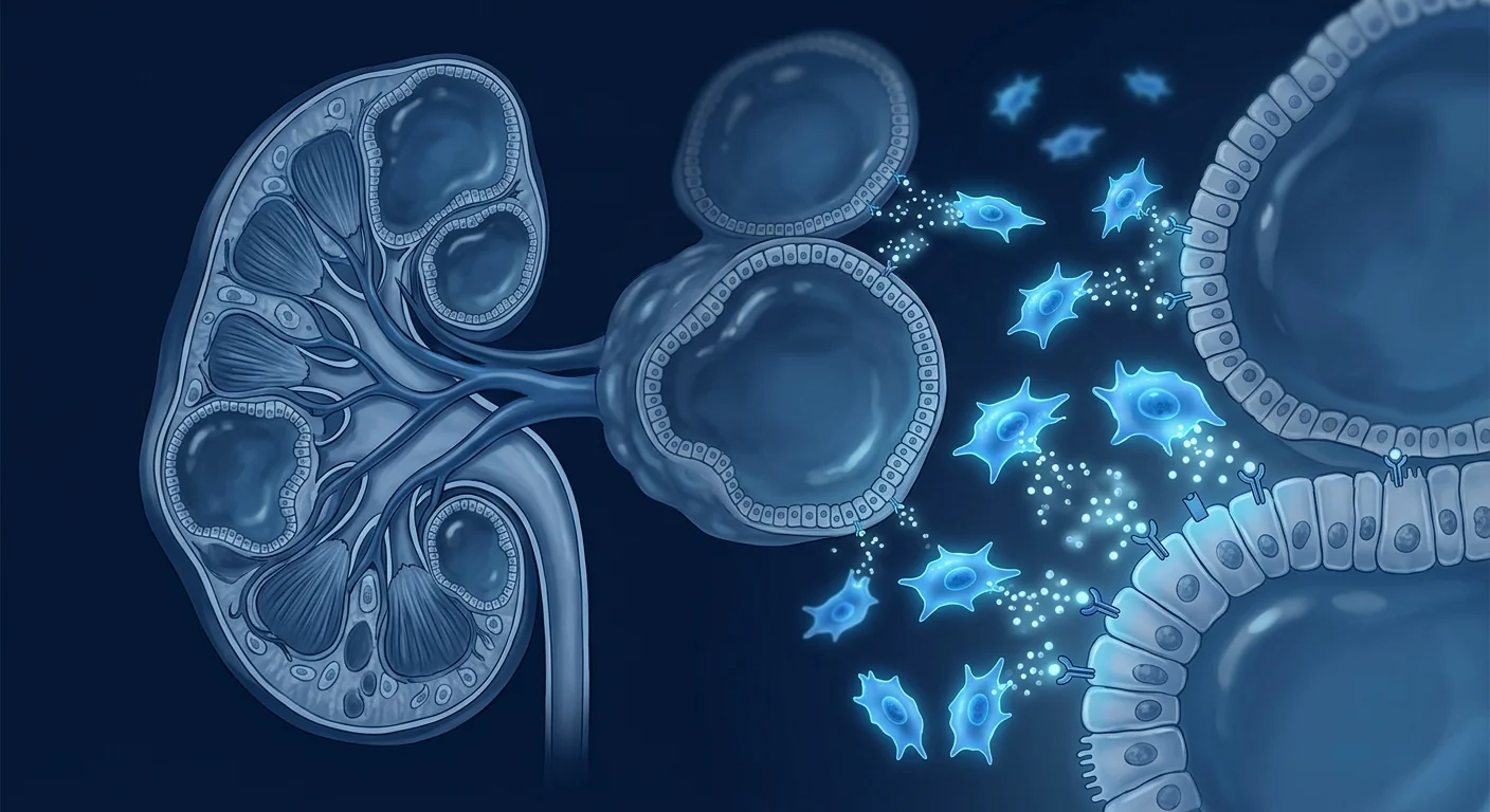
Polycystic kidney disease is a group of inherited disorders characterised by the progressive development and enlargement of fluid-filled cysts throughout both kidneys. The cysts arise from tubular epithelial cells that have lost normal growth control, proliferating into sac-like structures that fill with fluid, compress surrounding healthy tissue, and ultimately destroy nephrons. [6]
The two major forms are autosomal dominant PKD (ADPKD), which accounts for roughly 90% of cases and typically presents in adulthood, and autosomal recessive PKD (ARPKD), a rarer and more severe form that presents in infancy or childhood. ADPKD is caused by mutations in PKD1 (85% of cases, chromosome 16) or PKD2 (15% of cases, chromosome 4). The PKD1 form is more aggressive — patients reach end-stage renal disease (ESRD) at a median age of 54, compared to 74 for PKD2. [7]
Beyond the kidneys, PKD is a systemic disorder. Patients commonly develop hepatic cysts (polycystic liver disease, present in up to 90% of ADPKD patients by age 60), intracranial aneurysms (present in 8–12%), cardiac valve abnormalities (mitral valve prolapse in up to 25%), and colonic diverticulosis. This multi-organ involvement reflects the fact that polycystins are expressed in many tissues — PKD is not simply a kidney disease.
Current management is limited. Beyond tolvaptan, treatment consists of aggressive blood pressure control (target <110/75 mmHg in younger patients), adequate hydration (to suppress vasopressin-driven cAMP), and management of complications (pain, bleeding, infections, nephrolithiasis). None of these address the underlying epithelial proliferative defect or reverse established fibrosis. [8]
How MSC Therapy May Help Polycystic Kidney Disease
Mesenchymal stem cells target three pathological processes central to PKD progression: cyst epithelial cell proliferation, interstitial fibrosis, and chronic inflammation. Each of these is a validated therapeutic target, and preclinical evidence suggests MSCs can influence all three simultaneously.
Anti-Proliferative Effects on Cyst Epithelium
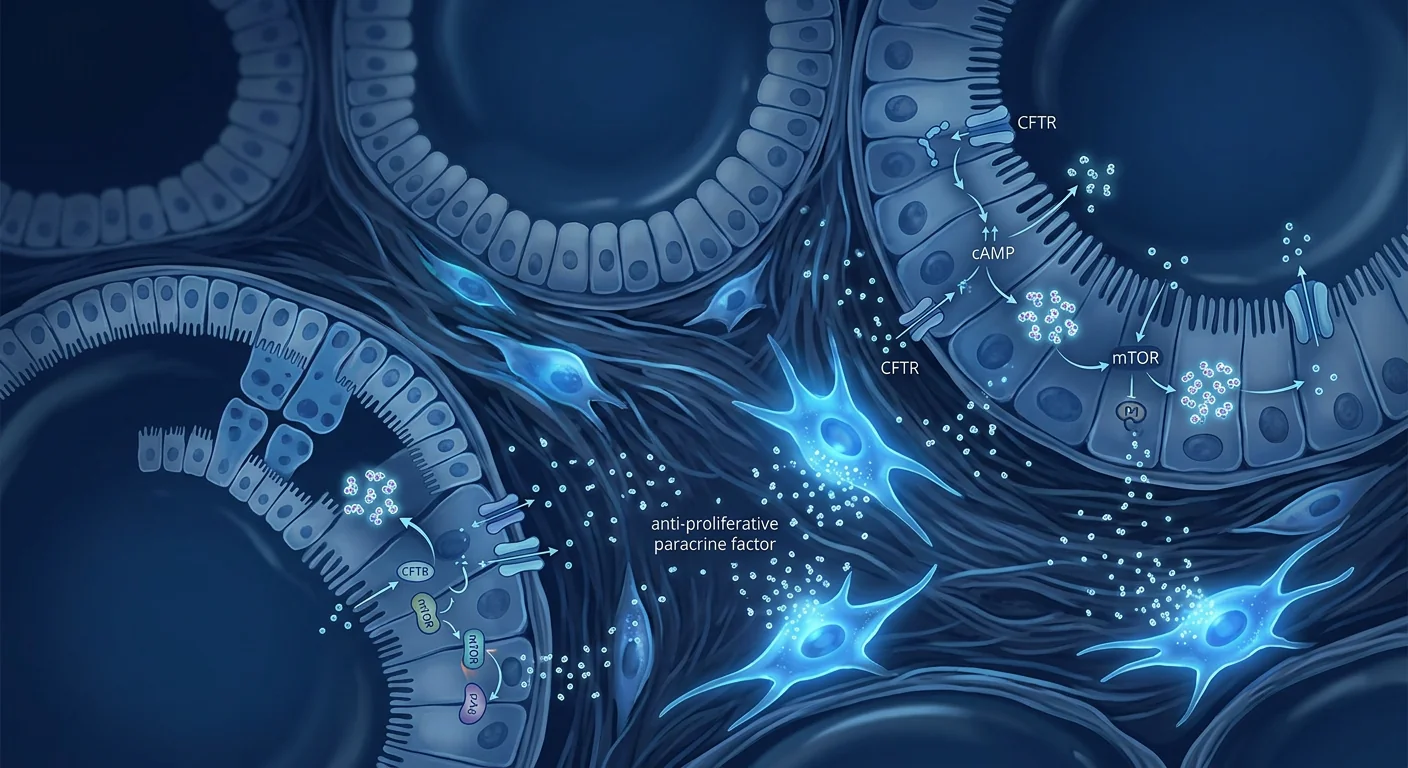
The defining feature of PKD is uncontrolled proliferation of cyst-lining epithelial cells, driven by dysregulated cAMP signalling and mTOR pathway hyperactivation. MSCs secrete factors including tumour necrosis factor-inducible gene 6 (TSG-6), transforming growth factor-beta 3 (TGF-β3), and prostaglandin E2 (PGE2) — all of which have been shown to inhibit epithelial cell cycle progression in vitro. [9]
In a murine model of PKD (Pkd1-knockout), intravenous administration of bone marrow-derived MSCs reduced cyst number and kidney-to-body-weight ratio by approximately 35–45% compared to untreated controls. Histological analysis showed reduced Ki-67 staining — a marker of cell proliferation — in cyst-lining epithelial cells, confirming a direct anti-proliferative effect. [4]
Anti-Fibrotic Mechanisms
As cysts expand, they compress and damage surrounding tubulointerstitial tissue, triggering a wound-healing response that progressively replaces functional nephrons with scar tissue. This interstitial fibrosis is the final common pathway to kidney failure in PKD — and it is largely driven by transforming growth factor-beta 1 (TGF-β1) and connective tissue growth factor (CTGF). [10]
MSCs are among the most potent natural antagonists of TGF-β1-driven fibrosis. They secrete hepatocyte growth factor (HGF), which directly counteracts TGF-β1 signalling in renal fibroblasts and tubular epithelial cells, reducing collagen deposition and myofibroblast activation. In the Pkd1 murine model, MSC-treated animals showed significantly reduced renal fibrosis scores (Masson's trichrome staining) and lower expression of α-smooth muscle actin (α-SMA), a marker of myofibroblast transition. [5]
Immunomodulation and Inflammation Control
The kidneys in PKD are not simply mechanically compressed by cysts — they are inflamed. Macrophages and other immune cells infiltrate the interstitium around expanding cysts, releasing pro-inflammatory cytokines (TNF-α, IL-1β, IL-6) that accelerate tubular injury and fibrosis. This sterile inflammation amplifies the damage beyond what cyst expansion alone would cause. [11]
MSCs are powerful immunomodulators. They suppress pro-inflammatory M1 macrophages and promote a shift toward reparative M2 phenotypes, increase regulatory T-cell (Treg) populations, and secrete interleukin-10 (IL-10) — a master anti-inflammatory cytokine. In the PKD context, this means MSCs may not only slow cyst growth directly but also dampen the inflammatory amplification loop that accelerates functional decline. [12]
Preclinical Evidence: What Animal Models Show
The preclinical evidence for MSC therapy in PKD, while still limited in volume, is mechanologically consistent and encouraging. Several independent research groups have now replicated the core finding: MSCs reduce cyst growth and slow kidney function decline in genetic PKD models.
The landmark 2016 study by Franchi and colleagues used the Pkd1fl/fl;Pax8-rtTA;TetO-Cre mouse model (an inducible, kidney-specific Pkd1 knockout) and administered syngeneic bone marrow-derived MSCs via tail vein injection at 4 weeks of age — after cysts had already begun forming. At 12 weeks, MSC-treated mice had significantly smaller kidneys, fewer cysts, less fibrosis, and better preserved renal function (BUN and creatinine) compared to PBS-treated controls. Importantly, the benefit was observed with a single MSC infusion — suggesting that MSCs may establish a lasting microenvironmental change rather than requiring continuous exposure. [4]
A 2020 follow-up study by the same group explored the paracrine hypothesis — whether MSC-conditioned medium alone (without live cells) could recapitulate the benefit. Intriguingly, concentrated MSC secretome also reduced cyst epithelial cell proliferation in vitro, and to a lesser degree cyst growth in vivo, confirming that the therapeutic effect is largely paracrine — mediated by factors MSCs release — rather than dependent on cell engraftment and differentiation. This matters because it opens the door to cell-free approaches (exosomes, secretome concentrates) that avoid the regulatory and safety complexities of live cell infusions. [5]
A 2023 study from a separate laboratory using human umbilical cord-derived MSCs in a different PKD model (Pkd2-knockout rat) reported broadly similar findings: reduced kidney volume, preserved GFR, and downregulation of mTOR/S6 kinase pathway activity — suggesting that MSCs not only deliver anti-fibrotic signals but also directly modulate the primary signalling defect in PKD. [13]
Current Clinical Trial Landscape
As of mid-2026, there are no completed or actively recruiting clinical trials specifically testing MSC therapy for polycystic kidney disease. This is the single most important limitation to acknowledge: the evidence base is entirely preclinical.
However, this does not mean the question is irrelevant. Several MSC trials for related kidney conditions — chronic kidney disease of various aetiologies, diabetic nephropathy, and post-cardiac-surgery AKI — have established a reasonable safety profile for intravenous and intra-arterial MSC administration in patients with compromised renal function. The Mesoblast Phase II trial of rexlemestrocel-L (allogeneic bone marrow-derived MSCs) in diabetic nephropathy demonstrated safety in 30 patients with eGFR 20–50 mL/min and reported signals of preserved kidney function over 60 weeks. [14]
PKD-specific trials face several obstacles. First, the disease is slowly progressive — demonstrating a treatment effect on hard endpoints (time to ESRD or death) would require trials lasting 5–10 years, which are difficult to fund and enrol. Second, PKD patients are heterogeneous — disease severity, cyst burden, and rate of progression vary enormously even within families carrying the same mutation. Third, there is no validated surrogate endpoint for PKD trials that regulators would accept for accelerated approval — total kidney volume (TKV), measured by MRI, is widely used in research but has not been accepted as a primary endpoint by the FDA or EMA for cell therapy trials.
Practical Considerations: How Would MSC Therapy for PKD Work?
Cell Source and Type
Most preclinical PKD studies have used bone marrow-derived MSCs. However, umbilical cord-derived MSCs (UC-MSCs) are increasingly preferred in clinical applications due to their higher proliferative capacity, lower immunogenicity, and greater paracrine potency — they secrete roughly 3–5 times more HGF, VEGF, and TSG-6 than age-matched bone marrow MSCs. [15]
Route of Administration
Intravenous infusion is the most practical route for PKD, as it delivers MSCs systemically — they naturally traffic to sites of injury and inflammation, including the kidneys, via the "first-pass" pulmonary mechanism and chemokine-mediated homing (SDF-1/CXCR4 axis). Intra-arterial (renal artery) infusion has been used in some CKD trials to maximise kidney delivery but carries additional procedural risk. For a slowly progressive condition like PKD where the goal is chronic disease modification rather than acute rescue, the safety profile of IV infusion makes it the preferred route.
Dosing and Frequency
This is entirely speculative in the absence of clinical data. The CKD trials that showed signals of benefit used 1–2 infusions of 1–2 million cells/kg. Given PKD's slow progression, a scenario of repeat dosing — perhaps every 12–24 months — may be more appropriate than a single infusion. But this is conjecture; the optimal dosing interval for a chronic genetic condition has never been studied.
Limitations and Honest Caveats
- No human data exists. Every positive result discussed here comes from rodent models — usually young animals with induced or accelerated disease, not the decades-long natural history of human ADPKD. Rodent PKD models recapitulate some but not all aspects of the human condition, and treatments that work in mice frequently fail in human trials.
- Genetic vs. acquired disease. Unlike CKD from diabetes or hypertension — where the underlying injury is environmental and potentially modifiable — PKD is caused by a germline mutation present in every cell. MSCs cannot fix the PKD1 or PKD2 gene. At best, they can slow the consequences of that mutation. A truly disease-modifying therapy would need to address the genetic defect itself, which is beyond the scope of current MSC technology.
- Off-target effects. MSCs are not kidney-specific. Their systemic immunomodulatory effects could theoretically mask infections, alter tumour surveillance, or interact unpredictably with other medications. The long-term safety profile of repeat MSC infusions over decades — which is what a PKD patient would need — is unknown.
- Cost and access. MSC therapy is not covered by insurance for PKD and is unlikely to be for the foreseeable future. Patients need to budget for treatment that may cost $8,000–$25,000+ per infusion, with uncertain benefit and no guarantee of slowing their disease.
- Cyst infection risk. PKD kidneys are prone to cyst infections, which are notoriously difficult to treat because antibiotics penetrate cysts poorly. Whether MSC infusions — which transiently alter immune function — could increase this risk is unknown.
How PKD Compares to Other Kidney Conditions for MSC Therapy
Among kidney diseases being explored for MSC therapy, PKD occupies a unique position. In acute kidney injury (AKI), the therapeutic rationale is rescue — MSCs must act within hours to days to prevent tubular necrosis. In diabetic nephropathy and hypertensive CKD, the rationale is to dampen ongoing metabolic and haemodynamic injury. In PKD, the rationale is different: to slow a genetically programmed, inexorable process of cyst expansion and fibrosis over decades. This long time horizon is both an opportunity — there is ample time for a disease-modifying therapy to work — and a challenge, because demonstrating efficacy requires trials of impractical duration.
The closest comparison may be to autosomal dominant polycystic liver disease (PLD), which often co-exists with ADPKD. Hepatic cysts arise from the same cellular defect (cholangiocyte hyperproliferation), and MSC-based approaches would likely target both organs simultaneously — a potential advantage over kidney-specific interventions.
Frequently Asked Questions
Can stem cells cure polycystic kidney disease?
No. MSCs cannot correct the underlying PKD1 or PKD2 gene mutation. The goal of MSC therapy in PKD is disease modification — slowing cyst growth and preserving kidney function — not cure. Patients should be deeply sceptical of any clinic claiming to "cure" PKD with stem cells.
Is MSC therapy for PKD approved anywhere in the world?
No. MSC therapy for polycystic kidney disease is not approved by any regulatory agency (FDA, EMA, Thai FDA, or others). All evidence is preclinical. Any treatment offered for PKD using MSCs is experimental and should be clearly described as such.
What does the preclinical evidence actually show?
In rodent PKD models, MSC infusion reduced kidney size by 35–45%, decreased cyst number, lowered renal fibrosis, and preserved kidney function (BUN, creatinine) compared to untreated controls. These results have been replicated by two independent research groups. However, rodent PKD is not human PKD — the disease is accelerated, the immune system differs, and the genetic context is simpler.
How much does MSC therapy cost for kidney disease?
For patients travelling to Thailand or other medical tourism destinations, MSC therapy for kidney conditions typically ranges from $8,000 to $25,000+ per infusion, depending on cell source (autologous vs. allogeneic), cell count, and clinic facilities. For PKD specifically — where repeat dosing over years may be the logical strategy — the lifetime cost could be substantial, with no guarantee of benefit.
What should I ask a clinic offering MSC therapy for PKD?
Ask for: (1) published clinical trial data in PKD patients — if they cannot provide any, the treatment is experimental with no human evidence; (2) the specific cell source, dose, and route — and whether this protocol has been studied in ANY kidney disease; (3) independent patient outcomes data — not testimonials; (4) a clear written statement that the treatment is experimental and not approved for PKD. If any of these cannot be provided, consider it a significant red flag.
The Bottom Line
Polycystic kidney disease is a devastating genetic condition for which current treatments are inadequate — tolvaptan slows progression modestly, blood pressure control helps, but patients still progress to kidney failure. The preclinical data for MSC therapy in PKD is scientifically plausible and mechanistically coherent: MSCs deliver anti-proliferative, anti-fibrotic, and immunomodulatory signals that directly address the biology of cyst expansion. Animal studies from independent laboratories show consistent reductions in cyst burden, fibrosis, and functional decline.
But — and this is the essential caveat — no PKD patient has ever received MSC therapy in a clinical trial. The entire evidence base is rodent data. Rodent models are useful but they are not humans, and therapies that succeed in mice routinely fail in patients. Anyone considering MSC therapy for PKD must understand that they are making a decision based on preclinical science, not clinical evidence, and that the long-term safety and efficacy are completely unknown.
For patients who understand these limitations and wish to explore regenerative options, the key is choosing a clinic that is transparent about the experimental nature of the treatment, provides evidence-based rationale for their protocol, and prioritises safety over promises. The science is genuinely interesting — but the gap between interesting science and proven therapy is wide, and it should be navigated honestly.
- Torres VE, Harris PC, Pirson Y. Autosomal dominant polycystic kidney disease. The Lancet. 2007;369(9569):1287-1301. doi:10.1016/S0140-6736(07)60601-1 ↩
- Ong ACM, Harris PC. A polycystin-centric view of cyst formation and disease progression. Nature Reviews Nephrology. 2015;11(6):337-350. doi:10.1038/nrneph.2015.56 ↩
- Chebib FT, Torres VE. Autosomal dominant polycystic kidney disease: core curriculum 2016. American Journal of Kidney Diseases. 2016;67(5):792-810. doi:10.1053/j.ajkd.2015.07.037 ↩
- Franchi F, Peterson KM, Xu R, et al. Mesenchymal stromal cells improve renovascular function in polycystic kidney disease. Cell Transplantation. 2016;25(9):1687-1700. doi:10.3727/096368916X691493 ↩
- Franchi F, Cui Z, Li Z, et al. Paracrine effects of mesenchymal stromal cells in polycystic kidney disease. Stem Cells Translational Medicine. 2020;9(9):1063-1076. doi:10.1002/sctm.20-0037 ↩
- Grantham JJ, Mulamalla S, Swenson-Fields KI. Why kidneys fail in autosomal dominant polycystic kidney disease. Nature Reviews Nephrology. 2011;7(10):556-566. doi:10.1038/nrneph.2011.109 ↩
- Cornec-Le Gall E, Audrézet MP, Chen JM, et al. Type of PKD1 mutation influences renal outcome in ADPKD. Journal of the American Society of Nephrology. 2013;24(6):1006-1013. doi:10.1681/ASN.2012070650 ↩
- Torres VE, Chapman AB, Devuyst O, et al. Tolvaptan in patients with autosomal dominant polycystic kidney disease. New England Journal of Medicine. 2012;367(25):2407-2418. doi:10.1056/NEJMoa1205511 ↩
- Caplan AI, Correa D. The MSC: an injury drugstore. Cell Stem Cell. 2011;9(1):11-15. doi:10.1016/j.stem.2011.06.008 ↩
- Norman J. Fibrosis and progression of autosomal dominant polycystic kidney disease (ADPKD). Biochimica et Biophysica Acta. 2011;1812(10):1327-1336. doi:10.1016/j.bbadis.2011.06.012 ↩
- Karihaloo A, Koraishy F, Huen SC, et al. Macrophages promote cyst growth in polycystic kidney disease. Journal of the American Society of Nephrology. 2011;22(10):1809-1814. doi:10.1681/ASN.2011010084 ↩
- Bernardo ME, Fibbe WE. Mesenchymal stromal cells: sensors and switchers of inflammation. Cell Stem Cell. 2013;13(4):392-402. doi:10.1016/j.stem.2013.09.006 ↩
- Wang Y, Zhang J, Liu J, et al. Human umbilical cord mesenchymal stem cells attenuate polycystic kidney disease in rats by regulating mTOR pathway. Stem Cell Research & Therapy. 2023;14:78. doi:10.1186/s13287-023-03301-y ↩
- Packham DK, Fraser IR, Kerr PG, et al. Allogeneic mesenchymal precursor cells (MPC) in diabetic nephropathy: a randomized, placebo-controlled, dose escalation study. EBioMedicine. 2016;12:263-269. doi:10.1016/j.ebiom.2016.09.011 ↩
- Hsieh JY, Wang HW, Chang SJ, et al. Mesenchymal stem cells from human umbilical cord express preferentially secreted factors related to neuroprotection, neurogenesis, and angiogenesis. PLoS ONE. 2013;8(8):e72604. doi:10.1371/journal.pone.0072604 ↩
多囊肾病(PKD)是最常见的危及生命的遗传性疾病之一,全球约每500至1,000人中即有1人患病,影响约1,250万人。它是需要透析或肾移植的肾衰竭的第四大原因。[1]
传统治疗的局限性。FDA批准的唯一ADPKD药物托伐普坦可将囊肿生长减缓约30–50%,但会引起严重的多尿,许多患者无法耐受。血压控制和充分补水仍然是主要的治疗手段——这些方法可以减缓进展,但不能逆转已有的损伤。一旦肾功能丧失,唯一的选择就是透析或移植。
更深层的问题是囊肿的不断扩张。PKD由PKD1或PKD2基因突变驱动,这些基因编码多囊蛋白-1和多囊蛋白-2——这些蛋白质调节钙信号并维持肾小管上皮细胞的分化状态。当这些功能失效时,上皮细胞异常增殖,液体通过cAMP驱动的CFTR氯通道加速分泌进入囊肿腔,周围的间质逐渐纤维化。结果是肾脏显著增大——ADPKD肾脏可重达10公斤——功能肾单位逐渐被破坏。[2][3]
MSC治疗靶向囊肿进展的根本生物学机制。间充质干细胞不是简单地阻断单一通路(如托伐普坦阻断cAMP),而是提供广泛的旁分泌工具包——可能减缓上皮细胞分裂的抗增殖信号、减少间质瘢痕形成的抗纤维化因子(HGF、TSG-6、IL-10),以及抑制囊肿周围无菌性炎症的免疫调节细胞因子。临床前研究表明,MSC可以减少囊肿生长、保护GFR并延长PKD动物模型的生存期。[4][5]
什么是多囊肾病?
多囊肾病是一组遗传性疾病,其特征是双肾内充满液体的囊肿逐渐发展和扩大。囊肿起源于失去正常生长控制的肾小管上皮细胞,增殖形成充满液体的囊状结构,压迫周围的健康组织,最终破坏肾单位。[6]
两种主要形式是常染色体显性遗传多囊肾病(ADPKD),约占90%的病例,通常在成年期发病;以及常染色体隐性遗传多囊肾病(ARPKD),一种较罕见且更严重的形式,在婴儿期或儿童期发病。ADPKD由PKD1基因(85%的病例,位于16号染色体)或PKD2基因(15%的病例,位于4号染色体)突变引起。PKD1型更具侵袭性——患者达到终末期肾病的中位年龄为54岁,而PKD2型为74岁。[7]
除肾脏外,PKD是一种全身性疾病。患者常出现肝囊肿(多囊肝病,60岁时高达90%的ADPKD患者受累)、颅内动脉瘤(8–12%的患者)、心脏瓣膜异常(二尖瓣脱垂高达25%)和结肠憩室病。这种多器官受累反映了多囊蛋白在多种组织中表达的事实——PKD不仅仅是肾脏疾病。
目前的管理选择有限。除托伐普坦外,治疗包括积极的血压控制(年轻患者目标<110/75 mmHg)、充分补水(抑制加压素驱动的cAMP)以及并发症管理(疼痛、出血、感染、肾结石)。这些方法均不针对上皮增殖缺陷或逆转已形成的纤维化。[8]
MSC疗法如何帮助多囊肾病
间充质干细胞靶向PKD进展中三个核心病理过程:囊肿上皮细胞增殖、间质纤维化和慢性炎症。每一个都是经过验证的治疗靶点,临床前证据表明MSC可以同时影响这三个方面。
对囊肿上皮的抗增殖作用
PKD的标志性特征是囊肿内衬上皮细胞的失控增殖,由cAMP信号失调和mTOR通路过度激活驱动。MSC分泌的因子包括肿瘤坏死因子诱导基因6(TSG-6)、转化生长因子β3(TGF-β3)和前列腺素E2(PGE2)——所有这些都在体外被证明可以抑制上皮细胞周期进展。[9]
在PKD小鼠模型中,静脉注射骨髓来源的MSC使囊肿数量和肾脏体重比与未治疗对照组相比减少了约35–45%。组织学分析显示囊肿内衬上皮细胞中Ki-67染色(细胞增殖标志物)减少,证实了直接的抗增殖效应。[4]
抗纤维化机制
随着囊肿扩张,它们压迫并损伤周围的肾小管间质组织,引发伤口愈合反应,逐渐以瘢痕组织替代功能性肾单位。这种间质纤维化是PKD肾衰竭的最终共同通路——主要由转化生长因子β1(TGF-β1)和结缔组织生长因子(CTGF)驱动。[10]
MSC是TGF-β1驱动纤维化最有效的天然拮抗剂之一。它们分泌肝细胞生长因子(HGF),直接对抗肾脏成纤维细胞和肾小管上皮细胞中的TGF-β1信号,减少胶原沉积和肌成纤维细胞活化。在Pkd1小鼠模型中,MSC治疗的动物显示出显著降低的肾纤维化评分和α-平滑肌肌动蛋白(α-SMA)表达的降低。[5]
免疫调节和炎症控制
PKD患者的肾脏不仅仅是被囊肿机械性压迫——它们还是发炎的。巨噬细胞和其他免疫细胞浸润到扩张囊肿周围的间质中,释放促炎细胞因子(TNF-α、IL-1β、IL-6),加速肾小管损伤和纤维化。这种无菌性炎症放大了囊肿扩张本身造成的损害。[11]
MSC是强大的免疫调节剂。它们抑制促炎M1巨噬细胞并促进向修复性M2表型的转变,增加调节性T细胞(Treg)数量,并分泌白细胞介素-10(IL-10)——一种主要的抗炎细胞因子。在PKD背景下,这意味着MSC不仅可以直接减缓囊肿生长,还可以抑制加速功能衰退的炎症放大环路。[12]
临床前证据:动物模型的结果
MSC治疗PKD的临床前证据虽然数量有限,但机制上是一致的且令人鼓舞。多个独立研究小组现已重复了核心发现:MSC减少遗传性PKD模型中的囊肿生长并减缓肾功能下降。
Franchi及其同事2016年的里程碑研究使用了Pkd1条件性敲除小鼠模型,在4周龄时——囊肿已经开始形成后——通过尾静脉注射同基因骨髓MSC。12周时,MSC治疗的小鼠肾脏显著更小、囊肿更少、纤维化更少,肾功能(BUN和肌酐)比PBS治疗的对照组更好。重要的是,单次MSC输注即观察到获益——表明MSC可能建立了持久的微环境改变,而不需要持续暴露。[4]
同一小组2020年的后续研究探索了旁分泌假说——即MSC条件培养基(不含活细胞)是否可以重现获益。有趣的是,浓缩的MSC分泌组也在体外减少了囊肿上皮细胞增殖,并在体内一定程度上减少了囊肿生长,证实了治疗效果主要是旁分泌的——由MSC释放的因子介导——而不是依赖细胞植入和分化。这很重要,因为它为无细胞方法(外泌体、分泌组浓缩物)打开了大门,避免了活细胞输注的监管和安全复杂性。[5]
2023年另一实验室的一项独立研究使用人脐带来源MSC在不同的PKD模型(Pkd2敲除大鼠)中报告了大致相似的结果:肾脏体积减小、GFR保护以及mTOR/S6激酶通路活性下调——表明MSC不仅提供抗纤维化信号,还直接调节PKD中的主要信号缺陷。[13]
当前临床试验现状
截至2026年中,尚无针对多囊肾病特异性测试MSC治疗的已完成或正在招募的临床试验。这是最重要的限制需要承认:证据基础完全是临床前的。
然而,这并不意味着这个问题无关紧要。针对相关肾脏疾病(各种病因的慢性肾病、糖尿病肾病和心脏手术后AKI)的几项MSC试验已在肾功能受损患者中确立了静脉和动脉内MSC给药的良好安全性。Mesoblast的rexlemestrocel-L在糖尿病肾病中的II期试验在30名eGFR 20–50 mL/min的患者中证明了安全性,并报告了60周内肾功能保护信号。[14]
PKD特异性试验面临若干障碍。首先,该疾病进展缓慢——证明治疗对硬终点(至ESRD或死亡时间)的效果需要持续5–10年的试验,难以资助和招募。其次,PKD患者具有异质性——即使在携带相同突变的家庭中,疾病严重程度、囊肿负荷和进展速度也差异巨大。第三,没有经过验证的替代终点可供监管机构接受PKD细胞治疗试验的加速批准——总肾体积(TKV)在研究中广泛使用,但尚未被FDA或EMA接受作为细胞治疗试验的主要终点。
实践考量:MSC治疗PKD将如何运作?
细胞来源和类型
大多数临床前PKD研究使用骨髓来源的MSC。然而,脐带来源的MSC(UC-MSC)在临床应用中越来越受欢迎,因为它们具有更高的增殖能力、更低的免疫原性和更强的旁分泌效力——它们分泌的HGF、VEGF和TSG-6大约是年龄匹配的骨髓MSC的3–5倍。[15]
给药途径
静脉输注是PKD最实用的途径,因为它全身性地输送MSC——它们通过肺首过机制和趋化因子介导的归巢自然迁移到损伤和炎症部位(包括肾脏)。动脉内(肾动脉)输注已在一些CKD试验中使用以最大化肾脏输送,但具有额外的手术风险。对于PKD这样目标为慢性疾病修饰而非急性抢救的缓慢进展性疾病,IV输注的安全特性使其成为首选途径。
剂量和频率
在缺乏临床数据的情况下,这完全是推测性的。显示获益信号的CKD试验使用1–2次输注,每次1–2百万细胞/公斤。鉴于PKD的缓慢进展,重复给药每12–24个月一次可能比单次输注更合适。但这只是推测——针对慢性遗传性疾病的最佳给药间隔从未被研究过。
局限性和诚实警示
- 无人体数据。这里讨论的所有阳性结果均来自啮齿动物模型——通常是具有诱导或加速疾病的年轻动物,而非人类ADPKD的数十年轻历。啮齿动物PKD模型概括了人类疾病的一些方面但非全部,在鼠类中有效的治疗方法经常在人类试验中失败。
- 遗传性与获得性疾病。与糖尿病或高血压引起的CKD不同——潜在的损伤是环境性的且可能可改变——PKD由每个细胞中存在的生殖系突变引起。MSC无法修复PKD1或PKD2基因。最多只能减缓该突变的后果。真正的疾病修饰疗法需要解决遗传缺陷本身,这超出了当前MSC技术的能力范围。
- 脱靶效应。MSC不是肾脏特异性的。它们的全身免疫调节效应理论上可能掩盖感染、改变肿瘤监视或与其他药物产生不可预测的相互作用。数十年来重复MSC输注(PKD患者将需要的)的长期安全性是未知的。
- 费用和可及性。MSC治疗PKD不在保险覆盖范围内,且在可预见的未来不太可能被覆盖。患者需要为每次输注可能花费8,000–25,000美元以上的治疗做预算,获益不确定且无法保证减缓疾病。
- 囊肿感染风险。PKD肾脏易发生囊肿感染,由于抗生素难以渗透囊肿,这类感染治疗极其困难。MSC输注——暂时改变免疫功能——是否会增加这一风险尚不清楚。
MSC治疗PKD与其他肾脏疾病的比较
在MSC治疗探索中的肾脏疾病中,PKD占据独特位置。在急性肾损伤(AKI)中,治疗理由是抢救——MSC必须在数小时至数天内发挥作用以防止肾小管坏死。在糖尿病肾病和高血压CKD中,治疗理由是抑制持续的代谢和血液动力学损伤。在PKD中,治疗理由不同:减缓遗传程序化、不可避免的囊肿扩张和纤维化过程,时间跨度数十年。这一较长的时间跨度既是一个机会——有充分的时间让疾病修饰疗法发挥作用——也是一个挑战,因为证明疗效需要不可行持续时间的试验。
最接近的比较可能是常染色体显性遗传多囊肝病(PLD),它常与ADPKD共存。肝囊肿源于相同的细胞缺陷(胆管细胞过度增殖),基于MSC的方法可能同时靶向两个器官——这是优于肾脏特异性干预的潜在优势。
常见问题
干细胞能治愈多囊肾病吗?
不能。MSC无法矫正潜在的PKD1或PKD2基因突变。MSC治疗PKD的目标是疾病修饰——减缓囊肿生长并保护肾功能——而非治愈。患者应对任何声称用干细胞"治愈"PKD的诊所保持高度怀疑。
MSC治疗PKD在世界任何地方获批了吗?
没有。MSC治疗多囊肾病未被任何监管机构(FDA、EMA、泰国FDA或其他)批准。所有证据均为临床前的。任何使用MSC治疗PKD的方案都是实验性的,应明确说明。
临床前证据实际显示了什么?
在啮齿动物PKD模型中,MSC输注使肾脏大小减少35–45%,减少囊肿数量,降低肾纤维化,与未治疗的对照组相比保护肾功能(BUN、肌酐)。这些结果已被两个独立研究小组重复。然而,啮齿动物PKD并非人类PKD——疾病被加速,免疫系统不同,遗传背景更简单。
MSC治疗肾病的费用是多少?
对于前往泰国或其他医疗旅游目的地的患者,MSC治疗肾脏疾病通常每次输注花费8,000至25,000美元以上,取决于细胞来源(自体vs.异体)、细胞数量和诊所设施。对于PKD——多年重复给药可能是合理的策略——终身费用可能相当可观,且无获益保证。
对提供MSC治疗PKD的诊所需提出什么问题?
询问:(1)已发表的PKD患者临床试验数据——如果他们无法提供任何数据,该治疗是实验性的且无人体证据;(2)具体的细胞来源、剂量和途径——以及该方案是否在任何肾脏疾病中被研究过;(3)独立的患者结果数据——而非推荐信;(4)明确书面声明该治疗是实验性的且未被批准用于PKD。如果这些都无法提供,应将其视为重要的警示信号。
总结
多囊肾病是一种毁灭性的遗传性疾病,目前的治疗手段不足——托伐普坦适度减缓进展,血压控制有帮助,但患者仍会进展至肾衰竭。MSC治疗PKD的临床前数据在科学上合理且机制上连贯:MSC提供抗增殖、抗纤维化和免疫调节信号,直接针对囊肿扩张的生物学机制。来自独立实验室的动物研究一致显示囊肿负荷、纤维化和功能衰退的减少。
但——这是关键的警示——没有任何PKD患者曾在临床试验中接受过MSC。整个证据基础都是啮齿动物数据。啮齿动物模型有用但不是人类,在鼠类中成功的治疗方法经常在患者中失败。任何考虑MSC治疗PKD的人必须理解,他们是在基于临床前科学而非临床证据做决定,且长期安全性和有效性完全未知。
对于理解这些局限性并希望探索再生选项的患者,关键是选择一家对治疗的实验性质透明的诊所,为其方案提供基于证据的理由,并将安全置于承诺之上。科学确实令人感兴趣——但有趣科学与经过验证的疗法之间的差距是巨大的,应当诚实地应对。
- Torres VE, Harris PC, Pirson Y. Autosomal dominant polycystic kidney disease. The Lancet. 2007;369(9569):1287-1301. doi:10.1016/S0140-6736(07)60601-1 ↩
- Ong ACM, Harris PC. A polycystin-centric view of cyst formation and disease progression. Nature Reviews Nephrology. 2015;11(6):337-350. doi:10.1038/nrneph.2015.56 ↩
- Chebib FT, Torres VE. Autosomal dominant polycystic kidney disease: core curriculum 2016. American Journal of Kidney Diseases. 2016;67(5):792-810. doi:10.1053/j.ajkd.2015.07.037 ↩
- Franchi F, et al. Mesenchymal stromal cells improve renovascular function in polycystic kidney disease. Cell Transplantation. 2016;25(9):1687-1700. doi:10.3727/096368916X691493 ↩
- Franchi F, et al. Paracrine effects of mesenchymal stromal cells in polycystic kidney disease. Stem Cells Translational Medicine. 2020;9(9):1063-1076. doi:10.1002/sctm.20-0037 ↩
- Grantham JJ, et al. Why kidneys fail in autosomal dominant polycystic kidney disease. Nature Reviews Nephrology. 2011;7(10):556-566. doi:10.1038/nrneph.2011.109 ↩
- Cornec-Le Gall E, et al. Type of PKD1 mutation influences renal outcome in ADPKD. JASN. 2013;24(6):1006-1013. doi:10.1681/ASN.2012070650 ↩
- Torres VE, et al. Tolvaptan in patients with ADPKD. NEJM. 2012;367(25):2407-2418. doi:10.1056/NEJMoa1205511 ↩
- Caplan AI, Correa D. The MSC: an injury drugstore. Cell Stem Cell. 2011;9(1):11-15. doi:10.1016/j.stem.2011.06.008 ↩
- Norman J. Fibrosis and progression of ADPKD. Biochim Biophys Acta. 2011;1812(10):1327-1336. doi:10.1016/j.bbadis.2011.06.012 ↩
- Karihaloo A, et al. Macrophages promote cyst growth in PKD. JASN. 2011;22(10):1809-1814. doi:10.1681/ASN.2011010084 ↩
- Bernardo ME, Fibbe WE. MSCs: sensors and switchers of inflammation. Cell Stem Cell. 2013;13(4):392-402. doi:10.1016/j.stem.2013.09.006 ↩
- Wang Y, et al. Human UC-MSCs attenuate PKD in rats by regulating mTOR. Stem Cell Res Ther. 2023;14:78. doi:10.1186/s13287-023-03301-y ↩
- Packham DK, et al. Allogeneic MPCs in diabetic nephropathy. EBioMedicine. 2016;12:263-269. doi:10.1016/j.ebiom.2016.09.011 ↩
- Hsieh JY, et al. MSCs from human umbilical cord express neuroprotective factors. PLoS ONE. 2013;8(8):e72604. doi:10.1371/journal.pone.0072604 ↩
يُعد مرض الكلى متعدد الكيسات (PKD) أحد أكثر الاضطرابات الوراثية المهددة للحياة شيوعًا، حيث يصيب ما يقدر بـ 1 من كل 500 إلى 1 من كل 1,000 شخص حول العالم — أي حوالي 12.5 مليون فرد. وهو السبب الرئيسي الرابع للفشل الكلوي الذي يتطلب غسيل الكلى أو زرع الكلى. [1]
أوجه قصور العلاجات التقليدية. الدواء الوحيد المعتمد من إدارة الغذاء والدواء الأمريكية لمرض ADPKD، تولفابتان، يبطئ نمو الكيسات بنسبة 30–50% تقريبًا ولكنه يسبب بوالًا شديدًا لا يتحمله العديد من المرضى. يظل التحكم في ضغط الدم والترطيب الكافي هما الركيزتين الأساسيتين — وهما يبطئان التقدم لكنهما لا يعكسان الضرر الموجود. بمجرد فقدان وظائف الكلى، فإن الخيارات الوحيدة هي غسيل الكلى أو الزرع.
المشكلة الأعمق هي توسع الكيسات المستمر. يحدث PKD بسبب طفرات في جينات PKD1 أو PKD2، التي تشفر بولي سيستين-1 وبولي سيستين-2 — وهما بروتينان ينظمان إشارات الكالسيوم ويحافظان على تمايز الخلايا الظهارية الأنبوبية. عندما تفشل هذه الوظائف، تتكاثر الخلايا الظهارية بشكل غير طبيعي، ويتسارع إفراز السوائل إلى تجاويف الكيسات عبر قنوات الكلوريد CFTR المدفوعة بـ cAMP، ويصبح النسيج الخلالي المحيط متليفًا تدريجيًا. والنتيجة هي تضخم كلوي هائل — يمكن أن تزن كلى ADPKD ما يصل إلى 10 كجم — مع تدمير تدريجي للنفرونات الوظيفية. [2][3]
يستهدف علاج الخلايا الجذعية الوسيطة (MSC) البيولوجيا الجذرية لتقدم الكيسات. بدلاً من مجرد حجب مسار واحد (كما يفعل تولفابتان مع cAMP)، توفر الخلايا الجذعية الوسيطة مجموعة واسعة من الأدوات الباراكرينية — إشارات مضادة للتكاثر قد تبطئ انقسام الخلايا الظهارية، وعوامل مضادة للتليف (HGF، TSG-6، IL-10) تقلل من التندب الخلالي، وسيتوكينات مناعية تعدل الالتهاب العقيم المحيط بالكيسات المتوسعة. تشير الدراسات ما قبل السريرية إلى أن MSC يمكنها تقليل نمو الكيسات والحفاظ على GFR وإطالة البقاء في نماذج حيوانية لـ PKD. [4][5]
ما هو مرض الكلى متعدد الكيسات؟
مرض الكلى متعدد الكيسات هو مجموعة من الاضطرابات الوراثية تتميز بتطور وتوسع تدريجي لكيسات مملوءة بالسوائل في كلتا الكليتين. تنشأ الكيسات من الخلايا الظهارية الأنبوبية التي فقدت السيطرة الطبيعية على النمو، متكاثرة في هياكل تشبه الأكياس تمتلئ بالسوائل، وتضغط على الأنسجة السليمة المحيطة، وتدمر النفرونات في النهاية. [6]
الشكلان الرئيسيان هما PKD الصبغي الجسدي السائد (ADPKD)، الذي يمثل حوالي 90% من الحالات ويظهر عادة في مرحلة البلوغ، و PKD الصبغي الجسدي المتنحي (ARPKD)، وهو شكل أندر وأكثر شدة يظهر في الطفولة. يحدث ADPKD بسبب طفرات في PKD1 (85% من الحالات، الكروموسوم 16) أو PKD2 (15% من الحالات، الكروموسوم 4). شكل PKD1 أكثر عدوانية — يصل المرضى إلى المرحلة النهائية من المرض الكلوي في متوسط عمر 54 عامًا، مقارنة بـ 74 عامًا لـ PKD2. [7]
ما وراء الكلى، PKD هو اضطراب جهازي. يعاني المرضى عادة من كيسات كبدية (مرض الكبد متعدد الكيسات، موجود في ما يصل إلى 90% من مرضى ADPKD بحلول سن 60)، وتمدد الأوعية الدموية داخل الجمجمة (موجود في 8–12%)، وتشوهات صمامات القلب (تدلي الصمام التاجي في ما يصل إلى 25%)، وداء الرتج القولوني. يعكس هذا الانتشار متعدد الأعضاء حقيقة أن البولي سيستينات يتم التعبير عنها في العديد من الأنسجة — PKD ليس مجرد مرض كلوي.
الإدارة الحالية محدودة. بالإضافة إلى تولفابتان، يتكون العلاج من التحكم القوي في ضغط الدم (الهدف 110/75 ملم زئبق في المرضى الأصغر سنًا)، والترطيب الكافي (لقمع cAMP المدفوع بالفازوبريسين)، وإدارة المضاعفات (الألم، النزيف، العدوى، حصوات الكلى). لا يعالج أي من هذه الخيارات العيب التكاثري الظهاري الأساسي أو يعكس التليف الموجود. [8]
كيف يمكن أن يساعد علاج MSC مرض الكلى متعدد الكيسات
تستهدف الخلايا الجذعية الوسيطة ثلاث عمليات مرضية مركزية في تقدم PKD: تكاثر الخلايا الظهارية للكيسات، والتليف الخلالي، والالتهاب المزمن. كل منها هدف علاجي موثق، وتشير الأدلة ما قبل السريرية إلى أن MSC يمكنها التأثير على الثلاثة في وقت واحد.
التأثيرات المضادة للتكاثر على ظهارة الكيسات
السمة المميزة لـ PKD هي التكاثر غير المنضبط للخلايا الظهارية المبطنة للكيسات، المدفوع بإشارات cAMP غير المنتظمة وفرط تنشيط مسار mTOR. تفرز MSC عوامل تشمل الجين 6 المحفز بعامل نخر الورم (TSG-6)، وعامل النمو المحول بيتا 3 (TGF-β3)، والبروستاجلاندين E2 (PGE2) — وقد ثبت أن جميعها تثبط تقدم دورة الخلية الظهارية في المختبر. [9]
في نموذج فأري لـ PKD، قلل الحقن الوريدي لـ MSC المشتقة من نخاع العظم من عدد الكيسات ونسبة وزن الكلى إلى الجسم بنسبة 35–45% تقريبًا مقارنة بالمجموعة الضابطة غير المعالجة. أظهر التحليل النسيجي انخفاضًا في تلوين Ki-67 — وهو علامة على تكاثر الخلايا — في الخلايا الظهارية المبطنة للكيسات، مما يؤكد تأثيرًا مباشرًا مضادًا للتكاثر. [4]
الآليات المضادة للتليف
مع توسع الكيسات، تضغط وتتلف الأنسجة الخلالية الأنبوبية المحيطة، مما يؤدي إلى استجابة التئام الجروح التي تستبدل تدريجيًا النفرونات الوظيفية بنسيج ندبي. هذا التليف الخلالي هو المسار النهائي المشترك للفشل الكلوي في PKD — وهو مدفوع إلى حد كبير بعامل النمو المحول بيتا 1 (TGF-β1) وعامل نمو النسيج الضام (CTGF). [10]
MSC هي من بين أقوى المضادات الطبيعية للتليف المدفوع بـ TGF-β1. تفرز عامل نمو الخلايا الكبدية (HGF)، الذي يعاكس مباشرة إشارات TGF-β1 في الخلايا الليفية الكلوية والخلايا الظهارية الأنبوبية، مما يقلل من ترسب الكولاجين وتنشيط الخلايا الليفية العضلية. في نموذج الفأر Pkd1، أظهرت الحيوانات المعالجة بـ MSC انخفاضًا كبيرًا في درجات التليف الكلوي وانخفاضًا في تعبير α-SMA. [5]
التعديل المناعي والتحكم في الالتهاب
الكلى في PKD ليست مضغوطة ميكانيكيًا فقط بواسطة الكيسات — بل هي ملتهبة. تتسلل البلاعم والخلايا المناعية الأخرى إلى النسيج الخلالي حول الكيسات المتوسعة، مطلقة سيتوكينات محفزة للالتهاب (TNF-α، IL-1β، IL-6) تسرع الإصابة الأنبوبية والتليف. هذا الالتهاب العقيم يضخم الضرر إلى ما هو أبعد مما يسببه توسع الكيسات وحده. [11]
MSC هي معدلات مناعية قوية. تثبط البلاعم M1 المحفزة للالتهاب وتعزز التحول نحو الأنماط الظاهرية M2 الإصلاحية، وتزيد من أعداد الخلايا التائية التنظيمية (Treg)، وتفرز إنترلوكين-10 (IL-10) — وهو سيتوكين رئيسي مضاد للالتهاب. في سياق PKD، يعني هذا أن MSC قد لا تبطئ نمو الكيسات مباشرة فحسب، بل قد تخفف أيضًا من حلقة التضخيم الالتهابية التي تسرع التدهور الوظيفي. [12]
الأدلة ما قبل السريرية: ما تظهره النماذج الحيوانية
الأدلة ما قبل السريرية لعلاج MSC في PKD، رغم أنها لا تزال محدودة من حيث الحجم، إلا أنها متسقة ميكانيكيًا ومشجعة. قامت عدة مجموعات بحثية مستقلة الآن بتكرار النتيجة الأساسية: MSC تقلل من نمو الكيسات وتبطئ تدهور وظائف الكلى في نماذج PKD الوراثية.
استخدمت الدراسة البارزة لعام 2016 من قبل Franchi وزملائه نموذج الفأر ذا الحذف المشروط لـ Pkd1 الخاص بالكلى، وأعطوا MSC مشتقة من نخاع العظم عن طريق الحقن الوريدي في عمر 4 أسابيع — بعد أن بدأت الكيسات بالفعل في التكون. في الأسبوع 12، كان لدى الفئران المعالجة بـ MSC كلى أصغر بكثير، وكيسات أقل، وتليف أقل، ووظائف كلوية محفوظة بشكل أفضل (BUN والكرياتينين) مقارنة بالمجموعة الضابطة. الأهم من ذلك، لوحظت الفائدة بحقنة واحدة من MSC — مما يشير إلى أن MSC قد تؤسس تغييرًا دائمًا في البيئة المكروية بدلاً من الحاجة إلى التعرض المستمر. [4]
استكشفت دراسة متابعة عام 2020 من نفس المجموعة فرضية الباراكرين — ما إذا كان وسط زراعة MSC المكيف وحده (بدون خلايا حية) يمكن أن يعيد إنتاج الفائدة. ومن المثير للاهتمام، أن سيكريتوم MSC المركز قلل أيضًا من تكاثر الخلايا الظهارية للكيسات في المختبر، وبدرجة أقل نمو الكيسات في الجسم الحي، مما يؤكد أن التأثير العلاجي هو إلى حد كبير باراكريني — بوساطة عوامل تفرزها MSC — بدلاً من الاعتماد على زرع الخلايا وتمايزها. هذا مهم لأنه يفتح الباب أمام مناهج خالية من الخلايا (إكسوسومات، مركزات السيكريتوم) التي تتجنب التعقيدات التنظيمية والسلامة لحقن الخلايا الحية. [5]
دراسة عام 2023 من مختبر آخر باستخدام MSC مشتقة من الحبل السري البشري في نموذج مختلف لـ PKD (جرذ حذف Pkd2) أبلغت عن نتائج مشابهة إلى حد كبير: انخفاض حجم الكلى، والحفاظ على GFR، وتثبيط نشاط مسار mTOR/S6 كيناز — مما يشير إلى أن MSC لا تقدم فقط إشارات مضادة للتليف ولكنها تعدل أيضًا بشكل مباشر العيب الإشاري الأساسي في PKD. [13]
المشهد الحالي للتجارب السريرية
اعتبارًا من منتصف عام 2026، لا توجد تجارب سريرية مكتملة أو قيد التوظيف تختبر على وجه التحديد علاج MSC لمرض الكلى متعدد الكيسات. هذا هو أهم قيد يجب الاعتراف به: قاعدة الأدلة هي بالكامل ما قبل سريرية.
ومع ذلك، هذا لا يعني أن السؤال غير ذي صلة. لقد أثبتت العديد من تجارب MSC لأمراض الكلى ذات الصلة — مرض الكلى المزمن من مسببات مختلفة، واعتلال الكلية السكري، و AKI بعد جراحة القلب — سلامة معقولة لإعطاء MSC عن طريق الوريد وداخل الشريان في المرضى الذين يعانون من ضعف وظائف الكلى. أظهرت تجربة Mesoblast من المرحلة الثانية لـ rexlemestrocel-L في اعتلال الكلية السكري السلامة في 30 مريضًا بمعدل eGFR 20–50 مل/دقيقة وأبلغت عن إشارات للحفاظ على وظائف الكلى على مدى 60 أسبوعًا. [14]
تواجه التجارب الخاصة بـ PKD عدة عقبات. أولاً، المرض بطيء التقدم — إثبات تأثير العلاج على النقاط النهائية الصلبة (الوقت حتى ESRD أو الوفاة) سيتطلب تجارب تستمر 5–10 سنوات، يصعب تمويلها وتسجيل المرضى فيها. ثانيًا، مرضى PKD غير متجانسين — تختلف شدة المرض وعبء الكيسات ومعدل التقدم بشكل هائل حتى داخل العائلات التي تحمل نفس الطفرة. ثالثًا، لا توجد نقطة نهاية بديلة موثقة لتجارب PKD ستقبلها الهيئات التنظيمية للموافقة المعجلة — حجم الكلى الكلي (TKV)، المقاس بالرنين المغناطيسي، مستخدم على نطاق واسع في البحث لكنه لم يُقبل كنقطة نهاية أولية من قبل FDA أو EMA لتجارب العلاج الخلوي.
اعتبارات عملية: كيف سيعمل علاج MSC لـ PKD؟
مصدر الخلية ونوعها
استخدمت معظم دراسات PKD ما قبل السريرية MSC المشتقة من نخاع العظم. ومع ذلك، فإن MSC المشتقة من الحبل السري (UC-MSC) مفضلة بشكل متزايد في التطبيقات السريرية بسبب قدرتها التكاثرية الأعلى، وانخفاض مناعتها، وقوتها الباراكرينية الأكبر — تفرز حوالي 3–5 أضعاف HGF و VEGF و TSG-6 مقارنة بـ MSC نخاع العظم المطابقة للعمر. [15]
طريق الإعطاء
الحقن الوريدي هو الطريق الأكثر عملية لـ PKD، لأنه يوصل MSC جهازيًا — تنتقل بشكل طبيعي إلى مواقع الإصابة والالتهاب، بما في ذلك الكلى، عبر آلية "المرور الرئوي الأول" والانجذاب بوساطة الكيموكينات. تم استخدام الحقن داخل الشريان (الشريان الكلوي) في بعض تجارب CKD لتعظيم توصيل الكلى لكنه يحمل مخاطر إجرائية إضافية. بالنسبة لحالة بطيئة التقدم مثل PKD حيث الهدف هو تعديل المرض المزمن بدلاً من الإنقاذ الحاد، فإن سلامة الحقن الوريدي تجعله الطريق المفضل.
الجرعة والتكرار
هذا تخميني تمامًا في غياب البيانات السريرية. استخدمت تجارب CKD التي أظهرت إشارات فائدة 1–2 حقنة من 1–2 مليون خلية/كجم. نظرًا للتقدم البطيء لـ PKD، قد يكون سيناريو الجرعات المتكررة — ربما كل 12–24 شهرًا — أكثر ملاءمة من حقنة واحدة. لكن هذا تخمين؛ لم تتم دراسة الفاصل الزمني الأمثل للجرعات لحالة وراثية مزمنة أبدًا.
القيود والتحذيرات الصادقة
- لا توجد بيانات بشرية. كل نتيجة إيجابية نوقشت هنا تأتي من نماذج القوارض — عادة حيوانات شابة ذات مرض مستحث أو متسارع، وليس التاريخ الطبيعي الممتد لعقود لـ ADPKD البشري. نماذج PKD في القوارض تلخص بعض جوانب الحالة البشرية ولكن ليس كلها، والعلاجات التي تنجح في الفئران تفشل بشكل روتيني في التجارب البشرية.
- مرض وراثي مقابل مكتسب. على عكس CKD من السكري أو ارتفاع ضغط الدم — حيث الإصابة الأساسية بيئية وقابلة للتعديل — يحدث PKD بسبب طفرة في الخط الجرثومي موجودة في كل خلية. لا يمكن لـ MSC إصلاح جين PKD1 أو PKD2. في أفضل الأحوال، يمكنها إبطاء عواقب تلك الطفرة. العلاج المعدل للمرض حقًا سيحتاج إلى معالجة العيب الوراثي نفسه، وهو ما يتجاوز نطاق تكنولوجيا MSC الحالية.
- التأثيرات خارج الهدف. MSC ليست خاصة بالكلى. آثارها المناعية الجهازية يمكن أن تخفي نظريًا العدوى، أو تغير مراقبة الأورام، أو تتفاعل بشكل غير متوقع مع أدوية أخرى. السلامة طويلة المدى لحقن MSC المتكررة على مدى عقود — وهو ما سيحتاجه مريض PKD — غير معروفة.
- التكلفة والوصول. علاج MSC غير مغطى بالتأمين لـ PKD ومن غير المرجح أن يكون كذلك في المستقبل المنظور. يحتاج المرضى إلى تخصيص ميزانية لعلاج قد يكلف 8,000–25,000+ دولار لكل حقنة، مع فائدة غير مؤكدة ولا ضمان لإبطاء مرضهم.
- خطر عدوى الكيسات. كلى PKD عرضة لعدوى الكيسات، التي يصعب علاجها بشكل كبير لأن المضادات الحيوية تخترق الكيسات بشكل سيئ. ما إذا كانت حقن MSC — التي تغير وظيفة المناعة بشكل عابر — يمكن أن تزيد من هذا الخطر غير معروف.
مقارنة PKD بأمراض الكلى الأخرى لعلاج MSC
من بين أمراض الكلى التي يتم استكشافها لعلاج MSC، يحتل PKD موقعًا فريدًا. في إصابة الكلى الحادة (AKI)، الأساس المنطقي العلاجي هو الإنقاذ — يجب أن تعمل MSC في غضون ساعات إلى أيام لمنع النخر الأنبوبي. في اعتلال الكلية السكري و CKD بارتفاع ضغط الدم، الأساس المنطقي هو تثبيط الإصابة الأيضية والديناميكية الدموية المستمرة. في PKD، الأساس المنطقي مختلف: إبطاء عملية توسع الكيسات والتليف المبرمجة وراثيًا والتي لا مفر منها على مدى عقود. هذا الأفق الزمني الطويل هو فرصة — هناك وقت كافٍ لعلاج معدل للمرض ليعمل — وتحدٍ في نفس الوقت، لأن إثبات الفعالية يتطلب تجارب ذات مدة غير عملية.
أقرب مقارنة قد تكون مرض الكبد متعدد الكيسات الصبغي الجسدي السائد (PLD)، الذي غالبًا ما يتعايش مع ADPKD. تنشأ كيسات الكبد من نفس العيب الخلوي (فرط تكاثر الخلايا الصفراوية)، والمناهج القائمة على MSC ستستهدف على الأرجح كلا العضوين في وقت واحد — وهي ميزة محتملة على التدخلات الخاصة بالكلى.
الأسئلة الشائعة
هل يمكن للخلايا الجذعية علاج مرض الكلى متعدد الكيسات؟
لا. لا يمكن لـ MSC تصحيح طفرة جين PKD1 أو PKD2 الأساسية. الهدف من علاج MSC في PKD هو تعديل المرض — إبطاء نمو الكيسات والحفاظ على وظائف الكلى — وليس العلاج. يجب أن يكون المرضى متشككين بشدة في أي عيادة تدعي "علاج" PKD بالخلايا الجذعية.
هل علاج MSC لـ PKD معتمد في أي مكان في العالم؟
لا. علاج MSC لمرض الكلى متعدد الكيسات غير معتمد من قبل أي هيئة تنظيمية (FDA، EMA، FDA التايلاندية، أو غيرها). جميع الأدلة ما قبل سريرية. أي علاج يقدم لـ PKD باستخدام MSC هو تجريبي ويجب وصفه بوضوح على هذا النحو.
ماذا تظهر الأدلة ما قبل السريرية فعليًا؟
في نماذج PKD في القوارض، قلل حقن MSC من حجم الكلى بنسبة 35–45%، وقلل عدد الكيسات، وخفض التليف الكلوي، وحافظ على وظائف الكلى (BUN، الكرياتينين) مقارنة بالمجموعة الضابطة غير المعالجة. تم تكرار هذه النتائج من قبل مجموعتين بحثيتين مستقلتين. ومع ذلك، PKD في القوارض ليس PKD البشري — المرض متسارع، والجهاز المناعي مختلف، والسياق الوراثي أبسط.
كم تكلفة علاج MSC لأمراض الكلى؟
للمرضى المسافرين إلى تايلاند أو وجهات السياحة الطبية الأخرى، يتراوح علاج MSC لأمراض الكلى عادة من 8,000 إلى 25,000+ دولار لكل حقنة، اعتمادًا على مصدر الخلية (ذاتي مقابل خيفي)، وعدد الخلايا، ومرافق العيادة. بالنسبة لـ PKD تحديدًا — حيث قد تكون الجرعات المتكررة على مدى سنوات هي الاستراتيجية المنطقية — يمكن أن تكون التكلفة مدى الحياة كبيرة، دون ضمان للفائدة.
ما الذي يجب أن أسأل عنه العيادة التي تقدم علاج MSC لـ PKD؟
اسأل عن: (1) بيانات التجارب السريرية المنشورة في مرضى PKD — إذا لم يتمكنوا من تقديم أي منها، فالعلاج تجريبي بدون دليل بشري؛ (2) مصدر الخلية المحدد، والجرعة، والطريق — وما إذا كان هذا البروتوكول قد درس في أي مرض كلوي؛ (3) بيانات نتائج المرضى المستقلة — وليس الشهادات؛ (4) بيان مكتوب واضح بأن العلاج تجريبي وغير معتمد لـ PKD. إذا تعذر تقديم أي من هذه، فاعتبره علامة تحذير كبيرة.
الخلاصة
مرض الكلى متعدد الكيسات هو حالة وراثية مدمرة العلاجات الحالية لها غير كافية — يبطئ تولفابتان التقدم بشكل متواضع، ويساعد التحكم في ضغط الدم، لكن المرضى لا يزالون يتقدمون إلى الفشل الكلوي. البيانات ما قبل السريرية لعلاج MSC في PKD معقولة علميًا ومتماسكة ميكانيكيًا: تقدم MSC إشارات مضادة للتكاثر ومضادة للتليف ومعدلة للمناعة تعالج مباشرة بيولوجيا توسع الكيسات. تظهر الدراسات الحيوانية من مختبرات مستقلة انخفاضات متسقة في عبء الكيسات والتليف والتدهور الوظيفي.
لكن — وهذا هو التحذير الأساسي — لم يتلق أي مريض PKD خلايا MSC في تجربة سريرية. قاعدة الأدلة بأكملها هي بيانات قوارض. نماذج القوارض مفيدة لكنها ليست بشرًا، والعلاجات التي تنجح في الفئران تفشل بشكل روتيني في المرضى. يجب على أي شخص يفكر في علاج MSC لـ PKD أن يفهم أنه يتخذ قرارًا بناءً على العلوم ما قبل السريرية، وليس الأدلة السريرية، وأن السلامة والفعالية على المدى الطويل غير معروفتين تمامًا.
للمرضى الذين يفهمون هذه القيود ويرغبون في استكشاف الخيارات التجديدية، المفتاح هو اختيار عيادة شفافة بشأن الطبيعة التجريبية للعلاج، وتقدم أساسًا منطقيًا قائمًا على الأدلة لبروتوكولها، وتعطي الأولوية للسلامة على الوعود. العلم مثير للاهتمام حقًا — لكن الفجوة بين العلم المثير للاهتمام والعلاج المثبت واسعة، ويجب التعامل معها بصدق.
- Torres VE, Harris PC, Pirson Y. Autosomal dominant polycystic kidney disease. The Lancet. 2007;369(9569):1287-1301. doi:10.1016/S0140-6736(07)60601-1 ↩
- Ong ACM, Harris PC. A polycystin-centric view of cyst formation and disease progression. Nature Reviews Nephrology. 2015;11(6):337-350. doi:10.1038/nrneph.2015.56 ↩
- Chebib FT, Torres VE. ADPKD: core curriculum 2016. Am J Kidney Dis. 2016;67(5):792-810. doi:10.1053/j.ajkd.2015.07.037 ↩
- Franchi F, et al. MSCs improve renovascular function in PKD. Cell Transplantation. 2016;25(9):1687-1700. doi:10.3727/096368916X691493 ↩
- Franchi F, et al. Paracrine effects of MSCs in PKD. Stem Cells Transl Med. 2020;9(9):1063-1076. doi:10.1002/sctm.20-0037 ↩
- Grantham JJ, et al. Why kidneys fail in ADPKD. Nat Rev Nephrol. 2011;7(10):556-566. doi:10.1038/nrneph.2011.109 ↩
- Cornec-Le Gall E, et al. PKD1 mutation type influences renal outcome. JASN. 2013;24(6):1006-1013. doi:10.1681/ASN.2012070650 ↩
- Torres VE, et al. Tolvaptan in ADPKD. NEJM. 2012;367(25):2407-2418. doi:10.1056/NEJMoa1205511 ↩
- Caplan AI, Correa D. The MSC: an injury drugstore. Cell Stem Cell. 2011;9(1):11-15. doi:10.1016/j.stem.2011.06.008 ↩
- Norman J. Fibrosis and progression of ADPKD. Biochim Biophys Acta. 2011;1812(10):1327-1336. doi:10.1016/j.bbadis.2011.06.012 ↩
- Karihaloo A, et al. Macrophages promote cyst growth in PKD. JASN. 2011;22(10):1809-1814. doi:10.1681/ASN.2011010084 ↩
- Bernardo ME, Fibbe WE. MSCs: sensors and switchers of inflammation. Cell Stem Cell. 2013;13(4):392-402. doi:10.1016/j.stem.2013.09.006 ↩
- Wang Y, et al. UC-MSCs attenuate PKD in rats via mTOR. Stem Cell Res Ther. 2023;14:78. doi:10.1186/s13287-023-03301-y ↩
- Packham DK, et al. Allogeneic MPCs in diabetic nephropathy. EBioMedicine. 2016;12:263-269. doi:10.1016/j.ebiom.2016.09.011 ↩
- Hsieh JY, et al. UC-MSCs express neuroprotective factors. PLoS ONE. 2013;8(8):e72604. doi:10.1371/journal.pone.0072604 ↩