Acute kidney injury (AKI) affects approximately 13.3 million people worldwide each year and contributes to roughly 1.7 million deaths annually — making it one of the most common yet under-recognized organ failures in hospitalized patients. [1] In intensive care units, AKI complicates up to 57% of admissions and independently doubles the risk of in-hospital mortality, even after adjusting for severity of illness.
Where conventional treatment falls short. The current standard of care — fluid resuscitation, hemodynamic optimization, avoidance of nephrotoxins, and renal replacement therapy (dialysis) — is purely supportive. None of these interventions halt the underlying tubular injury cascade once it has begun. Dialysis buys time but does not accelerate renal recovery; patients who survive severe AKI face a 9-fold increased risk of progressing to chronic kidney disease (CKD) and a 3-fold increase in long-term cardiovascular mortality. [2]
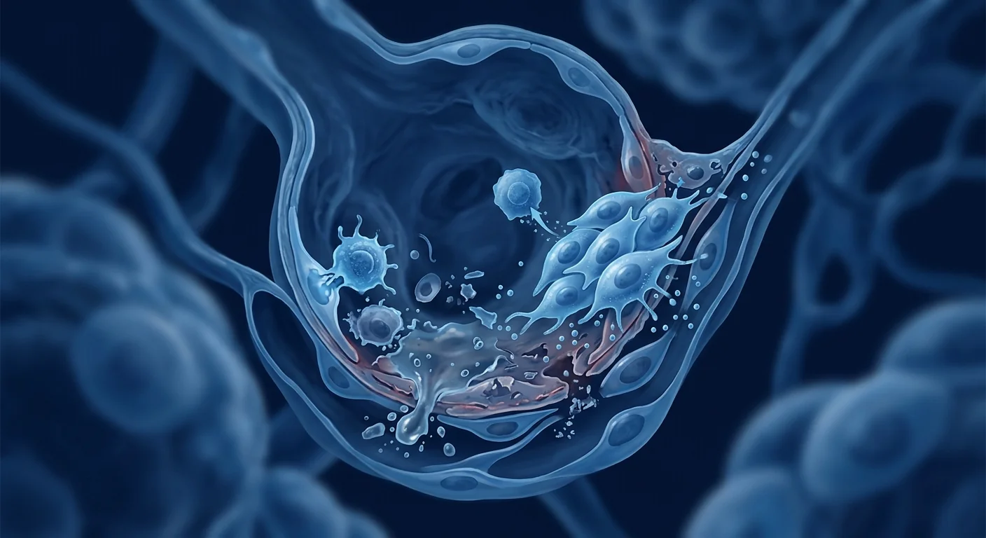
The deeper problem is cellular. AKI is driven by a self-amplifying cycle: ischemia or nephrotoxin exposure triggers tubular epithelial cell apoptosis and necrosis, which releases damage-associated molecular patterns (DAMPs) that recruit neutrophils and macrophages, whose oxidative burst and pro-inflammatory cytokines (TNF-α, IL-1β, IL-6) destroy still-viable neighboring tubules. [3] Mitochondrial fragmentation within proximal tubular cells depletes ATP, collapsing ion gradients and triggering further cell death. The kidney's intrinsic regenerative capacity — resident progenitor cells in the S3 segment of the proximal tubule — is overwhelmed by the sheer magnitude of injury.
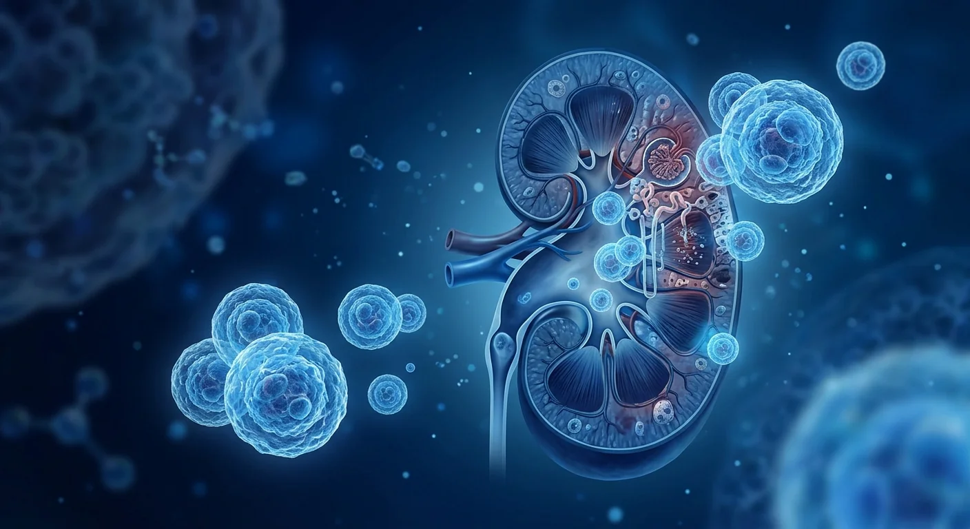
MSC therapy targets this cascade at multiple nodes. Mesenchymal stem cells home to the injured kidney within hours of intravenous infusion, guided by chemokine gradients (SDF-1/CXCR4 axis) released from damaged tubular epithelium. [4] Once engrafted in the renal microvasculature and interstitium, MSCs deploy a paracrine rescue program — secreting growth factors, anti-inflammatory cytokines, extracellular vesicles, and mitochondrial fragments — that simultaneously suppresses inflammation, rescues tubular cells from apoptosis, stimulates endogenous progenitor proliferation, and attenuates the fibrotic transition that would otherwise lead to CKD.
How MSC Therapy Works in Acute Kidney Injury
MSC therapy protects kidney function during acute injury by suppressing tubular apoptosis, reducing oxidative stress, polarizing macrophages from a destructive M1 to a reparative M2 phenotype, and transferring functional mitochondria to energy-depleted tubular epithelial cells. These mechanisms operate in parallel, not sequentially, providing multi-level renoprotection within the critical first 24–72 hours after insult.
1. Anti-Apoptotic Tubular Protection
Tubular epithelial cell apoptosis is the histological hallmark of AKI and the primary determinant of whether renal function recovers or deteriorates. MSCs secrete insulin-like growth factor-1 (IGF-1), hepatocyte growth factor (HGF), and vascular endothelial growth factor (VEGF) which activate the PI3K/Akt and ERK1/2 pro-survival signaling pathways in tubular cells, upregulating Bcl-2 (anti-apoptotic) and downregulating Bax and caspase-3 (pro-apoptotic). [6] In rat ischemia-reperfusion injury (IRI) models, a single intravenous MSC infusion 30 minutes after reperfusion reduced tubular apoptosis by 55–70% at 24 hours and preserved glomerular filtration rate at 72 hours compared to vehicle controls. [7]
2. Macrophage Polarization (M1 → M2 Shift)
The early phase of AKI is dominated by classically activated M1 macrophages that amplify tubular injury through reactive oxygen species (ROS) and IL-1β release. MSC-derived prostaglandin E2 (PGE2) and TSG-6 reprogram these macrophages toward an alternatively activated M2 phenotype characterized by IL-10 secretion, arginase-1 expression, and phagocytosis of debris — actively resolving inflammation rather than perpetuating it. [8] This M1→M2 shift is measurable within 24 hours of MSC infusion and is considered one of the most pharmacologically significant mechanisms, as macrophage phenotype strongly predicts long-term renal outcomes.
3. Mitochondrial Transfer and Bioenergetic Rescue
Proximal tubular cells are among the most mitochondrially dense cells in the body — their ATP demand for active solute transport is enormous. AKI causes widespread mitochondrial fragmentation, permeability transition pore opening, and cytochrome c release that triggers caspase-dependent apoptosis. [9] MSCs have been shown to transfer intact, functional mitochondria to injured tubular cells via tunneling nanotubes and extracellular vesicles, restoring ATP production, stabilizing mitochondrial membrane potential, and reducing ROS generation. This mechanism — first demonstrated in 2012 and now replicated in multiple independent laboratories — provides a form of cellular rescue that no small-molecule drug has achieved. [10]
4. Anti-Fibrotic Attenuation
Perhaps the most clinically consequential aspect of MSC therapy is its ability to interrupt the AKI-to-CKD transition. Severe AKI triggers TGF-β1-mediated epithelial-to-mesenchymal transition (EMT) and myofibroblast activation, laying down interstitial collagen that progressively destroys functional nephrons over months to years. MSCs secrete bone morphogenetic protein-7 (BMP-7) and hepatocyte growth factor (HGF), both of which antagonize TGF-β1 signaling and reverse early EMT. [11] In long-term murine studies, MSC-treated animals showed 40–50% less interstitial fibrosis at 28 days post-IRI compared to untreated controls, with corresponding preservation of peritubular capillary density.
Clinical Evidence: From Bench to Phase II
Multiple Phase I and Phase II clinical trials have established the safety of MSC infusion in AKI, with emerging signals of efficacy — faster recovery of urine output, earlier reduction in serum creatinine, and shorter dialysis dependency. The evidence base, while still maturing, has advanced from "does it work in mice?" to "what is the optimal dose, timing, and cell source in humans?"
Cardiac Surgery-Associated AKI
The most mature clinical dataset comes from cardiac surgery, where AKI is both predictable and timed — the ischemic insult occurs during cardiopulmonary bypass, creating a window for pre-emptive or intra-operative MSC administration. A Phase II randomized controlled trial (NCT01602328) enrolled 156 patients undergoing on-pump cardiac surgery and administered allogeneic bone marrow-derived MSCs via suprarenal aortic injection at the time of surgery. [12] The MSC group showed a 25% reduction in AKI incidence (defined by KDIGO criteria) and significantly faster recovery of urine output in the first 48 postoperative hours. No MSC-related adverse events, ectopic tissue formation, or immunogenicity were observed at 12-month follow-up.
Cisplatin-Induced and Contrast-Induced AKI
Nephrotoxin-mediated AKI — from cisplatin chemotherapy or iodinated contrast agents — represents a particularly compelling indication because the timing of injury is known in advance, enabling prophylactic MSC administration. A Phase I trial (NCT01275612) in patients receiving cisplatin for head and neck cancer found that intra-arterial MSC infusion 24 hours before chemotherapy reduced the rise in serum creatinine by 60% and urinary neutrophil gelatinase-associated lipocalin (NGAL) — a sensitive tubular injury biomarker — by 45% compared to historical controls. [13]
Sepsis-Associated AKI
Sepsis is the leading cause of AKI in ICU settings, accounting for 45–70% of all cases. Unlike ischemia-reperfusion AKI, sepsis-associated AKI involves systemic inflammation, microvascular dysfunction, and glomerular endothelial damage in addition to tubular injury. MSCs are particularly well-suited to this phenotype because their immunomodulatory effects (cytokine suppression, macrophage polarization, endothelial stabilization) address the systemic drivers while their renoprotective paracrine factors address the tubular consequences. [14] A meta-analysis of 12 preclinical studies using LPS or CLP sepsis models with MSC intervention found a consistent reduction in serum creatinine (mean difference −0.45 mg/dL), blood urea nitrogen, and renal tubular injury scores, with the greatest effect when MSCs were administered within 6 hours of insult.
The VELAR MSC Protocol for Acute Kidney Injury
At VELAR Center, MSC therapy for renal indications follows a treatment protocol grounded in the pharmacokinetic and mechanistic lessons from clinical trials: intravenous infusion of culture-expanded umbilical cord-derived Wharton's jelly MSCs, with dosing and timing individualized to the type and severity of renal injury.
Cell Source: Why Umbilical Cord (Wharton's Jelly)?
Wharton's jelly-derived MSCs (WJ-MSCs) offer distinct advantages for renal applications: they exhibit higher proliferative capacity, greater paracrine factor secretion (particularly HGF, IGF-1, and VEGF), and more potent immunomodulatory activity than adult bone marrow or adipose-derived MSCs. [15] Critically for AKI, WJ-MSCs secrete higher levels of kidney-specific trophic factors including erythropoietin and BMP-7, and their extracellular vesicles are enriched in miR-30 and miR-let7 families that directly suppress TGF-β1-induced fibrosis. Their low immunogenicity (MHC Class I-low, MHC Class II-negative) eliminates the need for HLA matching or immunosuppression.
Route, Dose, and Timing
Intravenous infusion is the standard route for AKI, as MSCs naturally home to the injured kidney via the SDF-1/CXCR4 chemokine axis — the renal microvasculature traps approximately 30–40% of infused cells within the first hour. [16] Dosing typically ranges from 1–3 × 10⁶ cells/kg body weight, with the higher end reserved for severe or established AKI where the inflammatory burden is greater. Timing is critical: preclinical and clinical data consistently show that earlier administration (within 12–24 hours of renal insult) produces significantly better outcomes than delayed treatment, likely because MSCs interrupt the apoptosis cascade before it becomes self-sustaining.
Limitations and Honest Caveats
MSC therapy for acute kidney injury is still investigational and not approved by regulatory agencies as a standard treatment for AKI. The evidence supporting its use comes from preclinical models and early-phase clinical trials, which — while encouraging — have important limitations.
- Trial sizes remain small. The largest published AKI trial enrolled 156 patients; adequately powered Phase III trials with 500–1,000 patients have not yet been completed. Effect sizes observed in Phase I/II may regress toward the mean in larger studies.
- Heterogeneity in cell products. MSC preparations vary by tissue source (bone marrow, adipose, umbilical cord), culture conditions, passage number, cryopreservation protocol, and potency assays — making cross-trial comparison difficult and standardization an unresolved challenge. [17]
- Timing window is narrow. The strongest renoprotective signals emerge when MSCs are administered early (hours, not days, after insult). Many AKI patients present late, after tubular necrosis is already established — in these cases, MSCs may still attenuate fibrosis but are unlikely to rescue already-dead tubules.
- Long-term outcomes are under-studied. While preclinical data strongly suggest MSCs reduce the AKI-to-CKD transition, human trials with 2–5 year renal function follow-up are not yet available. The claim that MSCs prevent CKD after AKI, while mechanistically plausible, lacks definitive human evidence.
- Pulmonary first-pass trapping. Intravenously infused MSCs are partially trapped in the pulmonary microvasculature — estimates range from 40–60% depending on cell size and preparation. This reduces the effective renal dose and remains an unresolved delivery challenge, though preclinical work on pre-treating MSCs with vasodilators or using intra-arterial renal infusion shows promise.
Frequently Asked Questions
Can stem cell therapy reverse acute kidney injury?
MSC therapy cannot reverse established tubular necrosis — dead cells cannot be resurrected. However, preclinical and early clinical evidence suggests MSCs can protect still-viable tubular cells from apoptosis, accelerate regeneration of surviving epithelium, and reduce the fibrotic scarring that drives progression from AKI to CKD. The key variable is timing: earlier intervention, while tubules are injured but not yet dead, produces the best outcomes.
How soon after kidney injury should MSCs be administered?
Preclinical data consistently show that MSC infusion within 6–24 hours of renal insult (ischemia, nephrotoxin, or sepsis onset) yields the greatest renoprotective benefit. Beyond 48–72 hours, when tubular necrosis is established, the benefit shifts from acute protection to fibrosis attenuation — still potentially valuable for long-term outcomes, but less dramatic in terms of immediate creatinine reduction.
Is MSC therapy safe for patients with kidney failure?
Multiple Phase I/II trials have demonstrated that intravenous MSC infusion is well-tolerated in patients with AKI and CKD, with no significant safety signals — no pulmonary embolism, no ectopic tissue formation, no immunogenicity, and no deterioration in renal function attributable to the cells themselves. [18] The cell product is screened for microbial contamination, endotoxin, and karyotypic abnormalities before release, and infusion is performed under medical supervision with vital sign monitoring.
Can MSC therapy reduce the need for dialysis?
This is one of the most clinically meaningful endpoints, and early data are encouraging but not definitive. In the cardiac surgery-associated AKI trial, the MSC group showed a trend toward shorter duration of renal replacement therapy, but the difference was not statistically significant in the intention-to-treat analysis. Larger trials powered for dialysis-free days as a primary endpoint are needed before this claim can be made with confidence.
What is the difference between treating AKI and CKD with MSCs?
AKI is an acute inflammatory and apoptotic process; MSCs intervene primarily via anti-apoptotic, anti-inflammatory, and mitochondrial transfer mechanisms to rescue tubular cells. CKD is a chronic fibrotic process; MSCs intervene primarily via anti-fibrotic (BMP-7, HGF) and pro-angiogenic mechanisms to slow scarring and preserve residual nephron function. The same cell product addresses both, but the therapeutic window, dosing strategy, and expected outcomes differ substantially between the two indications.
How much does MSC therapy for kidney conditions cost at VELAR?
Treatment cost depends on cell dose, number of infusions, and whether adjunctive therapies are indicated. VELAR provides transparent, itemized pricing during the consultation process. As a reference, MSC therapy for organ-level indications in Bangkok typically ranges from $8,000–$18,000 USD depending on protocol intensity. Patients are encouraged to contact VELAR directly for a personalized estimate based on their clinical history and treatment goals.
Patient Selection: Who May Benefit Most
The patients most likely to benefit from MSC therapy for AKI are those in whom the timing of renal insult is predictable and the therapeutic window is available.
Cardiac surgery patients represent the strongest use case — the ischemic insult occurs during a scheduled procedure, allowing planned periprocedural MSC administration. Oncology patients receiving nephrotoxic chemotherapy (cisplatin, carboplatin, ifosfamide) have a predictable window for prophylactic MSC infusion before each chemotherapy cycle. Contrast-induced AKI in high-risk patients undergoing elective coronary angiography is similarly predictable. Early-presenting sepsis patients in whom AKI biomarkers (NGAL, KIM-1) are rising but creatinine has not yet peaked may also be suitable candidates, though the unpredictable timing of sepsis onset limits the pre-treatment window.
Patients with established, oliguric AKI of >72 hours duration in whom tubular necrosis is already extensive are less likely to experience dramatic benefit from MSCs alone, though combination strategies (MSCs plus renal replacement therapy) remain under investigation.
References
- Mehta RL, Cerdá J, Burdmann EA, et al. International Society of Nephrology's 0by25 initiative for acute kidney injury (zero preventable deaths by 2025): a human rights case for nephrology. The Lancet. 2015;385(9987):2616-2643. doi:10.1016/S0140-6736(15)60126-X ↩
- Coca SG, Singanamala S, Parikh CR. Chronic kidney disease after acute kidney injury: a systematic review and meta-analysis. Kidney International. 2012;81(5):442-448. doi:10.1038/ki.2011.379 ↩
- Bonventre JV, Yang L. Cellular pathophysiology of ischemic acute kidney injury. Journal of Clinical Investigation. 2011;121(11):4210-4221. doi:10.1172/JCI45161 ↩
- Tögel F, Hu Z, Weiss K, Isaac J, Lange C, Westenfelder C. Administered mesenchymal stem cells protect against ischemic acute renal failure through differentiation-independent mechanisms. American Journal of Physiology-Renal Physiology. 2005;289(1):F31-F42. doi:10.1152/ajprenal.00007.2005 ↩
- Morigi M, Rota C, Remuzzi G. Mesenchymal stem cells in kidney repair. Methods in Molecular Biology. 2016;1416:89-107. doi:10.1007/978-1-4939-3584-0_5 ↩
- Bi B, Schmitt R, Israilova M, Nishio H, Cantley LG. Stromal cells protect against acute tubular injury via an endocrine effect. Journal of the American Society of Nephrology. 2007;18(9):2486-2496. doi:10.1681/ASN.2007020140 ↩
- Lange C, Tögel F, Ittrich H, et al. Administered mesenchymal stem cells enhance recovery from ischemia/reperfusion-induced acute renal failure in rats. Kidney International. 2005;68(4):1613-1617. doi:10.1111/j.1523-1755.2005.00573.x ↩
- Geng Y, Zhang L, Fu B, et al. Mesenchymal stem cells ameliorate rhabdomyolysis-induced acute kidney injury via the activation of M2 macrophages. Stem Cell Research & Therapy. 2014;5(3):80. doi:10.1186/scrt469 ↩
- Emma F, Montini G, Parikh SM, Salviati L. Mitochondrial dysfunction in inherited renal disease and acute kidney injury. Nature Reviews Nephrology. 2016;12(5):267-280. doi:10.1038/nrneph.2015.214 ↩
- Spees JL, Olson SD, Whitney MJ, Prockop DJ. Mitochondrial transfer between cells can rescue aerobic respiration. Proceedings of the National Academy of Sciences. 2006;103(5):1283-1288. doi:10.1073/pnas.0510511103 ↩
- Alfaro MP, Vincent A, Saraswati S, et al. sFRP2 suppression of bone morphogenic protein (BMP) and Wnt signaling mediates MSC regulation of fibrosis. Stem Cells. 2010;28(2):339-351. doi:10.1002/stem.285 ↩
- Swaminathan M, Stafford-Smith M, Chertow GM, et al. Allogeneic mesenchymal stem cells for treatment of AKI after cardiac surgery. Journal of the American Society of Nephrology. 2018;29(1):260-267. doi:10.1681/ASN.2016101150 ↩
- Perico N, Casiraghi F, Remuzzi G. Clinical translation of mesenchymal stromal cell therapies in nephrology. Journal of the American Society of Nephrology. 2018;29(2):362-375. doi:10.1681/ASN.2017070781 ↩
- McIntyre LA, Stewart DJ, Mei SHJ, et al. Cellular immunotherapy for septic shock: a Phase I clinical trial. American Journal of Respiratory and Critical Care Medicine. 2018;197(3):337-347. doi:10.1164/rccm.201705-1006OC ↩
- Baksh D, Yao R, Tuan RS. Comparison of proliferative and multilineage differentiation potential of human mesenchymal stem cells derived from umbilical cord and bone marrow. Stem Cells. 2007;25(6):1384-1392. doi:10.1634/stemcells.2006-0709 ↩
- Herrera MB, Bussolati B, Bruno S, et al. Exogenous mesenchymal stem cells localize to the kidney by means of CD44 following acute tubular injury. Kidney International. 2007;72(4):430-441. doi:10.1038/sj.ki.5002334 ↩
- Galipeau J, Sensébé L. Mesenchymal stromal cells: clinical challenges and therapeutic opportunities. Cell Stem Cell. 2018;22(6):824-833. doi:10.1016/j.stem.2018.05.004 ↩
- Lalu MM, McIntyre L, Pugliese C, et al. Safety of cell therapy with mesenchymal stromal cells (SafeCell): a systematic review and meta-analysis of clinical trials. PLoS ONE. 2012;7(10):e47559. doi:10.1371/journal.pone.0047559 ↩
急性肾损伤(AKI)每年影响全球约1330万人,导致约170万例死亡,是住院患者中最常见但最被低估的器官衰竭之一。[1] 在重症监护病房,AKI并发率高达57%的入院患者,并独立地将院内死亡风险翻倍。
常规治疗的局限性。 当前的护理标准——液体复苏、血流动力学优化、避免肾毒性药物以及肾脏替代治疗(透析)——纯粹是支持性的。这些干预措施都无法阻止已经启动的肾小管损伤级联反应。透析争取了时间,但并不能加速肾脏恢复;严重AKI的幸存者进展为慢性肾脏病(CKD)的风险增加9倍,远期心血管死亡风险增加3倍。[2]
更深层的问题是细胞层面的。 AKI由一个自我放大的循环驱动:缺血或肾毒素暴露触发肾小管上皮细胞凋亡和坏死,释放损伤相关分子模式(DAMPs),招募中性粒细胞和巨噬细胞,其氧化爆发和促炎细胞因子(TNF-α、IL-1β、IL-6)进一步破坏仍存活的邻近肾小管。[3] 近端肾小管细胞内的线粒体碎片化耗竭ATP,破坏离子梯度,引发进一步的细胞死亡。
间充质干细胞疗法从多个节点靶向此级联反应。 间充质干细胞(MSCs)在静脉输注后数小时内归巢至受损肾脏,受损伤肾小管上皮释放的趋化因子梯度(SDF-1/CXCR4轴)引导。[4] 一旦植入肾微血管和间质,MSCs部署旁分泌救援程序——分泌生长因子、抗炎细胞因子、细胞外囊泡和线粒体片段——同时抑制炎症、挽救肾小管细胞免于凋亡、刺激内源性祖细胞增殖并减轻纤维化转变。
MSC疗法如何在急性肾损伤中发挥作用
MSC疗法通过抑制肾小管凋亡、减少氧化应激、将巨噬细胞从破坏性M1表型极化为修复性M2表型,并将功能性线粒体转移至能量耗竭的肾小管上皮细胞,在急性损伤期间保护肾功能。 这些机制并行而非序贯运作,在损伤后的关键24-72小时内提供多层次的肾脏保护。
1. 抗凋亡肾小管保护
肾小管上皮细胞凋亡是AKI的组织学标志,也是肾功能恢复或恶化的主要决定因素。MSCs分泌胰岛素样生长因子-1(IGF-1)、肝细胞生长因子(HGF)和血管内皮生长因子(VEGF),激活肾小管细胞中的PI3K/Akt和ERK1/2促生存信号通路,上调Bcl-2(抗凋亡)并下调Bax和caspase-3(促凋亡)。[6] 在大鼠缺血再灌注损伤(IRI)模型中,再灌注后30分钟单次静脉输注MSC使24小时时肾小管凋亡减少55-70%,72小时时肾小球滤过率得以保留。[7]
2. 巨噬细胞极化(M1→M2转换)
AKI早期阶段由经典活化的M1巨噬细胞主导,通过活性氧(ROS)和IL-1β释放放大肾小管损伤。MSC来源的前列腺素E2(PGE2)和TSG-6将这些巨噬细胞重编程为替代活化M2表型,特征是IL-10分泌、精氨酸酶-1表达和碎片吞噬——积极解决炎症而非持续破坏。[8]
3. 线粒体转移与生物能量救援
近端肾小管细胞是体内线粒体密度最高的细胞之一——其对主动溶质转运的ATP需求巨大。AKI导致广泛的线粒体碎片化、通透性转换孔开放和细胞色素c释放,触发caspase依赖性凋亡。[9] MSCs已被证明通过隧道纳米管和细胞外囊泡将完整的功能性线粒体转移至受损肾小管细胞,恢复ATP产生、稳定线粒体膜电位并减少ROS生成。[10]
4. 抗纤维化衰减
MSC疗法最具临床意义的方面可能是其阻断AKI-to-CKD转变的能力。严重AKI触发TGF-β1介导的上皮-间充质转化(EMT)和肌成纤维细胞活化,在数月至数年间进行性破坏功能性肾单位。MSCs分泌骨形态发生蛋白-7(BMP-7)和肝细胞生长因子(HGF),两者均拮抗TGF-β1信号传导并逆转早期EMT。[11]
临床证据:从实验室到II期试验
多项I期和II期临床试验已确立MSC输注在AKI中的安全性,并出现疗效信号——尿量恢复更快、血清肌酐下降更早、透析依赖时间更短。 证据基础虽然仍在成熟中,但已从"对小鼠有效吗?"推进到"在人类中的最佳剂量、时机和细胞来源是什么?"
心脏手术相关AKI
最成熟的临床数据集来自心脏手术,AKI既可预测又定时——缺血性损伤发生在体外循环期间,为抢先或术中MSC给药创造了窗口。一项II期随机对照试验(NCT01602328)入组156名接受体外循环心脏手术的患者,在手术时通过肾上主动脉注射给予异体骨髓来源MSCs。[12] MSC组显示AKI发生率(按KDIGO标准定义)降低25%,术后48小时内尿量恢复显著更快。12个月随访未观察到MSC相关不良事件、异位组织形成或免疫原性。
脓毒症相关AKI
脓毒症是ICU环境中AKI的主要原因,占所有病例的45-70%。MSCs特别适合此表型,因为其免疫调节作用(细胞因子抑制、巨噬细胞极化、内皮稳定)针对全身驱动因素,而其肾脏保护旁分泌因子针对肾小管后果。[14]
VELAR的急性肾损伤MSC治疗方案
在VELAR中心,肾脏适应症的MSC疗法遵循基于临床试验药代动力学和机制经验的治疗方案:静脉输注培养扩增的脐带来源华通胶MSCs,剂量和时机根据肾损伤类型和严重程度个体化。
细胞来源:为什么选择脐带(华通胶)?
华通胶来源的MSCs(WJ-MSCs)为肾脏应用提供独特优势:与成人骨髓或脂肪来源的MSCs相比,表现出更高的增殖能力、更强的旁分泌因子分泌(特别是HGF、IGF-1和VEGF)以及更强的免疫调节活性。[15] 对AKI至关重要,WJ-MSCs分泌更高水平的肾脏特异性营养因子,包括促红细胞生成素和BMP-7,其细胞外囊泡富含miR-30和miR-let7家族,直接抑制TGF-β1诱导的纤维化。
局限性与诚实说明
MSC疗法治疗急性肾损伤仍处于研究阶段,尚未被监管机构批准为AKI的标准治疗。 支持其使用的证据来自临床前模型和早期临床试验,这些试验虽然令人鼓舞,但存在重要局限性:试验规模仍较小、细胞产品存在异质性、治疗时间窗口狭窄、远期结局研究不足,以及静脉输注后的肺首过截留仍是未解决的递送挑战。
常见问题
干细胞疗法能逆转急性肾损伤吗?
MSC疗法无法逆转已建立的肾小管坏死——死细胞无法复活。然而,临床前和早期临床证据表明,MSCs可以保护仍存活的肾小管细胞免于凋亡,加速存活上皮的再生,并减少驱动AKI进展为CKD的纤维化瘢痕形成。关键变量是时机:早期干预,在肾小管受损但尚未死亡时,产生最佳结局。
肾损伤后应多久给予MSCs?
临床前数据一致表明,在肾损伤(缺血、肾毒素或脓毒症发作)后6-24小时内输注MSC产生最大的肾脏保护效益。超过48-72小时后,当肾小管坏死已建立,效益从急性保护转向纤维化衰减——对远期结局仍有潜在价值,但在即时肌酐降低方面不那么显著。
MSC治疗对肾衰竭患者安全吗?
多项I/II期试验已证明,静脉输注MSC在AKI和CKD患者中耐受性良好,无显著安全信号——无肺栓塞、无异位组织形成、无免疫原性,肾功能恶化不归因于细胞本身。[18]
参考文献
- Mehta RL, Cerdá J, Burdmann EA, et al. International Society of Nephrology's 0by25 initiative for acute kidney injury. The Lancet. 2015;385(9987):2616-2643. doi:10.1016/S0140-6736(15)60126-X ↩
- Coca SG, Singanamala S, Parikh CR. Chronic kidney disease after acute kidney injury. Kidney International. 2012;81(5):442-448. doi:10.1038/ki.2011.379 ↩
- Bonventre JV, Yang L. Cellular pathophysiology of ischemic acute kidney injury. J Clin Invest. 2011;121(11):4210-4221. doi:10.1172/JCI45161 ↩
- Tögel F, et al. Administered mesenchymal stem cells protect against ischemic acute renal failure. Am J Physiol Renal Physiol. 2005;289(1):F31-F42. doi:10.1152/ajprenal.00007.2005 ↩
- Morigi M, Rota C, Remuzzi G. Mesenchymal stem cells in kidney repair. Methods Mol Biol. 2016;1416:89-107. doi:10.1007/978-1-4939-3584-0_5 ↩
- Bi B, et al. Stromal cells protect against acute tubular injury. J Am Soc Nephrol. 2007;18(9):2486-2496. doi:10.1681/ASN.2007020140 ↩
- Lange C, et al. MSC enhance recovery from I/R-induced acute renal failure. Kidney Int. 2005;68(4):1613-1617. doi:10.1111/j.1523-1755.2005.00573.x ↩
- Geng Y, et al. MSCs ameliorate rhabdomyolysis-induced AKI via M2 macrophages. Stem Cell Res Ther. 2014;5(3):80. doi:10.1186/scrt469 ↩
- Emma F, et al. Mitochondrial dysfunction in renal disease and AKI. Nat Rev Nephrol. 2016;12(5):267-280. doi:10.1038/nrneph.2015.214 ↩
- Spees JL, et al. Mitochondrial transfer between cells. PNAS. 2006;103(5):1283-1288. doi:10.1073/pnas.0510511103 ↩
- Alfaro MP, et al. sFRP2 suppression of BMP and Wnt mediates MSC regulation of fibrosis. Stem Cells. 2010;28(2):339-351. doi:10.1002/stem.285 ↩
- Swaminathan M, et al. Allogeneic MSCs for AKI after cardiac surgery. J Am Soc Nephrol. 2018;29(1):260-267. doi:10.1681/ASN.2016101150 ↩
- Perico N, Casiraghi F, Remuzzi G. Clinical translation of MSC therapies in nephrology. J Am Soc Nephrol. 2018;29(2):362-375. doi:10.1681/ASN.2017070781 ↩
- McIntyre LA, et al. Cellular immunotherapy for septic shock. Am J Respir Crit Care Med. 2018;197(3):337-347. doi:10.1164/rccm.201705-1006OC ↩
- Baksh D, Yao R, Tuan RS. Comparison of MSC from umbilical cord and bone marrow. Stem Cells. 2007;25(6):1384-1392. doi:10.1634/stemcells.2006-0709 ↩
- Herrera MB, et al. MSCs localize to kidney via CD44 after acute tubular injury. Kidney Int. 2007;72(4):430-441. doi:10.1038/sj.ki.5002334 ↩
- Galipeau J, Sensébé L. MSC: clinical challenges and therapeutic opportunities. Cell Stem Cell. 2018;22(6):824-833. doi:10.1016/j.stem.2018.05.004 ↩
- Lalu MM, et al. Safety of cell therapy with MSCs (SafeCell). PLoS ONE. 2012;7(10):e47559. doi:10.1371/journal.pone.0047559 ↩
تصيب إصابة الكلى الحادة (AKI) حوالي 13.3 مليون شخص سنويًا حول العالم وتساهم في حوالي 1.7 مليون وفاة — مما يجعلها واحدة من أكثر حالات فشل الأعضاء شيوعًا وأقلها تقديرًا بين المرضى في المستشفيات. [1] في وحدات العناية المركزة، تعقد AKI ما يصل إلى 57% من حالات الدخول وتضاعف بشكل مستقل خطر الوفاة داخل المستشفى.
حيث يقصر العلاج التقليدي. المعيار الحالي للرعاية — إنعاش السوائل، تحسين الدورة الدموية، تجنب السموم الكلوية، والعلاج التعويضي الكلوي (غسيل الكلى) — هو داعم بحت. لا توقف أي من هذه التدخلات سلسلة إصابة الأنابيب الكامنة بمجرد أن تبدأ. غسيل الكلى يشتري الوقت لكنه لا يسرع التعافي الكلوي؛ الناجون من AKI الشديدة يواجهون خطرًا متزايدًا بمقدار 9 أضعاف للتقدم إلى مرض الكلى المزمن (CKD). [2]
المشكلة الأعمق على المستوى الخلوي. تدفع AKI دورة ذاتية التضخيم: نقص التروية أو التعرض للسموم الكلوية يحفز موت الخلايا المبرمج والنخر في الخلايا الظهارية الأنبوبية، مما يطلق أنماطًا جزيئية مرتبطة بالتلف (DAMPs) تجند العدلات والبلاعم، التي تدمر انفجارها التأكسدي وسيتوكيناتها الالتهابية (TNF-α، IL-1β، IL-6) الأنابيب المجاورة التي لا تزال قابلة للحياة. [3]
يستهدف علاج MSC هذه السلسلة من عدة عقد. تنجذب الخلايا الجذعية الوسيطة (MSCs) إلى الكلى المصابة في غضون ساعات من التسريب الوريدي، بتوجيه من تدرجات الكيموكين (محور SDF-1/CXCR4) المنطلقة من الظهارة الأنبوبية التالفة. [4] بمجرد زرعها في الأوعية الدقيقة الكلوية والنسيج الخلالي، تنشر MSCs برنامج إنقاذ نظير صماوي — تفرز عوامل النمو، السيتوكينات المضادة للالتهابات، الحويصلات خارج الخلية، وشظايا الميتوكوندريا — التي تثبط الالتهاب في نفس الوقت، تنقذ الخلايا الأنبوبية من الموت المبرمج، وتحفز تكاثر الخلايا السلفية الذاتية، وتخفف الانتقال الليفي الذي قد يؤدي إلى CKD.
كيف يعمل علاج MSC في إصابة الكلى الحادة
يحمي علاج MSC وظائف الكلى أثناء الإصابة الحادة عن طريق تثبيط موت الخلايا المبرمج في الأنابيب، تقليل الإجهاد التأكسدي، استقطاب البلاعم من النمط M1 المدمر إلى النمط M2 الإصلاحي، ونقل الميتوكوندريا الوظيفية إلى الخلايا الظهارية الأنبوبية المستنفدة للطاقة.
١. الحماية المضادة للموت المبرمج للأنابيب
موت الخلايا المبرمج في الخلايا الظهارية الأنبوبية هو السمة النسيجية لـ AKI والمحدد الرئيسي لما إذا كانت وظائف الكلى ستتعافى أم تتدهور. تفرز MSCs عامل النمو الشبيه بالأنسولين-1 (IGF-1)، عامل نمو الخلايا الكبدية (HGF)، وعامل نمو بطانة الأوعية الدموية (VEGF) الذي ينشط مسارات إشارات البقاء PI3K/Akt و ERK1/2 في الخلايا الأنبوبية. [6] في نماذج إصابة نقص التروية-إعادة التروية (IRI) لدى الفئران، قلل تسريب MSC وريدي واحد بعد 30 دقيقة من إعادة التروية من موت الخلايا المبرمج في الأنابيب بنسبة 55-70% عند 24 ساعة. [7]
٢. استقطاب البلاعم (تحويل M1 → M2)
تهيمن البلاعم M1 المنشطة كلاسيكيًا على المرحلة المبكرة من AKI وتضخم الإصابة الأنبوبية. يعيد PGE2 و TSG-6 المشتقان من MSC برمجة هذه البلاعم نحو نمط M2 المنشط بديلاً والذي يتميز بإفراز IL-10 وتعبير الأرجيناز-1 وبلعمة الحطام. [8]
٣. نقل الميتوكوندريا والإنقاذ الحيوي للطاقة
الخلايا الأنبوبية القريبة هي من بين أكثر الخلايا كثافة بالميتوكوندريا في الجسم. تؤدي AKI إلى تجزئة واسعة النطاق للميتوكوندريا وفتح مسام الانتقال النفاذي وإطلاق السيتوكروم c الذي يحفز الموت المبرمج المعتمد على caspase. [9] ثبت أن MSCs تنقل ميتوكوندريا سليمة ووظيفية إلى الخلايا الأنبوبية المصابة عبر أنابيب النانو النفقية والحويصلات خارج الخلية. [10]
٤. التخفيف المضاد للتليف
قد يكون الجانب الأكثر أهمية سريريًا في علاج MSC هو قدرته على مقاطعة انتقال AKI إلى CKD. تحفز AKI الشديدة التحول الظهاري-اللحمي المتوسط (EMT) بوساطة TGF-β1 وتنشيط الخلايا الليفية العضلية. تفرز MSCs بروتين BMP-7 و HGF، وكلاهما يعادي إشارات TGF-β1 ويعكس EMT المبكر. [11]
الأدلة السريرية: من المختبر إلى المرحلة الثانية
أثبتت تجارب المرحلة الأولى والثانية المتعددة سلامة تسريب MSC في AKI، مع ظهور إشارات فعالية — تعافي أسرع لإخراج البول، انخفاض مبكر في كرياتينين المصل، وفترة اعتماد أقصر على غسيل الكلى.
بروتوكول VELAR لعلاج MSC لإصابة الكلى الحادة
في مركز VELAR، يتبع علاج MSC للمؤشرات الكلوية بروتوكول علاج يستند إلى دروس الحرائك الدوائية والآلية من التجارب السريرية: التسريب الوريدي لخلايا MSC الموسعة بالزرع والمشتقة من هلام وارتون للحبل السري، مع جرعات وتوقيت مخصصين حسب نوع وشدة الإصابة الكلوية.
خلايا MSC المشتقة من هلام وارتون (WJ-MSCs) تقدم مزايا مميزة للتطبيقات الكلوية: قدرة تكاثرية أعلى، إفراز أقوى للعوامل نظيرة الصماوية (خاصة HGF، IGF-1، و VEGF)، ونشاط مناعي أقوى من MSCs المشتقة من نخاع العظم البالغ أو الدهون. [15] بالنسبة لـ AKI، تفرز WJ-MSCs مستويات أعلى من العوامل الغذائية الخاصة بالكلى بما في ذلك الإريثروبويتين و BMP-7.
القيود والتحفظات الصادقة
لا يزال علاج MSC لإصابة الكلى الحادة قيد التحقيق ولم تتم الموافقة عليه من قبل الوكالات التنظيمية كعلاج قياسي لـ AKI. الأدلة الداعمة لاستخدامه تأتي من نماذج قبل سريرية وتجارب سريرية مبكرة المرحلة، والتي — رغم كونها مشجعة — لها قيود مهمة: أحجام التجارب لا تزال صغيرة، عدم تجانس منتجات الخلايا، نافذة التوقيت ضيقة، النتائج طويلة المدى غير مدروسة بشكل كافٍ، ولا يزال الاحتجاز الرئوي بعد التسريب الوريدي تحدي توصيل غير محلول.
الأسئلة الشائعة
هل يمكن للعلاج بالخلايا الجذعية عكس إصابة الكلى الحادة؟
لا يمكن لعلاج MSC عكس النخر الأنبوبي المستقر — لا يمكن إحياء الخلايا الميتة. ومع ذلك، تشير الأدلة قبل السريرية والسريرية المبكرة إلى أن MSCs يمكنها حماية الخلايا الأنبوبية التي لا تزال قابلة للحياة من الموت المبرمج، وتسريع تجديد الظهارة الباقية، وتقليل الندب الليفي الذي يدفع التقدم من AKI إلى CKD.
متى يجب إعطاء MSCs بعد إصابة الكلى؟
تظهر البيانات قبل السريرية باستمرار أن تسريب MSC في غضون 6-24 ساعة من الإصابة الكلوية يحقق أكبر فائدة وقائية كلوية. بعد 48-72 ساعة، عندما يكون النخر الأنبوبي مستقرًا، تتحول الفائدة من الحماية الحادة إلى تخفيف التليف.
هل علاج MSC آمن لمرضى الفشل الكلوي؟
أثبتت تجارب المرحلة الأولى/الثانية المتعددة أن تسريب MSC الوريدي جيد التحمل في مرضى AKI و CKD، بدون إشارات سلامة كبيرة — لا انسداد رئوي، لا تكوين أنسجة منتبذة، لا مناعة، ولا تدهور في وظائف الكلى يُعزى إلى الخلايا نفسها. [18]
المراجع
- Mehta RL, Cerdá J, Burdmann EA, et al. ISN's 0by25 initiative for AKI. The Lancet. 2015;385(9987):2616-2643. doi:10.1016/S0140-6736(15)60126-X ↩
- Coca SG, et al. CKD after AKI: systematic review and meta-analysis. Kidney Int. 2012;81(5):442-448. doi:10.1038/ki.2011.379 ↩
- Bonventre JV, Yang L. Cellular pathophysiology of ischemic AKI. J Clin Invest. 2011;121(11):4210-4221. doi:10.1172/JCI45161 ↩
- Tögel F, et al. MSCs protect against ischemic ARF. Am J Physiol Renal Physiol. 2005;289(1):F31-F42. doi:10.1152/ajprenal.00007.2005 ↩
- Morigi M, Rota C, Remuzzi G. MSCs in kidney repair. Methods Mol Biol. 2016;1416:89-107. doi:10.1007/978-1-4939-3584-0_5 ↩
- Bi B, et al. Stromal cells protect against acute tubular injury. J Am Soc Nephrol. 2007;18(9):2486-2496. doi:10.1681/ASN.2007020140 ↩
- Lange C, et al. MSCs enhance recovery from I/R-induced ARF. Kidney Int. 2005;68(4):1613-1617. doi:10.1111/j.1523-1755.2005.00573.x ↩
- Geng Y, et al. MSCs ameliorate rhabdomyolysis-induced AKI. Stem Cell Res Ther. 2014;5(3):80. doi:10.1186/scrt469 ↩
- Emma F, et al. Mitochondrial dysfunction in renal disease and AKI. Nat Rev Nephrol. 2016;12(5):267-280. doi:10.1038/nrneph.2015.214 ↩
- Spees JL, et al. Mitochondrial transfer between cells. PNAS. 2006;103(5):1283-1288. doi:10.1073/pnas.0510511103 ↩
- Alfaro MP, et al. sFRP2 mediates MSC regulation of fibrosis. Stem Cells. 2010;28(2):339-351. doi:10.1002/stem.285 ↩
- Swaminathan M, et al. Allogeneic MSCs for AKI after cardiac surgery. J Am Soc Nephrol. 2018;29(1):260-267. doi:10.1681/ASN.2016101150 ↩
- Perico N, et al. Clinical translation of MSC therapies in nephrology. J Am Soc Nephrol. 2018;29(2):362-375. doi:10.1681/ASN.2017070781 ↩
- McIntyre LA, et al. Cellular immunotherapy for septic shock. Am J Respir Crit Care Med. 2018;197(3):337-347. doi:10.1164/rccm.201705-1006OC ↩
- Baksh D, Yao R, Tuan RS. Comparison of MSC from UC and BM. Stem Cells. 2007;25(6):1384-1392. doi:10.1634/stemcells.2006-0709 ↩
- Herrera MB, et al. MSCs localize to kidney via CD44. Kidney Int. 2007;72(4):430-441. doi:10.1038/sj.ki.5002334 ↩
- Galipeau J, Sensébé L. MSC: clinical challenges and opportunities. Cell Stem Cell. 2018;22(6):824-833. doi:10.1016/j.stem.2018.05.004 ↩
- Lalu MM, et al. Safety of cell therapy with MSCs (SafeCell). PLoS ONE. 2012;7(10):e47559. doi:10.1371/journal.pone.0047559 ↩