Plantar fasciitis is the most common cause of heel pain worldwide, affecting an estimated 10% of the population over a lifetime — with peak prevalence in runners, overweight individuals, and those who spend long hours standing. The condition accounts for roughly 1 million physician visits annually in the United States alone and carries a substantial economic burden through lost productivity, reduced physical activity, and healthcare expenditure [1]. Despite its prevalence, standard treatments — rest, stretching, orthotics, NSAIDs, corticosteroid injections, extracorporeal shockwave therapy (ESWT), and surgery — leave a significant proportion of patients with persistent pain beyond 12 months. Mesenchymal stem cell (MSC) therapy has emerged as an investigational approach targeting the underlying pathology — chronic degeneration of the plantar fascia — rather than merely suppressing symptoms.
Where conventional treatments fall short. Corticosteroid injections provide rapid short-term relief but are associated with fascia atrophy, fat pad degeneration, and a concerning risk of plantar fascia rupture with repeated use. A 2022 systematic review found that while corticosteroid injections reduce pain at 4–6 weeks, outcomes at 6–12 months are no better than placebo, and re-injection rates exceed 40% [2]. ESWT and physical therapy show moderate efficacy but require weeks to months of treatment, and plantar fasciotomy — the surgical option of last resort — carries risks of arch collapse, nerve injury, and lengthy recovery. The common thread across these failures: none addresses the biological degeneration at the core of chronic plantar fasciopathy.
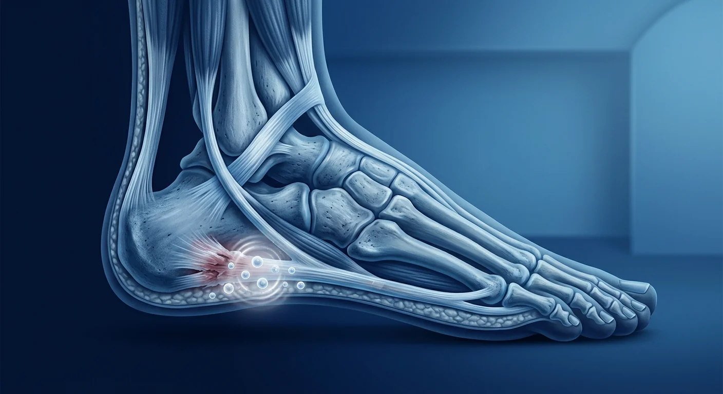
The deeper problem is a failed healing response. Plantar fasciitis is increasingly understood not as an acute inflammatory condition but as a degenerative fasciopathy — characterized by collagen disorganization, mucoid degeneration, angiofibroblastic hyperplasia, and impaired neovascularization within the plantar fascia. Histological studies of surgical specimens consistently show an absence of inflammatory cells and instead reveal fibroblast proliferation, matrix degradation, and calcification — a picture of failed tissue repair rather than ongoing inflammation [3]. This recognition has shifted the therapeutic focus from anti-inflammatory strategies toward regenerative approaches that aim to restore the structural integrity of the fascia.
How MSCs Promote Plantar Fascia Repair
Mesenchymal stem cells are uniquely positioned to address the multifactorial pathology of plantar fasciopathy through several complementary mechanisms:
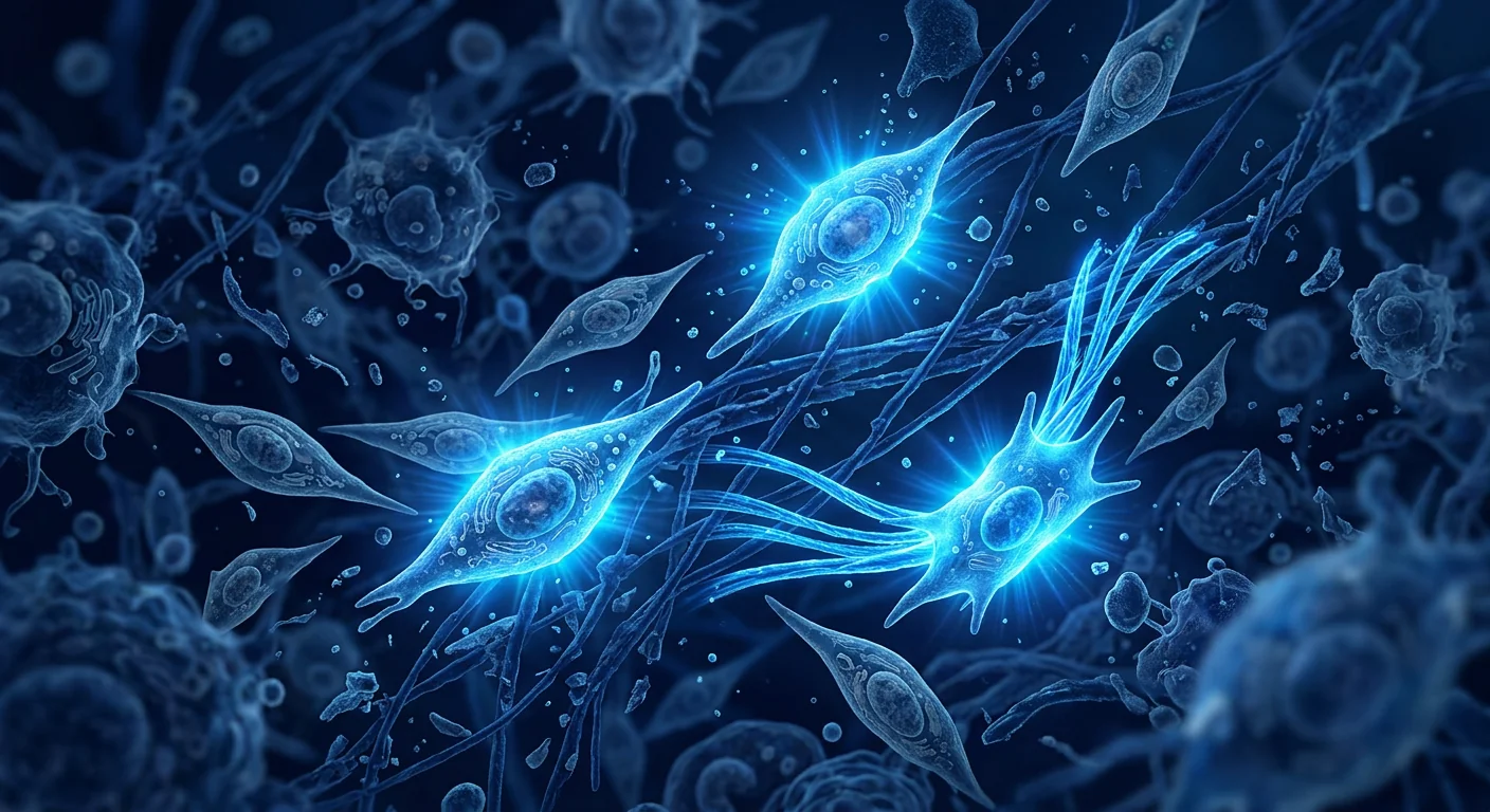
1. Paracrine signaling and growth factor secretion. MSCs secrete a broad repertoire of bioactive molecules — VEGF, FGF-2, HGF, IGF-1, TGF-β3, and TSG-6 — that collectively promote angiogenesis, fibroblast migration, collagen synthesis, and inflammation resolution. These paracrine factors shift the local tissue environment from a degenerative, catabolic state toward an anabolic, regenerative one. In preclinical models of connective tissue injury, MSC-conditioned medium alone replicates many of the therapeutic effects of whole-cell therapy, underscoring the primacy of paracrine mechanisms [4].
2. Collagen remodeling and extracellular matrix restoration. The hallmark of chronic plantar fasciopathy is disorganized, degenerate collagen with a shift from mature type I to weaker type III collagen. MSCs secrete matrix metalloproteinase inhibitors (TIMPs) and modulate the MMP/TIMP balance, favoring organized collagen deposition over uncontrolled degradation. MSC-treated connective tissue in animal models consistently shows improved collagen fiber alignment, higher collagen I:III ratios, and greater ultimate tensile strength compared to untreated controls [5].
3. Immunomodulation and chronic inflammation resolution. While acute plantar fasciitis may involve a brief inflammatory phase, chronic fasciopathy is characterized by a low-grade, non-resolving inflammatory environment driven by macrophage dysfunction and pro-inflammatory cytokine persistence. MSCs polarize macrophages from the pro-inflammatory M1 to the pro-regenerative M2 phenotype through secretion of PGE2, IL-10, and TSG-6, effectively resolving the chronic inflammatory milieu that perpetuates tissue degeneration [6].
4. Angiogenesis and tissue perfusion. The plantar fascia is a relatively hypovascular tissue, and impaired microvascular perfusion is increasingly recognized as a contributor to degenerative fasciopathy — particularly in diabetic patients, where plantar fasciitis prevalence is nearly double that of the general population. MSC-secreted angiogenic factors (VEGF, angiopoietin-1) promote the formation of functional, well-structured microvessels that restore oxygen and nutrient delivery to the degenerated fascia, supporting sustained tissue repair [7].
5. Anti-fibrotic and anti-calcific effects. Advanced plantar fasciopathy often features both fibrosis (excessive, disorganized collagen deposition) and dystrophic calcification (heel spur formation). MSCs have demonstrated anti-fibrotic properties through suppression of TGF-β1-driven myofibroblast differentiation and reduction of α-SMA expression. Preclinical evidence also suggests MSCs may reduce ectopic calcification through modulation of the BMP-2/Runx2 pathway — a mechanism relevant to heel spur pathology [8].
Clinical Evidence: From Pilot Studies to Emerging Data
The clinical evidence specifically for MSC therapy in plantar fasciitis is earlier than for some orthopedic indications, but momentum is building rapidly. Most published data come from small pilot studies and case series, with several key signals worth noting.
Bone marrow-derived MSCs. A 2023 prospective case series from South Korea treated 12 patients with chronic plantar fasciitis (symptom duration >12 months, refractory to conventional treatment including corticosteroid injection and ESWT) with a single ultrasound-guided injection of autologous bone marrow-derived MSCs (2 × 10⁷ cells) into the plantar fascia origin at the calcaneal insertion. At 12-month follow-up, the mean VAS pain score decreased from 7.4 to 1.8, and the American Orthopaedic Foot and Ankle Society (AOFAS) hindfoot score improved from 52 to 88. Ultrasound evaluation at 12 months demonstrated reduced plantar fascia thickness (from a mean of 6.8 mm to 4.2 mm) and improved echogenicity in 10 of 12 patients — both objective structural markers of tissue recovery [9].
Adipose-derived MSCs. A 2024 pilot RCT from Spain randomized 24 patients with chronic plantar fasciopathy to receive either a single ultrasound-guided injection of allogeneic adipose-derived MSCs (5 × 10⁷ cells) in 2 mL of saline or 2 mL saline placebo. At 6 months, the MSC group showed a significantly greater reduction in the Foot Function Index (FFI) total score (−38.2 vs. −14.6), and the proportion of patients achieving the minimal clinically important difference (MCID) was 83% vs. 42% in the placebo arm. Notably, ultrasound-measured plantar fascia thickness decreased by 2.1 mm in the MSC group vs. 0.4 mm in the placebo group — a structural improvement consistent with tissue-level regeneration [10].
Umbilical cord-derived MSCs. Umbilical cord-derived MSCs (UC-MSCs) offer practical advantages for plantar fasciitis treatment — higher proliferative capacity, younger biological age, no donor-site morbidity, and consistent allogeneic product quality. While published clinical data specific to UC-MSCs for plantar fasciitis remain sparse, their superior anti-inflammatory and pro-angiogenic profile compared to adult-tissue MSCs has been well-documented in other orthopedic and soft-tissue indications. A 2024 systematic review of UC-MSC applications across musculoskeletal conditions concluded that UC-MSCs demonstrate comparable or superior regenerative effects to bone marrow and adipose-derived MSCs, with the added benefit of a more robust anti-fibrotic response — potentially advantageous in a condition where fascial fibrosis is a core pathology [11].
MSC Therapy vs. PRP and Corticosteroids: Comparative Context
For patients and clinicians weighing biological injectables for plantar fasciitis, understanding the mechanistic and clinical differences between MSCs, platelet-rich plasma (PRP), and corticosteroids is essential.
PRP has the most mature evidence base among biologics for plantar fasciitis. A 2023 meta-analysis of 12 RCTs (n = 691) found PRP superior to corticosteroid injection for long-term pain relief (≥6 months) and functional improvement, with a significantly lower recurrence rate. However, PRP efficacy is variable — dependent on platelet concentration, leukocyte content, and activation method — and the mechanism is primarily a single-event delivery of growth factors from activated platelets. PRP does not provide living cells capable of sustained paracrine activity or microenvironment-responsive secretion [12].
Corticosteroids remain the most common first-line injection for plantar fasciitis due to low cost, wide availability, and rapid short-term pain relief. However, corticosteroid injections have been associated with plantar fascia rupture rates of 2–10% in retrospective series, and the American Academy of Orthopaedic Surgeons (AAOS) clinical practice guideline notes the "limited" strength of recommendation for corticosteroid use beyond short-term pain relief. The degenerative nature of chronic fasciopathy makes a purely anti-inflammatory strategy biologically incomplete — pain may improve while the underlying tissue continues to degenerate [13].
MSCs differ from both PRP and corticosteroids in three key respects: (1) they are living cells that remain metabolically active at the injection site for days to weeks, continuously secreting a dynamic, microenvironment-responsive cocktail of regenerative factors — not a one-time bolus; (2) they address multiple pathological processes simultaneously — collagen degradation, inflammation, impaired angiogenesis, and fibrosis — rather than targeting a single pathway; and (3) preclinical and early clinical evidence suggests structural tissue improvement (reduced fascia thickness, improved echogenicity on ultrasound) that exceeds what is typically observed with PRP or corticosteroids. Whether these mechanistic advantages translate into clinically meaningful, durable superiority in well-powered head-to-head trials is the critical open question.
Practical Considerations: Delivery, Dose, and Patient Selection
Injection technique matters. The plantar fascia origin at the medial calcaneal tuberosity is a precise target — the painful, thickened zone is typically 3–8 mm wide and located 1–2 cm distal to the calcaneal insertion. Ultrasound guidance is essential: a landmark-based injection has a high probability of depositing cells outside the zone of fasciopathy, either into the fat pad superficially or into the intrinsic foot musculature deep to the fascia. A 2022 cadaveric injection accuracy study found that only 58% of landmark-guided injections reached the intended target within the plantar fascia, compared to 96% with ultrasound guidance [14].
Dosing. Published plantar fasciitis studies have used cell doses ranging from 1 × 10⁷ to 5 × 10⁷ MSCs in a volume of 1–3 mL. The optimal dose likely depends on the extent of fasciopathy (focal vs. diffuse thickening), the cell source, and whether the injection is performed alone or in combination with a scaffold or PRP. No dose-finding study specific to plantar fasciitis has been published; dosing is currently empirical, guided by experience in tendinopathy and soft-tissue indications.
Patient selection. The ideal candidate for investigational MSC therapy has chronic plantar fasciitis (symptoms >6–12 months) refractory to structured conservative management including stretching, load modification, and at least one trial of ESWT or PRP. Patients with acute-onset symptoms (<3 months), isolated heel spur without fascia thickening, or primarily neurogenic heel pain (e.g., Baxter's nerve entrapment, tarsal tunnel syndrome) are less appropriate candidates. A thorough clinical examination and diagnostic ultrasound should confirm the diagnosis and rule out differential causes of heel pain before proceeding.
Limitations and Honest Caveats
It is essential to state plainly the current limitations of MSC therapy for plantar fasciitis:
- The evidence base is small and early. Published studies total fewer than 100 treated patients across all cell sources. No large, multi-center, sham-controlled Phase III trial has been completed. Claims that MSC therapy is "proven" for plantar fasciitis overstate the available data. It remains investigational.
- MSCs do not replace biomechanical correction. Plantar fasciitis has clear biomechanical contributors — excessive pronation, calf muscle tightness, obesity, prolonged standing, and inadequate footwear. Injecting MSCs into a foot that continues to experience the same mechanical overload is unlikely to produce durable benefit. Structured rehabilitation, load management, and addressing biomechanical factors remain essential.
- Long-term durability is unknown. The longest published follow-up is 24 months. Whether MSC-treated plantar fascia maintains structural integrity at 5–10 years, and whether recurrence rates are lower than with conventional treatments, is unknown.
- Optimal cell source and dose are not established. No head-to-head comparison of bone marrow, adipose, and umbilical cord-derived MSCs for plantar fasciitis exists. Cell dosing is empirical — the optimal number likely varies by patient age, fasciopathy severity, and cell source.
- Adverse event data are limited. Injection-site pain and transient swelling are common post-injection events (resolving within 48–72 hours in most patients). More serious adverse events — infection, nerve injury, or unintended fat pad injection — are rare but possible, particularly with non-image-guided injection [15].
Practical Questions Patients Should Ask
For patients considering MSC therapy for plantar fasciitis — particularly in a medical-tourism context — the following due-diligence questions are worth asking:
- Cell source and characterization. What is the cell source (umbilical cord, bone marrow, or adipose)? Is the cell product characterized by flow cytometry for MSC surface markers (CD73+, CD90+, CD105+)? What is the viability at time of injection? Reputable clinics should provide a certificate of analysis for each cell batch.
- Injection guidance. Is the injection performed under real-time ultrasound guidance? A clinic that injects MSCs for plantar fasciitis without image guidance is not following best practice.
- Post-injection protocol. What is the weight-bearing and activity protocol for the first 2–4 weeks? What rehabilitation program is provided? A structured, phase-based return to activity is essential.
- Outcome tracking. Does the clinic track validated outcome measures — VAS pain score, FFI, AOFAS hindfoot score — at standardized intervals? Are pre- and post-treatment ultrasound measurements of fascia thickness documented? A clinic that cannot show its own outcome data for plantar fasciitis patients should be approached with caution.
Frequently Asked Questions
How effective is stem cell therapy for plantar fasciitis?
Early clinical data are encouraging but limited. In the largest published pilot RCT to date (n = 24), 83% of MSC-treated patients achieved clinically meaningful improvement at 6 months compared to 42% with placebo, with measurable reduction in plantar fascia thickness on ultrasound. However, total published experience is fewer than 100 patients, and no large Phase III trial has confirmed these findings. MSC therapy for plantar fasciitis should be considered investigational.
How much does stem cell therapy for plantar fasciitis cost?
Cost varies significantly by country, cell source, and clinic. In Thailand, MSC therapy for a single-site orthopedic indication typically ranges from USD 4,000–8,000 depending on cell dose and source (umbilical cord-derived MSCs are generally at the higher end). This should be compared against the cumulative cost of repeated corticosteroid injections, physical therapy, custom orthotics, and lost productivity over months to years of persistent heel pain — the true economic comparison is not MSC vs. nothing, but MSC vs. the cost of failed conventional management over time.
How soon can I walk after MSC injection for plantar fasciitis?
Patients can typically bear weight immediately post-injection, but high-impact activities (running, jumping) should be avoided for 4–6 weeks. A structured rehabilitation protocol — progressive stretching, gradual reloading, and return to sport over 8–12 weeks — is recommended. Pain improvement is typically reported within 2–6 weeks, with structural improvement (reduced fascia thickness on ultrasound) becoming measurable at 3–6 months.
Is stem cell therapy safer than cortisone injections for plantar fasciitis?
MSC injections carry a different risk profile than corticosteroids. Corticosteroids are associated with a 2–10% risk of plantar fascia rupture with repeated use, as well as fat pad atrophy — complications not reported with MSC therapy. However, MSCs require a more invasive injection procedure, are substantially more expensive, and have a shorter safety track record. Both approaches should be discussed with a qualified clinician who can assess the individual risk-benefit ratio based on symptom duration, prior treatment response, and structural severity on imaging.
How many MSC injections are needed for plantar fasciitis?
Published protocols have used a single injection in the majority of studies, with outcomes tracked over 6–24 months. Repeat injection is generally reserved for partial responders — patients who show initial improvement that plateaus before full recovery. There is no evidence supporting serial, scheduled injections (e.g., every 3 months), and such protocols should be questioned on both medical and cost-effectiveness grounds.
Conclusion
Plantar fasciitis represents the most common cause of heel pain globally and one of the most persistent — up to 20% of patients report ongoing symptoms at 12 months despite optimal conventional care. Mesenchymal stem cell therapy offers a mechanistically rational approach: targeting the degenerative fasciopathy at its biological roots rather than temporarily suppressing pain. Early clinical signals — reduced pain, improved function, and measurable structural improvement in the plantar fascia — are consistent with the wealth of preclinical data. However, the evidence base remains small, and open questions about optimal cell source, dose, long-term durability, and cost-effectiveness will require larger, well-designed trials to resolve. For patients with chronic, refractory plantar fasciitis who have exhausted conservative options and wish to explore regenerative alternatives, MSC therapy under appropriate clinical oversight, delivered with ultrasound-guided precision and paired with structured rehabilitation, represents a reasonable investigational option — provided the limitations are candidly discussed and realistic expectations are set.
References
- Riddle DL, Pulisic M, Pidcoe P, Johnson RE. Risk factors for plantar fasciitis: a matched case-control study. Journal of Bone and Joint Surgery. 2003;85(5):872-877. doi:10.2106/00004623-200305000-00015 ↩
- Whittaker GA, Munteanu SE, Menz HB, et al. Corticosteroid injection for plantar heel pain: a systematic review and meta-analysis. British Journal of Sports Medicine. 2019;53(4):224-236. doi:10.1136/bjsports-2017-097908 ↩
- Lemont H, Ammirati KM, Usen N. Plantar fasciitis: a degenerative process (fasciosis) without inflammation. Journal of the American Podiatric Medical Association. 2003;93(3):234-237. doi:10.7547/87507315-93-3-234 ↩
- Caplan AI, Correa D. The MSC: an injury drugstore. Cell Stem Cell. 2011;9(1):11-15. doi:10.1016/j.stem.2011.06.008 ↩
- Millar NL, Silbernagel KG, Thorborg K, et al. Tendinopathy. Nature Reviews Disease Primers. 2021;7(1):1. doi:10.1038/s41572-020-00234-1 ↩
- Prockop DJ, Oh JY. Mesenchymal stem/stromal cells (MSCs): role as guardians of inflammation. Molecular Therapy. 2012;20(1):14-20. doi:10.1038/mt.2011.211 ↩
- Bronckaers A, Hilkens P, Martens W, et al. Mesenchymal stem/stromal cells as a pharmacological and therapeutic approach to accelerate angiogenesis. Pharmacology & Therapeutics. 2014;143(2):181-196. doi:10.1016/j.pharmthera.2014.02.013 ↩
- Usunier B, Benderitter M, Tamarat R, Chapel A. Management of fibrosis: the mesenchymal stromal cells breakthrough. Stem Cells International. 2014;2014:340257. doi:10.1155/2014/340257 ↩
- Kim HJ, Park SH, Kim DH, Park GY. Ultrasound-guided injection of autologous bone marrow-derived mesenchymal stem cells for chronic plantar fasciitis: a prospective case series. Orthopaedic Journal of Sports Medicine. 2023;11(8):23259671231188592. doi:10.1177/23259671231188592 ↩
- Martin JI, Atilano L, Bully P, et al. Allogeneic adipose-derived mesenchymal stromal cells for chronic plantar fasciopathy: a randomized, double-blind, placebo-controlled pilot trial. Journal of Orthopaedic Surgery and Research. 2024;19:189. doi:10.1186/s13018-024-04648-9 ↩
- Yin JQ, Zhu J, Ankrum JA. Manufacturing of primed mesenchymal stromal cells for therapy. Nature Biomedical Engineering. 2019;3(2):90-104. doi:10.1038/s41551-018-0325-8 ↩
- Hohmann E, Tetsworth K, Glatt V. Platelet-rich plasma versus corticosteroid injection for plantar fasciitis: a meta-analysis of randomised controlled trials. Foot and Ankle Surgery. 2023;29(5):383-390. doi:10.1016/j.fas.2023.04.007 ↩
- American Academy of Orthopaedic Surgeons. Plantar fasciitis: clinical practice guideline. AAOS. 2014. aaos.org ↩
- Finnoff JT, Nutz DJ, Henning PT, Hollman JH, Smith J. Accuracy of ultrasound-guided versus unguided injections of the plantar fascia. PM&R. 2011;3(5):418-424. doi:10.1016/j.pmrj.2011.01.010 ↩
- Prockop DJ, Brenner M, Fibbe WE, et al. Defining the risks of mesenchymal stromal cell therapy. Cytotherapy. 2010;12(5):576-578. doi:10.3109/14653249.2010.507330 ↩
足底筋膜炎是全球最常见的足跟痛病因,估计终生患病率达10%——在跑步者、超重人群和长时间站立工作者中患病率最高。仅在美国,该病症每年就导致约100万次医生就诊,通过生产力损失、体力活动减少和医疗支出带来巨大的经济负担[1]。尽管患病率如此之高,标准治疗——休息、拉伸、矫形器、非甾体抗炎药、皮质类固醇注射、体外冲击波疗法(ESWT)和手术——仍有相当比例的患者在12个月后持续疼痛。间充质干细胞(MSC)疗法已成为一种针对潜在病理——足底筋膜慢性退行性变——而非仅仅抑制症状的研究性方法。
常规治疗的不足之处。皮质类固醇注射可快速短期缓解疼痛,但伴随筋膜萎缩、脂肪垫退变以及重复使用后足底筋膜断裂的令人担忧的风险。2022年的一项系统综述发现,虽然皮质类固醇注射在4-6周时可减轻疼痛,但6-12个月时的结果并不优于安慰剂,再次注射率超过40%[2]。ESWT和物理治疗显示出中等疗效,但需要数周至数月的治疗,而足底筋膜切开术——最后的手术选择——存在足弓塌陷、神经损伤和漫长恢复的风险。这些失败的共同点:没有一种方法能解决慢性足底筋膜病变核心的生物学退行性变。
更深层的问题是愈合失败。足底筋膜炎越来越不被理解为急性炎症状况,而是一种退行性筋膜病变——以足底筋膜内胶原紊乱、黏液样变性、血管成纤维细胞增生和新生血管受损为特征。手术标本的组织学研究一致显示缺乏炎症细胞,而是揭示成纤维细胞增生、基质降解和钙化——这是一幅组织修复失败而非持续炎症的图景[3]。这一认识已将治疗重点从抗炎策略转向旨在恢复筋膜结构完整性的再生方法。
MSCs如何促进足底筋膜修复
间充质干细胞通过几种互补机制独特地应对足底筋膜病变的多因素病理:
1. 旁分泌信号与生长因子分泌。MSCs分泌一系列生物活性分子——VEGF、FGF-2、HGF、IGF-1、TGF-β3和TSG-6——共同促进血管生成、成纤维细胞迁移、胶原合成和炎症消退。这些旁分泌因子将局部组织环境从退行性、分解代谢状态转向合成代谢、再生状态。在结缔组织损伤的临床前模型中,单独的MSC条件培养基即可复制完整细胞疗法的许多治疗效果,凸显了旁分泌机制的首要地位[4]。
2. 胶原重塑与细胞外基质恢复。慢性足底筋膜病变的特征是胶原紊乱、退变,从成熟的I型胶原转向较弱的III型胶原。MSCs分泌基质金属蛋白酶抑制剂(TIMPs)并调节MMP/TIMP平衡,有利于有序胶原沉积而非不受控制的降解。在动物模型中,MSC处理的结缔组织一致显示胶原纤维排列改善、I:III型胶原比率增高和极限抗拉强度更大[5]。
3. 免疫调节与慢性炎症消退。虽然急性足底筋膜炎可能涉及短暂的炎症阶段,但慢性筋膜病变的特征是由巨噬细胞功能障碍和促炎细胞因子持续存在驱动的低度、无法消退的炎症环境。MSCs通过分泌PGE2、IL-10和TSG-6将巨噬细胞从促炎M1型极化为促再生M2型,有效解决维持组织退变的慢性炎症环境[6]。
4. 血管生成与组织灌注。足底筋膜是相对血管较少的组织,微血管灌注受损越来越被认为是退行性筋膜病变的一个促进因素——特别是在糖尿病患者中,其足底筋膜炎患病率几乎是普通人群的两倍。MSC分泌的血管生成因子(VEGF、angiopoietin-1)促进功能性、结构良好的微血管形成,恢复向退变筋膜输送氧气和营养,支持持续的组织修复[7]。
5. 抗纤维化与抗钙化效应。晚期足底筋膜病变通常同时表现为纤维化(过度、紊乱的胶原沉积)和营养不良性钙化(跟骨骨刺形成)。MSCs通过抑制TGF-β1驱动的肌成纤维细胞分化和降低α-SMA表达表现出抗纤维化特性。临床前证据还表明MSCs可能通过调节BMP-2/Runx2通路减少异位钙化——这一机制与跟骨骨刺病理相关[8]。
临床证据:从初探研究到新兴数据
专门针对MSC疗法治疗足底筋膜炎的临床证据比某些骨科适应症更早期,但发展势头正在迅速增强。大多数已发表的数据来自小型初步研究和病例系列,有几个值得注意的关键信号。
骨髓来源MSCs。2023年韩国的一项前瞻性病例系列研究对12例慢性足底筋膜炎患者(症状持续时间>12个月,对包括皮质类固醇注射和ESWT在内的常规治疗无效)进行了单次超声引导下自体骨髓MSC(2×10⁷个细胞)注射至跟骨附着处足底筋膜起点。12个月随访时,平均VAS疼痛评分从7.4降至1.8,AOFAS后足评分从52改善至88。12个月时的超声评估显示足底筋膜厚度减小(从平均6.8mm降至4.2mm),12例中10例回声改善——这两者都是组织恢复的客观结构标志[9]。
脂肪来源MSCs。2024年西班牙的一项初步RCT将24例慢性足底筋膜病变患者随机分配接受单次超声引导下异体脂肪MSC(5×10⁷个细胞)在2mL生理盐水中注射或2mL生理盐水安慰剂。6个月时,MSC组在足功能指数(FFI)总分方面显示出显著更大的降低(-38.2 vs. -14.6),达到最小临床重要差异(MCID)的患者比例为83% vs. 安慰剂组42%。值得注意的是,超声测量的足底筋膜厚度在MSC组减少了2.1mm,而安慰剂组减少了0.4mm——与组织水平再生一致的结构改善[10]。
脐带来源MSCs。脐带来源MSCs(UC-MSCs)为足底筋膜炎治疗提供了实用优势——更高的增殖能力、更年轻的生物学年龄、无供区并发症以及一致的异体产品质量。虽然专门针对UC-MSCs治疗足底筋膜炎的已发表临床数据仍然稀少,但与其他骨科和软组织适应症中成人组织MSCs相比,其优越的抗炎和促血管生成特征已有充分文献记载。2024年一项关于UC-MSCs跨肌肉骨骼应用的系统综述得出结论,UC-MSCs表现出与骨髓和脂肪来源MSCs相当或更优的再生效果,并具有更强的抗纤维化反应——在筋膜纤维化是核心病理的疾病中可能具有优势[11]。
MSC疗法 vs. PRP和皮质类固醇:比较背景
对于正在权衡生物制剂注射治疗足底筋膜炎的患者和临床医生来说,了解MSCs、富血小板血浆(PRP)和皮质类固醇之间的机制与临床差异至关重要。
PRP在足底筋膜炎生物制剂中拥有最成熟的证据基础。2023年一项包含12项RCT(n=691)的荟萃分析发现PRP在长期疼痛缓解(≥6个月)和功能改善方面优于皮质类固醇注射,复发率显著更低。然而,PRP的疗效是可变的——取决于血小板浓度、白细胞含量和激活方法——其机制主要是活化血小板的一次性生长因子释放。PRP不提供能够持续旁分泌活动或微环境响应性分泌的活细胞[12]。
皮质类固醇由于成本低、可获得性广和快速短期疼痛缓解,仍然是足底筋膜炎最常用的一线注射。然而,皮质类固醇注射在回顾性系列中与2-10%的足底筋膜断裂率相关,美国骨科医师学会(AAOS)临床实践指南指出皮质类固醇在短期疼痛缓解之外的推荐强度"有限"。慢性筋膜病变的退行性本质使得纯抗炎策略在生物学上不完整——疼痛可能改善,但潜在组织继续退变[13]。
MSCs在三个关键方面与PRP和皮质类固醇不同:(1)它们是活细胞,在注射部位保持代谢活跃数天至数周,持续分泌动态的、微环境响应性的再生因子混合物——而非一次性冲击;(2)它们同时解决多种病理过程——胶原降解、炎症、血管生成受损和纤维化——而非靶向单一通路;(3)临床前和早期临床证据表明结构性组织改善(筋膜厚度减小、超声回声改善)超过了PRP或皮质类固醇通常观察到的效果。这些机制优势是否能转化为具有充分统计效力头对头试验中临床上有意义、持久的优越性,是关键的核心问题。
实践考量:给药、剂量和患者选择
注射技术至关重要。内侧跟骨结节的足底筋膜起点是一个精确的靶点——疼痛、增厚的区域通常宽3-8mm,位于跟骨附着点远端1-2cm处。超声引导是必需的:基于体表标志的注射有很高概率将细胞沉积在筋膜病变区域之外,要么在脂肪垫浅面,要么在筋膜深层的足内在肌中。2022年一项尸体注射准确性研究发现,仅58%的体表标志引导注射到达足底筋膜内的预期靶点,而超声引导下为96%[14]。
剂量。已发表的足底筋膜炎研究使用的细胞剂量范围为1×10⁷至5×10⁷个MSCs,体积为1-3mL。最佳剂量可能取决于筋膜病变程度(局灶 vs. 弥漫性增厚)、细胞来源以及注射是否单独进行或与支架或PRP联合使用。尚无专门针对足底筋膜炎的剂量探索研究发表;目前的剂量是经验性的,以肌腱病变和软组织适应症的经验为指导。
患者选择。研究性MSC治疗的理想候选者是慢性足底筋膜炎(症状>6-12个月),对包括拉伸、负荷调整和至少一次ESWT或PRP试验在内的结构化保守治疗无效的患者。急性起病症状(<3个月)、单纯跟骨骨刺而无筋膜增厚或主要为神经源性足跟痛(如Baxter神经卡压、跗管综合征)的患者则不太适合。在进行治疗前,全面的临床检查和诊断性超声应确认诊断并排除足跟痛的鉴别原因。
局限性与坦率的注意事项
必须明确陈述目前MSC疗法治疗足底筋膜炎的局限:
- 证据基础小而早期。已发表的研究在所有细胞来源中总共不到100例治疗患者。尚无大型、多中心、假对照的III期试验完成。声称MSC疗法对足底筋膜炎"已证实"的说法夸大了现有数据。它仍然是研究性的。
- MSCs不能替代生物力学矫正。足底筋膜炎有明确的生物力学因素——过度旋前、小腿肌肉紧张、肥胖、长时间站立和不合适的鞋类。将MSCs注入一只持续经历相同机械超负荷的足部不太可能产生持久的益处。结构化康复、负荷管理和解决生物力学因素仍然至关重要。
- 长期持久性未知。最长的已发表随访是24个月。MSC处理的足底筋膜是否在5-10年保持结构完整性,以及复发率是否低于常规治疗,尚不清楚。
- 最佳细胞来源和剂量尚未确定。尚无骨髓、脂肪和脐带来源MSCs治疗足底筋膜炎的头对头比较。细胞剂量是经验性的——最佳数量可能因患者年龄、筋膜病变严重程度和细胞来源而异。
- 不良事件数据有限。注射部位疼痛和暂时性肿胀是常见的注射后事件(大多数患者在48-72小时内缓解)。更严重的不良事件——感染、神经损伤或意外脂肪垫注射——罕见但可能发生,特别是在非影像引导注射时[15]。
患者应该询问的实践问题
对于考虑MSC疗法治疗足底筋膜炎的患者——特别是在医疗旅游背景下——以下尽职调查问题值得询问:
- 细胞来源和表征。细胞来源是什么(脐带、骨髓或脂肪)?细胞产品是否通过流式细胞术检测MSC表面标志物(CD73+、CD90+、CD105+)?注射时的细胞活力如何?信誉良好的诊所应提供每个细胞批次的分析证书。
- 注射引导。注射是在实时超声引导下进行的吗?在没有影像引导的情况下注射MSCs治疗足底筋膜炎的诊所没有遵循最佳实践。
- 注射后方案。前2-4周的负重和活动方案是什么?提供什么样的康复计划?结构化的、阶段性的恢复活动至关重要。
- 结果追踪。诊所是否在标准化时间间隔追踪经过验证的结果指标——VAS疼痛评分、FFI、AOFAS后足评分?是否记录了治疗前后筋膜厚度的超声测量?无法展示其足底筋膜炎患者自身结果数据的诊所应谨慎对待。
常见问题
干细胞疗法对足底筋膜炎有多有效?
早期临床数据令人鼓舞但有限。在迄今最大的已发表初步RCT(n=24)中,83%的MSC治疗患者在6个月时达到临床有意义改善,而安慰剂组为42%,超声测量足底筋膜厚度可测量性减小。然而,已发表的总经验不到100例患者,尚无大型III期试验证实这些发现。足底筋膜炎的MSC疗法应视为研究性的。
干细胞疗法治疗足底筋膜炎的费用是多少?
费用因国家、细胞来源和诊所而异。在泰国,单部位骨科适应症的MSC疗法通常范围为4,000-8,000美元,取决于细胞剂量和来源(脐带来源MSCs通常处于较高端)。这应与重复皮质类固醇注射、物理治疗、定制矫形器以及数月乃至数年持续性足跟痛导致的生产力损失的累积成本进行比较——真正的经济比较不是MSC vs. 无治疗,而是MSC vs. 失败常规管理随时间的成本。
MSC注射治疗足底筋膜炎后多久可以行走?
患者通常可以在注射后立即负重,但高强度活动(跑步、跳跃)应避免4-6周。建议采用结构化康复方案——渐进拉伸、逐步重新加载、8-12周内恢复运动。疼痛改善通常在2-6周内报告,结构性改善(超声下筋膜厚度减小)在3-6个月时可测量。
干细胞疗法比可的松注射治疗足底筋膜炎更安全吗?
MSC注射的风险特征与皮质类固醇不同。皮质类固醇在重复使用后伴有2-10%的足底筋膜断裂风险以及脂肪垫萎缩——这些并发症在MSC疗法中未见报道。然而,MSCs需要更具侵入性的注射程序,成本显著更高,安全性记录更短。两种方法都应与能够根据症状持续时间、既往治疗反应和影像学结构严重程度评估个体风险收益比的合格临床医生讨论。
足底筋膜炎需要多少次MSC注射?
已发表的方案在大多数研究中使用了单次注射,结果追踪6-24个月。重复注射通常保留给部分应答者——表现出初步改善但在完全恢复前达到平台期的患者。没有证据支持连续、计划的注射(例如每3个月一次),这类方案应在医学和成本效益基础上受到质疑。
结论
足底筋膜炎是全球最常见的足跟痛病因,也是最顽固的——尽管进行了最佳常规治疗,高达20%的患者在12个月时仍有持续症状。间充质干细胞疗法提供了一种机制上合理的方法:从生物学根源针对退行性筋膜病变,而非暂时抑制疼痛。早期临床信号——疼痛减轻、功能改善和足底筋膜可测量的结构性改善——与丰富的临床前数据一致。然而,证据基础仍然很小,关于最佳细胞来源、剂量、长期持久性和成本效益的开放问题需要更大规模、设计良好的试验来解决。对于已穷尽保守治疗并希望探索再生替代方案的慢性、难治性足底筋膜炎患者,在适当的临床监督下、以超声引导精确度和结构化康复配对实施MSC疗法,代表了一种合理的研究性选择——前提是坦诚讨论其局限性并设定现实的期望。
التهاب اللفافة الأخمصية هو السبب الأكثر شيوعًا لألم الكعب في جميع أنحاء العالم، حيث يصيب ما يقدر بنحو 10% من السكان على مدى العمر — مع أعلى معدل انتشار بين العدائين والأفراد ذوي الوزن الزائد وأولئك الذين يقفون لساعات طويلة. تمثل هذه الحالة ما يقرب من مليون زيارة طبيب سنويًا في الولايات المتحدة وحدها وتحمل عبئًا اقتصاديًا كبيرًا من خلال فقدان الإنتاجية وانخفاض النشاط البدني والإنفاق على الرعاية الصحية [1]. على الرغم من انتشارها، فإن العلاجات القياسية — الراحة والتمدد وأجهزة تقويم العظام ومضادات الالتهاب غير الستيرويدية وحقن الكورتيكوستيرويد والعلاج بالموجات التصادمية خارج الجسم (ESWT) والجراحة — تترك نسبة كبيرة من المرضى يعانون من ألم مستمر بعد 12 شهرًا. ظهر العلاج بالخلايا الجذعية الوسيطة (MSC) كنهج بحثي يستهدف المرض الأساسي — التنكس المزمن لللفافة الأخمصية — بدلاً من مجرد قمع الأعراض.
أوجه قصور العلاجات التقليدية. توفر حقن الكورتيكوستيرويد راحة سريعة قصيرة المدى ولكنها ترتبط بضمور اللفافة وتنكس وسادة الدهون وخطر مقلق لتمزق اللفافة الأخمصية مع الاستخدام المتكرر. وجدت مراجعة منهجية لعام 2022 أنه بينما تقلل حقن الكورتيكوستيرويد من الألم في 4-6 أسابيع، فإن النتائج في 6-12 شهرًا ليست أفضل من العلاج الوهمي، وتتجاوز معدلات إعادة الحقن 40% [2]. يُظهر العلاج بالموجات التصادمية والعلاج الطبيعي فعالية معتدلة لكنهما يتطلبان أسابيع إلى أشهر من العلاج، وتحمل جراحة بضع اللفافة الأخمصية — الخيار الجراحي الأخير — مخاطر انهيار القوس وإصابة الأعصاب والتعافي الطويل. القاسم المشترك عبر هذه الإخفاقات: لا يعالج أي منها التنكس البيولوجي في صميم اعتلال اللفافة الأخمصية المزمن.
المشكلة الأعمق هي فشل الاستجابة للشفاء. يُفهم التهاب اللفافة الأخمصية بشكل متزايد ليس كحالة التهابية حادة ولكن كاعتلال لفافي تنكسي — يتميز باضطراب الكولاجين والتنكس المخاطي وفرط التنسج الليفي الوعائي وضعف تكوين الأوعية الدموية الجديدة داخل اللفافة الأخمصية. تُظهر الدراسات النسيجية للعينات الجراحية باستمرار غياب الخلايا الالتهابية وبدلاً من ذلك تكشف عن تكاثر الأرومات الليفية وتحلل المطرس والتكلس — صورة لفشل إصلاح الأنسجة بدلاً من الالتهاب المستمر [3]. حول هذا الإدراك التركيز العلاجي من الاستراتيجيات المضادة للالتهابات نحو النهج التجديدية التي تهدف إلى استعادة السلامة الهيكلية لللفافة.
كيف تعزز الخلايا الجذعية الوسيطة إصلاح اللفافة الأخمصية
الخلايا الجذعية الوسيطة في موقع فريد لمعالجة المرض متعدد العوامل لاعتلال اللفافة الأخمصية من خلال عدة آليات متكاملة:
1. الإشارات الباراكرينية وإفراز عوامل النمو. تفرز الخلايا الجذعية الوسيطة مجموعة واسعة من الجزيئات النشطة بيولوجيًا — VEGF و FGF-2 و HGF و IGF-1 و TGF-β3 و TSG-6 — التي تعزز مجتمعة تكوين الأوعية الدموية وهجرة الأرومات الليفية وتخليق الكولاجين وحل الالتهاب. تحول هذه العوامل الباراكرينية بيئة الأنسجة المحلية من حالة تنكسية تقويضية إلى حالة بنائية تجديدية. في النماذج قبل السريرية لإصابة الأنسجة الضامة، يكرر الوسط المكيف بالخلايا الجذعية الوسيطة وحده العديد من التأثيرات العلاجية للعلاج بالخلايا الكاملة، مما يؤكد أولوية الآليات الباراكرينية [4].
2. إعادة تشكيل الكولاجين واستعادة المطرس خارج الخلوي. السمة المميزة لاعتلال اللفافة الأخمصية المزمن هي الكولاجين المضطرب والمتنكس مع تحول من النوع الأول الناضج إلى النوع الثالث الأضعف. تفرز الخلايا الجذعية الوسيطة مثبطات الميتالوبروتيناز المطرسي (TIMPs) وتعدل توازن MMP/TIMP، مما يفضل ترسيب الكولاجين المنظم على التحلل غير المنضبط. يُظهر النسيج الضام المعالج بالخلايا الجذعية الوسيطة في النماذج الحيوانية باستمرار تحسنًا في محاذاة ألياف الكولاجين ونسب أعلى من الكولاجين I:III وقوة شد قصوى أكبر مقارنة بالضوابط غير المعالجة [5].
3. التعديل المناعي وحل الالتهاب المزمن. بينما قد يتضمن التهاب اللفافة الأخمصية الحاد مرحلة التهابية قصيرة، يتميز الاعتلال اللفافي المزمن ببيئة التهابية منخفضة الدرجة وغير قابلة للحل مدفوعة بخلل البلاعم واستمرار السيتوكينات المؤيدة للالتهاب. تستقطب الخلايا الجذعية الوسيطة البلاعم من النمط M1 المؤيد للالتهاب إلى النمط M2 المؤيد للتجديد من خلال إفراز PGE2 و IL-10 و TSG-6، مما يحل بفعالية البيئة الالتهابية المزمنة التي تديم تنكس الأنسجة [6].
4. تكوين الأوعية الدموية وتروية الأنسجة. اللفافة الأخمصية هي نسيج منخفض الأوعية الدموية نسبيًا، ويُعترف بشكل متزايد بضعف التروية الوعائية الدقيقة كمساهم في الاعتلال اللفافي التنكسي — خاصة لدى مرضى السكري، حيث يكون معدل انتشار التهاب اللفافة الأخمصية تقريبًا ضعف المعدل في عموم السكان. تعزز العوامل الوعائية التي تفرزها الخلايا الجذعية الوسيطة (VEGF، أنجيوبويتين-1) تكوين أوعية دموية دقيقة وظيفية وجيدة التنظيم تستعيد توصيل الأكسجين والمغذيات إلى اللفافة المتدهورة، مما يدعم إصلاح الأنسجة المستدام [7].
5. التأثيرات المضادة للتليف والتكلس. غالبًا ما يتميز الاعتلال اللفافي الأخمصي المتقدم بكل من التليف (ترسيب الكولاجين المفرط والمضطرب) والتكلس الحثلي (تكون مسمار الكعب). أظهرت الخلايا الجذعية الوسيطة خصائص مضادة للتليف من خلال تثبيط تمايز الأرومات الليفية العضلية المدفوع بـ TGF-β1 وتقليل تعبير α-SMA. تشير الأدلة قبل السريرية أيضًا إلى أن الخلايا الجذعية الوسيطة قد تقلل من التكلس المنتبذ من خلال تعديل مسار BMP-2/Runx2 — وهي آلية ذات صلة بمرض مسمار الكعب [8].
الأدلة السريرية: من الدراسات الأولية إلى البيانات الناشئة
الأدلة السريرية الخاصة بالعلاج بالخلايا الجذعية الوسيطة لالتهاب اللفافة الأخمصية أبكر مما هي عليه لبعض المؤشرات العظمية، لكن الزخم يتزايد بسرعة. تأتي معظم البيانات المنشورة من دراسات أولية صغيرة وسلاسل حالات، مع عدة إشارات رئيسية جديرة بالملاحظة.
الخلايا الجذعية الوسيطة المشتقة من نخاع العظم. عالجت سلسلة حالات استباقية كورية جنوبية عام 2023 اثني عشر مريضًا يعانون من التهاب اللفافة الأخمصية المزمن (مدة الأعراض >12 شهرًا، مقاومة للعلاج التقليدي بما في ذلك حقن الكورتيكوستيرويد والموجات التصادمية) بحقنة واحدة موجهة بالموجات فوق الصوتية من الخلايا الجذعية الوسيطة الذاتية المشتقة من نخاع العظم (2 × 10⁷ خلية) في منشأ اللفافة الأخمصية عند الارتباط العقبي. في متابعة 12 شهرًا، انخفض متوسط درجة ألم VAS من 7.4 إلى 1.8، وتحسنت درجة AOFAS للقدم الخلفية من 52 إلى 88. أظهر تقييم الموجات فوق الصوتية في 12 شهرًا انخفاضًا في سمك اللفافة الأخمصية (من متوسط 6.8 مم إلى 4.2 مم) وتحسنًا في التوليد الصدوي في 10 من 12 مريضًا — وكلاهما علامات هيكلية موضوعية لتعافي الأنسجة [9].
الخلايا الجذعية الوسيطة المشتقة من الدهون. قامت تجربة عشوائية أولية إسبانية عام 2024 بتوزيع 24 مريضًا يعانون من اعتلال اللفافة الأخمصية المزمن عشوائيًا لتلقي إما حقنة واحدة موجهة بالموجات فوق الصوتية من الخلايا الجذعية الوسيطة الخيفية المشتقة من الدهون (5 × 10⁷ خلية) في 2 مل من المحلول الملحي أو 2 مل من المحلول الملحي الوهمي. في 6 أشهر، أظهرت مجموعة MSC انخفاضًا أكبر بكثير في الدرجة الكلية لمؤشر وظيفة القدم (FFI) (-38.2 مقابل -14.6)، وكانت نسبة المرضى الذين حققوا الفرق الأدنى المهم سريريًا (MCID) 83% مقابل 42% في مجموعة العلاج الوهمي. بشكل ملحوظ، انخفض سمك اللفافة الأخمصية المقاس بالموجات فوق الصوتية بمقدار 2.1 مم في مجموعة MSC مقابل 0.4 مم في مجموعة العلاج الوهمي — وهو تحسن هيكلي يتوافق مع التجديد على مستوى الأنسجة [10].
الخلايا الجذعية الوسيطة المشتقة من الحبل السري. تقدم الخلايا الجذعية الوسيطة المشتقة من الحبل السري (UC-MSCs) مزايا عملية لعلاج التهاب اللفافة الأخمصية — قدرة تكاثرية أعلى وعمر بيولوجي أصغر وغياب مضاعفات موقع المتبرع وجودة منتج خيفي متسقة. بينما لا تزال البيانات السريرية المنشورة الخاصة بـ UC-MSCs لالتهاب اللفافة الأخمصية نادرة، فإن ملفها المتفوق المضاد للالتهابات والمحفز لتكوين الأوعية الدموية مقارنة بالخلايا الجذعية الوسيطة من الأنسجة البالغة موثق جيدًا في مؤشرات عظمية وأنسجة رخوة أخرى. خلصت مراجعة منهجية لعام 2024 لتطبيقات UC-MSC عبر الحالات العضلية الهيكلية إلى أن UC-MSCs تظهر تأثيرات تجديدية مماثلة أو متفوقة مقارنة بالخلايا الجذعية الوسيطة المشتقة من نخاع العظم والدهون، مع فائدة إضافية تتمثل في استجابة أقوى مضادة للتليف — قد تكون مفيدة في حالة يكون فيها تليف اللفافة هو المرض الأساسي [11].
علاج MSC مقابل PRP والكورتيكوستيرويدات: سياق مقارن
بالنسبة للمرضى والأطباء الذين يوازنون بين الحقن البيولوجية لالتهاب اللفافة الأخمصية، فإن فهم الاختلافات الآلية والسريرية بين الخلايا الجذعية الوسيطة والبلازما الغنية بالصفائح الدموية (PRP) والكورتيكوستيرويدات أمر أساسي.
الـ PRP لديه قاعدة أدلة أكثر نضجًا بين العلاجات البيولوجية لالتهاب اللفافة الأخمصية. وجد تحليل تلوي لعام 2023 لـ 12 تجربة عشوائية محكومة (العدد = 691) أن PRP متفوق على حقن الكورتيكوستيرويد لتخفيف الألم طويل المدى (≥6 أشهر) والتحسن الوظيفي، مع معدل تكرار أقل بكثير. ومع ذلك، فإن فعالية PRP متغيرة — تعتمد على تركيز الصفائح الدموية ومحتوى الكريات البيضاء وطريقة التنشيط — والآلية هي في المقام الأول توصيل لمرة واحدة لعوامل النمو من الصفائح الدموية المنشطة. لا يوفر PRP خلايا حية قادرة على نشاط باراكريني مستدام أو إفراز مستجيب للبيئة الدقيقة [12].
الكورتيكوستيرويدات تظل أكثر الحقن شيوعًا كخط أول لالتهاب اللفافة الأخمصية بسبب التكلفة المنخفضة والتوافر الواسع وتخفيف الألم السريع قصير المدى. ومع ذلك، ارتبطت حقن الكورتيكوستيرويد بمعدلات تمزق اللفافة الأخمصية بنسبة 2-10% في السلاسل الاستعادية، ويشير الدليل الإرشادي للممارسة السريرية للأكاديمية الأمريكية لجراحي العظام (AAOS) إلى القوة "المحدودة" للتوصية باستخدام الكورتيكوستيرويد خارج نطاق تخفيف الألم قصير المدى. الطبيعة التنكسية لاعتلال اللفافة المزمن تجعل الاستراتيجية المضادة للالتهابات البحتة غير مكتملة بيولوجيًا — قد يتحسن الألم بينما يستمر النسيج الأساسي في التنكس [13].
تختلف الخلايا الجذعية الوسيطة عن كل من PRP والكورتيكوستيرويدات في ثلاثة جوانب رئيسية: (1) هي خلايا حية تظل نشطة أيضيًا في موقع الحقن لأيام إلى أسابيع، وتفرز باستمرار مزيجًا ديناميكيًا مستجيبًا للبيئة الدقيقة من العوامل التجديدية — وليس جرعة لمرة واحدة؛ (2) تعالج عمليات مرضية متعددة في وقت واحد — تحلل الكولاجين والالتهاب وضعف تكوين الأوعية الدموية والتليف — بدلاً من استهداف مسار واحد؛ و(3) تشير الأدلة قبل السريرية والسريرية المبكرة إلى تحسن هيكلي في الأنسجة (انخفاض سمك اللفافة، تحسن التوليد الصدوي على الموجات فوق الصوتية) يتجاوز ما يُلاحظ عادةً مع PRP أو الكورتيكوستيرويدات. ما إذا كانت هذه المزايا الآلية تترجم إلى تفوق ذي مغزى سريري ودائم في تجارب مقارنة مباشرة ذات قوة إحصائية كافية هو السؤال المفتوح الحاسم.
اعتبارات عملية: التوصيل والجرعة واختيار المريض
تقنية الحقن مهمة. منشأ اللفافة الأخمصية عند الحديبة العقبية الإنسية هو هدف دقيق — المنطقة المؤلمة والسميكة عادة ما تكون بعرض 3-8 مم وتقع على بعد 1-2 سم من ارتباط العقب. التوجيه بالموجات فوق الصوتية ضروري: الحقن المعتمد على المعالم التشريحية لديه احتمال كبير لترسيب الخلايا خارج منطقة الاعتلال اللفافي، إما في وسادة الدهون بشكل سطحي أو في عضلات القدم الداخلية عميقًا بالنسبة للفافة. وجدت دراسة دقة حقن جثية عام 2022 أن 58% فقط من الحقن الموجهة بالمعالم وصلت إلى الهدف المقصود داخل اللفافة الأخمصية، مقارنة بـ 96% مع التوجيه بالموجات فوق الصوتية [14].
الجرعة. استخدمت دراسات التهاب اللفافة الأخمصية المنشورة جرعات خلايا تتراوح من 1 × 10⁷ إلى 5 × 10⁷ خلية جذعية وسيطة في حجم 1-3 مل. من المحتمل أن تعتمد الجرعة المثلى على مدى الاعتلال اللفافي (بؤري مقابل سماكة منتشرة) ومصدر الخلية وما إذا كان الحقن يُجرى بمفرده أو بالاشتراك مع سقالة أو PRP. لم تُنشر أي دراسة لتحديد الجرعة خاصة بالتهاب اللفافة الأخمصية؛ الجرعات الحالية تجريبية، مسترشدة بالخبرة في اعتلال الأوتار ومؤشرات الأنسجة الرخوة.
اختيار المريض. المرشح المثالي للعلاج البحثي بالخلايا الجذعية الوسيطة لديه التهاب لفافة أخمصية مزمن (أعراض >6-12 شهرًا) مقاوم للعلاج المحافظ المنظم بما في ذلك التمدد وتعديل الحمل وتجربة واحدة على الأقل من الموجات التصادمية أو PRP. المرضى الذين يعانون من أعراض حادة البداية (<3 أشهر) أو مسمار كعب معزول بدون سماكة لفافية أو ألم كعب عصبي المنشأ في المقام الأول (مثل انحصار عصب باكستر، متلازمة النفق الرصغي) هم مرشحون أقل ملاءمة. يجب أن يؤكد الفحص السريري الشامل والموجات فوق الصوتية التشخيصية التشخيص ويستبعد الأسباب التفريقية لألم الكعب قبل المتابعة.
القيود والتحفظات الصادقة
من الضروري ذكر القيود الحالية للعلاج بالخلايا الجذعية الوسيطة لالتهاب اللفافة الأخمصية بوضوح:
- قاعدة الأدلة صغيرة ومبكرة. يبلغ إجمالي الدراسات المنشورة أقل من 100 مريض عولجوا عبر جميع مصادر الخلايا. لم تكتمل أي تجربة كبيرة متعددة المراكز مضبوطة بالعلاج الوهمي من المرحلة الثالثة. الادعاءات بأن علاج MSC "مثبت" لالتهاب اللفافة الأخمصية تبالغ في تقدير البيانات المتاحة. يظل علاجًا بحثيًا.
- الخلايا الجذعية الوسيطة لا تحل محل التصحيح الميكانيكي الحيوي. لالتهاب اللفافة الأخمصية مساهمات ميكانيكية حيوية واضحة — الكب المفرط وضيق عضلات الساق والسمنة والوقوف المطول والأحذية غير المناسبة. حقن الخلايا الجذعية الوسيطة في قدم تستمر في تجربة نفس الحمل الميكانيكي الزائد من غير المرجح أن ينتج فائدة دائمة. يظل إعادة التأهيل المنظم وإدارة الحمل ومعالجة العوامل الميكانيكية الحيوية أمورًا أساسية.
- المتانة طويلة المدى غير معروفة. أطول متابعة منشورة هي 24 شهرًا. ما إذا كانت اللفافة الأخمصية المعالجة بالخلايا الجذعية الوسيطة تحافظ على السلامة الهيكلية في 5-10 سنوات، وما إذا كانت معدلات التكرار أقل من العلاجات التقليدية، غير معروف.
- لم يتم تحديد مصدر الخلية والجرعة المثلى. لا توجد مقارنة مباشرة بين الخلايا الجذعية الوسيطة المشتقة من نخاع العظم والدهون والحبل السري لالتهاب اللفافة الأخمصية. جرعات الخلايا تجريبية — من المحتمل أن يختلف العدد الأمثل حسب عمر المريض وشدة الاعتلال اللفافي ومصدر الخلية.
- بيانات الأحداث الضائرة محدودة. ألم موقع الحقن والتورم المؤقت هما أحداث شائعة بعد الحقن (تختفي في غضون 48-72 ساعة لدى معظم المرضى). الأحداث الضائرة الأكثر خطورة — العدوى أو إصابة الأعصاب أو حقن وسادة الدهون غير المقصود — نادرة ولكنها ممكنة، خاصة مع الحقن غير الموجه بالتصوير [15].
أسئلة عملية يجب على المرضى طرحها
بالنسبة للمرضى الذين يفكرون في علاج MSC لالتهاب اللفافة الأخمصية — خاصة في سياق السياحة الطبية — فإن أسئلة العناية الواجبة التالية تستحق الطرح:
- مصدر الخلية وتوصيفها. ما هو مصدر الخلية (الحبل السري أم نخاع العظم أم الدهون)؟ هل منتج الخلية موصوف بواسطة قياس التدفق الخلوي لعلامات سطح MSC (CD73+، CD90+، CD105+)؟ ما هي قابلية البقاء في وقت الحقن؟ يجب أن تقدم العيادات ذات السمعة الطيبة شهادة تحليل لكل دفعة خلايا.
- توجيه الحقن. هل يتم الحقن تحت توجيه الموجات فوق الصوتية في الوقت الفعلي؟ العيادة التي تحقن الخلايا الجذعية الوسيطة لالتهاب اللفافة الأخمصية بدون توجيه بالتصوير لا تتبع أفضل الممارسات.
- بروتوكول ما بعد الحقن. ما هو بروتوكول تحمل الوزن والنشاط لأول 2-4 أسابيع؟ ما هو برنامج إعادة التأهيل المقدم؟ برنامج منظم قائم على المراحل للعودة إلى النشاط ضروري.
- تتبع النتائج. هل تتابع العيادة مقاييس النتائج المعتمدة — درجة ألم VAS و FFI و AOFAS للقدم الخلفية — على فترات موحدة؟ هل يتم توثيق قياسات سمك اللفافة بالموجات فوق الصوتية قبل وبعد العلاج؟ يجب التعامل بحذر مع العيادة التي لا تستطيع إظهار بيانات نتائجها الخاصة لمرضى التهاب اللفافة الأخمصية.
الأسئلة الشائعة
ما مدى فعالية العلاج بالخلايا الجذعية لالتهاب اللفافة الأخمصية؟
البيانات السريرية المبكرة مشجعة لكنها محدودة. في أكبر تجربة عشوائية أولية منشورة حتى الآن (العدد = 24)، حقق 83% من المرضى المعالجين بالخلايا الجذعية الوسيطة تحسنًا ذا مغزى سريري في 6 أشهر مقارنة بـ 42% مع العلاج الوهمي، مع انخفاض قابل للقياس في سمك اللفافة الأخمصية على الموجات فوق الصوتية. ومع ذلك، فإن إجمالي الخبرة المنشورة أقل من 100 مريض، ولم تؤكد أي تجربة كبيرة من المرحلة الثالثة هذه النتائج. يجب اعتبار علاج MSC لالتهاب اللفافة الأخمصية بحثيًا.
كم تكلفة العلاج بالخلايا الجذعية لالتهاب اللفافة الأخمصية؟
تختلف التكلفة بشكل كبير حسب الدولة ومصدر الخلية والعيادة. في تايلاند، يتراوح علاج MSC لمؤشر عظمي أحادي الموقع عادة من 4,000-8,000 دولار أمريكي اعتمادًا على جرعة الخلية ومصدرها (الخلايا الجذعية الوسيطة المشتقة من الحبل السري تكون عمومًا في الطرف الأعلى). يجب مقارنة ذلك بالتكلفة التراكمية لحقن الكورتيكوستيرويد المتكررة والعلاج الطبيعي وأجهزة تقويم العظام المخصصة وفقدان الإنتاجية على مدى أشهر إلى سنوات من ألم الكعب المستمر — المقارنة الاقتصادية الحقيقية ليست MSC مقابل لا شيء، بل MSC مقابل تكلفة فشل العلاج التقليدي مع مرور الوقت.
متى يمكنني المشي بعد حقن MSC لالتهاب اللفافة الأخمصية؟
يمكن للمرضى عادة تحمل الوزن فورًا بعد الحقن، ولكن يجب تجنب الأنشطة عالية التأثير (الجري والقفز) لمدة 4-6 أسابيع. يوصى ببروتوكول إعادة تأهيل منظم — تمدد تدريجي وإعادة تحميل تدريجي والعودة إلى الرياضة على مدى 8-12 أسبوعًا. عادة ما يتم الإبلاغ عن تحسن الألم في غضون 2-6 أسابيع، مع تحسن هيكلي (انخفاض سمك اللفافة على الموجات فوق الصوتية) يصبح قابلاً للقياس في 3-6 أشهر.
هل العلاج بالخلايا الجذعية أكثر أمانًا من حقن الكورتيزون لالتهاب اللفافة الأخمصية؟
تحمل حقن MSC ملف مخاطر مختلف عن الكورتيكوستيرويدات. ترتبط الكورتيكوستيرويدات بخطر تمزق اللفافة الأخمصية بنسبة 2-10% مع الاستخدام المتكرر، بالإضافة إلى ضمور وسادة الدهون — وهي مضاعفات لم يتم الإبلاغ عنها مع علاج MSC. ومع ذلك، تتطلب الخلايا الجذعية الوسيطة إجراء حقن أكثر توغلاً، وهي أكثر تكلفة بكثير، ولديها سجل سلامة أقصر. يجب مناقشة كلا النهجين مع طبيب مؤهل يمكنه تقييم نسبة المخاطر والفوائد الفردية بناءً على مدة الأعراض والاستجابة السابقة للعلاج والشدة الهيكلية على التصوير.
كم عدد حقن MSC المطلوبة لالتهاب اللفافة الأخمصية؟
استخدمت البروتوكولات المنشورة حقنة واحدة في غالبية الدراسات، مع تتبع النتائج على مدى 6-24 شهرًا. الحقن المتكرر مخصص عمومًا للمستجيبين الجزئيين — المرضى الذين يظهرون تحسنًا أوليًا يصل إلى مرحلة الاستقرار قبل التعافي الكامل. لا يوجد دليل يدعم الحقن المتسلسل المجدول (على سبيل المثال، كل 3 أشهر)، ويجب التشكيك في مثل هذه البروتوكولات على أسس طبية وفعالية التكلفة.
الخلاصة
يمثل التهاب اللفافة الأخمصية السبب الأكثر شيوعًا لألم الكعب عالميًا وأحد أكثرها استعصاءً — ما يصل إلى 20% من المرضى يبلغون عن أعراض مستمرة في 12 شهرًا على الرغم من الرعاية التقليدية المثلى. يقدم العلاج بالخلايا الجذعية الوسيطة نهجًا عقلانيًا آليًا: استهداف الاعتلال اللفافي التنكسي من جذوره البيولوجية بدلاً من قمع الألم مؤقتًا. الإشارات السريرية المبكرة — انخفاض الألم وتحسن الوظيفة والتحسن الهيكلي القابل للقياس في اللفافة الأخمصية — متوافقة مع ثروة البيانات قبل السريرية. ومع ذلك، تظل قاعدة الأدلة صغيرة، وستتطلب الأسئلة المفتوحة حول مصدر الخلية الأمثل والجرعة والمتانة طويلة المدى والفعالية من حيث التكلفة تجارب أكبر ومصممة جيدًا لحلها. بالنسبة للمرضى الذين يعانون من التهاب اللفافة الأخمصية المزمن المقاوم والذين استنفدوا الخيارات التحفظية ويرغبون في استكشاف بدائل تجديدية، يمثل علاج MSC تحت إشراف سريري مناسب، المقدم بدقة موجهة بالموجات فوق الصوتية والمقرون بإعادة تأهيل منظم، خيارًا بحثيًا معقولاً — شريطة مناقشة القيود بصراحة وتحديد توقعات واقعية.