Periodontal disease is the most common chronic inflammatory condition in humans — affecting nearly half of adults over 30 and approximately 70% of those over 65. Yet it is frequently underestimated as simply "gum problems." In reality, periodontitis is a destructive process in which bacterial biofilms trigger a host inflammatory response that progressively destroys the periodontal ligament, alveolar bone, and gingival connective tissue — the very structures that anchor teeth in place. Unlike caries, which attacks the tooth crown, periodontitis attacks the tooth's foundation. For decades, treatment has been largely mechanical — scaling and root planing, flap surgery, bone grafting, and guided tissue regeneration (GTR) with barrier membranes. These approaches can arrest disease progression, but they rarely achieve true regeneration of the lost periodontium. The question that mesenchymal stem cell (MSC) research is now asking is whether the biology of repair can be meaningfully directed to regrow what has been lost.
What actually happens inside a periodontally compromised site
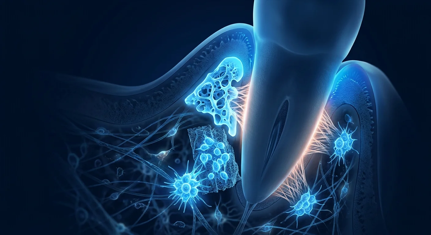
The healthy periodontium is a precisely organised complex of four tissues: the gingiva (gum), the periodontal ligament (PDL — a collagenous sling that connects tooth root to bone), the cementum (a thin mineralised layer covering the root surface), and the alveolar bone. Together they form a functional unit that absorbs occlusal forces, seals the tooth against bacterial ingress, and maintains the tooth's position within the arch. In periodontitis, a polymicrobial biofilm accumulates in the subgingival sulcus, and the host's inflammatory response — driven by neutrophils, macrophages, and T cells — releases matrix metalloproteinases (MMPs), IL-1β, TNF-α, and RANKL. RANKL in particular activates osteoclasts, leading to progressive resorption of alveolar bone [1]. The PDL is degraded, the junctional epithelium migrates apically, and a periodontal pocket forms — a deepened space between tooth and gum that harbours the biofilm and perpetuates the cycle of destruction.
What was once considered a simple bacterial infection is now understood to be an inflammatory dysbiosis — the disease is driven less by the bacteria themselves than by the host's own disproportionate immune response. This recognition is important because it explains why mechanical debridement alone is often insufficient: removing the biofilm does not automatically reset the host response, and it does nothing to regenerate the tissues that have already been destroyed. The therapeutic goal has therefore evolved from simply "cleaning the pocket" to attempting true periodontal regeneration — the formation of new cementum, new PDL fibres inserted into that cementum, and new alveolar bone — a histological outcome known as "new attachment."
Why mesenchymal stem cells are a candidate for periodontal regeneration
The rationale for MSCs in periodontal repair rests on four overlapping capabilities that map directly onto the regenerative requirements of the periodontium. First, MSCs can differentiate into multiple periodontal lineages — under appropriate signalling, they can become cementoblast-like cells, PDL fibroblast-like cells, and osteoblasts, each of which is required for the corresponding tissue compartment [2]. Dental pulp stem cells (DPSCs), periodontal ligament stem cells (PDLSCs), and gingival mesenchymal stem cells (GMSCs) are tissue-resident MSC populations that have been identified within the oral cavity itself, and they exhibit particularly strong odontogenic and cementogenic differentiation potential. Umbilical-cord and bone-marrow MSCs, while not oral-tissue-resident, have also been shown to support periodontal regeneration when delivered locally.
Second, MSCs are potently immunomodulatory in the periodontal microenvironment. They suppress the activation of inflammatory macrophages, shift the T-cell balance from Th17 toward regulatory T cells (Tregs), and downregulate the expression of RANKL — the master driver of osteoclast-mediated bone resorption [3]. In the periodontitis context, where bone loss is driven precisely by RANKL-expressing inflammatory cells, this immunomodulation amounts to a direct intervention on the disease mechanism, not merely a symptomatic one.
Third, MSCs secrete a rich cocktail of trophic and angiogenic factors — VEGF, HGF, IGF-1, FGF-2, and PDGF — that promote the formation of new blood vessels, attract endogenous progenitor cells, and support the survival of the cells already present in the wound [4]. Periodontal regeneration is a highly vascular process, and the angiogenic capacity of MSCs is thought to be as important as any direct differentiation they undergo.
Fourth, MSC-derived exosomes and extracellular vesicles have emerged as a cell-free alternative with substantial preclinical support. These nanoscale particles carry microRNAs, growth factors, and signalling lipids that recapitulate many of the regenerative and immunomodulatory effects of the parent cells without the logistical complexity of live-cell transplantation [5]. Several groups have reported that MSC exosomes alone can reduce periodontal inflammation and promote bone and ligament regeneration in animal models, opening a potentially simpler regulatory path.
What the clinical evidence actually shows
The clinical literature on MSCs for periodontal regeneration is considerably smaller than for orthopaedic indications, but it is growing steadily and the signals are consistent. Systematic reviews aggregating the available human studies — predominantly small pilot trials and case series — report that MSC-based approaches are associated with statistically significant gains in clinical attachment level (CAL), probing depth (PD) reduction, and radiographic bone fill when compared with open-flap debridement alone or with scaffold-only controls [6][7].
Several studies merit attention. A 2020 randomised controlled trial by Sánchez and colleagues evaluated autologous PDL-derived MSCs delivered on a collagen scaffold for the treatment of intrabony defects, reporting significantly greater CAL gain and radiographic bone fill in the MSC group at 12 months compared with the scaffold-only group [8]. A 2016 trial by Chen et al. used autologous PDL stem cell sheets for intrabony defects — the cell-sheet approach preserves extracellular matrix and cell-cell junctions — and demonstrated not only clinical improvement but also histological evidence of new cementum, PDL, and bone formation in a subset of patients who consented to re-entry [9]. These histological data are critical: they confirm that the radiographic improvements seen on X-rays correspond to genuine regeneration, not simply scar repair.
The cell sources studied span the full MSC landscape. Autologous PDLSCs and DPSCs have produced the most compelling histological data, likely because they are developmentally programmed for periodontal differentiation. Bone-marrow MSCs (both autologous and allogeneic) have been used in several trials with favourable clinical outcomes, while umbilical-cord MSCs — favoured for their high proliferative capacity, low immunogenicity, and off-the-shelf availability — are the subject of active investigation [10]. Gingival MSCs (GMSCs), which can be harvested from a small gingival biopsy under local anaesthesia, represent an attractive autologous option with minimal donor-site morbidity and are being evaluated in early-phase trials [11].
The honest headline
As of mid-2026, no MSC product has received regulatory approval as a standard-of-care treatment for periodontal regeneration. The clinical evidence base consists primarily of small RCTs and case series with follow-up typically limited to 12 months. The signals for CAL gain and bone fill are consistent and biologically plausible, but the field has not yet produced the large, multicentre, randomised trials that would be required for regulatory approval or guideline inclusion. The responsible description is advanced investigational therapy with encouraging early clinical signals, not a proven standard of care. Any clinic promising guaranteed bone regeneration or complete periodontal restoration is misrepresenting the data.
How periodontal outcomes are measured — and what MSCs actually move
To weigh the evidence seriously requires knowing what is being measured. In periodontal trials, the gold-standard clinical outcome is the clinical attachment level (CAL) — the distance from the cementoenamel junction to the base of the periodontal pocket, measured with a calibrated periodontal probe. CAL gain (a reduction in this distance) is the strongest indicator that new attachment has occurred. Probing depth (PD) measures pocket depth alone and can improve from gingival recession as well as from true gain of attachment, so it is a secondary rather than primary endpoint. Bleeding on probing (BOP) indicates active inflammation. On the radiographic side, intraoral periapical radiographs and cone-beam computed tomography (CBCT) can quantify the linear bone fill within an intrabony defect, expressed as a percentage of the original defect depth [12].
Across the published trials, MSC-treated sites show mean CAL gains in the range of 2–4 mm and radiographic bone fill of 30–60% of the original defect depth at 6–12 months — both exceeding the results typically achieved with open-flap debridement alone (CAL gain ~1–2 mm, bone fill ~10–20%). These are clinically meaningful numbers: a 3 mm CAL gain can shift a tooth from a "hopeless" prognosis to one that is maintainable. The strongest outcomes have been reported in three-wall intrabony defects — the contained anatomy of a 3-wall defect provides a natural scaffold for the regenerative process — while the weakest signals are seen in suprabony (horizontal) defects where there is no bony wall to contain the graft material [13].
What the evidence supports — and what it doesn't
A fair reading of the data supports several conclusions. MSC-based therapies for intrabony periodontal defects have a well-established short-term safety record, with adverse events generally limited to transient post-operative swelling and discomfort. They consistently improve clinical attachment levels and radiographic bone fill relative to controls in the available studies, with effect sizes that are clinically meaningful. There is histological confirmation from a small number of studies that the gains represent true regeneration (new cementum, PDL, and bone) rather than simple repair (long junctional epithelium).
What the evidence does NOT yet support is equally important. It does not establish that MSCs can regenerate the entire periodontium in advanced horizontal bone loss — the structural containment of a defect matters enormously. It does not confirm durability beyond 12–24 months, as long-term follow-up data are scarce. It does not identify a single optimal cell type, dose, or delivery method — PDLSCs, DPSCs, BM-MSCs, UC-MSCs, and GMSCs have each been studied in different protocols with different scaffolds, making direct comparisons impossible. And it does not demonstrate that MSC therapy is superior to established regenerative techniques such as enamel matrix derivative (Emdogain®) or GTR with barrier membranes — the trials have largely compared MSCs against open-flap debridement, not against the best available conventional regenerative treatment.
- Defect morphology matters. Three-wall intrabony defects are the population in whom the strongest signals have been seen. Horizontal bone loss with a suprabony configuration is less amenable to a contained regenerative approach.
- Scaffold choice is non-trivial. The delivery vehicle — collagen sponge, β-TCP, hyaluronic acid gel, or cell sheet — influences cell retention, viability, and differentiation. A poor scaffold can negate the benefits of a good cell product.
- Cell-free approaches are the wildcard. MSC-derived exosomes and conditioned medium have shown comparable regenerative effects in preclinical models and would be substantially easier to manufacture, store, and regulate than live cells. This is an area to watch closely in the next 3–5 years.
Periodontal regeneration is not about filling a hole in bone — it is about reconstructing a complex, multi-tissue interface at the micron scale. The honest contribution of MSC therapy is likely to be in defects that still have bony walls to contain the regenerative process.
— VELAR Clinical Team
How to evaluate any offer responsibly
If you are considering MSC therapy for periodontal disease, the diligence follows the same principles that apply across all of regenerative medicine. Ask what cell type and source are used — oral-tissue-derived MSCs (PDLSCs, DPSCs, GMSCs) have the strongest rationale for periodontal applications, but umbilical-cord and bone-marrow MSCs also have data. Ask whether the provider can cite published data for their specific protocol, not just general references to stem cell biology. Ask what outcome measures are tracked — a provider who measures your CAL, PD, and BOP before and after treatment, and who takes standardised radiographs, is operating at a different level of rigour from one offering only testimonials. Ask about cell characterisation and sterility testing: ISCT-compliant identity and viability testing are minimum standards for any clinical MSC product. And ask what scaffold or delivery system is used — MSCs injected as a liquid suspension into a periodontal pocket will largely wash out; a scaffold that retains cells at the defect site for days to weeks is likely essential for a meaningful regenerative effect. Be deeply sceptical of claims of "full regeneration," of promises that horizontally compromised teeth with minimal remaining bone can be saved, and of any suggestion that MSC therapy replaces meticulous oral hygiene and regular professional maintenance — it does not.
The VELAR perspective
At VELAR Center, we approach periodontal regeneration with the same disciplined conservatism we apply to every indication. The oral cavity is an immunologically complex, microbiologically active environment, and the regenerative bar is high — new cementum, new PDL, new bone, all in the correct spatial relationship. We view MSC therapy for periodontal defects as a biologically rational intervention with encouraging early data, but one that is still in an active phase of evidence accumulation. We use umbilical-cord-derived MSCs manufactured under cGMP conditions with ISCT-compliant characterisation and third-party sterility release, and we work closely with the patient's periodontist to ensure that any biologic intervention is integrated into a comprehensive treatment plan that includes definitive mechanical debridement, occlusal management, and long-term maintenance. Every consultation begins with an honest conversation about what the evidence says, what it does not say, and whether, given your specific defect anatomy and periodontal status, a biologic approach is a rational complement to conventional care — or whether the data suggest you would be better served by established regenerative techniques alone. That is the standard we would want for our own families, and it is the only standard we offer.
References
- Hajishengallis G. Periodontitis: from microbial immune subversion to systemic inflammation. Nature Reviews Immunology. 2015;15(1):30-44. doi:10.1038/nri3785 ↩
- Seo BM, Miura M, Gronthos S, et al. Investigation of multipotent postnatal stem cells from human periodontal ligament. The Lancet. 2004;364(9429):149-155. doi:10.1016/S0140-6736(04)16627-0 ↩
- Liu Y, Wang L, Kikuiri T, et al. Mesenchymal stem cell-based tissue regeneration is governed by recipient T lymphocytes via IFN-γ and TNF-α. Nature Medicine. 2011;17(12):1594-1601. doi:10.1038/nm.2542 ↩
- Du J, Li Y, Li S, et al. Allogeneic bone marrow mesenchymal stem cell-derived exosomes promote periodontal regeneration in a rat model. Stem Cell Research & Therapy. 2022;13:123. doi:10.1186/s13287-022-02797-y ↩
- Chew JRJ, Chuah SJ, Teo KYW, et al. Mesenchymal stem cell exosomes enhance periodontal ligament cell functions and promote periodontal regeneration. Acta Biomaterialia. 2019;89:252-264. doi:10.1016/j.actbio.2019.03.021 ↩
- Bright R, Hynes K, Gronthos S, Bartold PM. Periodontal ligament-derived cells for periodontal regeneration in animal models: a systematic review. Journal of Periodontal Research. 2015;50(2):160-172. doi:10.1111/jre.12205 ↩
- Bassir SH, Wisitrasameewong W, Raanan J, et al. Potential for stem cell-based periodontal therapy. Journal of Cellular Physiology. 2016;231(1):50-61. doi:10.1002/jcp.25067 ↩
- Sánchez N, Fierravanti L, Núñez J, et al. Periodontal regeneration using a xenogeneic bone substitute seeded with autologous periodontal ligament-derived mesenchymal stem cells: a 12-month quasi-randomized controlled pilot clinical trial. Journal of Clinical Periodontology. 2020;47(11):1391-1402. doi:10.1111/jcpe.13368 ↩
- Chen FM, Gao LN, Tian BM, et al. Treatment of periodontal intrabony defects using autologous periodontal ligament stem cells: a randomized clinical trial. Stem Cell Research & Therapy. 2016;7:33. doi:10.1186/s13287-016-0288-1 ↩
- Yamada Y, Nakamura-Yamada S, Kusano K, Baba S. Clinical potential and current progress of dental pulp stem cells for various systemic diseases in regenerative medicine: a concise review. International Journal of Molecular Sciences. 2019;20(5):1132. doi:10.3390/ijms20051132 ↩
- Zhang Q, Shi S, Liu Y, et al. Mesenchymal stem cells derived from human gingiva are capable of immunomodulatory functions and ameliorate inflammation-related tissue destruction in experimental colitis. Journal of Immunology. 2009;183(12):7787-7798. doi:10.4049/jimmunol.0902318 ↩
- Cortellini P, Tonetti MS. Clinical concepts for regenerative therapy in intrabony defects. Periodontology 2000. 2015;68(1):282-307. doi:10.1111/prd.12048 ↩
- Bartold PM, Gronthos S, Ivanovski S, Fisher A, Hutmacher DW. Tissue engineered periodontal products. Journal of Periodontal Research. 2016;51(1):1-15. doi:10.1111/jre.12275 ↩
- Iwata T, Yamato M, Tsuchioka H, et al. Periodontal regeneration with multi-layered periodontal ligament-derived cell sheets in a canine model. Biomaterials. 2009;30(14):2716-2723. doi:10.1016/j.biomaterials.2009.01.032 ↩
- Nuñez J, Vignoletti F, Caffesse RG, Sanz M. Cellular therapy in periodontal regeneration. Periodontology 2000. 2019;79(1):107-116. doi:10.1111/prd.12250 ↩
牙周病是人类最常见的慢性炎症性疾病——影响近半数30岁以上成人和约70%的65岁以上人群。然而它常被低估为仅仅是"牙龈问题"。实际上,牙周炎是一种破坏性过程,细菌生物膜触发宿主炎症反应,逐步破坏牙周韧带、牙槽骨和牙龈结缔组织——这些正是将牙齿固定在位的结构。与攻击牙冠的龋齿不同,牙周炎攻击的是牙齿的基础。几十年来,治疗主要是机械性的——刮治和根面平整、翻瓣手术、骨移植以及屏障膜引导组织再生(GTR)。这些方法可以阻止疾病进展,但很少实现丢失牙周组织的真正再生。间充质干细胞(MSC)研究现在提出的问题是:修复生物学能否被有意义地引导来再生已丢失的组织。
牙周受损部位内部究竟发生了什么
健康的牙周组织是一个精确组织的四种组织复合体:牙龈、牙周韧带(PDL——连接牙根与骨的胶原悬带)、牙骨质(覆盖根面的薄矿化层)和牙槽骨。它们共同形成一个功能单位,吸收咬合力,封闭牙齿防止细菌侵入,并维持牙齿在牙弓中的位置。在牙周炎中,多微生物生物膜在龈下沟积聚,宿主的炎症反应——由中性粒细胞、巨噬细胞和T细胞驱动——释放基质金属蛋白酶(MMPs)、IL-1β、TNF-α和RANKL。RANKL特别激活破骨细胞,导致牙槽骨的进行性吸收 [1]。PDL被降解,结合上皮向根方迁移,形成牙周袋——牙齿与牙龈之间的加深空间,藏匿生物膜并延续破坏循环。
曾经被认为是简单细菌感染的牙周炎,现在被理解为一种炎症性菌群失调——疾病更多由宿主自身不成比例的免疫反应驱动,而非细菌本身。这一认识很重要,因为它解释了为什么单纯机械清创通常不够:去除生物膜并不会自动重置宿主反应,也不会再生已经被破坏的组织。因此,治疗目标已从简单的"清理牙周袋"演变为尝试真正的牙周再生——形成新的牙骨质、插入该牙骨质的新PDL纤维和新牙槽骨——一种被称为"新附着"的组织学结果。
为何间充质干细胞是牙周再生的候选
MSC用于牙周修复的理论基础基于四个相互重叠的能力,直接映射到牙周组织的再生需求。第一,MSC可以分化为多种牙周谱系——在适当信号下,它们可以成为成牙骨质细胞样细胞、PDL成纤维细胞样细胞和成骨细胞,每种都是相应组织区室所需的 [2]。牙髓干细胞(DPSCs)、牙周韧带干细胞(PDLSCs)和牙龈间充质干细胞(GMSCs)是已在口腔内鉴定出的组织驻留MSC群体,它们表现出特别强的成牙本质和成牙骨质分化潜能。脐带和骨髓MSC虽非口腔组织驻留,但在局部递送时也已被证明支持牙周再生。
第二,MSC在牙周微环境中具有强大的免疫调节作用。它们抑制炎症巨噬细胞的激活,将T细胞平衡从Th17转向调节性T细胞(Tregs),并下调RANKL的表达——破骨细胞介导的骨吸收的主要驱动因子 [3]。在牙周炎背景下,骨丢失正是由表达RANKL的炎症细胞驱动的,这种免疫调节相当于对疾病机制的直接干预,而不仅仅是对症治疗。
第三,MSC分泌丰富的营养和血管生成因子——VEGF、HGF、IGF-1、FGF-2和PDGF——促进新血管形成,吸引内源性祖细胞,并支持伤口中已有细胞的存活 [4]。牙周再生是一个高度血管化的过程,MSC的血管生成能力被认为与其经历的任何直接分化同等重要。
第四,MSC来源的外泌体和细胞外囊泡已成为一种无细胞替代方案,拥有大量临床前支持。这些纳米级颗粒携带microRNA、生长因子和信号脂质,再现了亲代细胞的许多再生和免疫调节效应,而无需活细胞移植的后勤复杂性 [5]。多个研究组报告称,MSC外泌体单独使用即可在动物模型中减少牙周炎症并促进骨和韧带再生,开辟了一条可能更简单的监管路径。
临床证据实际显示了什么
MSC用于牙周再生的临床文献比骨科适应症少得多,但正在稳步增长,信号是一致的。汇总现有的人类研究(主要是小型试验性试验和病例系列)的系统评价报告称,基于MSC的方法与单独开放翻瓣清创或仅使用支架的对照相比,与临床附着水平(CAL)的统计学显著改善、探诊深度(PD)减少和影像学骨填充相关 [6][7]。
几项研究值得关注。Sánchez及其同事2020年的一项随机对照试验评估了在胶原支架上递送的自体PDL来源MSC治疗骨内缺损,报告12个月时MSC组的CAL获得和影像学骨填充显著大于仅使用支架的组 [8]。Chen等人2016年的一项试验使用自体PDL干细胞片治疗骨内缺损——细胞片方法保留了细胞外基质和细胞间连接——不仅展示了临床改善,还在同意再次进入的亚组患者中展示了新牙骨质、PDL和骨形成的组织学证据 [9]。这些组织学数据至关重要:它们确认X光片上看到的影像学改善对应真正的再生,而不仅仅是瘢痕修复。
研究的细胞来源涵盖整个MSC谱系。自体PDLSCs和DPSCs产生了最引人注目的组织学数据,可能是因为它们在发育上被编程为牙周分化。骨髓MSC(自体和异体)已在多项试验中使用,临床结果良好,而脐带MSC——因其高增殖能力、低免疫原性和即用性而受到青睐——是积极研究的对象 [10]。牙龈MSC(GMSCs)可通过局麻下小牙龈活检获取,是一种有吸引力的自体选择,供区并发症最小,正在早期试验中评估 [11]。
诚实的标题
截至2026年中,没有任何MSC产品获得监管批准作为牙周再生的标准治疗。临床证据基础主要由小型RCT和病例系列组成,随访通常限于12个月。CAL获得和骨填充的信号是一致的且在生物学上合理的,但该领域尚未产生监管批准或指南纳入所需的大型、多中心、随机试验。负责任的描述是具有令人鼓舞的早期临床信号的先进研究性疗法,而非已证实的标准治疗。任何承诺保证骨再生或完全牙周修复的诊所都在歪曲数据。
牙周结果如何衡量——以及MSC实际改变了什么
要认真评估证据,需要知道衡量的是什么。在牙周试验中,金标准的临床结果是临床附着水平(CAL)——从釉牙骨质界到牙周袋基底的距离,使用校准的牙周探针测量。CAL获得(该距离的减少)是新附着已经发生的最强指标。探诊深度(PD)仅测量袋深,可因牙龈退缩和真正的附着获得而改善,因此是次要而非主要终点。探诊出血(BOP)指示活动性炎症。在影像学方面,口内根尖片和锥束CT(CBCT)可以量化骨内缺损内的线性骨填充量,以原始缺损深度的百分比表示 [12]。
在已发表的试验中,MSC治疗部位在6–12个月时显示平均CAL获得2–4 mm,影像学骨填充为原始缺损深度的30–60%——两者均超过仅开放翻瓣清创通常达到的结果(CAL获得~1–2 mm,骨填充~10–20%)。这些是具有临床意义的数字:3 mm的CAL获得可以将一颗牙齿从"无望"的预后转变为可维持的。最强的结果在三壁骨内缺损中报告——三壁缺损的封闭解剖为再生过程提供了天然支架——而最弱的信号见于骨上(水平)缺损,其中没有骨壁来容纳移植材料 [13]。
证据支持什么——又不支持什么
对数据的公允解读支持几个结论。基于MSC的骨内牙周缺损治疗拥有确立良好的短期安全记录,不良事件通常限于短暂的术后肿胀和不适。在现有研究中,它们相对于对照组持续改善临床附着水平和影像学骨填充,效应量具有临床意义。有来自少量研究的组织学确认,表明这些改善代表真正的再生(新牙骨质、PDL和骨)而非简单修复(长结合上皮)。
证据尚未支持的内容同样重要。它并未确立MSC可以在晚期水平骨丢失中再生整个牙周组织——缺损的结构封闭性至关重要。它并未确认超过12–24个月的持久性,因为长期随访数据稀缺。它并未确定单一最佳细胞类型、剂量或递送方法——PDLSCs、DPSCs、BM-MSCs、UC-MSCs和GMSCs各自在不同方案中使用不同支架进行研究,无法进行直接比较。它并未证明MSC治疗优于已确立的再生技术,如釉基质衍生物(Emdogain®)或带屏障膜的GTR——试验主要将MSC与开放翻瓣清创进行比较,而非与最佳可用的常规再生治疗进行比较。
- 缺损形态很重要。三壁骨内缺损是观察到最强信号的人群。具有骨上构型的水平骨丢失不太适合封闭的再生方法。
- 支架选择非小事。递送载体——胶原海绵、β-TCP、透明质酸凝胶或细胞片——影响细胞保留、活性和分化。差的支架可以抵消良好细胞产品的益处。
- 无细胞方法是变数。MSC来源的外泌体和条件培养基在临床前模型中显示出可比的再生效应,且制造、储存和监管将比活细胞显著更简单。这是未来3–5年值得密切关注的领域。
牙周再生不是填充骨中的洞——而是在微米尺度上重建一个复杂的多组织界面。MSC治疗的诚实贡献可能在于仍有骨壁容纳再生过程的缺损中。
— VELAR 临床团队
如何负责任地评估任何提议
如果您正在考虑牙周病的MSC治疗,所需的审慎遵循适用于整个再生医学领域的相同原则。询问使用何种细胞类型和来源——口腔组织来源的MSC(PDLSCs、DPSCs、GMSCs)对牙周应用具有最强的理论基础,但脐带和骨髓MSC也有数据。询问提供者能否为其特定方案引用已发表的数据,而非仅对干细胞生物学的一般性引用。询问追踪什么结果测量——一个在治疗前后测量您的CAL、PD和BOP,并拍摄标准化X光片的提供者,其操作严谨程度与仅提供患者感言的诊所不同。询问细胞鉴定和无菌检测:ISCT合规的身份和活性检测是任何临床MSC产品的最低标准。询问使用何种支架或递送系统——作为液体悬液注射到牙周袋中的MSC将大部分被冲走;一种能将细胞在缺损部位保留数天到数周的支架对于有意义的再生效应可能是必需的。对"完全再生"的声称、对骨量极少且水平受损的牙齿能被挽救的承诺,以及对MSC治疗替代细致的口腔卫生和定期专业维护的任何暗示,都要深度怀疑——它不能。
VELAR 的观点
在VELAR中心,我们对待牙周再生的态度与我们对待每个适应症的严谨保守主义相同。口腔是一个免疫学复杂、微生物活跃的环境,再生的门槛很高——新牙骨质、新PDL、新骨,全部在正确的空间关系中。我们将MSC治疗牙周缺损视为一种具有令人鼓舞早期数据的生物学合理干预,但仍处于证据积累的活跃阶段。我们使用在cGMP条件下制造、具有ISCT合规鉴定和第三方无菌放行的脐带来源MSC,并与患者的牙周科医生密切合作,确保任何生物学干预被整合到包括确定性机械清创、咬合管理和长期维护在内的综合治疗计划中。每次咨询都以关于证据说了什么、没说什么的诚实对话开始,以及根据您的具体缺损解剖和牙周状态,生物学方法是否是对常规治疗的合理补充——还是数据表明您更好地仅由已确立的再生技术来服务。这是我们希望为自家家人设定的标准,也是我们提供的唯一标准。
参考文献
- Hajishengallis G. Periodontitis: from microbial immune subversion to systemic inflammation. Nature Reviews Immunology. 2015;15(1):30-44. doi:10.1038/nri3785 ↩
- Seo BM, Miura M, Gronthos S, et al. Investigation of multipotent postnatal stem cells from human periodontal ligament. The Lancet. 2004;364(9429):149-155. doi:10.1016/S0140-6736(04)16627-0 ↩
- Liu Y, Wang L, Kikuiri T, et al. Mesenchymal stem cell-based tissue regeneration is governed by recipient T lymphocytes via IFN-γ and TNF-α. Nature Medicine. 2011;17(12):1594-1601. doi:10.1038/nm.2542 ↩
- Du J, Li Y, Li S, et al. Allogeneic bone marrow mesenchymal stem cell-derived exosomes promote periodontal regeneration in a rat model. Stem Cell Research & Therapy. 2022;13:123. doi:10.1186/s13287-022-02797-y ↩
- Chew JRJ, Chuah SJ, Teo KYW, et al. Mesenchymal stem cell exosomes enhance periodontal ligament cell functions and promote periodontal regeneration. Acta Biomaterialia. 2019;89:252-264. doi:10.1016/j.actbio.2019.03.021 ↩
- Bright R, Hynes K, Gronthos S, Bartold PM. Periodontal ligament-derived cells for periodontal regeneration in animal models: a systematic review. Journal of Periodontal Research. 2015;50(2):160-172. doi:10.1111/jre.12205 ↩
- Bassir SH, Wisitrasameewong W, Raanan J, et al. Potential for stem cell-based periodontal therapy. Journal of Cellular Physiology. 2016;231(1):50-61. doi:10.1002/jcp.25067 ↩
- Sánchez N, Fierravanti L, Núñez J, et al. Periodontal regeneration using a xenogeneic bone substitute seeded with autologous periodontal ligament-derived mesenchymal stem cells: a 12-month quasi-randomized controlled pilot clinical trial. Journal of Clinical Periodontology. 2020;47(11):1391-1402. doi:10.1111/jcpe.13368 ↩
- Chen FM, Gao LN, Tian BM, et al. Treatment of periodontal intrabony defects using autologous periodontal ligament stem cells: a randomized clinical trial. Stem Cell Research & Therapy. 2016;7:33. doi:10.1186/s13287-016-0288-1 ↩
- Yamada Y, Nakamura-Yamada S, Kusano K, Baba S. Clinical potential and current progress of dental pulp stem cells for various systemic diseases in regenerative medicine: a concise review. International Journal of Molecular Sciences. 2019;20(5):1132. doi:10.3390/ijms20051132 ↩
- Zhang Q, Shi S, Liu Y, et al. Mesenchymal stem cells derived from human gingiva are capable of immunomodulatory functions and ameliorate inflammation-related tissue destruction in experimental colitis. Journal of Immunology. 2009;183(12):7787-7798. doi:10.4049/jimmunol.0902318 ↩
- Cortellini P, Tonetti MS. Clinical concepts for regenerative therapy in intrabony defects. Periodontology 2000. 2015;68(1):282-307. doi:10.1111/prd.12048 ↩
- Bartold PM, Gronthos S, Ivanovski S, Fisher A, Hutmacher DW. Tissue engineered periodontal products. Journal of Periodontal Research. 2016;51(1):1-15. doi:10.1111/jre.12275 ↩
- Iwata T, Yamato M, Tsuchioka H, et al. Periodontal regeneration with multi-layered periodontal ligament-derived cell sheets in a canine model. Biomaterials. 2009;30(14):2716-2723. doi:10.1016/j.biomaterials.2009.01.032 ↩
- Nuñez J, Vignoletti F, Caffesse RG, Sanz M. Cellular therapy in periodontal regeneration. Periodontology 2000. 2019;79(1):107-116. doi:10.1111/prd.12250 ↩
مرض دواعم السن هو أكثر الحالات الالتهابية المزمنة شيوعاً لدى البشر — يصيب قرابة نصف البالغين فوق 30 عاماً ونحو 70% ممن تجاوزوا 65 عاماً. ومع ذلك كثيراً ما يُستخف به على أنه مجرد "مشاكل لثة". في الواقع، التهاب دواعم السن عملية تدميرية تثير فيها الأغشية الحيوية البكتيرية استجابة التهابية من المضيف تدمر تدريجياً الرباط الدواعمي والعظم السنخي والنسيج الضام اللثوي — وهي تحديداً البنى التي تثبّت الأسنان في مكانها. وبخلاف التسوس الذي يهاجم تاج السن، يهاجم التهاب دواعم السن أساس السن. ولعقود، كان العلاج ميكانيكياً إلى حد كبير — تقليح وكشط الجذور وجراحة السديلة وتطعيم العظم وتجديد النسيج الموجَّه (GTR) بأغشية حاجزة. يمكن لهذه المقاربات أن توقف تقدم المرض، لكنها نادراً ما تحقق تجديداً حقيقياً للنسيج الدواعمي المفقود. والسؤال الذي تطرحه أبحاث الخلايا الجذعية الوسيطة (MSC) الآن هو: هل يمكن توجيه بيولوجيا الإصلاح بشكل ذي معنى لإعادة إنماء ما فُقد.
ما الذي يحدث فعلياً داخل الموقع المصاب دواعمياً
النسيج الدواعمي السليم معقد منظم بدقة من أربعة أنسجة: اللثة والرباط الدواعمي (PDL — حبال كولاجينية تصل جذر السن بالعظم) والملاط (طبقة رقيقة متمعدنة تغطي سطح الجذر) والعظم السنخي. تشكل معاً وحدة وظيفية تمتص قوى الإطباق وتغلق السن ضد الدخول البكتيري وتحافظ على وضع السن داخل القوس. في التهاب دواعم السن، تتراكم أغشية حيوية متعددة الميكروبات في التلم تحت اللثوي، وتطلق الاستجابة الالتهابية للمضيف — المدفوعة بالعدلات والبلاعم والخلايا التائية — بروتيِنات المطرس المعدنية (MMPs) وIL-1β وTNF-α وRANKL. ينشط RANKL تحديداً ناقضات العظم، مؤدياً إلى ارتشاف تقدمي للعظم السنخي [1]. يتحلل PDL وتهاجر الظهارة الموصلية ذروياً ويتكون جيب دواعمي — حيّز متعمق بين السن واللثة يأوي الغشاء الحيوي ويديم دورة التدمير.
ما كان يُعتبر يوماً عدوى بكتيرية بسيطة صار يُفهم الآن على أنه خلل توازن ميكروبي التهابي — المرض تدفعه استجابة المضيف المناعية غير المتناسبة أكثر مما تدفعه البكتيريا نفسها. وهذا الإدراك مهم لأنه يفسر لماذا لا يكفي التنضير الميكانيكي وحده في كثير من الأحيان: إزالة الغشاء الحيوي لا تعيد ضبط استجابة المضيف تلقائياً، ولا تفعل شيئاً لتجديد الأنسجة التي دُمرت بالفعل. لذلك تطور الهدف العلاجي من مجرد "تنظيف الجيب" إلى محاولة تحقيق تجديد دواعمي حقيقي — تكوين ملاط جديد وألياف PDL جديدة مغروزة في ذلك الملاط وعظم سنخي جديد — نتيجة نسيجية تُعرف بـ "الارتباط الجديد".
لماذا الخلايا الجذعية الوسيطة مرشحة لتجديد دواعم السن
يقوم الأساس المنطقي لاستخدام MSCs في إصلاح دواعم السن على أربع قدرات متداخلة تنطبق مباشرة على المتطلبات التجديدية للنسيج الدواعمي. أولاً، يمكن لـ MSCs أن تتمايز إلى سلاسل دواعمية متعددة — تحت إشارات مناسبة، يمكنها أن تصبح خلايا شبيهة بالأرومة الملاطية وخلايا شبيهة بأرومة PDL الليفية وبانيات عظم، وكل منها مطلوب للحجرة النسيجية المناظرة [2]. الخلايا الجذعية للب السني (DPSCs) والخلايا الجذعية للرباط الدواعمي (PDLSCs) والخلايا الجذعية الوسيطة اللثوية (GMSCs) هي جمهرات MSC مقيمة في النسيج تم تحديدها داخل جوف الفم نفسه، وتظهر إمكانات تمايز سنية وملاطية قوية بشكل خاص. وخلايا MSC الحبل السري ونخاع العظم، رغم أنها ليست مقيمة في النسيج الفموي، أظهرت أيضاً أنها تدعم التجديد الدواعمي عند توصيلها موضعياً.
ثانياً، MSCs معدِّلة مناعياً بقوة في البيئة الدقيقة الدواعمية. تثبّط تنشيط البلاعم الالتهابية، وتنقل توازن الخلايا التائية من Th17 نحو الخلايا التائية التنظيمية (Tregs)، وتخفض التعبير عن RANKL — المحرك الرئيس لارتشاف العظم المتوسط بناقضات العظم [3]. وفي سياق التهاب دواعم السن، حيث يُدفع فقدان العظم تحديداً بالخلايا الالتهابية المعبّرة عن RANKL، فإن هذا التعديل المناعي يرقى إلى تدخل مباشر على آلية المرض، لا مجرد تدخل عرضي.
ثالثاً، تفرز MSCs مزيجاً غنياً من العوامل الغذائية والمولّدة للأوعية — VEGF وHGF وIGF-1 وFGF-2 وPDGF — تعزز تكوين أوعية دموية جديدة وتجذب الخلايا السلفية الذاتية وتدعم بقاء الخلايا الموجودة أصلاً في الجرح [4]. التجديد الدواعمي عملية عالية التوعية، ويُعتقد أن قدرة MSCs المولّدة للأوعية لا تقل أهمية عن أي تمايز مباشر تخضع له.
رابعاً، برزت الإكسوزومات والحويصلات خارج الخلوية مشتقة MSC كبديل خالٍ من الخلايا مع دعم سريري مسبق كبير. تحمل هذه الجسيمات النانوية microRNA وعوامل نمو وليبيدات إشارية تستنسخ كثيراً من التأثيرات التجديدية والمعدِّلة للمناعة للخلايا الأم دون التعقيد اللوجستي لزرع الخلايا الحية [5]. أبلغت مجموعات عدة أن إكسوزومات MSC وحدها يمكنها تقليل الالتهاب الدواعمي وتعزيز تجديد العظم والرباط في النماذج الحيوانية، فاتحةً طريقاً تنظيمياً أبسط محتملاً.
ماذا تظهر الأدلة السريرية فعلياً
الأدبيات السريرية حول MSCs لتجديد دواعم السن أصغر بكثير مما هي عليه لاستطبابات العظام، لكنها تنمو باطراد والإشارات متسقة. تفيد المراجعات المنهجية التي تجمع الدراسات البشرية المتاحة — وغالباً تجارب استطلاعية صغيرة وسلاسل حالات — بأن المقاربات المعتمدة على MSC ترتبط بمكاسب ذات دلالة إحصائية في مستوى الارتباط السريري (CAL) وتقليل عمق السبر (PD) وملء عظمي شعاعي عند مقارنتها بالتنضير بالسديلة المفتوحة وحده أو بشواهد السقالة فقط [6][7].
تستحق دراسات عدة الانتباه. قيّمت تجربة معشاة مضبوطة أجراها Sánchez وزملاؤه عام 2020 خلايا PDL MSC ذاتية موصَّلة على سقالة كولاجين لعلاج العيوب داخل العظم، وأبلغت عن مكسب CAL وملء عظمي شعاعي أكبر بكثير في مجموعة MSC عند 12 شهراً مقارنة بمجموعة السقالة فقط [8]. واستخدمت تجربة أجراها Chen وزملاؤه عام 2016 صفائح خلايا PDL جذعية ذاتية للعيوب داخل العظم — تحافظ مقاربة الصفيحة الخلوية على المطرس خارج الخلوي والوصلات بين الخلوية — وأظهرت ليس فقط تحسناً سريرياً بل أيضاً دليلاً نسجياً على تكوين ملاط جديد وPDL وعظم في مجموعة فرعية من المرضى وافقوا على إعادة الدخول [9]. وهذه البيانات النسيجية حاسمة: فهي تؤكد أن التحسنات الشعاعية المرئية على الأشعة تقابل تجديداً حقيقياً، لا مجرد إصلاح نَدَبي.
مصادر الخلايا المدروسة تغطي كامل طيف MSC. أنتجت PDLSCs وDPSCs الذاتية أكثر البيانات النسيجية إقناعاً، ربما لأنها مبرمجة تطورياً للتمايز الدواعمي. واستُخدمت MSCs نخاع العظم (الذاتية والخيفية) في تجارب عدة بنتائج سريرية مواتية، بينما MSCs الحبل السري — المفضَّلة لقدرتها التكاثرية العالية وانخفاض مناعيتها وتوفرها الجاهز — هي موضوع بحث نشط [10]. وتمثل MSCs اللثوية (GMSCs)، التي يمكن حصادها من خزعة لثوية صغيرة تحت تخدير موضعي، خياراً ذاتياً جذاباً بأقل مراضة للموقع المانح وتُقيَّم في تجارب المرحلة المبكرة [11].
العنوان الصادق
حتى منتصف 2026، لم يحصل أي منتج MSC على اعتماد تنظيمي كعلاج معياري لتجديد دواعم السن. تتكون قاعدة الأدلة السريرية أساساً من تجارب RCTs صغيرة وسلاسل حالات مع متابعة محدودة عادةً بـ 12 شهراً. إشارات مكسب CAL والملء العظمي متسقة ومعقولة بيولوجياً، لكن الحقل لم ينتج بعد التجارب الكبيرة متعددة المراكز المعشّاة المطلوبة للاعتماد التنظيمي أو الإدراج في التوجيهات الإرشادية. الوصف المسؤول هو علاج بحثي متقدم ذو إشارات سريرية مبكرة مشجّعة، لا معيار رعاية مثبت. أي عيادة تعد بتجديد عظمي مضمون أو استعادة دواعمية كاملة تسيء تمثيل البيانات.
كيف تُقاس النتائج الدواعمية — وما الذي تحرّكه MSCs فعلاً
لوزن الأدلة بجدية يتطلب معرفة ما يُقاس. في التجارب الدواعمية، النتيجة السريرية المعيارية الذهبية هي مستوى الارتباط السريري (CAL) — المسافة من الملتقى الملاطي المينائي إلى قاعدة الجيب الدواعمي، مقيسةً بمسبر دواعمي معايَر. مكسب CAL (نقصان هذه المسافة) هو أقوى مؤشر على حدوث ارتباط جديد. عمق السبر (PD) يقيس عمق الجيب وحده ويمكن أن يتحسن من الانحسار اللثوي وكذلك من المكسب الحقيقي للارتباط، لذا فهو نقطة نهاية ثانوية لا أولية. النزف عند السبر (BOP) يشير إلى التهاب نشط. وعلى الجانب الشعاعي، يمكن للصور الشعاعية الذروية داخل الفموية والتصوير المقطعي المحوسب بالحزمة المخروطية (CBCT) أن يكمّم الملء العظمي الخطي داخل عيب داخل العظم، معبَّراً عنه كنسبة مئوية من عمق العيب الأصلي [12].
عبر التجارب المنشورة، تظهر المواقع المعالَجة بـ MSC مكاسب CAL وسطية في حدود 2–4 مم وملء عظمي شعاعي بنسبة 30–60% من عمق العيب الأصلي عند 6–12 شهراً — وكلاهما يتجاوز النتائج المحققة عادةً بالتنضير بالسديلة المفتوحة وحده (مكسب CAL ~1–2 مم، ملء عظمي ~10–20%). وهذه أرقام ذات أهمية سريرية: مكسب CAL مقداره 3 مم يمكنه نقل سن من إنذار "ميؤوس منه" إلى إنذار قابل للصيانة. أبلغت أقوى النتائج في العيوب داخل العظم ثلاثية الجدران — التشريح المحتوى للعيب ثلاثي الجدران يوفر سقالة طبيعية لعملية التجديد — بينما شوهدت أضعف الإشارات في العيوب فوق العظمية (الأفقية) حيث لا يوجد جدار عظمي لاحتواء مادة الطعم [13].
ما تدعمه الأدلة — وما لا تدعمه
القراءة المنصفة للبيانات تدعم عدة استنتاجات. العلاجات المعتمدة على MSC للعيوب الدواعمية داخل العظم تملك سجل أمان قصير المدى راسخاً، مع أحداث ضائرة تقتصر عموماً على تورم وانزعاج عابرين بعد الجراحة. وهي تحسّن باستمرار مستويات الارتباط السريري والملء العظمي الشعاعي نسبةً للشواهد في الدراسات المتاحة، بأحجام أثر ذات أهمية سريرية. وهناك تأكيد نسيجي من عدد صغير من الدراسات بأن المكاسب تمثل تجديداً حقيقياً (ملاط وPDL وعظم جديد) لا مجرد إصلاح بسيط (ظهارة موصلية طويلة).
وما لا تدعمه الأدلة بعد لا يقل أهمية. فهي لا تثبت أن MSCs يمكنها تجديد كامل النسيج الدواعمي في الفقدان العظمي الأفقي المتقدم — احتواء العيب البنيوي مهم للغاية. ولا تؤكد الاستدامة بعد 12–24 شهراً، إذ تندر بيانات المتابعة طويلة المدى. ولا تحدد نمطاً خلوياً أو جرعة أو طريقة توصيل مثلى وحيدة — دُرس كل من PDLSCs وDPSCs وBM-MSCs وUC-MSCs وGMSCs في بروتوكولات مختلفة مع سقالات مختلفة، مما يجعل المقارنات المباشرة مستحيلة. ولا تبرهن أن علاج MSC أفضل من تقنيات التجديد الراسخة كمشتق مطرس المينا (Emdogain®) أو GTR بأغشية حاجزة — قارنت التجارب إلى حد كبير MSCs ضد التنضير بالسديلة المفتوحة، لا ضد أفضل علاج تجديدي تقليدي متاح.
- شكل العيب مهم. العيوب داخل العظم ثلاثية الجدران هي الفئة التي شوهدت فيها أقوى الإشارات. الفقدان العظمي الأفقي بتكوين فوق عظمي أقل قابلية لمقاربة تجديدية محتواة.
- اختيار السقالة ليس تافهاً. مركبة التوصيل — إسفنجة كولاجين أو β-TCP أو هلام حمض الهيالورونيك أو صفيحة خلوية — تؤثر على احتباس الخلايا وحيويتها وتمايزها. السقالة الرديئة يمكنها إبطال فوائد منتج خلوي جيد.
- المقاربات الخالية من الخلايا هي الورقة الرابحة. أظهرت الإكسوزومات مشتقة MSC والوسط المكيّف تأثيرات تجديدية مقارنة في النماذج قبل السريرية وستكون أسهل بكثير في التصنيع والتخزين والتنظيم من الخلايا الحية. هذا مجال يستحق المتابعة عن كثب في السنوات 3–5 القادمة.
التجديد الدواعمي ليس عن ملء ثقب في العظم — إنه عن إعادة بناء واجهة معقدة متعددة الأنسجة على مقياس الميكرون. الإسهام الصادق لعلاج MSC يرجّح أن يكون في العيوب التي لا يزال فيها جدران عظمية لاحتواء عملية التجديد.
— الفريق السريري في VELAR
كيف تقيّم أي عرض بمسؤولية
إذا كنت تفكر في علاج MSC لمرض دواعم السن، فالتدقيق المطلوب يتبع المبادئ نفسها التي تنطبق على كامل الطب التجديدي. اسأل عن نمط الخلية ومصدرها المستخدمَين — MSCs مشتقة النسيج الفموي (PDLSCs، DPSCs، GMSCs) تملك أقوى أساس منطقي للتطبيقات الدواعمية، لكن MSCs الحبل السري ونخاع العظم لديها بيانات أيضاً. واسأل إن كان بمقدور المزوّد أن يستشهد ببيانات منشورة لبروتوكوله المحدد، لا مجرد إشارات عامة لبيولوجيا الخلايا الجذعية. واسأل عن مقاييس النتائج المتابَعة — مزوّد يقيس CAL وPD وBOP لديك قبل العلاج وبعده، ويأخذ صوراً شعاعية معيارية، يعمل بمستوى دقة مختلف عن عيادة تقدّم الشهادات الشخصية فقط. واسأل عن توصيف الخلايا واختبار التعقيم: اختبار الهوية والحيوية المتوافق مع ISCT هما معايير دنيا لأي منتج MSC سريري. واسأل عن السقالة أو نظام التوصيل المستخدم — MSCs المحقونة كمعلّق سائل في جيب دواعمي ستُغسل إلى حد كبير؛ سقالة تحتفظ بالخلايا في موقع العيب لأيام إلى أسابيع ضرورية على الأرجح لتأثير تجديدي ذي معنى. كن شديد الشك تجاه ادعاءات "التجديد الكامل"، وتجاه الوعود بأن الأسنان المتضررة أفقياً ذات الحد الأدنى من العظم المتبقي يمكن إنقاذها، وتجاه أي إيحاء بأن علاج MSC يستبدل النظافة الفموية الدقيقة والصيانة المهنية المنتظمة — إنه لا يفعل.
منظور VELAR
في مركز VELAR، نقارب تجديد دواعم السن بنفس التحفظ المنضبط الذي نطبقه على كل استطباب. جوف الفم بيئة معقدة مناعياً ونشطة ميكروبياً، وعتبة التجديد عالية — ملاط جديد وPDL جديد وعظم جديد، كلها في العلاقة المكانية الصحيحة. ننظر إلى علاج MSC للعيوب الدواعمية كتدخل منطقي بيولوجياً ذي بيانات مبكرة مشجّعة، لكنه لا يزال في مرحلة نشطة من تراكم الأدلة. نستخدم MSCs مشتقة الحبل السري مصنَّعة تحت ظروف cGMP مع توصيف متوافق مع ISCT وإطلاق تعقيم من طرف ثالث، ونعمل عن كثب مع اختصاصي دواعم سن المريض لضمان دمج أي تدخل بيولوجي في خطة علاج شاملة تتضمن تنضيراً ميكانيكياً قطعياً وإدارة إطباقية وصيانة طويلة المدى. وكل استشارة تبدأ بحوار صادق حول ما تقوله الأدلة وما لا تقوله، وما إذا كان النهج البيولوجي، نظراً لتشريح عيبك المحدد وحالتك الدواعمية، مكملاً منطقياً للرعاية التقليدية — أم أن البيانات تشير إلى أنك ستُخدم أفضل بتقنيات التجديد الراسخة وحدها. هذا هو المعيار الذي نريده لعائلاتنا، وهو المعيار الوحيد الذي نقدمه.
المراجع
- Hajishengallis G. Periodontitis: from microbial immune subversion to systemic inflammation. Nature Reviews Immunology. 2015;15(1):30-44. doi:10.1038/nri3785 ↩
- Seo BM, Miura M, Gronthos S, et al. Investigation of multipotent postnatal stem cells from human periodontal ligament. The Lancet. 2004;364(9429):149-155. doi:10.1016/S0140-6736(04)16627-0 ↩
- Liu Y, Wang L, Kikuiri T, et al. Mesenchymal stem cell-based tissue regeneration is governed by recipient T lymphocytes via IFN-γ and TNF-α. Nature Medicine. 2011;17(12):1594-1601. doi:10.1038/nm.2542 ↩
- Du J, Li Y, Li S, et al. Allogeneic bone marrow mesenchymal stem cell-derived exosomes promote periodontal regeneration in a rat model. Stem Cell Research & Therapy. 2022;13:123. doi:10.1186/s13287-022-02797-y ↩
- Chew JRJ, Chuah SJ, Teo KYW, et al. Mesenchymal stem cell exosomes enhance periodontal ligament cell functions and promote periodontal regeneration. Acta Biomaterialia. 2019;89:252-264. doi:10.1016/j.actbio.2019.03.021 ↩
- Bright R, Hynes K, Gronthos S, Bartold PM. Periodontal ligament-derived cells for periodontal regeneration in animal models: a systematic review. Journal of Periodontal Research. 2015;50(2):160-172. doi:10.1111/jre.12205 ↩
- Bassir SH, Wisitrasameewong W, Raanan J, et al. Potential for stem cell-based periodontal therapy. Journal of Cellular Physiology. 2016;231(1):50-61. doi:10.1002/jcp.25067 ↩
- Sánchez N, Fierravanti L, Núñez J, et al. Periodontal regeneration using a xenogeneic bone substitute seeded with autologous periodontal ligament-derived mesenchymal stem cells: a 12-month quasi-randomized controlled pilot clinical trial. Journal of Clinical Periodontology. 2020;47(11):1391-1402. doi:10.1111/jcpe.13368 ↩
- Chen FM, Gao LN, Tian BM, et al. Treatment of periodontal intrabony defects using autologous periodontal ligament stem cells: a randomized clinical trial. Stem Cell Research & Therapy. 2016;7:33. doi:10.1186/s13287-016-0288-1 ↩
- Yamada Y, Nakamura-Yamada S, Kusano K, Baba S. Clinical potential and current progress of dental pulp stem cells for various systemic diseases in regenerative medicine: a concise review. International Journal of Molecular Sciences. 2019;20(5):1132. doi:10.3390/ijms20051132 ↩
- Zhang Q, Shi S, Liu Y, et al. Mesenchymal stem cells derived from human gingiva are capable of immunomodulatory functions and ameliorate inflammation-related tissue destruction in experimental colitis. Journal of Immunology. 2009;183(12):7787-7798. doi:10.4049/jimmunol.0902318 ↩
- Cortellini P, Tonetti MS. Clinical concepts for regenerative therapy in intrabony defects. Periodontology 2000. 2015;68(1):282-307. doi:10.1111/prd.12048 ↩
- Bartold PM, Gronthos S, Ivanovski S, Fisher A, Hutmacher DW. Tissue engineered periodontal products. Journal of Periodontal Research. 2016;51(1):1-15. doi:10.1111/jre.12275 ↩
- Iwata T, Yamato M, Tsuchioka H, et al. Periodontal regeneration with multi-layered periodontal ligament-derived cell sheets in a canine model. Biomaterials. 2009;30(14):2716-2723. doi:10.1016/j.biomaterials.2009.01.032 ↩
- Nuñez J, Vignoletti F, Caffesse RG, Sanz M. Cellular therapy in periodontal regeneration. Periodontology 2000. 2019;79(1):107-116. doi:10.1111/prd.12250 ↩