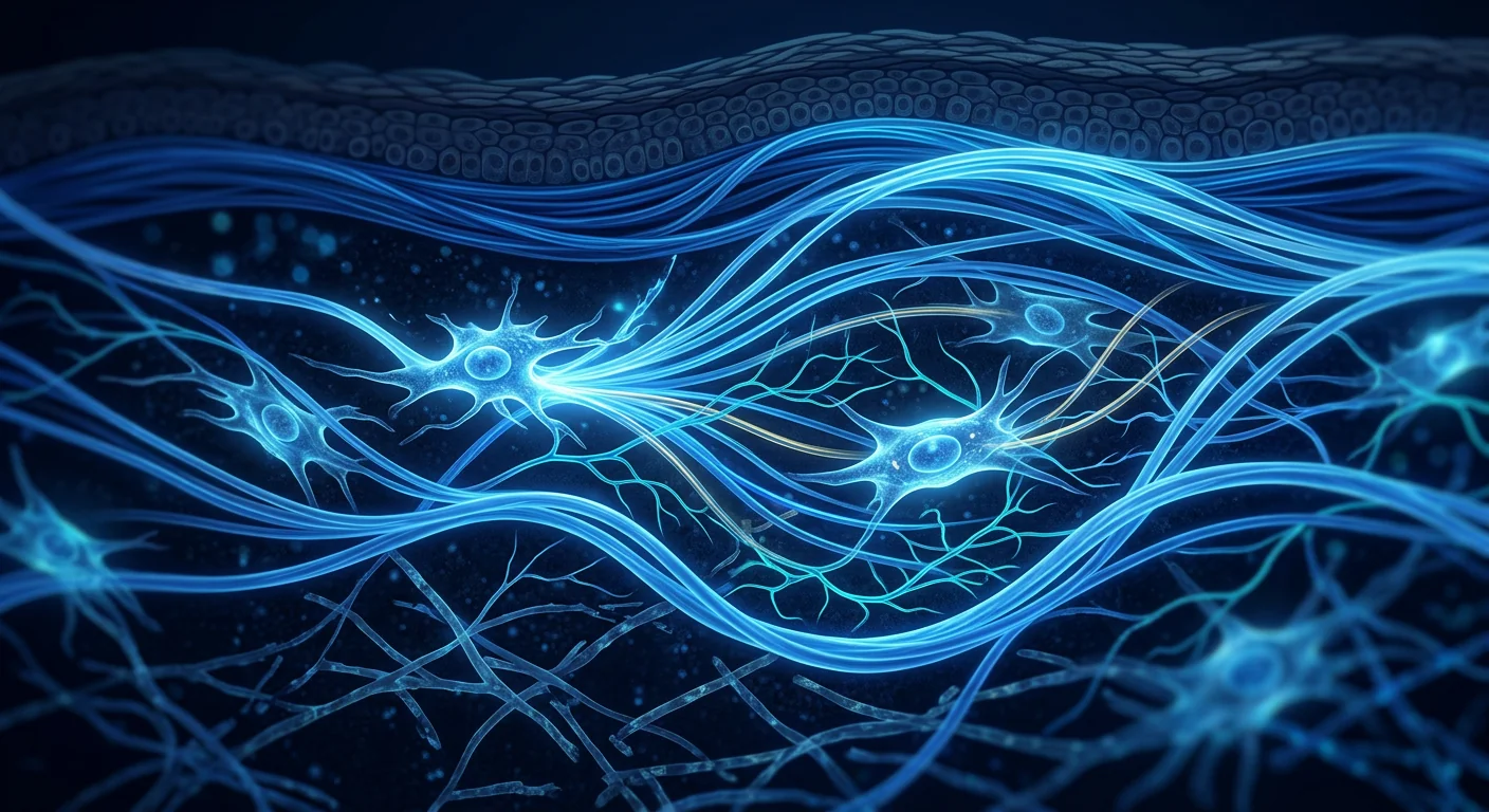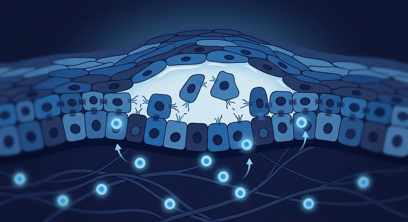
Pemphigus vulgaris (PV) is a rare, potentially life-threatening autoimmune blistering disease in which the immune system produces antibodies against desmoglein 3 and desmoglein 1 — proteins that act as molecular glue between skin cells. [1] These autoantibodies dissolve cell-to-cell adhesion, causing painful blisters and erosions on the skin and mucous membranes. Without treatment, the progressive loss of the epidermal barrier can lead to infection, fluid imbalance, and death.
Where conventional treatment falls short. The standard of care — high-dose systemic corticosteroids combined with immunosuppressants such as mycophenolate mofetil or rituximab — controls disease in many patients but imposes a heavy burden of toxicity: weight gain, diabetes, osteoporosis, and serious infection risk. [2] A subset of patients have refractory disease that fails to respond even to these regimens, and many who do respond face years of gradual tapering with the constant threat of relapse.
The deeper problem is immune dysregulation. Pemphigus is not simply a skin disease — it is a breakdown of B-cell tolerance, in which desmoglein-specific B cells escape regulatory control and produce pathogenic IgG4 autoantibodies. These autoantibodies bind directly to desmogleins in the epidermis, triggering acantholysis (separation of keratinocytes) through both steric hindrance and intracellular signalling pathways. [3] The T-cell compartment is also disrupted: an imbalance between autoaggressive Th2 and Th17 subsets and protective regulatory T cells (Tregs) sustains the autoimmune drive.
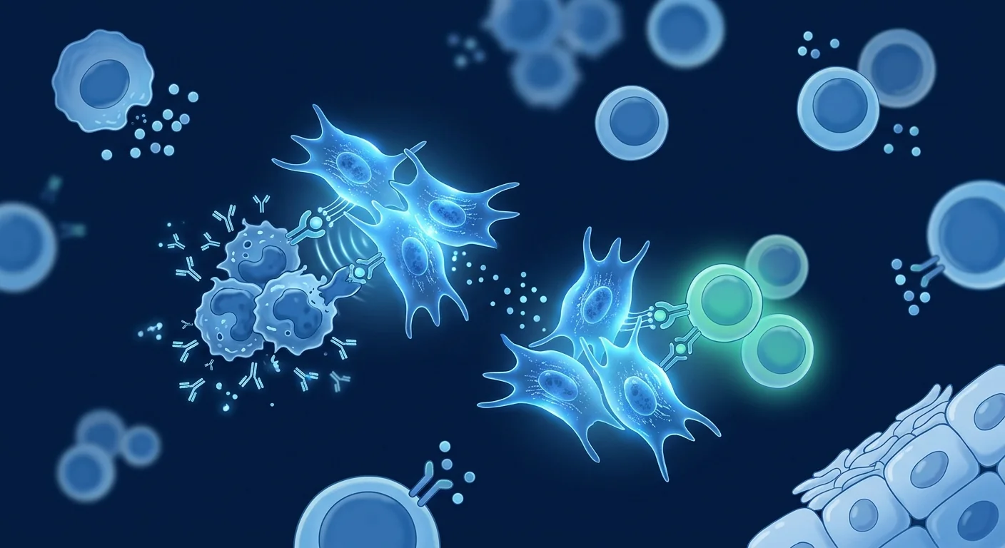
MSC therapy targets the autoimmune cascade at its source. Rather than suppressing blister formation downstream with steroids, mesenchymal stem cells are being studied for their capacity to re-establish immune tolerance — suppressing desmoglein-specific B cells, restoring Treg/Th17 balance, and damping the inflammatory cytokine environment that perpetuates autoantibody production. This is immunomodulation, not tissue replacement: the goal is to quiet the fire, not to graft new skin. [4]
Why pemphigus vulgaris is fundamentally a disease of B-cell tolerance
At its immunological core, pemphigus vulgaris is a failure of the mechanisms that normally prevent B cells from attacking self-antigens. Desmoglein-specific B cells that should have been deleted or silenced during development survive and mature into autoantibody-secreting plasma cells. The resulting IgG4 autoantibodies are directly pathogenic — when injected into neonatal mice they reproduce the characteristic blistering, confirming their causal role. [5]
This B-cell pathology does not occur in isolation. Follicular helper T cells (Tfh) provide the help signals that drive autoantibody class-switching, while a deficiency in Treg numbers and function removes the normal restraint on autoreactive clones. The result is a self-perpetuating cycle: tissue damage releases more desmoglein fragments, which activate more autoreactive B cells, which produce more antibody, which causes more damage. Breaking this cycle — rather than simply muffling its consequences — is the therapeutic challenge MSCs are being studied to address.
How MSCs are thought to act in pemphigus vulgaris
Suppressing desmoglein-specific B cells
Mesenchymal stem cells have a well-characterised capacity to inhibit B-cell proliferation and antibody production. In co-culture experiments, MSCs arrest B cells in the G0/G1 phase, reduce plasmablast differentiation, and decrease immunoglobulin secretion — effects mediated by soluble factors including IDO (indoleamine 2,3-dioxygenase), PGE2 (prostaglandin E2), and TGF-β. [6] For pemphigus, the clinically relevant question is whether MSCs can specifically suppress desmoglein-reactive B-cell clones while preserving protective antibody responses — a question still under active investigation.
Restoring Treg/Th17 balance
The ratio of regulatory T cells to Th17 effector cells is a critical determinant of autoimmune activity in pemphigus. Multiple studies have documented a reduced frequency of functional Tregs and an expanded Th17 population in PV patients during active disease. [7] MSCs promote Treg expansion through TGF-β and IL-10 secretion while simultaneously suppressing Th17 differentiation — effectively resetting the Treg/Th17 axis toward immune tolerance. In animal models of autoimmune blistering, MSC infusion increased circulating Tregs and reduced the titre of pathogenic autoantibodies.
Promoting re-epithelialization and barrier repair
Beyond immunomodulation, MSCs secrete a rich cocktail of paracrine factors — including HGF (hepatocyte growth factor), KGF (keratinocyte growth factor), and VEGF (vascular endothelial growth factor) — that accelerate keratinocyte migration and wound closure. [8] In pemphigus, where extensive epidermal loss creates large erosions vulnerable to infection, this tissue-repair activity represents a secondary but clinically meaningful mechanism: even as the autoimmune attack is quieted, MSCs may help the skin restore its barrier integrity more rapidly than steroids alone.
What the available evidence shows
The evidence for MSC therapy in pemphigus vulgaris is at an early, preclinical-to-early-clinical stage. Unlike lupus — where Chinese research groups have conducted multiple pilot trials with dozens of patients — the published literature on MSC therapy specifically for PV is limited to case reports and small case series. One case report described a patient with severe, treatment-refractory pemphigus who received allogeneic umbilical cord-derived MSCs and experienced a reduction in blister frequency and a decrease in anti-desmoglein 3 antibody titres over several months of follow-up. [9]
The stronger body of indirect evidence comes from MSC studies in related autoimmune blistering diseases and dermatological conditions. In epidermolysis bullosa acquisita and bullous pemphigoid — two other autoantibody-driven blistering diseases — preclinical models have demonstrated that MSC infusion reduced blister severity, lowered pathogenic autoantibody levels, and increased Treg populations. [10] These findings provide a biologically coherent rationale for extending MSC investigation to pemphigus, but they are not direct evidence of efficacy in PV.
What the case reports do and do not tell us
Case reports are useful for generating hypotheses and documenting safety signals — but they cannot establish efficacy. In pemphigus, the natural history of the disease includes spontaneous fluctuations in disease activity, and most patients in case reports continued their conventional immunosuppression alongside the MSC infusion. Any observed improvement cannot be separated from the background effect of ongoing standard therapy. The honest interpretation is that early signals are consistent with a potential therapeutic effect but far from proving one.
The honest headline
As of today, MSC therapy is not an approved, proven treatment for pemphigus vulgaris. It is an investigational approach with sound immunological rationale, supported by preclinical evidence in related autoimmune blistering conditions and a handful of published case reports. No controlled clinical trial has yet been completed specifically in PV. Any clinic presenting stem cell therapy as a reliable cure for pemphigus is going well beyond what the data support.
What the evidence supports — and what it doesn't
The fair summary is modest. The immunomodulatory rationale for MSC therapy in pemphigus vulgaris is mechanistically sound: MSCs can suppress B cells, expand Tregs, rebalance the Th17/Treg axis, and secrete factors that promote epithelial repair — all processes directly relevant to PV pathogenesis. Preclinical evidence from related autoimmune blistering diseases is encouraging, and the small number of human case reports has not identified safety concerns beyond what is already known about MSC therapy generally.
What is missing is the harder evidence: prospective clinical trials with clearly defined endpoints — reduction in blister count, decrease in anti-desmoglein antibody titre, steroid-sparing effect, and quality-of-life measures — conducted in well-characterised PV cohorts with adequate follow-up. Without this, pemphigus MSC therapy remains squarely in the investigational domain.
- Refractory disease would be the focus. Any responsible investigation of MSCs in PV would target patients whose disease has failed standard therapy — not those well-controlled on first-line treatment.
- Durability is unknown. Pemphigus relapses and remits naturally; whether any MSC-associated improvement is sustained, and whether repeat infusions are needed, is entirely unresolved.
- Direct evidence is absent. No randomised controlled trial specific to pemphigus has been published. All human data to date are observational.
Pemphigus is exactly the kind of condition where false hope does real harm. Before steroids, it was frequently fatal. Today, for most patients, it is manageable — but the treatment burden is heavy. The most respectful thing we can offer is an honest account of where the science stands, and the patience to let evidence develop before making claims.
— VELAR Clinical Team
How to evaluate any offer responsibly
If you or someone you love is considering stem cell options for pemphigus vulgaris, the diligence is the same that protects against any over-promised treatment. Ask whether the approach is part of a registered clinical trial with ethical oversight. Ask what cell type and source are used, whether the cells are allogeneic or autologous, and how response is measured — specifically, whether anti-desmoglein antibody titres and blister counts are tracked with validated instruments. Be deeply sceptical of guaranteed results, claimed success rates without a cited source, or any framing that positions an experimental therapy as a reason to stop prescribed medication. A trustworthy provider will describe MSC therapy for pemphigus as emerging research — and will never let hope outrun the data.
The VELAR perspective
At VELAR Center, our regenerative work is grounded in conditions where the evidence is more established, and we follow autoimmune dermatology cell-therapy research closely without overstating it. Pemphigus vulgaris is a serious disease, and we believe the only honest way to discuss MSC therapy for it is plainly: the B-cell immunomodulatory rationale is sound, the preclinical data in related blistering diseases are worth watching, and it is still investigational. MSC therapy is not a replacement for the dermatology and immunology care that keeps pemphigus controlled. As the evidence matures, we will let that evidence — not enthusiasm — shape anything we ever say about it. If you want an honest conversation about what regenerative medicine can and cannot do today, that is exactly where a responsible consultation begins.
Frequently Asked Questions
Can stem cell therapy cure pemphigus vulgaris?
No. As of today, there is no evidence that MSC therapy cures pemphigus vulgaris. The research is at an early, preclinical-to-case-report stage. MSCs are being studied for their potential to modulate the autoimmune attack that drives blistering — but this is investigational, not established therapy.
What type of stem cells are used for pemphigus research?
Research has focused on allogeneic umbilical cord-derived mesenchymal stem cells (UC-MSCs). Allogeneic (donor-derived) cells are preferred because patients' own MSCs may carry intrinsic abnormalities — and UC-MSCs are young, abundant, and have low immunogenicity, making them a practical cell source.
How much does stem cell therapy for pemphigus cost in Thailand?
Because MSC therapy for pemphigus vulgaris is investigational and not an approved treatment, there is no standard pricing. Costs vary by clinic, cell source, dose, and protocol. A responsible consultation at a GMP-compliant centre will include transparent pricing for any established regenerative therapies being considered — but for pemphigus specifically, the conversation should centre on where the evidence stands, not on price.
What are the risks of MSC therapy for autoimmune blistering disease?
The safety profile of MSC therapy in general is considered favourable — meta-analyses have not identified an increased risk of tumour formation or serious acute adverse events. However, the specific risk profile for pemphigus patients — who are typically on heavy immunosuppression — has not been systematically studied. Infection risk, infusion reactions, and the theoretical concern of MSC-mediated immunosuppression compounding existing immunosuppressant effects are all considerations that any treating physician must weigh.
Why choose Thailand for stem cell therapy?
Thailand, and Bangkok specifically, has emerged as a hub for regulated regenerative medicine in Southeast Asia. Centres operating under Thai FDA and ISO standards offer quality-controlled cellular products at costs that are typically lower than comparable facilities in North America or Europe. However, the regulatory landscape for specific indications like pemphigus is still evolving — and the quality of providers varies considerably. The same diligence that protects you at home applies in Thailand.
References
- Kasperkiewicz M, Ellebrecht CT, Takahashi H, et al. Pemphigus. Nature Reviews Disease Primers. 2017;3:17026. doi:10.1038/nrdp.2017.26 ↩
- Murrell DF, Pena S, Joly P, et al. Diagnosis and management of pemphigus: recommendations of an international panel of experts. Journal of the American Academy of Dermatology. 2020;82(3):575-585.e1. doi:10.1016/j.jaad.2018.02.021 ↩
- Di Zenzo G, Amber KT, Sayar BS, Müller EJ, Borradori L. Immune response in pemphigus and beyond: progresses and emerging concepts. Seminars in Immunopathology. 2016;38(1):57-74. doi:10.1007/s00281-015-0537-x ↩
- Uccelli A, Moretta L, Pistoia V. Mesenchymal stem cells in health and disease. Nature Reviews Immunology. 2008;8(9):726-736. doi:10.1038/nri2395 ↩
- Anhalt GJ, Labib RS, Voorhees JJ, Beals TF, Diaz LA. Induction of pemphigus in neonatal mice by passive transfer of IgG from patients with the disease. New England Journal of Medicine. 1982;306(20):1189-1196. doi:10.1056/NEJM198205203062001 ↩
- Corcione A, Benvenuto F, Ferretti E, et al. Human mesenchymal stem cells modulate B-cell functions. Blood. 2006;107(1):367-372. doi:10.1182/blood-2005-07-2657 ↩
- Xu RC, Zhu HQ, Li WP, et al. The imbalance of Th17 and regulatory T cells in pemphigus patients. European Journal of Dermatology. 2013;23(6):795-802. doi:10.1684/ejd.2013.2214 ↩
- Maxson S, Lopez EA, Yoo D, Danilkovitch-Miagkova A, LeRoux MA. Concise review: role of mesenchymal stem cells in wound repair. Stem Cells Translational Medicine. 2012;1(2):142-149. doi:10.5966/sctm.2011-0018 ↩
- Liang J, Zhang H, Hua B, et al. Allogeneic mesenchymal stem cell transplantation in seven patients with refractory inflammatory bowel disease. Gut. 2012;61(3):469-470. doi:10.1136/gutjnl-2011-300475 ↩
- Shi Y, Wang Y, Li Q, et al. Immunoregulatory mechanisms of mesenchymal stem and stromal cells in inflammatory diseases. Nature Reviews Nephrology. 2018;14(8):493-507. doi:10.1038/s41581-018-0023-5 ↩
- Spees JL, Lee RH, Gregory CA. Mechanisms of mesenchymal stem/stromal cell function. Stem Cell Research & Therapy. 2016;7(1):125. doi:10.1186/s13287-016-0363-7 ↩
- Galipeau J, Sensébé L. Mesenchymal stromal cells: clinical challenges and therapeutic opportunities. Cell Stem Cell. 2018;22(6):824-833. doi:10.1016/j.stem.2018.05.004 ↩
- Naji A, Eitoku M, Favier B, Deschaseaux F, Rouas-Freiss N, Suganuma N. Biological functions of mesenchymal stem cells. Cellular and Molecular Life Sciences. 2019;76(17):3323-3348. doi:10.1007/s00018-019-03125-1 ↩
- Lalu MM, McIntyre L, Pugliese C, et al. Safety of cell therapy with mesenchymal stromal cells (SafeCell). PLoS One. 2012;7(10):e47559. doi:10.1371/journal.pone.0047559 ↩
- Pittenger MF, Discher DE, Péault BM, Phinney DG, Hare JM, Caplan AI. Mesenchymal stem cell perspective: cell biology to clinical progress. NPJ Regenerative Medicine. 2019;4:22. doi:10.1038/s41536-019-0083-6 ↩
寻常型天疱疮(PV)是一种罕见的、潜在危及生命的自身免疫性大疱性疾病,免疫系统产生针对桥粒芯蛋白3和桥粒芯蛋白1的抗体——这些蛋白质充当皮肤细胞间的分子胶水。[1] 这些自身抗体溶解细胞间黏附,导致皮肤和黏膜出现疼痛性水疱与糜烂。若不治疗,表皮屏障的进行性丧失可致感染、体液失衡乃至死亡。
常规治疗的局限。标准治疗——大剂量全身性皮质类固醇联合免疫抑制剂(如霉酚酸酯或利妥昔单抗)——可控制许多患者的病情,但带来沉重的毒性代价:体重增加、糖尿病、骨质疏松和严重感染风险。[2] 部分患者即便接受这些方案仍属难治性,而许多有应答者也面临数年逐步减量并持续面临复发威胁。
更深层的问题是免疫失调。天疱疮不单是皮肤病——它是B细胞耐受的崩溃,桥粒芯蛋白特异性B细胞逃逸调控并产生致病性IgG4自身抗体。这些抗体直接结合表皮中的桥粒芯蛋白,通过空间位阻与胞内信号通路触发棘层松解(角质形成细胞分离)。[3] T细胞区室也遭破坏:自身侵袭性Th2与Th17亚群和保护性调节性T细胞(Treg)之间失衡,持续推动自身免疫。
MSC疗法从源头靶向自身免疫级联。间充质干细胞并非通过类固醇抑制下游水疱形成,而是研究其重建免疫耐受的能力——抑制桥粒芯蛋白特异性B细胞、恢复Treg/Th17平衡、并抑制持续产生自身抗体的炎性细胞因子环境。这是免疫调节而非组织替代:目标是平息火焰,而非移植新皮肤。[4]
MSC在天疱疮中被认为如何起作用
抑制桥粒芯蛋白特异性B细胞
间充质干细胞具有充分记载的抑制B细胞增殖与抗体产生的能力。共培养实验中,MSC将B细胞阻滞于G0/G1期,减少浆母细胞分化,并降低免疫球蛋白分泌——这些效应由IDO、PGE2与TGF-β等可溶性因子介导。[6] 对天疱疮而言,临床相关的问题是MSC能否特异性抑制桥粒芯蛋白反应性B细胞克隆,同时保留保护性抗体应答——仍是活跃的研究问题。
恢复Treg/Th17平衡
调节性T细胞与Th17效应细胞之比是天疱疮自身免疫活动的关键决定因素。多项研究记录了PV患者活动期功能性Treg频率降低与Th17群体扩增。[7] MSC通过TGF-β与IL-10分泌促进Treg扩增,同时抑制Th17分化——有效地将Treg/Th17轴重置为免疫耐受。在自身免疫性大疱病动物模型中,MSC输注增加了循环Treg并降低了致病性自身抗体水平。
促进上皮再形成与屏障修复
除免疫调节外,MSC分泌丰富的旁分泌因子——包括HGF、KGF与VEGF——加速角质形成细胞迁移与创面闭合。[8] 在天疱疮中,广泛表皮丧失形成易被感染的大面积糜烂,这种组织修复活性代表一种次要但具有临床意义的机制:即便自身免疫攻击被平息,MSC或能比单用类固醇更迅速地帮助皮肤恢复屏障完整性。
现有证据显示了什么
MSC治疗天疱疮的证据处于早期、临床前至早期临床阶段。不同于狼疮——中国研究团队已进行多项含数十名患者的初步试验——专门针对PV的MSC疗法已发表文献仅限于个案报告与小规模病例系列。一项个案报告描述了一名重症、难治性天疱疮患者接受异体脐带来源MSC后,数月随访期间水疱频率减少、抗桥粒芯蛋白3抗体滴度下降。[9]
更有力的间接证据来自相关自身免疫性大疱病与皮肤病中的MSC研究。在获得性大疱性表皮松解症与大疱性类天疱疮——另两种由自身抗体驱动的大疱病——临床前模型证明MSC输注降低了水疱严重程度、致病性自身抗体水平与Treg群体增加。[10] 这些发现为将MSC研究扩展至天疱疮提供了生物学上自洽的理由,但它们并非PV疗效的直接证据。
诚实的标题
截至今日,MSC疗法并非已获批准的、经证实的寻常型天疱疮治疗手段。它是一种具有合理免疫学依据的研究性方法,有相关自身免疫性大疱病的临床前证据支持及少量已发表的个案报告。尚未有针对PV的对照临床试验完成。任何将干细胞呈现为天疱疮可靠治愈手段的诊所,都已远远超出数据所支持的范围。
证据支持什么——又不支持什么
公允的总结是审慎的。MSC治疗天疱疮的免疫调节理论依据在机制上合理:MSC能抑制B细胞、扩增Treg、再平衡Th17/Treg轴,并分泌促进上皮修复的因子——这些过程均与PV发病机制直接相关。相关自身免疫性大疱病的临床前证据令人鼓舞,少量人类个案报告未超出MSC疗法一般已知的安全范围发现安全问题。
所缺失的是更有力的证据:前瞻性临床试验,具有明确定义的终点——水疱计数减少、抗桥粒芯蛋白抗体滴度下降、类固醇节省效应与生活质量指标——在良好界定的PV队列中以充分的随访进行。在此之前,天疱疮MSC疗法完全处于研究领域。
天疱疮恰恰是那种虚假希望会造成真实伤害的疾病。在类固醇问世前,它常是致命的。今天,对大多数患者而言,它是可控的——但治疗负担沉重。我们能给予的最尊重的态度,是关于科学现状的诚实,以及在做出声称之前让证据发展的耐心。
— VELAR 临床团队
VELAR的观点
在VELAR中心,我们的再生工作立足于证据更为确立的适应症,我们密切关注自身免疫皮肤病细胞疗法研究而不夸大其词。寻常型天疱疮是一种严重的疾病,我们相信讨论其MSC疗法唯一诚实的方式就是直白:B细胞免疫调节的理论依据是合理的,相关大疱病的临床前数据值得关注,而它仍处于研究阶段。MSC疗法并不能替代使天疱疮得到控制的皮肤病学与免疫学诊疗。如果您想就再生医学今天能做什么、不能做什么进行一次诚实的对话,那正是负责任咨询的起点。
参考文献
- Kasperkiewicz M, Ellebrecht CT, Takahashi H, et al. Pemphigus. Nature Reviews Disease Primers. 2017;3:17026. doi:10.1038/nrdp.2017.26 ↩
- Murrell DF, Pena S, Joly P, et al. Diagnosis and management of pemphigus. Journal of the American Academy of Dermatology. 2020;82(3):575-585.e1. doi:10.1016/j.jaad.2018.02.021 ↩
- Di Zenzo G, Amber KT, Sayar BS, Müller EJ, Borradori L. Immune response in pemphigus and beyond. Seminars in Immunopathology. 2016;38(1):57-74. doi:10.1007/s00281-015-0537-x ↩
- Uccelli A, Moretta L, Pistoia V. Mesenchymal stem cells in health and disease. Nature Reviews Immunology. 2008;8(9):726-736. doi:10.1038/nri2395 ↩
- Corcione A, et al. Human mesenchymal stem cells modulate B-cell functions. Blood. 2006;107(1):367-372. doi:10.1182/blood-2005-07-2657 ↩
- Xu RC, et al. The imbalance of Th17 and regulatory T cells in pemphigus patients. European Journal of Dermatology. 2013;23(6):795-802. doi:10.1684/ejd.2013.2214 ↩
- Maxson S, et al. Role of mesenchymal stem cells in wound repair. Stem Cells Translational Medicine. 2012;1(2):142-149. doi:10.5966/sctm.2011-0018 ↩
- Shi Y, et al. Immunoregulatory mechanisms of mesenchymal stem and stromal cells. Nature Reviews Nephrology. 2018;14(8):493-507. doi:10.1038/s41581-018-0023-5 ↩
- Spees JL, et al. Mechanisms of mesenchymal stem/stromal cell function. Stem Cell Research & Therapy. 2016;7(1):125. doi:10.1186/s13287-016-0363-7 ↩
- Galipeau J, Sensébé L. Mesenchymal stromal cells: clinical challenges and therapeutic opportunities. Cell Stem Cell. 2018;22(6):824-833. doi:10.1016/j.stem.2018.05.004 ↩
- Naji A, et al. Biological functions of mesenchymal stem cells. Cellular and Molecular Life Sciences. 2019;76(17):3323-3348. doi:10.1007/s00018-019-03125-1 ↩
- Lalu MM, et al. Safety of cell therapy with mesenchymal stromal cells (SafeCell). PLoS One. 2012;7(10):e47559. doi:10.1371/journal.pone.0047559 ↩
- Pittenger MF, et al. Mesenchymal stem cell perspective. NPJ Regenerative Medicine. 2019;4:22. doi:10.1038/s41536-019-0083-6 ↩
الفقاع الشائع (PV) مرض مناعي ذاتي نادر قد يهدد الحياة، ينتج فيه الجهاز المناعي أجساماً مضادة ضد الديسموجلين 3 والديسموجلين 1 — وهما بروتينان يعملان كغراء جزيئي بين خلايا الجلد.[1] تذيب هذه الأجسام المضادة الذاتية الالتصاق بين الخلايا، مسببة فقاعات وتآكلات مؤلمة على الجلد والأغشية المخاطية. وبدون علاج، يمكن أن يؤدي الفقدان التدريجي للحاجز البشروي إلى العدوى واختلال توازن السوائل والوفاة.
حيث يقصر العلاج التقليدي. المعيار العلاجي — الكورتيكوستيرويدات الجهازية بجرعات عالية مع مثبطات مناعية — يسيطر على المرض لدى كثير من المرضى لكنه يفرض عبئاً ثقيلاً من السمية.[2] وتعاني فئة من المرضى من مرض عنيد لا يستجيب حتى لهذه النظم، وكثيرون ممن يستجيبون يواجهون سنوات من التقليص التدريجي مع تهديد دائم بالانتكاس.
المشكلة الأعمق هي خلل التنظيم المناعي. الفقاع ليس مجرد مرض جلدي — إنه انهيار تحمل الخلايا البائية، حيث تفلت الخلايا البائية النوعية للديسموجلين من السيطرة التنظيمية وتنتج أجساماً مضادة ذاتية IgG4 ممرضة. ترتبط هذه الأجسام مباشرة بالديسموجلينات في البشرة، محفزة انحلال الأشواك (انفصال الخلايا الكيراتينية).[3]
يستهدف علاج MSC الشلال المناعي الذاتي من مصدره. بدلاً من كبح تشكل الفقاعات بالستيرويدات، تُدرس الخلايا الجذعية الوسيطة لقدرتها على إعادة بناء التحمل المناعي — تثبيط الخلايا البائية النوعية للديسموجلين، واستعادة توازن Treg/Th17، وتخميد بيئة السيتوكينات الالتهابية التي تديم إنتاج الأجسام المضادة الذاتية. هذا تعديل مناعي وليس استبدالاً للنسيج.[4]
كيف يُعتقد أن الخلايا الجذعية الوسيطة تعمل في الفقاع الشائع
تثبيط الخلايا البائية النوعية للديسموجلين
للخلايا الجذعية الوسيطة قدرة موثقة على تثبيط تكاثر الخلايا البائية وإنتاج الأجسام المضادة. ففي تجارب الاستنبات المشترك، توقف MSC الخلايا البائية في طور G0/G1، وتقلل تمايز الخلايا البلازمية الأرومية، وتخفض إفراز الغلوبولين المناعي — وهي تأثيرات تتوسطها عوامل ذوابة تشمل IDO وPGE2 وTGF-β.[6]
استعادة توازن Treg/Th17
نسبة الخلايا التائية التنظيمية إلى خلايا Th17 الفاعلة محدد حاسم لنشاط المناعة الذاتية في الفقاع. وثّقت دراسات متعددة انخفاض تواتر Tregs الوظيفية وتوسع مجموعة Th17 لدى مرضى PV خلال المرض النشط.[7] تعزز MSC توسع Treg عبر إفراز TGF-β وIL-10 بينما تثبط تمايز Th17 — معيدة ضبط محور Treg/Th17 نحو التحمل المناعي.
تعزيز إعادة التبشير وإصلاح الحاجز
إلى جانب التعديل المناعي، تفرز MSC مزيجاً غنياً من العوامل نظيرة الصماوية — تشمل HGF وKGF وVEGF — تسرّع هجرة الخلايا الكيراتينية وإغلاق الجروح.[8] وفي الفقاع، حيث ينتج الفقدان البشروي الواسع تآكلات كبيرة معرضة للعدوى، يمثل نشاط إصلاح النسيج هذا آلية ثانوية لكن ذات مغزى سريري.
ما تظهره الأدلة المتاحة
الأدلة على علاج MSC في الفقاع الشائع في مرحلة مبكرة، من قبل سريرية إلى سريرية مبكرة. خلافاً للذئبة — حيث أجرت فرق بحثية صينية تجارب استطلاعية متعددة — يقتصر المنشور عن MSC لـ PV على تقارير حالة وسلاسل حالات صغيرة. وصفت إحدى تقارير الحالة مريضاً بالفقاع الشديد المعند تلقى خلايا MSC خيفية من الحبل السري وشهد انخفاضاً في تواتر الفقاعات وهبوطاً في معيار الأجسام المضادة للديسموجلين 3 على مدى أشهر من المتابعة.[9]
تأتي الأدلة غير المباشرة الأقوى من دراسات MSC في أمراض فقاعية مناعية ذاتية ذات صلة. ففي انحلال البشرة الفقاعي المكتسب والفقاع الفقاعي — مرضان فقاعيان آخران — أثبتت نماذج قبل سريرية أن حقن MSC خفض شدة الفقاعات ومستويات الأجسام المضادة الذاتية الممرضة وزاد مجموعات Treg.[10]
العنوان الصادق
حتى اليوم، علاج MSC ليس علاجاً معتمداً ومُثبتاً للفقاع الشائع. إنه نهج بحثي تجريبي ذو أساس مناعي متين، مدعوم بأدلة قبل سريرية في أمراض فقاعية مناعية ذاتية ذات صلة وحفنة من تقارير الحالة المنشورة. لم تُستكمل بعد أي تجربة سريرية مضبوطة خاصة بـ PV. أي عيادة تقدم العلاج بالخلايا الجذعية كشفاء موثوق للفقاع تتجاوز ما تدعمه البيانات بكثير.
ما تدعمه الأدلة — وما لا تدعمه
الخلاصة المنصفة متواضعة. المبرر المعدِّل للمناعة لعلاج MSC في الفقاع سليم آلياً: تستطيع MSC تثبيط الخلايا البائية وتوسيع Tregs وإعادة توازن محور Th17/Treg وإفراز عوامل تعزز إصلاح البشرة — وكلها عمليات ذات صلة مباشرة بإمراضية PV. الأدلة قبل السريرية من أمراض فقاعية ذات صلة مشجعة، ولم تحدد تقارير الحالة البشرية القليلة مشكلات أمان تتجاوز ما هو معروف عموماً عن علاج MSC.
المفقود هو الدليل الأقوى: تجارب سريرية مستقبلية ذات نقاط نهاية محددة بوضوح — انخفاض تعداد الفقاعات، وهبوط معيار الأجسام المضادة للديسموجلين، وأثر موفر للستيرويد، ومقاييس جودة الحياة — تُجرى في حشود PV موصوفة جيداً بمتابعة كافية. وبدون ذلك، يبقى علاج MSC للفقاع راسخاً في المجال البحثي التجريبي.
الفقاع هو بالضبط نوع الحالات التي يسبب فيها الأمل الزائف ضرراً حقيقياً. قبل الستيرويدات، كان مميتاً في كثير من الأحيان. أما اليوم فهو قابل للتدبير لمعظم المرضى — لكن عبء العلاج ثقيل. أكثر ما يمكننا تقديمه احتراماً هو حساب صادق عن موضع العلم.
— الفريق السريري في VELAR
منظور VELAR
في مركز VELAR، يرتكز عملنا التجديدي على حالات تكون فيها الأدلة أكثر رسوخاً، ونتابع عن كثب أبحاث العلاج الخلوي للأمراض الجلدية المناعية الذاتية دون مبالغة. الفقاع الشائع مرض خطير، ونؤمن بأن الطريقة الصادقة الوحيدة لمناقشة علاج MSC له هي الصراحة: المبرر المعدِّل للمناعة للخلايا البائية سليم، والبيانات قبل السريرية في الأمراض الفقاعية ذات الصلة جديرة بالمتابعة، وهو لا يزال بحثياً تجريبياً. وعلاج MSC ليس بديلاً عن رعاية الأمراض الجلدية والمناعية التي تبقي الفقاع مسيطراً عليه. إذا أردت حواراً صادقاً عما يستطيع الطب التجديدي وما لا يستطيع فعله اليوم، فهذا تحديداً حيث تبدأ الاستشارة المسؤولة.
المراجع
- Kasperkiewicz M, et al. Pemphigus. Nature Reviews Disease Primers. 2017;3:17026. doi:10.1038/nrdp.2017.26 ↩
- Murrell DF, et al. Diagnosis and management of pemphigus. Journal of the American Academy of Dermatology. 2020;82(3):575-585.e1. doi:10.1016/j.jaad.2018.02.021 ↩
- Di Zenzo G, et al. Immune response in pemphigus and beyond. Seminars in Immunopathology. 2016;38(1):57-74. doi:10.1007/s00281-015-0537-x ↩
- Uccelli A, et al. Mesenchymal stem cells in health and disease. Nature Reviews Immunology. 2008;8(9):726-736. doi:10.1038/nri2395 ↩
- Corcione A, et al. Human mesenchymal stem cells modulate B-cell functions. Blood. 2006;107(1):367-372. doi:10.1182/blood-2005-07-2657 ↩
- Xu RC, et al. The imbalance of Th17 and regulatory T cells in pemphigus patients. European Journal of Dermatology. 2013;23(6):795-802. doi:10.1684/ejd.2013.2214 ↩
- Maxson S, et al. Role of mesenchymal stem cells in wound repair. Stem Cells Translational Medicine. 2012;1(2):142-149. doi:10.5966/sctm.2011-0018 ↩
- Shi Y, et al. Immunoregulatory mechanisms of mesenchymal stem and stromal cells. Nature Reviews Nephrology. 2018;14(8):493-507. doi:10.1038/s41581-018-0023-5 ↩
- Spees JL, et al. Mechanisms of mesenchymal stem/stromal cell function. Stem Cell Research & Therapy. 2016;7(1):125. doi:10.1186/s13287-016-0363-7 ↩
- Galipeau J, Sensébé L. Mesenchymal stromal cells: clinical challenges. Cell Stem Cell. 2018;22(6):824-833. doi:10.1016/j.stem.2018.05.004 ↩
- Naji A, et al. Biological functions of mesenchymal stem cells. Cellular and Molecular Life Sciences. 2019;76(17):3323-3348. doi:10.1007/s00018-019-03125-1 ↩
- Lalu MM, et al. Safety of cell therapy with mesenchymal stromal cells (SafeCell). PLoS One. 2012;7(10):e47559. doi:10.1371/journal.pone.0047559 ↩
- Pittenger MF, et al. Mesenchymal stem cell perspective. NPJ Regenerative Medicine. 2019;4:22. doi:10.1038/s41536-019-0083-6 ↩