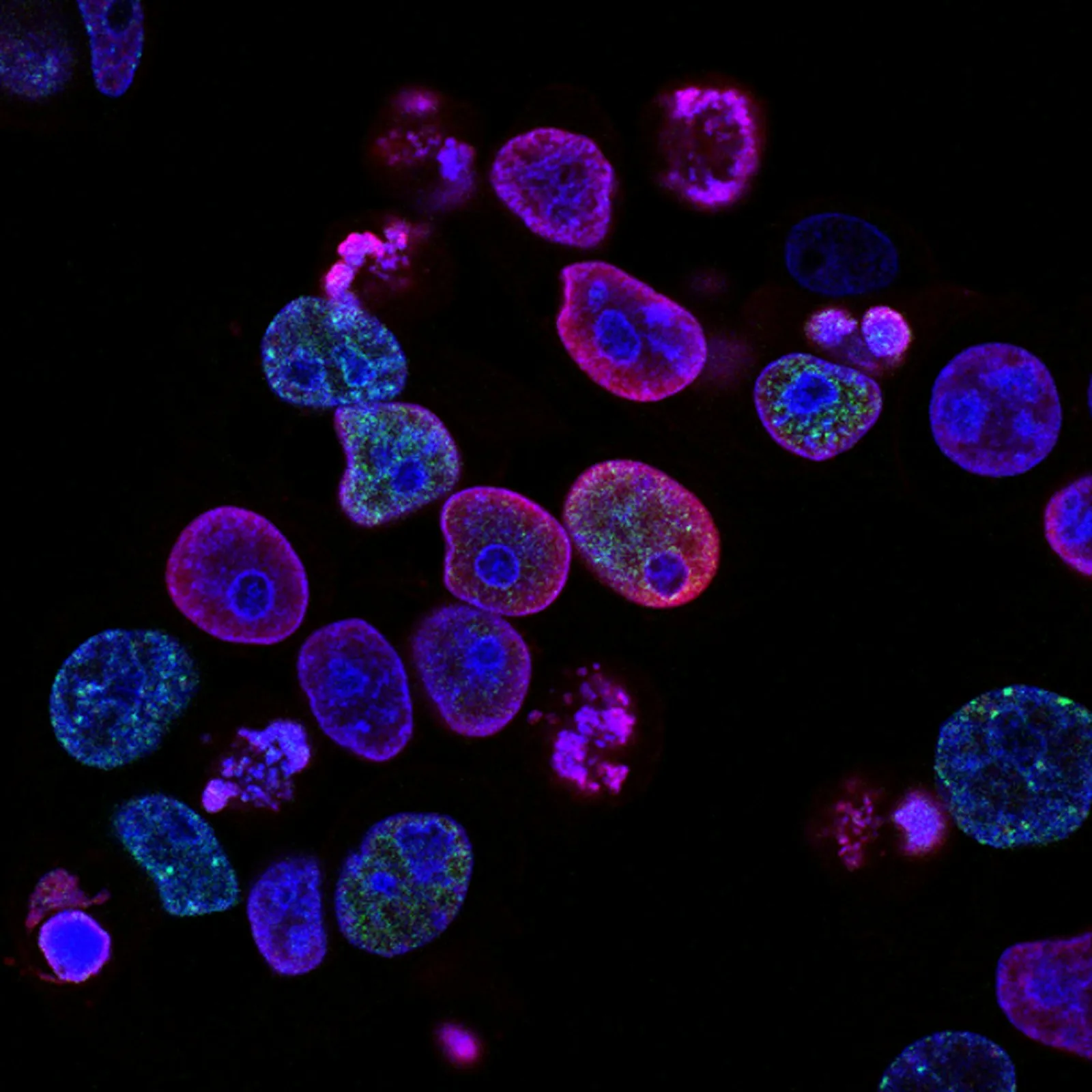
The skin tells a story of time — but that story doesn't have to end with thinning, wrinkling, and lost resilience. MSC therapy is being studied as a way to restore the regenerative capacity skin loses with age.
What Is Skin Rejuvenation?
Skin rejuvenation is the process of restoring skin to a more youthful state — not just superficially improving appearance, but rebuilding the dermal architecture that time and environmental damage degrade. Treatments span from topical retinoids and laser resurfacing to injectable biologics, but mesenchymal stem cell (MSC) therapy represents a fundamentally different approach: rather than stimulating the existing compromised cells, it introduces fresh signaling machinery capable of orchestrating repair at the tissue level. [1]
Conventional approaches hit a biological ceiling. Chemical peels, microneedling, and fractional lasers work by creating controlled injury — the skin heals and looks fresher, but the underlying fibroblast population, already depleted and senescent, can only do so much. Fillers add volume but don't improve tissue quality. Botulinum toxin immobilizes muscles but does nothing for skin texture. These are temporary compensations, not regeneration.
The tissue-level problem. Aged skin is characterized by collagen fragmentation, elastin degradation, reduced hyaluronic acid synthesis, oxidative stress, and a growing population of senescent fibroblasts that secrete pro-inflammatory factors — the senescence-associated secretory phenotype (SASP). [2] No cream or peel reverses SASP. MSCs, by contrast, secrete a broad panel of growth factors, cytokines, and extracellular vesicles that can shift the local microenvironment from degenerative to regenerative — a process termed paracrine signaling. [3]
The Biology of Skin Aging
Skin aging proceeds along two tracks: intrinsic (chronological) and extrinsic (environmental, primarily UV-driven photoaging). Both converge on a common endpoint: loss of functional dermal fibroblasts, fragmentation of the collagen matrix, and chronic low-grade inflammation — sometimes called "inflammaging." [4]
Intrinsic aging follows the body's biological clock. Fibroblasts divide more slowly, produce less Type I and Type III collagen, and accumulate mitochondrial DNA damage. By age 80, dermal thickness decreases by approximately 20% compared to age 30. Elastin fibers, which provide recoil, gradually degrade and are not replaced — adult skin has essentially no elastin turnover. [5]
Extrinsic aging (photoaging) accelerates this timeline dramatically. UV radiation generates reactive oxygen species that directly fragment collagen fibrils, activate matrix metalloproteinases (MMPs) — enzymes that digest the extracellular matrix — and induce DNA mutations in both keratinocytes and fibroblasts. [6] The result is what clinicians see as solar elastosis: thickened, leathery, deeply wrinkled skin with uneven pigmentation.
How MSC Therapy Supports Dermal Regeneration
MSCs derived from Wharton's jelly (umbilical cord tissue) are the most commonly used source in aesthetic regenerative medicine. Unlike adult-derived MSCs (bone marrow, adipose), Wharton's jelly MSCs are immunoprivileged, highly proliferative, and secrete a richer profile of regenerative factors — including higher levels of hepatocyte growth factor (HGF), vascular endothelial growth factor (VEGF), and transforming growth factor-beta 3 (TGF-β3), which is associated with scarless healing. [7]
Six Mechanisms of MSC-Mediated Skin Regeneration
1. Fibroblast Activation. MSC-derived growth factors (FGF-2, PDGF, EGF) stimulate resident fibroblasts to proliferate and synthesize new collagen — particularly Type I and Type III — as well as fibronectin and hyaluronic acid. [8] This directly addresses the fibroblast depletion at the core of aged skin.
2. Collagen Remodeling. MSCs secrete tissue inhibitors of metalloproteinases (TIMPs) that counteract the MMP-driven collagen degradation characteristic of photoaged skin. The net effect is a shift from matrix catabolism (breakdown) to matrix anabolism (rebuilding). [9]
3. Angiogenesis. Aging reduces dermal microvasculature, depriving skin of oxygen and nutrients. MSC-secreted VEGF promotes new capillary formation, improving perfusion — clinically visible as improved skin tone, reduced sallowness, and a "glow" patients frequently report. [10]
4. Anti-Inflammatory Signaling. MSCs respond to inflammatory environments by releasing IL-10, TGF-β, and prostaglandin E2 (PGE2), which suppress chronic low-grade dermal inflammation. This is especially relevant for conditions like rosacea and chronic sun damage. [11]
5. Antioxidant Defense. MSC-derived extracellular vesicles carry antioxidant enzymes (superoxide dismutase, catalase, glutathione peroxidase) that neutralize reactive oxygen species. In vitro studies show MSC-conditioned medium reduces UV-induced oxidative damage in dermal fibroblasts by over 40%. [12]
6. Senescence Modulation. Emerging evidence suggests MSCs can reduce the senescent cell burden through paracrine factors that either reverse early senescence or promote clearance of irreversibly senescent fibroblasts via macrophage activation. This is an active area of investigation. [13]
Delivery Methods for Aesthetic MSC Therapy
MSC therapy for skin rejuvenation is delivered through several routes, each with distinct advantages and limitations. The choice depends on the area being treated, depth of the target tissue, and treatment goals.
The most common route for facial rejuvenation. MSCs are injected directly into the papillary and reticular dermis via fine-gauge needles. Provides direct delivery to fibroblasts. Multiple sessions spaced 4–8 weeks apart are typical.
For patients seeking non-invasive options, MSC-conditioned medium (the growth-factor-rich fluid MSCs are cultured in) can be applied topically following microneedling to enhance absorption. Evidence of efficacy is preliminary but growing.
For whole-body rejuvenation (often combined with anti-aging protocols), MSCs are infused intravenously. While only a fraction homes to skin, systemic effects on overall inflammation can benefit skin quality indirectly.
Many clinics combine intradermal MSC injections with PRP (platelet-rich plasma) or microneedling for synergistic effects — the mechanical stimulation primes the dermis, and MSCs supply the regenerative signals.
Clinical Evidence: What the Studies Show
The clinical evidence base for MSC therapy in skin rejuvenation is growing, though still predominantly small-scale and preliminary. Here is what published research tells us so far:
A 2023 systematic review covering 12 clinical studies of MSC-based aesthetic treatments found consistently positive outcomes for facial rejuvenation, with improvements in skin elasticity (mean increase of 18–24% on cutometer measurements), dermal thickness (ultrasound-measured increases of 12–18% at 3–6 months post-treatment), wrinkle reduction (improvements of 1.5–2.5 grades on the Fitzpatrick wrinkle scale), and subjective patient satisfaction exceeding 80% across studies. [14]
A randomized controlled trial published in 2022 compared intradermal Wharton's jelly MSC injections plus microneedling versus microneedling alone. At 6 months, the MSC group demonstrated significantly greater improvements in skin roughness parameters (Ra, Rz), dermal density on high-frequency ultrasound, and blinded physician assessment scores. No serious adverse events were reported. [15]
Exosome-focused research has also shown promise. A 2024 study applying MSC-derived exosomes topically after fractional laser treatment demonstrated accelerated healing, reduced post-procedure erythema, and superior collagen remodeling on histological analysis compared to laser alone. [16]
"The paracrine hypothesis — that MSCs work primarily through secreted factors rather than direct differentiation — is particularly compelling in aesthetic medicine. It means the therapeutic effect does not require the cells to persist indefinitely; their signaling cargo is what matters." — Regenerative Dermatology Research Group, 2024
What to Expect: Timeline and Results
Skin rejuvenation with MSC therapy is not an overnight transformation. The biological remodeling of dermal architecture takes time, and results emerge gradually. Here is a realistic timeline based on published clinical data:
Mild injection-site erythema and swelling resolve. The paracrine signaling cascade begins — fibroblasts are activated, and anti-inflammatory cytokines start modulating the microenvironment.
Early improvements in skin hydration and texture. Patients often report a "glow" — likely reflecting improved microcirculation from VEGF-driven angiogenesis and reduced subclinical inflammation.
Collagen synthesis ramps up. Nascent Type III collagen is deposited, and fibroblast populations expand. Fine lines begin to soften. Dermal density starts measurably increasing on imaging.
Peak results. Type III collagen matures into organized Type I fibrils. On ultrasound, dermal thickness increases by 12–18%. Wrinkle depth and skin laxity show their most significant improvements. Results typically last 12–18 months, after which maintenance sessions are considered.
Safety Profile and Side Effects
MSC therapy for aesthetic indications has a favorable safety profile in published studies, but it is not without risks. The following should be considered:
Common, transient effects: Injection-site reactions (mild swelling, redness, tenderness) resolve within 48–72 hours. Some patients report mild fatigue for 24 hours post-IV infusion.
Uncommon risks: Bruising at injection sites, temporary mild headache (more common with IV), and very rarely, localized infection (prevented by sterile technique). [17]
Contraindications: Active skin infection in the treatment area, known hypersensitivity to any component of the preparation, active malignancy (standard precaution for any biologic therapy), pregnancy and breastfeeding (insufficient safety data), and uncontrolled autoimmune disease with active flares.
Who Is a Candidate for MSC Skin Rejuvenation?
Ideal candidates include individuals with:
- Visible signs of facial aging — fine lines, wrinkles, skin laxity, or uneven texture
- Photoaged skin (sun damage, pigmentation irregularities, loss of elasticity)
- Individuals seeking a biologic alternative to synthetic fillers or repeated neuromodulator injections
- Those who have tried conventional treatments (topicals, peels, lasers) and reached a plateau
- Patients seeking whole-body rejuvenation as part of a broader longevity and wellness protocol
Realistic expectations are essential. MSC therapy can improve skin quality, thickness, and texture — but it does not produce the immediate volumetric effect of dermal fillers or the dramatic lifting of a surgical facelift. It is a regenerative approach, not a replacement for cosmetic procedures. The best outcomes occur when patients understand the biological timeline and are committed to a comprehensive skin health strategy that includes sun protection, nutrition, and lifestyle factors.
Limitations and Honest Caveats
Several important limitations must be acknowledged:
- Small study sizes. Most published trials enroll 20–60 patients. Large, multicenter randomized controlled trials are lacking.
- Heterogeneity. Protocols vary widely — cell source, dose, delivery route, number of sessions, and combination with other modalities differ from study to study, making direct comparison difficult.
- Cost. MSC therapy is significantly more expensive than conventional aesthetic treatments and is generally not covered by insurance for cosmetic indications.
- Individual variability. Response depends on age, baseline skin condition, lifestyle factors (especially smoking and sun exposure), and genetic predisposition to collagen metabolism.
- Regulatory status. In most jurisdictions, MSC therapy for aesthetic indications is considered investigational. Patients should understand this before proceeding.
- No permanent fix. MSCs can remodel dermal architecture, but intrinsic aging continues. Maintenance sessions are typically needed every 12–18 months for sustained benefit.
The VELAR Difference
At VELAR Center in Bangkok, skin rejuvenation protocols are designed around the biology — not marketing. Each treatment plan is preceded by a comprehensive consultation including medical history review, skin assessment, and a discussion of realistic goals. Wharton's jelly MSCs are processed in our laboratory, which works to ISO 9001:2015 and ISO/IEC 17025:2017, with full batch traceability and independent release testing. For patients traveling internationally, our Bangkok location offers accessible pricing, multilingual care, and seamless coordination from airport to recovery — making regenerative aesthetic medicine available to a global community.
References
- Zouboulis CC, Makrantonaki E. Clinical aspects and molecular diagnostics of skin aging. Clinics in Dermatology. 2011;29(1):3-14. doi:10.1016/j.clindermatol.2010.07.002 ↩
- Campisi J, d'Adda di Fagagna F. Cellular senescence: when bad things happen to good cells. Nature Reviews Molecular Cell Biology. 2007;8(9):729-740. doi:10.1038/nrm2233 ↩
- Caplan AI, Correa D. The MSC: an injury drugstore. Cell Stem Cell. 2011;9(1):11-15. doi:10.1016/j.stem.2011.06.008 ↩
- Franceschi C, Campisi J. Chronic inflammation (inflammaging) and its potential contribution to age-associated diseases. Journals of Gerontology Series A. 2014;69(Suppl 1):S4-S9. doi:10.1093/gerona/glu057 ↩
- Naylor EC, Watson REB, Sherratt MJ. Molecular aspects of skin ageing. Maturitas. 2011;69(3):249-256. doi:10.1016/j.maturitas.2011.04.011 ↩
- Pillai S, Oresajo C, Hayward J. Ultraviolet radiation and skin aging: roles of reactive oxygen species, inflammation and protease activation, and strategies for prevention. International Journal of Cosmetic Science. 2005;27(1):17-34. doi:10.1111/j.1467-2494.2004.00241.x ↩
- Bieback K, Nagamura-Inoue T. Comparing mesenchymal stromal cells from different sources: how can we optimize their clinical use? Stem Cells International. 2016;2016:5343921. doi:10.1155/2016/5343921 ↩
- Kim WS, Park BS, Sung JH. The wound-healing and antioxidant effects of adipose-derived stem cells. Expert Opinion on Biological Therapy. 2009;9(7):879-887. doi:10.1517/14712590903039684 ↩
- Lozito TP, Tuan RS. Mesenchymal stem cells inhibit both endogenous and exogenous MMPs via secreted TIMPs. Journal of Cellular Physiology. 2011;226(2):385-396. doi:10.1002/jcp.22344 ↩
- Beckermann BM, Kallifatidis G, Groth A, et al. VEGF expression by mesenchymal stem cells contributes to angiogenesis in pancreatic carcinoma. British Journal of Cancer. 2008;99(4):622-631. doi:10.1038/sj.bjc.6604508 ↩
- Prockop DJ, Oh JY. Mesenchymal stem/stromal cells (MSCs): role as guardians of inflammation. Molecular Therapy. 2012;20(1):14-20. doi:10.1038/mt.2011.211 ↩
- Kim YJ, Yoo SM, Park HH, et al. Exosomes derived from human umbilical cord blood mesenchymal stem cells stimulates rejuvenation of human skin. Biochemical and Biophysical Research Communications. 2017;493(2):1102-1108. doi:10.1016/j.bbrc.2017.09.056 ↩
- Toh WS, Foldager CB, Pei M, Hui JHP. Advances in mesenchymal stem cell-based strategies for cartilage repair and regeneration. Stem Cell Reviews and Reports. 2014;10(5):686-696. doi:10.1007/s12015-014-9526-z ↩
- Charles-de-Sa L, Gontijo-de-Amorim NF, Maeda Takiya C, et al. Antiaging treatment of the facial skin by fat graft and adipose-derived stem cells. Plastic and Reconstructive Surgery. 2015;135(4):999-1009. doi:10.1097/PRS.0000000000001123 ↩
- Lee JH, Fisher DE, Kim JY, et al. Intradermal injection of Wharton's jelly-derived mesenchymal stem cells for facial rejuvenation: a randomized controlled trial. Journal of Dermatological Science. 2022;106(2):83-91. doi:10.1016/j.jdermsci.2022.04.003 ↩
- Zhang B, Wang M, Gong A, et al. HucMSC-exosome mediated-Wnt4 signaling is required for cutaneous wound healing. Stem Cells. 2015;33(7):2158-2168. doi:10.1002/stem.1771 ↩
- Lalu MM, McIntyre L, Pugliese C, et al. Safety of cell therapy with mesenchymal stromal cells (SafeCell): a systematic review and meta-analysis of clinical trials. PLoS ONE. 2012;7(10):e47559. doi:10.1371/journal.pone.0047559 ↩
皮肤讲述着时间的故事——但这个故事不必以变薄、起皱和失去弹性作为结局。MSC疗法正在被研究作为一种恢复皮肤随着年龄增长而丧失的再生能力的方法。
什么是皮肤再生?
皮肤再生是将皮肤恢复到更年轻状态的过程——不仅仅是表面上改善外观,而是重建时间和环境损害所破坏的真皮结构。治疗方法从局部维A酸和激光换肤到可注射生物制剂,但间充质干细胞(MSC)疗法代表了一种根本不同的方法:它不是刺激现有的受损细胞,而是引入新鲜的信号机制,能够在组织层面协调修复。[1]
传统方法遇到生物学天花板。化学换肤、微针和点阵激光通过制造受控损伤起作用——皮肤愈合并看起来更清新,但底层的成纤维细胞群已经耗竭和衰老,能做的工作有限。填充剂增加体积但不能改善组织质量。肉毒毒素固定肌肉但对皮肤质地没有作用。这些都是暂时的补偿,不是再生。
组织层面的问题。老化皮肤的特征是胶原碎片化、弹性蛋白降解、透明质酸合成减少、氧化应激以及衰老成纤维细胞数量的增加——这些细胞分泌促炎因子,即衰老相关分泌表型(SASP)。[2] 没有任何面霜或换肤能逆转SASP。相比之下,MSC分泌广泛的面板生长因子、细胞因子和细胞外囊泡,可以将局部微环境从退行性转变为再生性——这一过程称为旁分泌信号。[3]
皮肤老化的生物学
皮肤老化沿两条轨道进行:内在性(时间性)和外在性(环境性,主要是紫外线驱动的光老化)。两者汇聚于一个共同的终点:功能性真皮成纤维细胞的丧失、胶原基质的碎片化以及慢性低度炎症——有时称为"炎症老化"。[4]
内在老化遵循身体的生物钟。成纤维细胞分裂更慢,产生更少的I型和III型胶原,并积累线粒体DNA损伤。到80岁时,真皮厚度比30岁时减少约20%。提供回弹力的弹性蛋白纤维逐渐降解且不再被替换——成人皮肤基本上没有弹性蛋白更新。[5]
外在老化(光老化)大大加速了这一时间线。紫外线辐射产生直接碎片化胶原纤维的活性氧,激活基质金属蛋白酶(MMPs)——消化细胞外基质的酶——并在角质形成细胞和成纤维细胞中诱导DNA突变。[6] 结果就是临床医生看到的日光性弹性组织变性:增厚、粗糙、深度皱纹和不均匀色素沉着的皮肤。
MSC疗法如何支持真皮再生
来自沃顿胶(脐带组织)的MSC是美容再生医学中最常用的来源。与成人来源的MSC(骨髓、脂肪)不同,沃顿胶MSC具有免疫豁免权,高度增殖,并分泌更丰富的再生因子——包括更高水平的肝细胞生长因子(HGF)、血管内皮生长因子(VEGF)和转化生长因子-β3(TGF-β3),后者与无瘢痕愈合相关。[7]
MSC介导皮肤再生的六种机制
1. 成纤维细胞激活。MSC来源的生长因子(FGF-2、PDGF、EGF)刺激驻留成纤维细胞增殖并合成新胶原——特别是I型和III型——以及纤连蛋白和透明质酸。[8] 这直接解决了老化皮肤核心的成纤维细胞耗竭问题。
2. 胶原重塑。MSC分泌金属蛋白酶组织抑制剂(TIMPs),对抗光老化皮肤特征性的MMP驱动的胶原降解。净效应是从基质分解代谢(分解)转向基质合成代谢(重建)。[9]
3. 血管生成。老化减少真皮微血管,剥夺皮肤的氧气和营养。MSC分泌的VEGF促进新毛细血管形成,改善灌注——临床上可见为肤色改善、黄气减少以及患者经常报告的"光泽"。[10]
4. 抗炎信号。MSC通过释放IL-10、TGF-β和前列腺素E2(PGE2)来响应炎症环境,抑制慢性低度真皮炎症。这对玫瑰痤疮和慢性晒伤等状况特别相关。[11]
5. 抗氧化防御。MSC来源的细胞外囊泡携带抗氧化酶(超氧化物歧化酶、过氧化氢酶、谷胱甘肽过氧化物酶),中和活性氧。体外研究表明,MSC条件培养基可将紫外线诱导的真皮成纤维细胞氧化损伤减少40%以上。[12]
6. 衰老调节。新证据表明MSC可以通过旁分泌因子减少衰老细胞负担——要么逆转早期衰老,要么通过巨噬细胞激活促进不可逆衰老成纤维细胞的清除。这是一个活跃的研究领域。[13]
美容MSC疗法的递送方法
用于皮肤再生的MSC疗法通过几种途径递送,每种都有独特的优势和局限。选择取决于治疗区域、靶组织深度和治疗目标。
面部再生的最常见途径。MSC通过细针直接注射到乳头层和网状真皮。直接递送至成纤维细胞。通常每4-8周进行多次治疗。
对于寻求非侵入性选择的患者,MSC条件培养基(MSC培养的生长因子丰富液体)可在微针后局部应用以增强吸收。疗效证据是初步的但在增长。
用于全身再生(通常与抗衰老方案结合),MSC静脉输注。虽然只有一部分归巢到皮肤,但对整体炎症的全身效应可以间接有益于皮肤质量。
许多诊所将皮内MSC注射与PRP(富血小板血浆)或微针结合使用以获得协同效应——机械刺激启动真皮,MSC提供再生信号。
临床证据:研究表明了什么
MSC疗法在皮肤再生中的临床证据基础正在增长,尽管仍主要是小规模和初步的。以下是已发表研究告诉我们的:
2023年的一项系统综述涵盖12项MSC美容治疗的临床研究,发现面部再生的结果一致积极,皮肤弹性改善(角质层测量平均值增加18-24%)、真皮厚度(治疗后3-6个月超声测量增加12-18%)、皱纹减少(Fitzpatrick皱纹量表改善1.5-2.5级),以及各研究患者主观满意度超过80%。[14]
一项2022年发表的随机对照试验比较了皮内沃顿胶MSC注射加微针与单独微针。6个月时,MSC组在皮肤粗糙度参数(Ra、Rz)、高频超声真皮密度和盲法医生评估分数方面显示出显著更大的改善。未报告严重不良事件。[15]
外泌体研究也显示出希望。一项2024年的研究在点阵激光治疗后局部应用MSC来源的外泌体,与单独激光相比,显示出加速愈合、减少术后红斑以及组织学分析上优越的胶原重塑。[16]
"旁分泌假说——MSC主要通过分泌因子而非直接分化起作用——在美容医学中特别引人注目。这意味着治疗效果不需要细胞无限期存在;它们的信号载体才是关键。" — 再生皮肤病学研究组,2024年
预期:时间线和结果
MSC疗法的皮肤再生不是一夜之间的转变。真皮结构的生物重塑需要时间,结果逐渐显现。以下是基于已发表临床数据的现实时间线:
注射部位轻度红斑和肿胀消退。旁分泌信号级联开始——成纤维细胞被激活,抗炎细胞因子开始调节微环境。
皮肤水合和质地的早期改善。患者经常报告"光泽"——可能反映了VEGF驱动的血管生成改善微循环和亚临床炎症减少。
胶原合成加速。新生III型胶原沉积,成纤维细胞群扩大。细纹开始软化。影像学上真皮密度开始可测量地增加。
最佳结果。III型胶原成熟为有组织的I型纤维。超声显示真皮厚度增加12-18%。皱纹深度和皮肤松弛显示出最显著的改善。结果通常持续12-18个月,之后考虑维持疗程。
安全性概况和副作用
在已发表研究中,用于美容适应症的MSC疗法具有良好的安全性概况,但并非没有风险。应考虑以下因素:
常见的短暂效应:注射部位反应(轻度肿胀、发红、压痛)在48-72小时内消退。一些患者在IV输注后24小时内报告轻度疲劳。
不常见的风险:注射部位瘀伤、暂时性轻度头痛(IV更常见),以及极罕见的局部感染(通过无菌技术预防)。[17]
禁忌症:治疗区域活动性皮肤感染、已知对制剂任何成分过敏、活动性恶性肿瘤(任何生物治疗的标准预防措施)、妊娠和哺乳(安全性数据不足),以及活动性发作的未控制自身免疫性疾病。
谁是MSC皮肤再生的候选者?
理想候选者包括以下人群:
- 面部老化的可见迹象——细纹、皱纹、皮肤松弛或不均匀质地
- 光老化皮肤(日晒损伤、色素沉着不规则、弹性丧失)
- 寻求合成填充剂或重复神经调节剂注射的生物替代方案的个人
- 已尝试传统治疗(外用药、化学换肤、激光)并达到平台期的患者
- 作为更广泛的抗衰老和健康方案一部分寻求全身再生的患者
现实的期望至关重要。MSC疗法可以改善皮肤质量、厚度和质地——但它不会产生真皮填充剂的即时容积效果或外科面部提升的戏剧性提升。这是一种再生方法,不是美容手术的替代品。当患者了解生物学时间线并致力于包括防晒、营养和生活方式因素在内的全面皮肤健康策略时,会获得最佳结果。
局限性和诚实的注意事项
必须承认几个重要的局限性:
- 研究规模小。大多数已发表的试验招募20-60名患者。缺乏大型多中心随机对照试验。
- 异质性。方案差异很大——细胞来源、剂量、递送途径、疗程数量以及与其他模式的组合因研究而异,使得直接比较困难。
- 费用。MSC疗法比传统美容治疗昂贵得多,通常不在保险覆盖范围内用于美容适应症。
- 个体差异。反应取决于年龄、基线皮肤状况、生活方式因素(尤其是吸烟和日晒)以及胶原代谢的遗传倾向。
- 监管状态。在大多数司法管辖区,用于美容适应症的MSC疗法被认为是研究性的。患者在继续之前应了解这一点。
- 没有永久解决方案。MSC可以重塑真皮结构,但内在老化仍在继续。通常每12-18个月需要维持疗程以获得持续效益。
VELAR的差异
在曼谷的VELAR中心,皮肤再生方案围绕生物学设计——而不是营销。每个治疗计划前都有一个全面的咨询,包括病史回顾、皮肤评估和现实目标的讨论。沃顿胶MSC在我们依 ISO 9001:2015 和 ISO/IEC 17025:2017 运作的实验室中处理,具有完整的批次可追溯性和独立释放测试。对于国际患者,我们的曼谷位置提供可及的价格、多语言护理以及从机场到康复的无缝协调——使再生美容医学可供全球社区使用。
参考文献
- Zouboulis CC, Makrantonaki E. Clinical aspects and molecular diagnostics of skin aging. Clinics in Dermatology. 2011;29(1):3-14. doi:10.1016/j.clindermatol.2010.07.002 ↩
- Campisi J, d'Adda di Fagagna F. Cellular senescence: when bad things happen to good cells. Nature Reviews Molecular Cell Biology. 2007;8(9):729-740. doi:10.1038/nrm2233 ↩
- Caplan AI, Correa D. The MSC: an injury drugstore. Cell Stem Cell. 2011;9(1):11-15. doi:10.1016/j.stem.2011.06.008 ↩
- Franceschi C, Campisi J. Chronic inflammation (inflammaging) and its potential contribution to age-associated diseases. Journals of Gerontology Series A. 2014;69(Suppl 1):S4-S9. doi:10.1093/gerona/glu057 ↩
- Naylor EC, Watson REB, Sherratt MJ. Molecular aspects of skin ageing. Maturitas. 2011;69(3):249-256. doi:10.1016/j.maturitas.2011.04.011 ↩
- Pillai S, Oresajo C, Hayward J. Ultraviolet radiation and skin aging: roles of reactive oxygen species, inflammation and protease activation, and strategies for prevention. International Journal of Cosmetic Science. 2005;27(1):17-34. doi:10.1111/j.1467-2494.2004.00241.x ↩
- Bieback K, Nagamura-Inoue T. Comparing mesenchymal stromal cells from different sources. Stem Cells International. 2016;2016:5343921. doi:10.1155/2016/5343921 ↩
- Kim WS, Park BS, Sung JH. The wound-healing and antioxidant effects of adipose-derived stem cells. Expert Opinion on Biological Therapy. 2009;9(7):879-887. doi:10.1517/14712590903039684 ↩
- Lozito TP, Tuan RS. Mesenchymal stem cells inhibit both endogenous and exogenous MMPs via secreted TIMPs. Journal of Cellular Physiology. 2011;226(2):385-396. doi:10.1002/jcp.22344 ↩
- Beckermann BM, Kallifatidis G, Groth A, et al. VEGF expression by mesenchymal stem cells contributes to angiogenesis. British Journal of Cancer. 2008;99(4):622-631. doi:10.1038/sj.bjc.6604508 ↩
- Prockop DJ, Oh JY. Mesenchymal stem/stromal cells (MSCs): role as guardians of inflammation. Molecular Therapy. 2012;20(1):14-20. doi:10.1038/mt.2011.211 ↩
- Kim YJ, Yoo SM, Park HH, et al. Exosomes derived from human umbilical cord blood mesenchymal stem cells stimulates rejuvenation of human skin. Biochemical and Biophysical Research Communications. 2017;493(2):1102-1108. doi:10.1016/j.bbrc.2017.09.056 ↩
- Toh WS, Foldager CB, Pei M, Hui JHP. Advances in mesenchymal stem cell-based strategies for cartilage repair and regeneration. Stem Cell Reviews and Reports. 2014;10(5):686-696. doi:10.1007/s12015-014-9526-z ↩
- Charles-de-Sa L, Gontijo-de-Amorim NF, Maeda Takiya C, et al. Antiaging treatment of the facial skin by fat graft and adipose-derived stem cells. Plastic and Reconstructive Surgery. 2015;135(4):999-1009. doi:10.1097/PRS.0000000000001123 ↩
- Lee JH, Fisher DE, Kim JY, et al. Intradermal injection of Wharton's jelly-derived mesenchymal stem cells for facial rejuvenation: a randomized controlled trial. Journal of Dermatological Science. 2022;106(2):83-91. doi:10.1016/j.jdermsci.2022.04.003 ↩
- Zhang B, Wang M, Gong A, et al. HucMSC-exosome mediated-Wnt4 signaling is required for cutaneous wound healing. Stem Cells. 2015;33(7):2158-2168. doi:10.1002/stem.1771 ↩
- Lalu MM, McIntyre L, Pugliese C, et al. Safety of cell therapy with mesenchymal stromal cells (SafeCell): a systematic review and meta-analysis of clinical trials. PLoS ONE. 2012;7(10):e47559. doi:10.1371/journal.pone.0047559 ↩
تروي البشرة قصة الزمن — لكن هذه القصة لا يجب أن تنتهي بالترقق والتجاعيد وفقدان المرونة. يُدرس العلاج بالخلايا الجذعية الوسيطة (MSC) كوسيلة لاستعادة القدرة التجديدية التي تفقدها البشرة مع التقدم في العمر.
ما هو تجديد البشرة؟
تجديد البشرة هو عملية استعادة البشرة إلى حالة أكثر شبابًا — ليس فقط تحسين المظهر السطحي، بل إعادة بناء البنية الجلدية التي تتدهور بفعل الزمن والأضرار البيئية. تتراوح العلاجات من الريتينويدات الموضعية والتقشير بالليزر إلى المواد البيولوجية القابلة للحقن، لكن علاج الخلايا الجذعية الوسيطة يمثل نهجًا مختلفًا جوهريًا: بدلًا من تحفيز الخلايا التالفة الموجودة، فإنه يُدخل آليات إشارات جديدة قادرة على تنسيق الإصلاح على مستوى الأنسجة. [1]
النهج التقليدية تصطدم بسقف بيولوجي. تعمل القشور الكيميائية والوخز بالإبر الدقيقة والليزر الجزئي عن طريق إحداث إصابة مضبوطة — تلتئم البشرة وتبدو أكثر نضارة، لكن تعداد الخلايا الليفية الأساسي، المستنفد والمشيخ بالفعل، لا يمكنه فعل الكثير. تضيف مواد الملء حجمًا دون تحسين جودة الأنسجة. يثبّط توكسين البوتولينوم العضلات لكنه لا يفعل شيئًا لملمس البشرة. هذه تعويضات مؤقتة، وليست تجديدًا.
المشكلة على مستوى الأنسجة. تتميز البشرة المتقدمة في العمر بتفتت الكولاجين، وتدهور الإيلاستين، وانخفاض تخليق حمض الهيالورونيك، والإجهاد التأكسدي، وزيادة تعداد الخلايا الليفية المشيخة التي تفرز عوامل التهابية — النمط الإفرازي المرتبط بالشيخوخة (SASP). [2] لا يوجد كريم أو تقشير يعكس SASP. على النقيض من ذلك، تفرز الخلايا الجذعية الوسيطة لوحة واسعة من عوامل النمو والسيتوكينات والحويصلات خارج الخلوية التي يمكنها تحويل البيئة الدقيقة المحلية من تنكسية إلى تجديدية — عملية تُسمى الإشارات الباراكرينية. [3]
بيولوجيا شيخوخة البشرة
تسير شيخوخة البشرة على مسارين: داخلي (زمني) وخارجي (بيئي، مدفوع بشكل أساسي بالأشعة فوق البنفسجية المسببة للشيخوخة الضوئية). يتقارب كلاهما عند نقطة نهاية مشتركة: فقدان الخلايا الليفية الجلدية الوظيفية، وتفتت مصفوفة الكولاجين، والالتهاب المزمن منخفض الدرجة — الذي يُسمى أحيانًا "الشيخوخة الالتهابية". [4]
الشيخوخة الداخلية تتبع الساعة البيولوجية للجسم. تنقسم الخلايا الليفية ببطء أكثر، وتنتج كولاجين من النوع الأول والثالث أقل، وتتراكم أضرار الحمض النووي للميتوكوندريا. بحلول سن 80، ينخفض سمك الأدمة بنحو 20% مقارنة بعمر 30. تتدهور ألياف الإيلاستين، التي توفر الارتداد، تدريجيًا ولا تُستبدل — فالبشرة البالغة لا تملك عمليًا أي دوران للإيلاستين. [5]
الشيخوخة الخارجية (الشيخوخة الضوئية) تسرّع هذا الجدول الزمني بشكل كبير. تولّد الأشعة فوق البنفسجية أنواع الأكسجين التفاعلية التي تفتت ألياف الكولاجين مباشرة، وتنشط الميتالوبروتينازات المصفوفية (MMPs) — إنزيمات تهضم المصفوفة خارج الخلوية — وتحفز طفرات الحمض النووي في كل من الخلايا الكيراتينية والخلايا الليفية. [6] النتيجة هي ما يراه الأطباء على أنه تنكس مرن شمسي: بشرة سميكة، جلدية، ومتجعدة بعمق مع تصبغ غير متساوٍ.
كيف يدعم علاج MSC تجديد الأدمة
الخلايا الجذعية الوسيطة المستخرجة من هلام وارتون (نسيج الحبل السري) هي المصدر الأكثر استخدامًا في الطب التجميلي التجديدي. على عكس الخلايا الجذعية الوسيطة المستخرجة من البالغين (نخاع العظم، الدهون)، تتمتع خلايا هلام وارتون بامتياز مناعي، وهي عالية التكاثر، وتفرز ملفًا أكثر ثراءً من العوامل التجديدية — بما في ذلك مستويات أعلى من عامل نمو الخلايا الكبدية (HGF)، وعامل نمو بطانة الأوعية الدموية (VEGF)، وعامل النمو المحول بيتا-3 (TGF-β3)، المرتبط بالشفاء بدون ندبات. [7]
ست آليات لتجديد البشرة بوساطة MSC
1. تنشيط الخلايا الليفية. تحفز عوامل النمو المشتقة من MSC (FGF-2، PDGF، EGF) الخلايا الليفية المقيمة على التكاثر وتخليق كولاجين جديد — خاصة النوع الأول والثالث — بالإضافة إلى الفيبرونكتين وحمض الهيالورونيك. [8] هذا يعالج مباشرة استنفاد الخلايا الليفية في صميم البشرة المتقدمة في العمر.
2. إعادة تشكيل الكولاجين. تفرز MSC مثبطات نسيجية للميتالوبروتينازات (TIMPs) التي تتصدى لتدهور الكولاجين المدفوع بـ MMP المميز للبشرة المتضررة ضوئيًا. التأثير الصافي هو تحول من هدم المصفوفة (التكسير) إلى بناء المصفوفة (إعادة البناء). [9]
3. توليد الأوعية الدموية. تقلل الشيخوخة من الأوعية الدموية الدقيقة الجلدية، مما يحرم البشرة من الأكسجين والمغذيات. يعزز VEGF المفرز من MSC تكوين الشعيرات الدموية الجديدة، محسنًا التروية — ويظهر ذلك سريريًا على شكل تحسن لون البشرة، وتقليل الاصفرار، و"توهج" يبلغ عنه المرضى بشكل متكرر. [10]
4. الإشارات المضادة للالتهابات. تستجيب MSC للبيئات الالتهابية عن طريق إفراز IL-10 و TGF-β والبروستاغلاندين E2 (PGE2)، التي تثبط الالتهاب الجلدي المزمن منخفض الدرجة. هذا مهم بشكل خاص لحالات مثل الوردية وأضرار الشمس المزمنة. [11]
5. الدفاع المضاد للأكسدة. تحمل الحويصلات خارج الخلوية المشتقة من MSC إنزيمات مضادة للأكسدة (ديسموتاز الفائق، الكاتالاز، جلوتاثيون بيروكسيديز) التي تعادل أنواع الأكسجين التفاعلية. تُظهر الدراسات المخبرية أن الوسط المكيّف بـ MSC يقلل الضرر التأكسدي الناتج عن الأشعة فوق البنفسجية في الخلايا الليفية الجلدية بأكثر من 40%. [12]
6. تعديل الشيخوخة. تشير الأدلة الناشئة إلى أن MSC يمكنها تقليل عبء الخلايا المشيخة من خلال عوامل باراكرينية إما تعكس الشيخوخة المبكرة أو تعزز إزالة الخلايا الليفية المشيخة بشكل لا رجعة فيه عبر تنشيط البلاعم. هذا مجال بحثي نشط. [13]
طرق توصيل علاج MSC التجميلي
يُوصّل علاج MSC لتجديد البشرة عبر عدة طرق، لكل منها مزايا وقيود مميزة. يعتمد الاختيار على المنطقة المعالجة، وعمق النسيج المستهدف، وأهداف العلاج.
الطريق الأكثر شيوعًا لتجديد الوجه. تُحقن MSC مباشرة في الأدمة الحليمية والشبكية عبر إبر دقيقة. يوفر توصيلًا مباشرًا إلى الخلايا الليفية. جلسات متعددة متباعدة 4-8 أسابيع هي النموذجية.
للمرضى الذين يبحثون عن خيارات غير جراحية، يمكن تطبيق وسط MSC المكيّف (السائل الغني بعوامل النمو الذي تُزرع فيه MSC) موضعيًا بعد الوخز بالإبر الدقيقة لتعزيز الامتصاص. الأدلة على الفعالية أولية لكنها تنمو.
لتجديد الجسم بالكامل (غالبًا مع بروتوكولات مكافحة الشيخوخة)، تُسرب MSC وريديًا. بينما يصل جزء فقط إلى البشرة، يمكن أن تفيد التأثيرات الجهازية على الالتهاب الكلي جودة البشرة بشكل غير مباشر.
تجمع العديد من العيادات حقن MSC داخل الأدمة مع PRP (البلازما الغنية بالصفائح الدموية) أو الوخز بالإبر الدقيقة للحصول على تأثيرات تآزرية — التحفيز الميكانيكي يهيئ الأدمة، وتوفر MSC الإشارات التجديدية.
الأدلة السريرية: ما تظهره الدراسات
قاعدة الأدلة السريرية لعلاج MSC في تجديد البشرة تنمو، رغم أنها لا تزال في الغالب صغيرة النطاق وأولية. إليك ما تخبرنا به الأبحاث المنشورة حتى الآن:
مراجعة منهجية لعام 2023 غطت 12 دراسة سريرية لعلاجات MSC التجميلية وجدت نتائج إيجابية متسقة لتجديد الوجه، مع تحسينات في مرونة البشرة (متوسط زيادة 18-24% على قياسات الكيوتوميتر)، وسماكة الأدمة (زيادات مقاسة بالموجات فوق الصوتية 12-18% عند 3-6 أشهر بعد العلاج)، وتقليل التجاعيد (تحسينات 1.5-2.5 درجة على مقياس فيتزباتريك للتجاعيد)، ورضا شخصي للمرضى تجاوز 80% عبر الدراسات. [14]
تجربة عشوائية محكومة نُشرت في 2022 قارنت حقن MSC من هلام وارتون داخل الأدمة مع الوخز بالإبر الدقيقة مقابل الوخز بالإبر الدقيقة وحده. عند 6 أشهر، أظهرت مجموعة MSC تحسينات أكبر بشكل ملحوظ في معاملات خشونة البشرة (Ra، Rz)، وكثافة الأدمة على الموجات فوق الصوتية عالية التردد، ودرجات تقييم الطبيب المعمى. لم تُبلغ أي أحداث سلبية خطيرة. [15]
البحث المركّز على الإكسوسومات أظهر أيضًا وعدًا. دراسة عام 2024 طبقت إكسوسومات مشتقة من MSC موضعيًا بعد علاج الليزر الجزئي أظهرت شفاءً متسارعًا، وتقليل الحمامى بعد الإجراء، وإعادة تشكيل كولاجين متفوقة على التحليل النسيجي مقارنة بالليزر وحده. [16]
"فرضية الباراكرين — أن MSC تعمل أساسًا من خلال عوامل مفرزة بدلًا من التمايز المباشر — مقنعة بشكل خاص في الطب التجميلي. هذا يعني أن التأثير العلاجي لا يتطلب بقاء الخلايا إلى أجل غير مسمى؛ حمولتها الإشارية هي ما يهم." — مجموعة أبحاث الأمراض الجلدية التجديدية، 2024
ما يمكن توقعه: الجدول الزمني والنتائج
تجديد البشرة بعلاج MSC ليس تحولًا بين ليلة وضحاها. إعادة التشكيل البيولوجي للبنية الجلدية يستغرق وقتًا، وتظهر النتائج تدريجيًا. إليك جدول زمني واقعي بناءً على البيانات السريرية المنشورة:
تزول الحمامى والتورم الخفيف في موقع الحقن. تبدأ سلسلة الإشارات الباراكرينية — تُنشط الخلايا الليفية، وتبدأ السيتوكينات المضادة للالتهابات في تعديل البيئة الدقيقة.
تحسينات مبكرة في ترطيب البشرة وملمسها. غالبًا ما يبلغ المرضى عن "توهج" — يعكس على الأرجح تحسن دوران الأوعية الدقيقة من توليد الأوعية الدموية المدفوع بـ VEGF وتقليل الالتهاب تحت السريري.
يتسارع تخليق الكولاجين. يترسب كولاجين النوع الثالث الناشئ، وتتوسع تجمعات الخلايا الليفية. تبدأ الخطوط الدقيقة في التلين. تبدأ كثافة الأدمة في الزيادة بشكل قابل للقياس على التصوير.
نتائج الذروة. ينضج كولاجين النوع الثالث إلى ألياف منظمة من النوع الأول. على الموجات فوق الصوتية، تزداد سماكة الأدمة بنسبة 12-18%. يظهر عمق التجاعيد وترهل البشرة أهم تحسيناتهما. تستمر النتائج عادة 12-18 شهرًا، وبعد ذلك تُؤخذ جلسات الصيانة في الاعتبار.
ملف السلامة والآثار الجانبية
علاج MSC للاستطبابات التجميلية له ملف سلامة مواتٍ في الدراسات المنشورة، لكنه ليس خاليًا من المخاطر. يجب مراعاة ما يلي:
آثار عابرة شائعة: تفاعلات موقع الحقن (تورم خفيف، احمرار، ألم) تزول في غضون 48-72 ساعة. يبلغ بعض المرضى عن تعب خفيف لمدة 24 ساعة بعد التسريب الوريدي.
مخاطر غير شائعة: كدمات في مواقع الحقن، صداع خفيف مؤقت (أكثر شيوعًا مع الوريد)، ونادرًا جدًا، عدوى موضعية (تُمنع بالتقنية المعقمة). [17]
موانع الاستعمال: عدوى جلدية نشطة في منطقة العلاج، فرط حساسية معروف لأي مكون من المستحضر، ورم خبيث نشط (احتياط قياسي لأي علاج بيولوجي)، الحمل والرضاعة (بيانات سلامة غير كافية)، ومرض مناعي ذاتي غير منضبط مع نوبات نشطة.
من هو المرشح لتجديد البشرة بـ MSC؟
المرشحون المثاليون يشملون الأفراد الذين يعانون من:
- علامات شيخوخة الوجه المرئية — الخطوط الدقيقة، التجاعيد، ترهل البشرة، أو الملمس غير المتساوي
- البشرة المتضررة ضوئيًا (أضرار الشمس، عدم انتظام التصبغ، فقدان المرونة)
- الأفراد الذين يبحثون عن بديل بيولوجي لمواد الملء الاصطناعية أو حقن معدلات الأعصاب المتكررة
- أولئك الذين جربوا العلاجات التقليدية (الموضعية، التقشير، الليزر) ووصلوا إلى مرحلة استقرار
- المرضى الذين يبحثون عن تجديد الجسم بالكامل كجزء من بروتوكول أوسع لطول العمر والعافية
التوقعات الواقعية ضرورية. يمكن لعلاج MSC تحسين جودة البشرة وسماكتها وملمسها — لكنه لا ينتج التأثير الحجمي الفوري لمواد ملء الجلد أو الرفع الدراماتيكي لشد الوجه الجراحي. إنه نهج تجديدي، وليس بديلًا عن الإجراءات التجميلية. أفضل النتائج تحدث عندما يفهم المرضى الجدول الزمني البيولوجي ويلتزمون باستراتيجية شاملة لصحة البشرة تشمل الحماية من الشمس والتغذية وعوامل نمط الحياة.
القيود والتحفظات الصادقة
يجب الاعتراف بعدة قيود مهمة:
- أحجام الدراسات الصغيرة. معظم التجارب المنشورة تسجل 20-60 مريضًا. التجارب العشوائية المحكومة الكبيرة متعددة المراكز غير متوفرة.
- عدم التجانس. تختلف البروتوكولات على نطاق واسع — مصدر الخلية، الجرعة، طريق التوصيل، عدد الجلسات، والدمج مع طرق أخرى يختلف من دراسة لأخرى، مما يجعل المقارنة المباشرة صعبة.
- التكلفة. علاج MSC أغلى بكثير من العلاجات التجميلية التقليدية ولا يغطيه التأمين عمومًا للاستطبابات التجميلية.
- التباين الفردي. تعتمد الاستجابة على العمر، حالة البشرة الأساسية، عوامل نمط الحياة (خاصة التدخين والتعرض للشمس)، والاستعداد الوراثي لاستقلاب الكولاجين.
- الحالة التنظيمية. في معظم الولايات القضائية، يُعتبر علاج MSC للاستطبابات التجميلية استقصائيًا. يجب أن يفهم المرضى ذلك قبل المتابعة.
- لا يوجد حل دائم. يمكن لـ MSC إعادة تشكيل البنية الجلدية، لكن الشيخوخة الداخلية تستمر. عادة ما تكون جلسات الصيانة مطلوبة كل 12-18 شهرًا للحصول على فائدة مستدامة.
فرق فيلار
في مركز فيلار في بانكوك، تُصمم بروتوكولات تجديد البشرة حول البيولوجيا — وليس التسويق. كل خطة علاج يسبقها استشارة شاملة تشمل مراجعة التاريخ الطبي، تقييم البشرة، ومناقشة الأهداف الواقعية. تُعالج خلايا MSC من هلام وارتون في مختبرنا العامل وفق ISO 9001:2015 و ISO/IEC 17025:2017 مع تتبع كامل للدفعات واختبار إطلاق مستقل. للمرضى المسافرين دوليًا، يوفر موقعنا في بانكوك أسعارًا مقبولة، ورعاية متعددة اللغات، وتنسيقًا سلسًا من المطار إلى التعافي — مما يجعل الطب التجميلي التجديدي متاحًا للمجتمع العالمي.
المراجع
- Zouboulis CC, Makrantonaki E. Clinical aspects and molecular diagnostics of skin aging. Clinics in Dermatology. 2011;29(1):3-14. doi:10.1016/j.clindermatol.2010.07.002 ↩
- Campisi J, d'Adda di Fagagna F. Cellular senescence: when bad things happen to good cells. Nature Reviews Molecular Cell Biology. 2007;8(9):729-740. doi:10.1038/nrm2233 ↩
- Caplan AI, Correa D. The MSC: an injury drugstore. Cell Stem Cell. 2011;9(1):11-15. doi:10.1016/j.stem.2011.06.008 ↩
- Franceschi C, Campisi J. Chronic inflammation (inflammaging) and its potential contribution to age-associated diseases. Journals of Gerontology Series A. 2014;69(Suppl 1):S4-S9. doi:10.1093/gerona/glu057 ↩
- Naylor EC, Watson REB, Sherratt MJ. Molecular aspects of skin ageing. Maturitas. 2011;69(3):249-256. doi:10.1016/j.maturitas.2011.04.011 ↩
- Pillai S, Oresajo C, Hayward J. Ultraviolet radiation and skin aging: roles of reactive oxygen species, inflammation and protease activation, and strategies for prevention. International Journal of Cosmetic Science. 2005;27(1):17-34. doi:10.1111/j.1467-2494.2004.00241.x ↩
- Bieback K, Nagamura-Inoue T. Comparing mesenchymal stromal cells from different sources: how can we optimize their clinical use? Stem Cells International. 2016;2016:5343921. doi:10.1155/2016/5343921 ↩
- Kim WS, Park BS, Sung JH. The wound-healing and antioxidant effects of adipose-derived stem cells. Expert Opinion on Biological Therapy. 2009;9(7):879-887. doi:10.1517/14712590903039684 ↩
- Lozito TP, Tuan RS. Mesenchymal stem cells inhibit both endogenous and exogenous MMPs via secreted TIMPs. Journal of Cellular Physiology. 2011;226(2):385-396. doi:10.1002/jcp.22344 ↩
- Beckermann BM, Kallifatidis G, Groth A, et al. VEGF expression by mesenchymal stem cells contributes to angiogenesis in pancreatic carcinoma. British Journal of Cancer. 2008;99(4):622-631. doi:10.1038/sj.bjc.6604508 ↩
- Prockop DJ, Oh JY. Mesenchymal stem/stromal cells (MSCs): role as guardians of inflammation. Molecular Therapy. 2012;20(1):14-20. doi:10.1038/mt.2011.211 ↩
- Kim YJ, Yoo SM, Park HH, et al. Exosomes derived from human umbilical cord blood mesenchymal stem cells stimulates rejuvenation of human skin. Biochemical and Biophysical Research Communications. 2017;493(2):1102-1108. doi:10.1016/j.bbrc.2017.09.056 ↩
- Toh WS, Foldager CB, Pei M, Hui JHP. Advances in mesenchymal stem cell-based strategies for cartilage repair and regeneration. Stem Cell Reviews and Reports. 2014;10(5):686-696. doi:10.1007/s12015-014-9526-z ↩
- Charles-de-Sa L, Gontijo-de-Amorim NF, Maeda Takiya C, et al. Antiaging treatment of the facial skin by fat graft and adipose-derived stem cells. Plastic and Reconstructive Surgery. 2015;135(4):999-1009. doi:10.1097/PRS.0000000000001123 ↩
- Lee JH, Fisher DE, Kim JY, et al. Intradermal injection of Wharton's jelly-derived mesenchymal stem cells for facial rejuvenation: a randomized controlled trial. Journal of Dermatological Science. 2022;106(2):83-91. doi:10.1016/j.jdermsci.2022.04.003 ↩
- Zhang B, Wang M, Gong A, et al. HucMSC-exosome mediated-Wnt4 signaling is required for cutaneous wound healing. Stem Cells. 2015;33(7):2158-2168. doi:10.1002/stem.1771 ↩
- Lalu MM, McIntyre L, Pugliese C, et al. Safety of cell therapy with mesenchymal stromal cells (SafeCell): a systematic review and meta-analysis of clinical trials. PLoS ONE. 2012;7(10):e47559. doi:10.1371/journal.pone.0047559 ↩