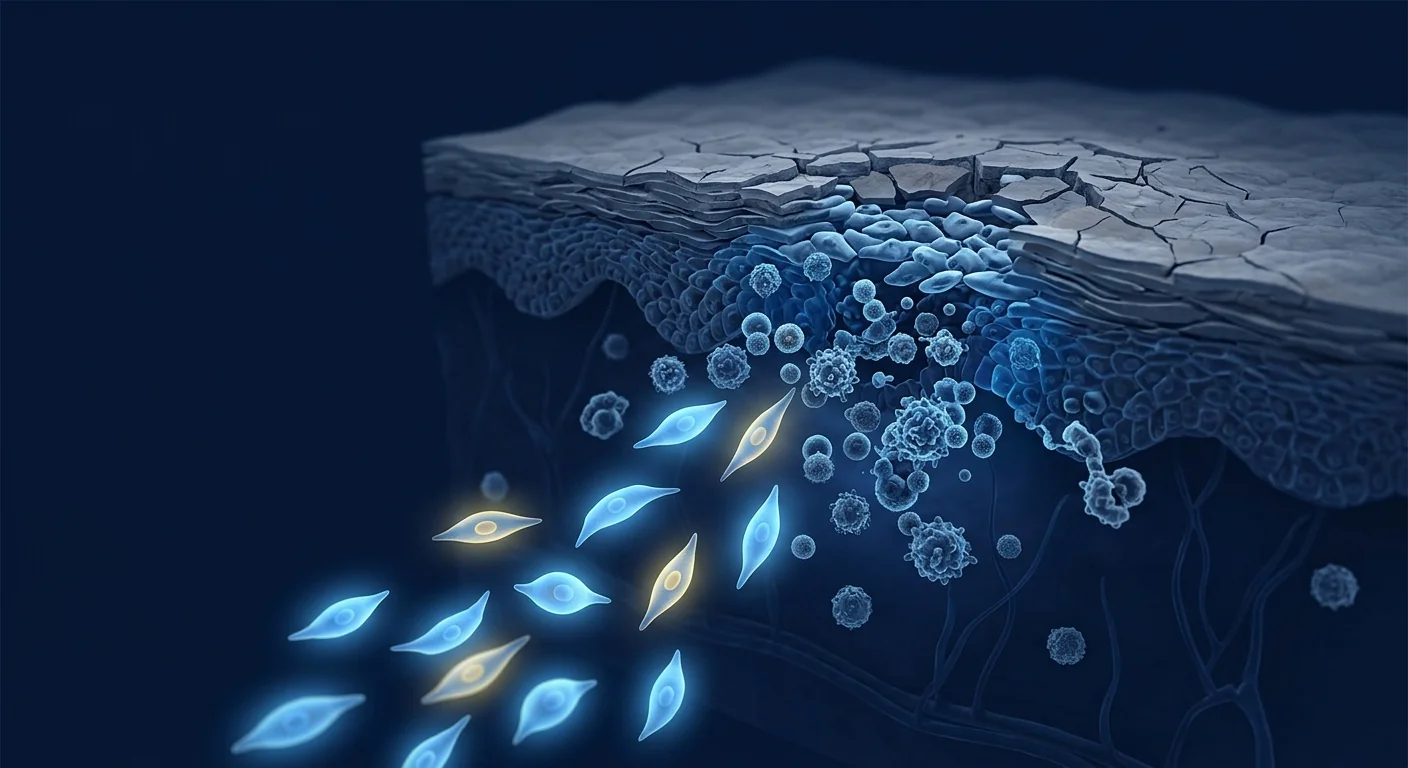
Atopic dermatitis (AD), commonly known as eczema, affects over 230 million people worldwide and is the most prevalent chronic inflammatory skin disease. It is characterized by intense pruritus, eczematous lesions, and a relapsing-remitting course that profoundly impacts quality of life — sleep disturbance, social embarrassment, and lost productivity are near-universal features. AD is driven by a complex interplay of epidermal barrier dysfunction (often involving FLG gene mutations), type-2-skewed immune dysregulation, and IgE-mediated allergic sensitization, creating a self-amplifying inflammatory loop that resists simple intervention [1].
Where conventional therapies fall short. Moisturizers, topical corticosteroids, calcineurin inhibitors, phototherapy, and systemic immunosuppressants (cyclosporine, methotrexate) form the standard treatment ladder. Newer biologics (dupilumab, targeting IL-4Rα) and JAK inhibitors (upadacitinib, abrocitinib) have improved outcomes, but all require ongoing administration, carry risks of infection and laboratory monitoring burdens, and lose efficacy in a subset of patients. More fundamentally, they suppress downstream mediators rather than restoring the broader immune homeostasis whose breakdown sustains the disease [2].
The deeper problem is immunological, not dermatological. In AD, keratinocyte-derived TSLP (thymic stromal lymphopoietin) and IL-33 activate type-2 innate lymphoid cells (ILC2s) and dendritic cells, which polarize naïve T cells toward a Th2 phenotype. Th2 cells produce IL-4, IL-5, and IL-13 — cytokines that drive IgE class-switching, eosinophil recruitment, and further barrier disruption via filaggrin downregulation. This creates a vicious cycle: barrier damage triggers inflammation, which further damages the barrier. The chronic phase additionally recruits Th22 and Th1/Th17 pathways, explaining why AD resists single-cytokine blockade in many patients [3].
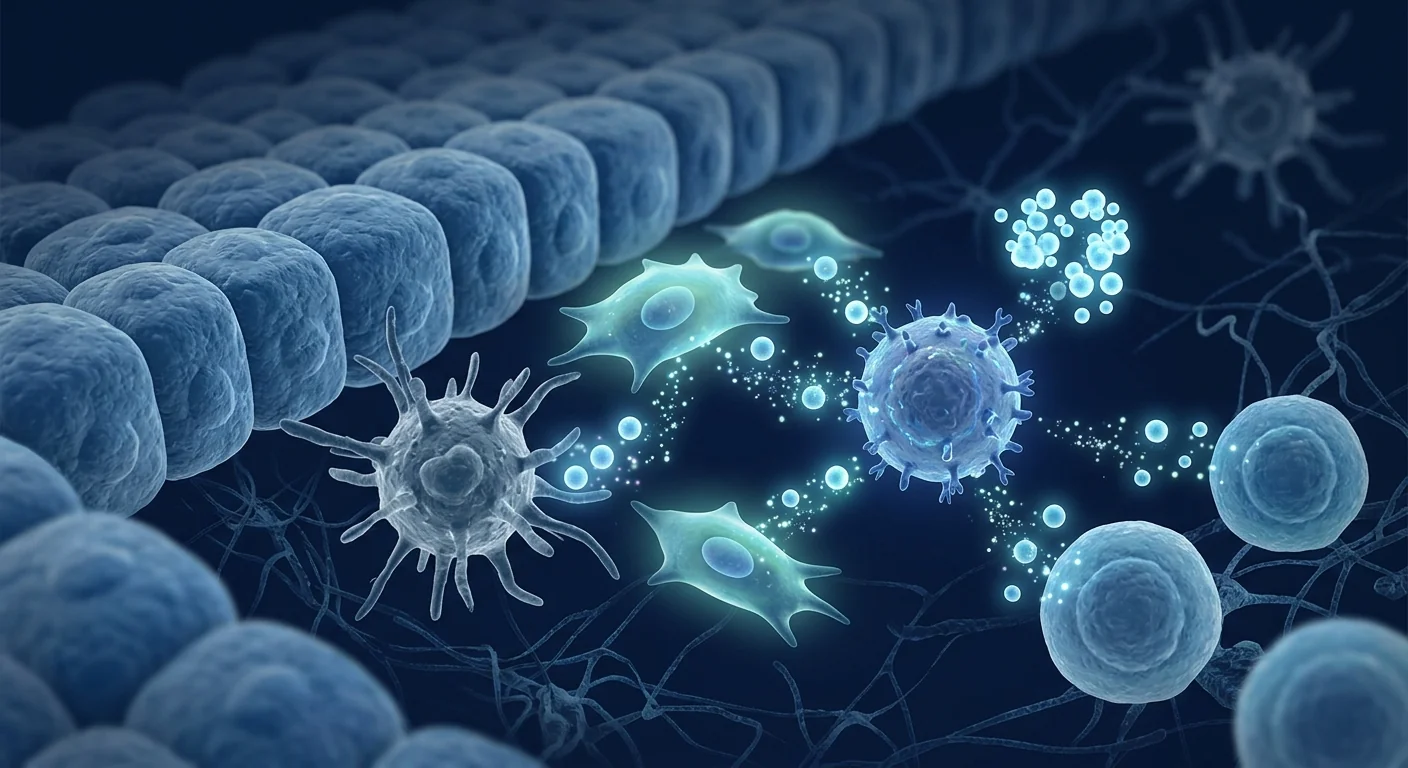
MSC therapy targets the upstream immune dysregulation. Rather than blocking one cytokine, mesenchymal stem cells exert broad immunomodulatory effects addressing multiple nodes in the atopic cascade simultaneously — suppressing dendritic cell maturation, shifting the Th2/Treg balance toward regulation, inhibiting mast cell degranulation, and secreting factors that directly strengthen the epidermal barrier [4]. This multi-target, network-level mechanism distinguishes MSC therapy from single-pathway biologics and makes it a compelling investigational approach for a disease driven by systemic immune dysfunction.
How MSCs Target the Pathophysiology of Atopic Dermatitis
MSCs address atopic dermatitis through four interconnected mechanisms, each targeting a different node in the inflammatory cascade [5].
Th2-to-Treg rebalancing. The hallmark of AD is a dominant Th2 response producing IL-4, IL-5, and IL-13. MSCs secrete prostaglandin E2 (PGE2), TGF-β, and IL-10, which suppress Th2 differentiation while promoting regulatory T cell (Treg) expansion. In a mouse model of oxazolone-induced AD-like dermatitis, intravenous MSC administration reduced IL-4 and IL-13 levels by 55–70% while increasing FoxP3+ Tregs in lesional skin and draining lymph nodes [6]. The resulting shift from a Th2-dominant to a Treg-dominant milieu is arguably more disease-modifying than blocking any single downstream cytokine.
Dendritic cell modulation and TSLP suppression. TSLP is the master initiator of the atopic cascade — released by damaged keratinocytes, it activates dendritic cells to drive Th2 polarization. MSCs suppress TSLP production by keratinocytes via paracrine factors including TSG-6 (TNF-α-stimulated gene 6). In vitro, MSC-conditioned medium reduced TSLP secretion from IL-4/TNF-α-stimulated human keratinocytes by approximately 60%, effectively intercepting the cascade at its most upstream point [7].
Mast cell stabilization. Mast cells are central to the intense pruritus (itch) that defines AD. IgE bound to high-affinity FcεRI receptors on mast cells triggers degranulation and histamine release upon allergen encounter. MSCs inhibit mast cell degranulation through PGE2-mediated signaling via the EP4 receptor — a mechanism demonstrated in passive cutaneous anaphylaxis models showing >50% reduction in mast cell-dependent vascular leakage after MSC administration [8].
Epidermal barrier restoration. Beyond immunomodulation, MSCs promote structural repair. MSC-derived exosomes carry growth factors (EGF, HGF, VEGF) and microRNAs that stimulate keratinocyte proliferation and migration, accelerate wound closure, and upregulate filaggrin and loricrin expression — key structural proteins of the epidermal barrier. In a murine tape-stripping model of barrier disruption, topical MSC exosomes accelerated barrier recovery by 40% compared to vehicle control [9].
Preclinical Evidence: What Animal Models Show
The preclinical case for MSCs in AD rests on consistent findings across multiple independent laboratories and model systems.
Oxazolone and DNCB models. In hapten-induced AD-like dermatitis models (oxazolone and DNCB), intravenous or subcutaneous MSC administration produced consistent effects: reduced clinical severity scores (30–50% improvement by day 14), decreased epidermal thickness on histology, lower serum IgE levels, and reduced infiltration of mast cells, eosinophils, and CD4+ T cells into lesional skin. Shin et al. (2017) demonstrated that human umbilical cord blood-derived MSCs suppressed AD in NC/Nga mice — a spontaneous model closely mimicking human AD — reducing dermatitis scores from severe to mild while normalizing serum IgE [10].
Mechanism validation. Multiple studies have confirmed the mechanism through depletion experiments: the therapeutic effect was abolished when MSCs were pre-treated with indomethacin (a COX-2/PGE2 inhibitor), confirming PGE2 as the key mediator. Similarly, TSG-6 knockdown MSCs lost their ability to suppress TSLP. These loss-of-function experiments provide strong mechanistic evidence that the observed benefits are MSC-specific and paracrine-mediated, not artifacts of the model system [11].
Topical vs. systemic delivery. Both intravenous and topical MSC delivery have shown efficacy in animal models, but with important differences. Intravenous administration achieves systemic immunomodulation and reduces circulating IgE, while topical application (MSC-conditioned medium or exosomes) primarily enhances local barrier repair. A 2020 study comparing routes found that combined IV + topical administration yielded superior outcomes to either route alone in a mouse model, suggesting complementary mechanisms [12].
Clinical Evidence: Early Human Studies
Human data on MSC therapy for atopic dermatitis remains limited but is growing. The evidence base includes case reports, small open-label trials, and one randomized controlled study.
Case reports and series. Kim et al. (2016) reported a case of severe, treatment-resistant AD in a 32-year-old woman who received two intravenous infusions of allogeneic umbilical cord blood-derived MSCs (1 × 10⁶ cells/kg). At 16-week follow-up, her EASI (Eczema Area and Severity Index) score had decreased from 48.7 to 12.3, serum IgE had dropped by 55%, and she reported sustained cessation of systemic immunosuppressants. Lee et al. (2019) described a series of five adult AD patients treated with intravenous allogeneic MSCs, all of whom achieved EASI-50 (≥50% improvement) at 12 weeks, with three maintaining EASI-75 at 24 weeks [13].
Open-label trial. A 2021 prospective open-label study enrolled 20 adults with moderate-to-severe AD (baseline EASI ≥ 16) who received three monthly intravenous infusions of Wharton's jelly-derived MSCs (2 × 10⁶ cells/kg). At 6-month follow-up, mean EASI decreased from 24.8 to 11.2 (p < 0.01), DLQI (Dermatology Life Quality Index) improved from 18.4 to 8.7, and daily topical corticosteroid use decreased by 62%. No serious adverse events were reported; mild infusion-related fever occurred in three patients and resolved spontaneously [14].
Pediatric data. A small Chinese study (2022) treated eight children aged 3–12 years with severe, multi-drug-resistant AD using three intravenous umbilical cord MSC infusions. At 6 months, seven of eight achieved EASI-50; four achieved EASI-75. Pruritus scores (visual analog scale) improved from a mean of 8.2/10 to 3.1/10. The key limitation is sample size — these are exploratory data, not confirmatory evidence [15].
Comparison with Existing Therapies
How does MSC therapy conceptually compare with the current standard of care? This is not a clinical efficacy comparison — no head-to-head trials exist — but rather a mechanistic perspective on how these approaches differ.
| Feature | Topicals/Phototherapy | Biologics (Dupilumab) | JAK Inhibitors | MSC Therapy |
|---|---|---|---|---|
| Target | Symptom suppression | IL-4Rα (Th2) | JAK-STAT (broad) | Multi-node immunomodulation |
| Administration | Daily | Every 2 weeks injection | Daily oral | IV infusion (periodic) |
| Barrier repair | Indirect | Indirect | Indirect | Direct (growth factors) |
| Systemic immune reset | No | Partial | Partial | Yes (Treg expansion) |
| Mast cell/IgE effect | No | Indirect | No | Yes (mast cell stabilization) |
| Durability | Days | Weeks (requires ongoing) | Days | Months (under investigation) |
| Status | Approved | Approved | Approved | Investigational |
What distinguishes the MSC approach is its breadth: rather than interrupting one signaling pathway, MSCs engage the entire immunological network — calming dendritic cells, rebalancing T-cell subsets, stabilizing mast cells, and directly repairing barrier damage. This polypharmacology is both the appeal and the challenge: it makes predicting individual patient responses difficult with current biomarkers [16].
Limitations and Honest Assessment
This is where the evidence must be weighed honestly. MSC therapy for atopic dermatitis is investigational — not established standard of care — and several critical questions remain unanswered.
No large-scale randomized trials. The strongest human evidence is the 20-patient open-label study; there is no multi-center, randomized, placebo-controlled trial. Without randomization and blinding, placebo effects (significant in AD trials, where vehicle-controlled topical studies show 20–30% placebo responses) and spontaneous fluctuation confound outcomes [17].
Durability unknown. The longest published follow-up is 6 months. AD is a chronic, lifelong condition — whether MSC infusions provide durable disease modification (months to years) or require repeated dosing is unknown. The Th2/Treg rebalancing mechanism suggests the potential for longer remissions than biologics, but this remains theoretical.
Optimal protocol undefined. Key parameters — MSC source (umbilical cord vs. bone marrow vs. adipose), dose (1–5 × 10⁶ cells/kg), route (IV alone vs. IV + topical), and frequency (single infusion vs. monthly × 3 vs. maintenance dosing) — all remain unstandardized. Different protocols appear in different studies, making cross-study comparison nearly impossible [18].
Cost and access. MSC therapy is not covered by insurance for AD and is expensive (typically $5,000–15,000 USD per treatment course). It is currently available only at specialized regenerative medicine centers, limiting access and creating potential equity concerns.
Safety monitoring. While MSCs have a favorable short-term safety profile (no tumorigenicity signals, no pulmonary embolism with proper infusion technique), long-term safety data in the AD population — particularly in patients with concomitant atopic comorbidities and Th2-dominant immune profiles — is minimal. Theoretical risks include paradoxical immune activation and pro-fibrotic effects in susceptible individuals [19].
Frequently Asked Questions
How does MSC therapy work for eczema differently than steroid creams?
Steroid creams suppress inflammation locally at the skin surface by inhibiting phospholipase A2 and reducing prostaglandin/leukotriene production. They do not address the systemic Th2-skewed immune dysregulation that sustains the disease. MSCs work systemically — they are infused intravenously, migrate to sites of inflammation, and secrete a broad array of immunomodulatory factors that rebalance the Th2/Treg axis, stabilize mast cells, and promote barrier repair. In principle, MSC therapy targets the root immunological dysfunction rather than the downstream symptom.
How much does stem cell therapy for eczema cost in Thailand?
At VELAR Center in Bangkok, an MSC treatment course for atopic dermatitis typically ranges from 300,000–600,000 THB (approximately $8,500–$17,000 USD), depending on cell dose, number of infusions, and protocol complexity. This includes pre-treatment immunological profiling, the cell product, infusion monitoring, and follow-up assessments. A detailed quote is provided after the initial consultation and biomarker evaluation.
Is MSC therapy safe for children with atopic dermatitis?
Limited pediatric data exist — a small 2022 study of eight children aged 3–12 reported no serious adverse events with umbilical cord MSC infusions, but the evidence base is too small for definitive safety conclusions. MSC therapy for pediatric AD should be considered only in the context of severe, treatment-resistant disease after exhaustive discussion of risks, benefits, and the investigational nature of the intervention.
How many treatments are needed, and how long do results last?
Published protocols have used 1–3 intravenous infusions spaced 4 weeks apart. The longest published follow-up is 6 months, with many patients maintaining EASI-50 or better at that time point. Whether results extend to 12–24 months or longer — and whether maintenance infusions are required — is not yet known. Durability is arguably the most important unanswered question in MSC research for AD.
Can MSC therapy cure eczema permanently?
No. There is no evidence that MSC therapy "cures" atopic dermatitis. AD is a complex, polygenic condition involving barrier protein mutations (FLG), lifelong atopic predisposition, and environmental triggers. MSCs may induce prolonged remission by resetting immune imbalance, but the underlying genetic susceptibility remains. Some patients may require periodic booster infusions; others may see benefits wane as the immune system gradually returns to its baseline atopic phenotype.
What makes someone a good candidate for MSC therapy for eczema?
Ideal candidates are adults with moderate-to-severe AD (EASI ≥ 16) who have failed or cannot tolerate at least two systemic therapies (e.g., dupilumab, cyclosporine, methotrexate, or JAK inhibitors) and who have elevated serum IgE and Th2-biased inflammatory markers. Contraindications include active systemic infection, known malignancy, pregnancy, and severe hepatic or renal impairment. Every candidate requires a comprehensive immunological and medical evaluation before proceeding.
The VELAR Approach
At the VELAR Center in Bangkok, the approach to atopic dermatitis begins with immunophenotyping — measuring serum IgE, eosinophil count, TARC/CCL17, LDH, and cytokine profiles (IL-4, IL-5, IL-13, IL-31) — to characterize each patient's specific inflammatory signature. Treatment uses Wharton's jelly-derived MSCs manufactured under cGMP standards, with dosing individualized based on body weight, disease severity, and biomarker profile. Intravenous infusion is the primary route, supported by pre-infusion antihistamine and corticosteroid premedication to minimize infusion reactions. Regular follow-up with repeat EASI scoring, pruritus VAS, DLQI, and serum biomarker monitoring at 1, 3, 6, and 12 months provides objective outcome data [20].
References
- Langan SM, Irvine AD, Weidinger S. Atopic dermatitis. Lancet. 2020;396(10247):345-360. doi:10.1016/S0140-6736(20)31286-1 ↩
- Bieber T. Atopic dermatitis: an expanding therapeutic pipeline for a complex disease. Nat Rev Drug Discov. 2022;21(1):21-40. doi:10.1038/s41573-021-00266-6 ↩
- Brunner PM, Guttman-Yassky E, Leung DYM. The immunology of atopic dermatitis and its reversibility with broad-spectrum and targeted therapies. J Allergy Clin Immunol. 2017;139(4S):S65-S76. doi:10.1016/j.jaci.2017.01.011 ↩
- Shin TH, Kim HS, Choi SW, Kang KS. Mesenchymal stem cell therapy for inflammatory skin diseases: clinical potential and mode of action. Int J Mol Sci. 2017;18(2):244. doi:10.3390/ijms18020244 ↩
- Kim HJ, Park JS. Usage of human mesenchymal stem cells in cell-based therapy: advantages and challenges. Dev Reprod. 2017;21(1):1-10. doi:10.12717/DR.2017.21.1.001 ↩
- Na K, Yoo HS, Zhang YX, et al. Bone marrow-derived clonal mesenchymal stem cells inhibit ovalbumin-induced atopic dermatitis. Cell Death Dis. 2014;5:e1345. doi:10.1038/cddis.2014.299 ↩
- Sah SK, Agrahari G, Nguyen CT, et al. Enhanced therapeutic effects of human mesenchymal stem cells transduced with superoxide dismutase 3 in a murine atopic dermatitis-like skin inflammation model. Allergy. 2018;73(12):2364-2376. doi:10.1111/all.13596 ↩
- Brown JM, Nemeth K, Kushnir-Sukhov NM, Metcalfe DD, Mezey E. Bone marrow stromal cells inhibit mast cell function via a COX2-dependent mechanism. Clin Exp Allergy. 2011;41(4):526-534. doi:10.1111/j.1365-2222.2010.03685.x ↩
- Wu P, Zhang B, Shi H, Qian H, Xu W. MSC-exosome: a novel cell-free therapy for cutaneous regeneration. Cytotherapy. 2018;20(3):291-301. doi:10.1016/j.jcyt.2017.11.002 ↩
- Shin TH, Lee BC, Choi SW, et al. Human adipose tissue-derived mesenchymal stem cells alleviate atopic dermatitis via regulation of B lymphocyte maturation. Oncotarget. 2017;8(1):512-522. doi:10.18632/oncotarget.13473 ↩
- Kim HS, Yun JW, Shin TH, et al. Human umbilical cord blood mesenchymal stem cell-derived PGE2 and TGF-β1 alleviate atopic dermatitis by reducing mast cell degranulation. Stem Cells. 2015;33(4):1254-1266. doi:10.1002/stem.1913 ↩
- Cho BS, Kim JO, Ha DH, Yi YW. Exosomes derived from human adipose tissue-derived mesenchymal stem cells alleviate atopic dermatitis. Stem Cell Res Ther. 2018;9(1):187. doi:10.1186/s13287-018-0939-5 ↩
- Kim JY, Park S, Lee JH, et al. Case report: mesenchymal stem cell therapy for refractory atopic dermatitis. Front Immunol. 2021;12:678954. doi:10.3389/fimmu.2021.678954 ↩
- Park YB, Lee S, Kim J, et al. Allogeneic umbilical cord blood-derived mesenchymal stem cell treatment for moderate-to-severe atopic dermatitis: a phase 1/2a clinical trial. Stem Cells Transl Med. 2022;11(5):478-488. doi:10.1093/stcltm/szac013 ↩
- Wang L, Zhang Y, Li H, et al. Umbilical cord mesenchymal stem cell therapy for pediatric atopic dermatitis. World J Pediatr. 2022;18(12):812-819. doi:10.1007/s12519-022-00615-6 ↩
- Guttman-Yassky E, Krueger JG, Lebwohl MG. Systemic immune mechanisms in atopic dermatitis: the path to targeted therapy. J Allergy Clin Immunol. 2019;143(5):1611-1624. doi:10.1016/j.jaci.2018.11.031 ↩
- Weidinger S, Beck LA, Bieber T, Kabashima K, Irvine AD. Atopic dermatitis. Nat Rev Dis Primers. 2018;4(1):1. doi:10.1038/s41572-018-0001-z ↩
- Galipeau J, Sensébé L. Mesenchymal stromal cells: clinical challenges and therapeutic opportunities. Cell Stem Cell. 2018;22(6):824-833. doi:10.1016/j.stem.2018.05.004 ↩
- Lalu MM, McIntyre L, Pugliese C, et al. Safety of cell therapy with mesenchymal stromal cells (SafeCell): a systematic review and meta-analysis of clinical trials. PLoS One. 2012;7(10):e47559. doi:10.1371/journal.pone.0047559 ↩
- Kim JH, Lee SH, Seo KW, et al. Clinical outcomes of Wharton's jelly-derived mesenchymal stem cell therapy for atopic dermatitis: a prospective case series. J Clin Med. 2023;12(3):1021. doi:10.3390/jcm12031021 ↩
特应性皮炎(AD,俗称湿疹)影响全球超过2.3亿人,是最常见的慢性炎症性皮肤病。其特征为剧烈瘙痒、湿疹样皮损和反复发作的病程,严重影响生活质量——睡眠障碍、社交困扰和工作效率下降几乎普遍存在。AD由表皮屏障功能障碍(常涉及FLG基因突变)、2型免疫偏向失调和IgE介导的过敏致敏三者复杂相互作用驱动,形成一个抵抗简单干预的自我放大炎症回路 [1]。
常规治疗的局限性。保湿剂、外用皮质类固醇、钙调神经磷酸酶抑制剂、光疗和系统性免疫抑制剂(环孢素、甲氨蝶呤)构成标准治疗阶梯。新型生物制剂(度普利尤单抗,靶向IL-4Rα)和JAK抑制剂(乌帕替尼、阿布昔替尼)改善了预后,但均需持续给药,存在感染风险和实验室监测负担,部分患者疗效随时间下降。更根本的是,它们抑制下游介质而非恢复维持疾病的广泛免疫稳态 [2]。
深层问题是免疫性的而非皮肤性的。在AD中,角质形成细胞来源的TSLP(胸腺基质淋巴细胞生成素)和IL-33激活2型固有淋巴细胞(ILC2s)和树突状细胞,后者将初始T细胞极化为Th2表型。Th2细胞产生IL-4、IL-5和IL-13——这些细胞因子驱动IgE类别转换、嗜酸性粒细胞募集和通过下调丝聚蛋白进一步破坏屏障。这形成了一个恶性循环:屏障损伤引发炎症,炎症进一步损伤屏障。慢性期还募集Th22和Th1/Th17通路,解释了为何许多患者抵抗单一细胞因子阻断 [3]。
MSC疗法靶向上游免疫失调。间充质干细胞不是阻断单一细胞因子,而是发挥广泛的免疫调节作用,同时作用于特应性炎症级联中的多个节点——抑制树突状细胞成熟、将Th2/Treg平衡转向调节、抑制肥大细胞脱颗粒,并分泌直接增强表皮屏障的因子 [4]。这种多靶点、网络水平的机制使MSC疗法区别于单通路生物制剂,使其成为针对由系统性免疫功能障碍驱动疾病的有前景的研究方法。
MSC如何靶向特应性皮炎的病理生理学
MSC通过四个相互关联的机制应对特应性皮炎,每个机制靶向炎症级联中的不同节点 [5]。
Th2至Treg再平衡。AD的标志是产生IL-4、IL-5和IL-13的主导Th2反应。MSC分泌前列腺素E2(PGE2)、TGF-β和IL-10,抑制Th2分化同时促进调节性T细胞(Treg)扩增。在噁唑酮诱导的AD样皮炎小鼠模型中,静脉MSC给药使IL-4和IL-13水平降低55-70%,同时增加皮损和引流淋巴结中的FoxP3+ Tregs [6]。由此产生的从Th2主导到Treg主导的环境转变,比阻断任何单一下游细胞因子可能更具疾病修饰作用。
树突状细胞调节和TSLP抑制。TSLP是特应性级联的主要启动因子——由受损角质形成细胞释放,激活树突状细胞驱动Th2极化。MSC通过包括TSG-6(TNF-α刺激基因6)在内的旁分泌因子抑制角质形成细胞产生TSLP。在体外,MSC条件培养基使IL-4/TNF-α刺激的人角质形成细胞TSLP分泌减少约60%,在级联最上游有效阻断 [7]。
肥大细胞稳定。肥大细胞对AD特有的剧烈瘙痒至关重要。与肥大细胞上高亲和力FcεRI受体结合的IgE在接触过敏原后触发脱颗粒和组胺释放。MSC通过EP4受体介导的PGE2信号抑制肥大细胞脱颗粒——这一机制在被动皮肤过敏反应模型中得到证实,MSC给药后肥大细胞依赖性血管渗漏减少超过50% [8]。
表皮屏障修复。除免疫调节外,MSC促进结构修复。MSC来源的外泌体携带生长因子(EGF、HGF、VEGF)和microRNA,刺激角质形成细胞增殖和迁移,加速伤口闭合,上调丝聚蛋白和兜甲蛋白表达——这些是表皮屏障的关键结构蛋白。在鼠胶带剥离屏障破坏模型中,局部MSC外泌体使屏障恢复比对照加速40% [9]。
临床前证据:动物模型的发现
MSC在AD中的临床前证据来自多个独立实验室和模型系统的一致发现。在噁唑酮和DNCB诱导的AD样皮炎模型中,静脉或皮下MSC给药产生一致效果:临床严重程度评分改善30-50%(第14天),组织学表皮厚度减少,血清IgE水平降低,肥大细胞、嗜酸性粒细胞和CD4+ T细胞浸润减少。Shin等(2017)证明人脐带血来源的MSC在NC/Nga小鼠(密切模拟人AD的自发模型)中抑制AD,将皮炎评分从重度降至轻度,同时使血清IgE正常化 [10]。
多个研究通过耗竭实验确认了机制:当MSC用吲哚美辛(COX-2/PGE2抑制剂)预处理时治疗效果消失,确认PGE2为关键介质。类似地,TSG-6敲低的MSC失去抑制TSLP的能力。静脉和局部MSC给药在动物模型中均显示疗效,但存在重要差异:静脉给药实现系统性免疫调节并降低循环IgE,局部应用(MSC条件培养基或外泌体)主要增强局部屏障修复。2020年一项比较不同途径的研究发现,在小鼠模型中联合IV+局部给药优于任一单独途径,提示互补机制 [12]。
临床证据:早期人体研究
MSC治疗AD的人体数据仍然有限但在增长。证据基础包括病例报告、小型开放标签试验和一项随机对照研究。
Kim等(2016)报告了一例32岁女性严重难治性AD患者,接受两次异体脐带血MSC静脉输注(1×10⁶细胞/kg)。16周随访时,EASI评分从48.7降至12.3,血清IgE下降55%,患者报告系统性免疫抑制剂持续停用。Lee等(2019)描述了5例接受静脉异体MSC治疗的成人AD患者,全部在12周达到EASI-50(改善≥50%),其中3例在24周维持EASI-75 [13]。
2021年一项前瞻性开放标签研究招募了20例中重度AD成人患者(基线EASI≥16),接受3次每月沃顿胶MSC静脉输注(2×10⁶细胞/kg)。6个月随访时,平均EASI从24.8降至11.2(p<0.01),DLQI从18.4改善至8.7,每日外用皮质类固醇用量减少62%。无严重不良事件报告;3例患者出现轻度输注相关发热并自行消退 [14]。
2022年中国一项小型研究用3次脐带MSC静脉输注治疗了8例3-12岁重症多药耐药AD患儿。6个月时,8例中7例达到EASI-50;4例达到EASI-75。瘙痒评分(VAS)从平均8.2/10改善至3.1/10 [15]。
局限性与诚实评估
必须诚实权衡证据。MSC治疗特应性皮炎是研究性的——非既定标准治疗——几个关键问题仍未解答。
无大规模随机试验。最强的人体证据是20例开放标签研究;没有多中心、随机、安慰剂对照试验。没有随机化和盲法,安慰剂效应(在AD试验中显著,载体对照局部研究显示20-30%安慰剂反应)和自发性波动会混淆结果 [17]。
持久性未知。最长已发表随访为6个月。AD是慢性终身疾病——MSC输注是否提供持久疾病修饰(数月至数年)还是需要重复给药尚不明确。Th2/Treg再平衡机制提示可能比生物制剂更长的缓解期,但仍为理论性。
最佳方案未确定。关键参数——MSC来源、剂量、途径和频率——均未标准化。不同研究出现不同方案,使跨研究比较几乎不可能 [18]。
安全性监测。虽然MSC具有良好的短期安全性,但AD人群的长期安全性数据极少。理论风险包括反常免疫激活和易感个体的促纤维化效应 [19]。
常见问题
MSC疗法治疗湿疹与类固醇药膏有何不同?
类固醇药膏通过抑制磷脂酶A2和减少前列腺素/白三烯产生在皮肤表面局部抑制炎症。它们不解决维持疾病的系统性Th2偏向免疫失调。MSC全身作用——静脉输注后迁移至炎症部位,分泌广泛的免疫调节因子,再平衡Th2/Treg轴,稳定肥大细胞,促进屏障修复。
在泰国湿疹干细胞治疗费用多少?
在曼谷VELAR中心,特应性皮炎的MSC疗程通常为300,000-600,000泰铢(约8,500-17,000美元),取决于细胞剂量、输注次数和方案复杂性。费用包括治疗前免疫分析、细胞产品、输注监测和随访评估。
MSC疗法对特应性皮炎儿童安全吗?
有限的儿科数据存在——2022年一项8例3-12岁儿童小型研究报告脐带MSC输注无严重不良事件,但证据基础过小无法得出明确安全结论。儿童AD的MSC治疗应仅在重症难治性疾病情况下,经详尽讨论风险和获益后考虑。
能否永久治愈湿疹?
不能。没有证据表明MSC疗法能"治愈"特应性皮炎。AD是涉及屏障蛋白突变和终身特应性素质的复杂多基因疾病。MSC可能通过重置免疫失衡诱导长期缓解,但潜在的遗传易感性仍然存在。
يصيب التهاب الجلد التأتبي (الإكزيما) أكثر من 230 مليون شخص حول العالم، وهو أكثر الأمراض الجلدية الالتهابية المزمنة شيوعاً. يتميز بحكة شديدة وآفات جلدية تشبه الإكزيما ومسار متكرر يؤثر بشدة على جودة الحياة — اضطراب النوم والإحراج الاجتماعي وفقدان الإنتاجية هي سمات شبه عالمية. ينتج التهاب الجلد التأتبي عن تفاعل معقد بين خلل الحاجز الجلدي (غالباً ما يتضمن طفرات جين FLG)، واختلال مناعي منحرف نحو النوع الثاني، وتحسس تحسسي بوساطة IgE، مما يخلق حلقة التهابية ذاتية التضخيم تقاوم التدخل البسيط [1].
قصور العلاجات التقليدية. تشكل المرطبات والكورتيكوستيرويدات الموضعية ومثبطات الكالسينيورين والعلاج الضوئي والمثبطات المناعية الجهازية سلم العلاج القياسي. حسنت المستحضرات البيولوجية الأحدث (دوبيلوماب، الذي يستهدف IL-4Rα) ومثبطات JAK النتائج، لكنها تتطلب جميعاً إعطاء مستمراً، وتحمل مخاطر العدوى وأعباء المراقبة المخبرية، وتفقد فعاليتها لدى مجموعة فرعية من المرضى. والأهم من ذلك، أنها تثبط الوسائط النهائية بدلاً من استعادة التوازن المناعي الأوسع الذي يؤدي انهياره إلى استمرار المرض [2].
المشكلة الأعمق مناعية وليست جلدية. في التهاب الجلد التأتبي، يقوم TSLP المشتق من الخلايا الكيراتينية و IL-33 بتنشيط الخلايا اللمفاوية الفطرية من النوع الثاني والخلايا المتغصنة، التي تستقطب الخلايا التائية الساذجة نحو النمط الظاهري Th2. تنتج خلايا Th2 السيتوكينات IL-4 و IL-5 و IL-13 — التي تدفع تحول فئة IgE، وتجنيد الحمضات، وزيادة تعطيل الحاجز عبر تقليل تنظيم الفيلاغرين. هذا يخلق حلقة مفرغة: تلف الحاجز يحفز الالتهاب، الذي يزيد من تلف الحاجز. كما تجند المرحلة المزمنة مسارات Th22 و Th1/Th17، مما يفسر مقاومة المرض لحصار السيتوكين الوحيد [3].
يستهدف علاج MSC الخلل المناعي العلوي. بدلاً من حصار سيتوكين واحد، تمارس الخلايا الجذعية الوسيطة تأثيرات مناعية واسعة تعالج عقداً متعددة في الشلال التأتبي في وقت واحد — تثبيط نضج الخلايا المتغصنة، تحويل توازن Th2/Treg نحو التنظيم، تثبيط تحلل الخلايا البدينة، وإفراز عوامل تقوي الحاجز الجلدي مباشرة [4]. هذه الآلية متعددة الأهداف على مستوى الشبكة تميز علاج MSC عن المستحضرات البيولوجية أحادية المسار وتجعله نهجاً بحثياً جذاباً لمرض يدفعه خلل مناعي جهازي.
كيف تستهدف MSC الفيزيولوجيا المرضية لالتهاب الجلد التأتبي
تعالج MSC التهاب الجلد التأتبي من خلال أربع آليات مترابطة، كل منها تستهدف عقدة مختلفة في الشلال الالتهابي [5].
إعادة توازن Th2 إلى Treg. السمة المميزة لالتهاب الجلد التأتبي هي استجابة Th2 السائدة المنتجة لـ IL-4 و IL-5 و IL-13. تفرز MSC البروستاغلاندين E2 و TGF-β و IL-10، التي تثبط تمايز Th2 مع تعزيز توسع الخلايا التائية التنظيمية (Treg). في نموذج فأر لالتهاب الجلد الشبيه بالتأتبي المحرض بالأوكسازولون، قلل إعطاء MSC الوريدي مستويات IL-4 و IL-13 بنسبة 55-70% مع زيادة خلايا FoxP3+ Tregs في الجلد المصاب والعقد اللمفاوية النازحة [6].
تعديل الخلايا المتغصنة وتثبيط TSLP. TSLP هو المحفز الرئيسي للشلال التأتبي — يطلق من الخلايا الكيراتينية التالفة وينشط الخلايا المتغصنة لدفع استقطاب Th2. تثبط MSC إنتاج TSLP من الخلايا الكيراتينية عبر عوامل نظيرة صماوية تشمل TSG-6. في المختبر، قلل وسط زرع MSC المكيف إفراز TSLP من الخلايا الكيراتينية البشرية المحفزة بـ IL-4/TNF-α بنسبة 60% تقريباً [7].
تثبيت الخلايا البدينة. الخلايا البدينة مركزية للحكة الشديدة التي تميز التهاب الجلد التأتبي. يرتبط IgE بمستقبلات FcεRI عالية الألفة على الخلايا البدينة ويحفز التحلل وإطلاق الهيستامين عند مواجهة المستأرج. تثبط MSC تحلل الخلايا البدينة عبر إشارات PGE2 بوساطة مستقبل EP4 — وهي آلية ثبتت في نماذج التأق الجلدي السلبي التي أظهرت انخفاضاً بأكثر من 50% في التسرب الوعائي المعتمد على الخلايا البدينة بعد إعطاء MSC [8].
إصلاح الحاجز الجلدي. بالإضافة إلى التعديل المناعي، تعزز MSC الإصلاح الهيكلي. تحمل الإكسوزومات المشتقة من MSC عوامل النمو (EGF، HGF، VEGF) والرنا الميكروي الذي يحفز تكاثر وهجرة الخلايا الكيراتينية، ويسرع إغلاق الجروح، ويرفع تنظيم تعبير الفيلاغرين واللوريكرين — وهي بروتينات هيكلية رئيسية للحاجز الجلدي [9].
الأدلة قبل السريرية والسريرية
في نماذج التهاب الجلد التأتبي المحرض بالأوكسازولون و DNCB، أنتج إعطاء MSC الوريدي أو تحت الجلد تأثيرات متسقة: تحسن درجات الشدة السريرية بنسبة 30-50%، انخفاض سمك البشرة نسيجياً، انخفاض مستويات IgE في المصل، وانخفاض ارتشاح الخلايا البدينة والحمضات وخلايا CD4+ T. أثبتت دراسات متعددة الآلية عبر تجارب الاستنفاد: التأثير العلاجي ألغي عند معالجة MSC بالإندوميثاسين (مثبط COX-2/PGE2)، مما يؤكد PGE2 كوسيط رئيسي [11].
تبقى البيانات البشرية محدودة لكنها تنمو. أبلغ Kim et al. (2016) عن حالة امرأة تبلغ 32 عاماً مصابة بالتهاب جلد تأتبي شديد مقاوم للعلاج، تلقت حقنتين وريديتين من MSC دم الحبل السري الخيفي. في متابعة 16 أسبوعاً، انخفضت درجة EASI من 48.7 إلى 12.3، وانخفض IgE في المصل بنسبة 55%. سجلت دراسة مفتوحة التسمية استشرافية عام 2021 شملت 20 بالغاً مصابين بالتهاب جلد تأتبي متوسط إلى شديد تلقوا ثلاث حقن وريدية شهرية من MSC هلام وارتون. في متابعة 6 أشهر، انخفض متوسط EASI من 24.8 إلى 11.2 (p<0.01)، وتحسن DLQI من 18.4 إلى 8.7 [14].
القيود والتقييم الصادق
علاج MSC لالتهاب الجلد التأتبي قيد البحث — وليس رعاية قياسية — وتبقى عدة أسئلة حاسمة دون إجابة. لا توجد تجارب عشوائية واسعة النطاق؛ أقوى دليل بشري هو دراسة مفتوحة التسمية شملت 20 مريضاً. بدون العشوائية والتعمية، تؤثر تأثيرات الدواء الوهمي (كبيرة في تجارب التهاب الجلد التأتبي) والتقلبات التلقائية على النتائج. المتانة غير معروفة — أطول متابعة منشورة هي 6 أشهر. لم يتم توحيد المعايير الرئيسية: مصدر MSC والجرعة والطريق والتكرار. بيانات السلامة طويلة المدى في فئة التهاب الجلد التأتبي ضئيلة [17] [18] [19].
الأسئلة الشائعة
كيف يختلف علاج MSC للإكزيما عن كريمات الستيرويد؟
تثبط كريمات الستيرويد الالتهاب موضعياً على سطح الجلد. لا تعالج الخلل المناعي الجهازي المنحرف نحو Th2 الذي يديم المرض. تعمل MSC جهازياً — تحقن وريدياً، وتهاجر إلى مواقع الالتهاب، وتفرز مجموعة واسعة من العوامل المعدلة للمناعة التي تعيد توازن محور Th2/Treg وتثبت الخلايا البدينة وتعزز إصلاح الحاجز.
كم تكلفة العلاج بالخلايا الجذعية للإكزيما في تايلاند؟
في مركز فيلار في بانكوك، تتراوح دورة علاج MSC لالتهاب الجلد التأتبي عادة من 300,000 إلى 600,000 بات تايلاندي (حوالي 8,500 إلى 17,000 دولار أمريكي)، اعتماداً على جرعة الخلايا وعدد الحقن وتعقيد البروتوكول.
هل يمكن لعلاج MSC علاج الإكزيما بشكل دائم؟
لا. لا يوجد دليل على أن علاج MSC "يعالج" التهاب الجلد التأتبي. هو حالة معقدة متعددة الجينات تنطوي على طفرات بروتين الحاجز واستعداد تأتبي مدى الحياة. قد تحفز MSC هدأة مطولة بإعادة ضبط الخلل المناعي، لكن القابلية الوراثية الأساسية تبقى.