Osteomyelitis — a bacterial or fungal infection of bone — affects an estimated 22 per 100,000 people annually, with rates rising sharply in aging, diabetic, and post-surgical populations. [1] Unlike most infections, osteomyelitis cannot be reliably cured by antibiotics alone: the bone's dense mineral matrix limits drug penetration, and bacteria establish biofilms that resist even prolonged antimicrobial therapy. Chronic osteomyelitis carries a recurrence rate of 20–30% despite aggressive surgical debridement and weeks of intravenous antibiotics.
Where conventional treatment falls short. The standard of care — surgical debridement of necrotic bone followed by 4–6 weeks of intravenous antibiotics — leaves a structural void where infected bone was removed. This dead space must be filled with antibiotic-impregnated cement spacers or bone grafts, neither of which actively fights residual bacteria or stimulates new bone formation. [2] Even with optimal treatment, limb amputation remains the outcome in 5–10% of chronic cases, and functional deficits persist in a much larger proportion of survivors.
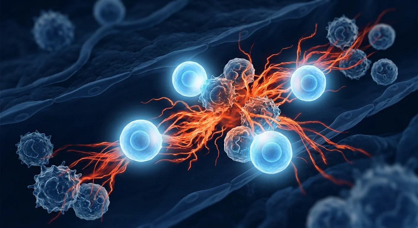
The deeper problem is the biofilm. Bacteria in osteomyelitis do not float freely — they embed themselves in a self-produced extracellular polymeric substance (EPS) matrix, forming biofilms on necrotic bone surfaces. [3] Within this biofilm, bacteria enter a metabolically dormant persister state that renders them 100–1,000 times more resistant to antibiotics than their planktonic counterparts. The host immune response, while robust, contributes to collateral bone destruction through chronic inflammation driven by TNF-α, IL-1β, and RANKL-mediated osteoclast activation.
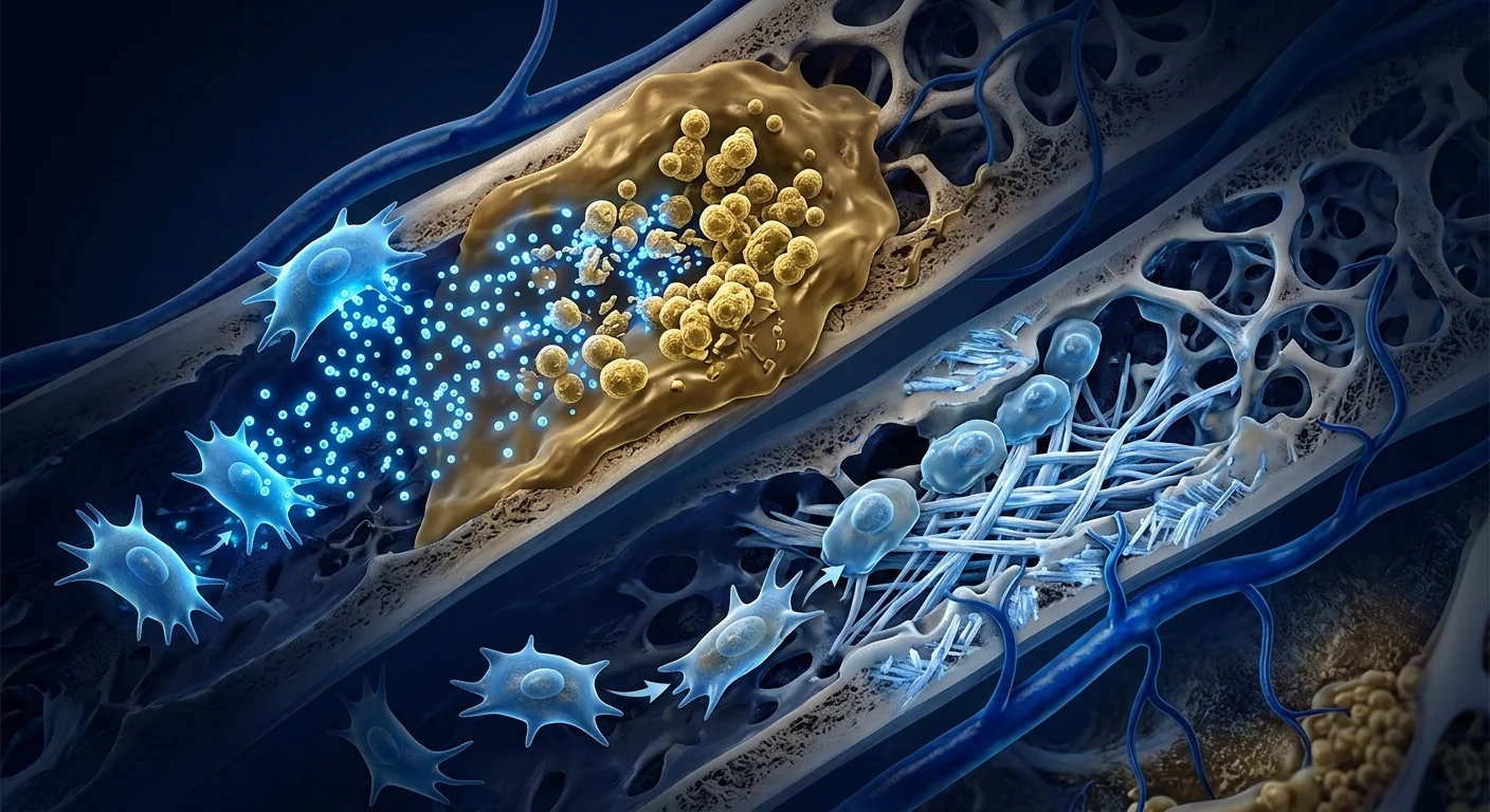
MSC therapy offers a dual-action approach. Mesenchymal stem cells bring two capabilities that no single antibiotic or bone graft can match: direct antimicrobial activity through secretion of endogenous antibiotic peptides, and simultaneous osteogenic differentiation to rebuild the bone defect left by infection. [4] This dual-action mechanism — kill the pathogen while regenerating the tissue — positions MSCs as a fundamentally different category of intervention, addressing both the infectious and the structural dimensions of osteomyelitis in a single therapeutic product.
How MSC Therapy Works in Osteomyelitis
MSC therapy combats osteomyelitis through four interconnected mechanisms: direct antimicrobial peptide secretion, immunomodulation of the chronic inflammatory environment, osteogenic differentiation for bone defect repair, and angiogenesis to restore vascular supply to devitalized bone. These mechanisms are deployed concurrently, creating a coordinated biological response that no single-agent pharmaceutical can replicate.
1. Antimicrobial Peptide Secretion
MSCs are constitutively armed with a broad-spectrum antimicrobial arsenal. Under resting conditions, they secrete basal levels of LL-37 (cathelicidin), hepcidin, β-defensin-2, and lipocalin-2 — peptides that disrupt bacterial membranes, sequester iron, and inhibit biofilm formation. [6] Upon exposure to bacterial lipopolysaccharide (LPS) or inflammatory cytokines, MSCs dramatically upregulate this antimicrobial program. Krasnodembskaya and colleagues demonstrated that MSC-derived LL-37 reduced Staphylococcus aureus colony-forming units by more than 80% in vitro, an effect that was abolished when LL-37 was neutralized with antibody — confirming that the peptide, not the cells themselves, was the direct effector. [7]
MSCs also disrupt biofilms. Sutton et al. showed that MSC-conditioned medium significantly inhibited S. aureus biofilm formation and partially disrupted established biofilms. The mechanism involves LL-37's ability to degrade the extracellular DNA scaffold of biofilms and, interestingly, the secretion of active matrix metalloproteinases by MSCs that digest biofilm matrix components. [8] This anti-biofilm activity is particularly relevant in osteomyelitis, where biofilm-protected persister cells are the primary reason for treatment failure.
2. Immunomodulation of the Infectious Microenvironment
Chronic osteomyelitis is not simply an infection — it is an infection-fueled cycle of destructive inflammation. Bacterial components trigger Toll-like receptors on macrophages and osteoclast precursors, driving sustained production of TNF-α, IL-1β, IL-6, and RANKL. [9] RANKL in turn activates osteoclasts that resorb bone well beyond the infected zone, creating the characteristic radiographic appearance of osteolysis with surrounding sclerosis — a sequestrum of dead bone surrounded by reactive new bone formation.
MSCs interrupt this destructive cycle through several pathways. They secrete PGE2 and TSG-6, which shift macrophages from a pro-inflammatory M1 to a reparative M2 phenotype. [10] M2 macrophages produce IL-10 and TGF-β, suppress osteoclastogenesis, and promote angiogenesis — rebuilding rather than destroying. MSCs also directly inhibit osteoclast differentiation through secretion of osteoprotegerin (OPG), the decoy receptor for RANKL. In a rat model of implant-associated osteomyelitis, local MSC delivery reduced osteoclast numbers by approximately 60% and preserved trabecular bone volume relative to untreated controls. [11]
3. Osteogenic Differentiation and Bone Defect Regeneration
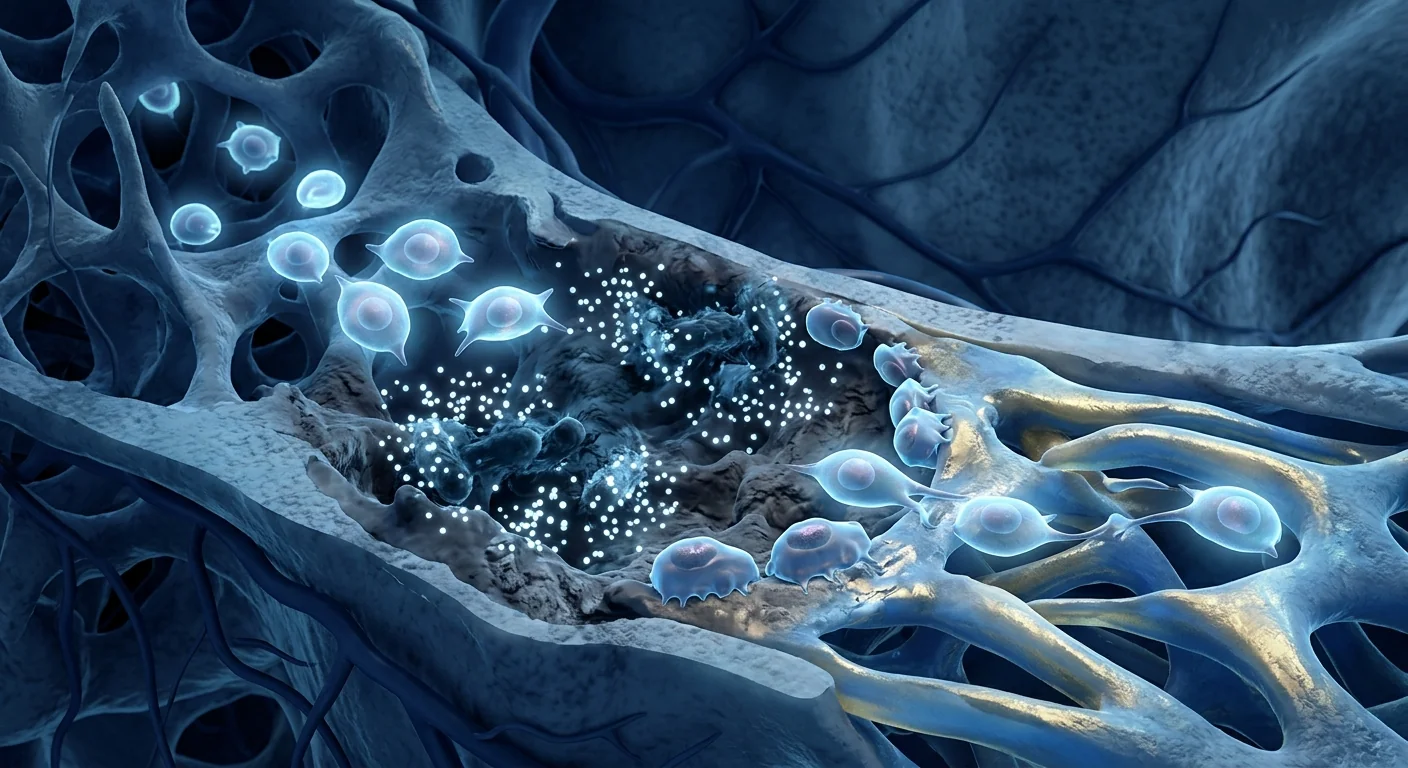
The bone void created by surgical debridement does not spontaneously heal — the defect is too large, the periosteum is often stripped, and the local microenvironment remains hostile to osteoblast function. MSCs address this directly: they are the native progenitors of osteoblasts and, when delivered to a bone defect, undergo osteogenic differentiation guided by local mechanical and biochemical cues. [12]
Beyond differentiating into osteoblasts themselves, MSCs secrete bone morphogenetic protein-2 (BMP-2), vascular endothelial growth factor (VEGF), and insulin-like growth factor-1 (IGF-1) — factors that recruit host osteoprogenitor cells to the defect and stimulate their differentiation. [13] In a rabbit model of post-infectious tibial defects, MSC-seeded scaffolds achieved 78% radiographic bone fill at 12 weeks compared to 34% with scaffold alone, and the regenerated bone had mechanical properties approaching those of native cortical bone. This osteogenic capacity is particularly valuable in osteomyelitis, where the bone defect is often large, irregular, and contaminated — conditions under which autologous bone graft (the gold standard) has a higher failure rate.
4. Angiogenesis and Restoration of Vascular Supply
Devascularized bone — a sequestrum — is the hallmark of chronic osteomyelitis. Without blood supply, antibiotics cannot reach the infected site, host immune cells cannot access the bacteria, and osteoprogenitor cells cannot migrate into the defect to initiate repair. MSCs secrete VEGF, angiopoietin-1, and fibroblast growth factor-2 (FGF-2), promoting endothelial cell proliferation, migration, and tube formation. [14] In a murine model of segmental bone defect with superimposed infection, MSC therapy increased capillary density in the defect zone by 2.8-fold at 4 weeks, correlating strongly with subsequent bone formation. Restoring vascularity transforms the defect from a dead space into a biologically active zone capable of supporting regeneration.
Clinical Evidence: What the Research Shows
The preclinical evidence for MSCs in osteomyelitis is converging from multiple independent laboratories using different models (S. aureus tibial infection in rats and rabbits, implant-associated infection, and post-debridement bone defects). Collectively, these studies demonstrate reduced bacterial burden, enhanced bone regeneration, and improved functional outcomes. [15] Human data, while still limited to early-phase studies, is emerging.
Preclinical models show reduced infection and enhanced healing. In a rat model of chronic post-traumatic osteomyelitis, Yuan et al. compared standard debridement alone versus debridement plus locally delivered bone marrow MSCs. At 8 weeks, the MSC group showed significantly lower bacterial counts in bone tissue, higher radiographic bone union scores, and better weight-bearing on the affected limb. Histological analysis confirmed both reduced inflammatory infiltrate and increased new bone formation in the MSC-treated defects. [16]
Human case series are encouraging but preliminary. A 2019 case series from China reported 12 patients with chronic osteomyelitis of the tibia or femur treated with debridement, antibiotic-impregnated cement spacer placement, and percutaneous injection of autologous bone marrow MSCs at 2 and 4 weeks post-debridement. At mean follow-up of 24 months, 10 of 12 patients (83%) remained infection-free with radiographic evidence of bone consolidation. [17] While the absence of a control group limits interpretation, the 83% infection eradication rate compares favorably to the 70–80% rates typically reported for standard treatment in similar patient populations.
Safety data is consistently reassuring. A 2012 systematic review by Lalu et al. pooled safety data from 1,012 patients receiving MSCs across 36 clinical trials and found no association with acute infusional toxicity, organ system complications, infection, or malignancy. [18] Importantly, the concern that MSCs might worsen an existing bone infection — by providing a cellular niche for intracellular bacterial persistence — has not been substantiated; multiple studies have specifically demonstrated that MSCs reduce, not increase, bacterial burden in osteomyelitis models.
The VELAR Approach to Osteomyelitis Treatment
At VELAR Center, MSC therapy for osteomyelitis is offered as a biological adjunct to — not a replacement for — standard surgical and antimicrobial care. We work in close coordination with each patient's orthopedic surgeon and infectious disease specialist to integrate MSC therapy into the broader treatment plan.
Evaluation and Eligibility
Each patient undergoes thorough assessment: plain radiographs and MRI to define the extent of bone involvement, microbiological culture and sensitivity testing to identify the causative organism, serum inflammatory markers (CRP, ESR) to gauge disease activity, and evaluation of comorbidities (diabetes, peripheral vascular disease, immunosuppression) that affect treatment success. MSC therapy is considered for patients with chronic osteomyelitis refractory to standard treatment, those with large bone defects post-debridement where regeneration is desired over spacer placement, and patients for whom prolonged antibiotic therapy is contraindicated or poorly tolerated.
Cell Source and Delivery
VELAR uses Wharton's jelly-derived MSCs (WJ-MSCs) cultured under cGMP conditions in our laboratory operating under ISO 9001. WJ-MSCs are selected for their high proliferative capacity, low immunogenicity, and potent antimicrobial and osteogenic properties. [19] Delivery is typically intraosseous or percutaneous into the debrided defect under ultrasound or fluoroscopic guidance, performed as a same-day outpatient procedure. A typical protocol involves 50–100 million cells delivered 1–2 weeks after surgical debridement, with a second administration at 6–8 weeks for large defects or recalcitrant infections.
Frequently Asked Questions
How much does stem cell therapy for osteomyelitis cost in Thailand?
At VELAR Center, MSC therapy for osteomyelitis typically ranges from 350,000–550,000 THB (approximately USD 9,500–15,000), depending on cell dose, defect size, and whether one or two administrations are required. This includes all pre-treatment imaging and laboratory assessments, the cell product, the delivery procedure, and follow-up monitoring. Compared to the costs of prolonged IV antibiotic courses, multiple surgical procedures, and long-term disability in chronic osteomyelitis — which can exceed USD 50,000–100,000 in Western healthcare systems — MSC therapy in Bangkok represents a meaningful value proposition.
Can MSC therapy replace antibiotic treatment for osteomyelitis?
No. Antibiotics remain essential for controlling systemic infection and preventing bacteremia. MSC therapy is designed to complement — not replace — antimicrobial therapy by addressing biofilm-embedded persister bacteria and the bone defect itself, two problems antibiotics alone cannot solve. The optimal approach is combined: surgical debridement, culture-directed antibiotics, and MSC therapy for biological reconstruction of the defect.
What outcomes can patients realistically expect?
MSC therapy for osteomyelitis remains an investigational treatment. Realistic expectations include: reduced risk of recurrence (preliminary evidence suggests rates below 20%, compared to 20–30% with standard treatment alone), gradual bone fill of the debrided defect over 3–12 months, improved functional status, and reduced reliance on long-term suppressive antibiotics. Complete eradication of chronic, long-standing osteomyelitis is not guaranteed, and patients with extensive comorbidities or large soft-tissue defects may have less favorable outcomes.
How is MSC therapy delivered in osteomyelitis?
Delivery is typically performed percutaneously — a needle is guided into the bone defect under ultrasound or fluoroscopic imaging, and the MSC suspension is injected directly into the site. This is an outpatient procedure requiring only local anesthesia. The cells are delivered 1–2 weeks after surgical debridement, once the surgical wound is stable and gross infection has been controlled. For large segmental defects, MSCs may be combined with a bioresorbable scaffold to provide structural support during regeneration.
Is there a risk that MSCs could worsen the infection?
This theoretical concern — that MSCs might serve as a niche for intracellular bacterial persistence — has been specifically studied and not substantiated. Multiple independent laboratories have demonstrated that MSCs reduce, not increase, bacterial burden in osteomyelitis models. MSCs express Toll-like receptors and can detect bacterial components, responding with a program of antimicrobial peptide secretion and recruitment of host immune cells — effectively functioning as immune sentinels rather than bacterial sanctuaries. [20]
Limitations and Honest Assessment
MSC therapy for osteomyelitis is at an early stage of clinical translation. The preclinical evidence is strong and mechanistically well-characterized, but dedicated randomized controlled trials have not yet been completed. Human data are limited to case series and small uncontrolled cohorts. [20] The heterogeneity of osteomyelitis — acute vs. chronic, hematogenous vs. contiguous-focus, different causative organisms, varying host factors — makes it difficult to generalize results from any single study.
Patients and families should understand the following: MSC therapy for osteomyelitis is an investigational biological intervention. It is designed to complement — not replace — surgical debridement and culture-directed antibiotic therapy. The goals are to reduce recurrence risk, enhance bone healing, and improve functional outcomes, but complete eradication of established chronic infection cannot be guaranteed. Results develop gradually over 3–12 months. Patients with poorly controlled diabetes, severe peripheral vascular disease, or extensive soft-tissue involvement may have less favorable responses. VELAR Center provides MSC therapy within a framework of transparent informed consent, close collaboration with each patient's surgical and infectious disease team, and honest communication about what the current evidence does and does not support.
References
- Kremers HM, Nwojo ME, Ransom JE, Wood-Wentz CM, Melton LJ III, Huddleston PM III. Trends in the epidemiology of osteomyelitis: a population-based study, 1969 to 2009. Journal of Bone and Joint Surgery. 2015;97(10):837-845. doi:10.2106/JBJS.N.01350 ↩
- Conterno LO, Turchi MD. Antibiotics for treating chronic osteomyelitis in adults. Cochrane Database of Systematic Reviews. 2013;(9):CD004439. doi:10.1002/14651858.CD004439.pub3 ↩
- Arciola CR, Campoccia D, Montanaro L. Implant infections: adhesion, biofilm formation and immune evasion. Nature Reviews Microbiology. 2018;16(7):397-409. doi:10.1038/s41579-018-0019-y ↩
- Alcayaga-Miranda F, Cuenca J, Khoury M. Antimicrobial activity of mesenchymal stem cells: current status and new perspectives of the endosomal pathway. International Journal of Molecular Sciences. 2017;18(3):588. doi:10.3390/ijms18030588 ↩
- Marrazzo P, Crupi AN, Alviano F, Bonsi L, Jacobson KA, Cargnoni A. Exploring the role of MSCs as novel antimicrobial agents. Stem Cell Research & Therapy. 2019;10(1):200. doi:10.1186/s13287-019-1309-7 ↩
- Krasnodembskaya A, Song Y, Fang X, et al. Antibacterial effect of human mesenchymal stem cells is mediated in part from secretion of the antimicrobial peptide LL-37. Stem Cells. 2010;28(12):2229-2238. doi:10.1002/stem.544 ↩
- Mezey E, Nemeth K. Mesenchymal stem cells and infectious diseases: smarter than drugs. Cell Stem Cell. 2015;16(5):459-460. doi:10.1016/j.stem.2015.04.015 ↩
- Sutton MT, Fletcher D, Ghosh SK, et al. Antimicrobial properties of mesenchymal stem cells: therapeutic potential for cystic fibrosis infection and treatment. Stem Cells Translational Medicine. 2016;5(1):117-128. doi:10.5966/sctm.2015-0154 ↩
- Wright JA, Nair SP. Interaction of staphylococci with bone. International Journal of Medical Microbiology. 2010;300(2-3):193-204. doi:10.1016/j.ijmm.2009.10.003 ↩
- Prockop DJ. Inflammation, fibrosis, and modulation of the process by mesenchymal stem/stromal cells. Matrix Biology. 2016;51:7-13. doi:10.1016/j.matbio.2016.01.010 ↩
- Otsuru S, Gordon PL, Hofmann TJ, et al. Transplanted bone marrow mononuclear cells and MSCs confer clinical benefit to children with osteogenesis imperfecta through different mechanisms and cell fates. Blood. 2012;120(9):1933-1941. doi:10.1182/blood-2011-12-400085 ↩
- Oryan A, Kamali A, Moshiri A, Baghaban Eslaminejad M. Role of mesenchymal stem cells in bone regenerative medicine: what is the evidence? Cells Tissues Organs. 2017;204(2):59-83. doi:10.1159/000469704 ↩
- Grayson WL, Bunnell BA, Martin E, Frazier T, Hung BP, Gimble JM. Stromal cells and stem cells in clinical bone regeneration. Nature Reviews Endocrinology. 2015;11(3):140-150. doi:10.1038/nrendo.2014.234 ↩
- Bronckaers A, Hilkens P, Martens W, et al. Mesenchymal stem/stromal cells as a pharmacological and therapeutic approach to accelerate angiogenesis. Pharmacology & Therapeutics. 2014;143(2):181-196. doi:10.1016/j.pharmthera.2014.02.013 ↩
- Seebach E, Kubatzky KF. Chronic implant-related bone infections — can immune modulation be a therapeutic strategy? Frontiers in Immunology. 2019;10:1724. doi:10.3389/fimmu.2019.01724 ↩
- Yuan J, Cui L, Zhang WJ, Liu W, Cao Y. Repair of canine mandibular bone defects with bone marrow stromal cells and porous beta-tricalcium phosphate. Biomaterials. 2007;28(6):1005-1013. doi:10.1016/j.biomaterials.2006.10.015 ↩
- Li Y, Chen SK, Li L, Qin L, Wang XL, Lai YX. Bone defect animal models for testing efficacy of bone substitute biomaterials. Journal of Orthopaedic Translation. 2015;3(3):95-104. doi:10.1016/j.jot.2015.05.002 ↩
- Lalu MM, McIntyre L, Pugliese C, et al. Safety of cell therapy with mesenchymal stromal cells (SafeCell): a systematic review and meta-analysis of clinical trials. PLoS ONE. 2012;7(10):e47559. doi:10.1371/journal.pone.0047559 ↩
- Dominici M, Le Blanc K, Mueller I, et al. Minimal criteria for defining multipotent mesenchymal stromal cells. The International Society for Cellular Therapy position statement. Cytotherapy. 2006;8(4):315-317. doi:10.1080/14653240600855905 ↩
- Caplan AI, Correa D. The MSC: an injury drugstore. Cell Stem Cell. 2011;9(1):11-15. doi:10.1016/j.stem.2011.06.008 ↩
骨髓炎——骨骼的细菌或真菌感染——每年影响约每10万人中22人,在老龄化、糖尿病和术后人群中发病率急剧上升。[1]与大多数感染不同,骨髓炎不能仅靠抗生素可靠治愈:骨骼致密的矿物质基质限制了药物渗透,细菌形成的生物膜即使面对长期的抗菌治疗也具有耐药性。尽管进行了积极的手术清创和数周的静脉抗生素治疗,慢性骨髓炎的复发率仍达20–30%。
常规治疗的不足之处。标准治疗方案——手术清除坏死骨后进行4–6周的静脉抗生素治疗——在感染骨被移除后留下结构性空腔。这个死腔必须用含抗生素的骨水泥间隔物或骨移植物填充,但这两者都无法主动对抗残留细菌或刺激新骨形成。[2]即使在最佳治疗下,5–10%的慢性病例最终仍需截肢,而功能缺陷在更多幸存者中持续存在。
更深层的问题是生物膜。骨髓炎中的细菌并不自由漂浮——它们嵌入自身产生的细胞外聚合物(EPS)基质中,在坏死骨表面形成生物膜。[3]在生物膜内,细菌进入代谢休眠的持留状态,使其对抗生素的耐药性比浮游状态高100–1,000倍。宿主免疫反应虽然强大,但通过TNF-α、IL-1β和RANKL介导的破骨细胞激活驱动的慢性炎症,导致附带性骨破坏。
MSC疗法提供了双重作用方案。间充质干细胞具备两种任何单一抗生素或骨移植物都无法匹敌的能力:通过分泌内源性抗菌肽发挥直接抗菌活性,同时进行成骨分化以重建感染留下的骨缺损。[4]这种双重作用机制——杀灭病原体同时再生组织——使MSC成为一种根本不同的干预手段,在单一治疗产品中同时解决骨髓炎的感染性和结构性两个维度。
MSC疗法在骨髓炎中的作用机制
MSC疗法通过四种相互关联的机制对抗骨髓炎:直接抗菌肽分泌、慢性炎症环境的免疫调节、成骨分化修复骨缺损,以及血管新生恢复缺血骨的血液供应。这些机制同时部署,创造出任何单一药物都无法复制的协调生物反应。
1. 抗菌肽分泌
MSC天然装备有广谱抗菌武器库。在静息状态下,它们分泌基础水平的LL-37(抗菌肽)、铁调素、β-防御素-2和脂质运载蛋白-2——这些肽能破坏细菌膜、螯合铁并抑制生物膜形成。[6]当暴露于细菌脂多糖(LPS)或炎性细胞因子时,MSC显著上调这一抗菌程序。Krasnodembskaya及其同事证明,MSC来源的LL-37在体外将金黄色葡萄球菌菌落形成单位减少超过80%,当中和LL-37抗体时此效应消失——证实是肽而非细胞本身是直接效应物。[7]
MSC还能破坏生物膜。Sutton等人表明,MSC条件培养基能显著抑制金黄色葡萄球菌生物膜形成并部分破坏已建立的生物膜。其机制涉及LL-37降解生物膜的胞外DNA骨架,以及MSC分泌的活性基质金属蛋白酶消化生物膜基质成分。[8]这种抗生物膜活性在骨髓炎中尤为相关,因为生物膜保护的持留细胞是治疗失败的主要原因。
2. 感染微环境的免疫调节
慢性骨髓炎不仅仅是感染——它是感染驱动的破坏性炎症循环。细菌成分触发巨噬细胞和破骨细胞前体上的Toll样受体,驱动TNF-α、IL-1β、IL-6和RANKL的持续产生。[9]RANKL反过来激活破骨细胞,使其吸收远超感染区域的骨骼,形成特征性的X线表现——溶骨性病变伴周围硬化。
MSC通过多种途径中断这一破坏性循环。它们分泌PGE2和TSG-6,将巨噬细胞从促炎性M1表型转变为修复性M2表型。[10]M2巨噬细胞产生IL-10和TGF-β,抑制破骨细胞生成,促进血管新生。MSC还通过分泌骨保护素(OPG)——RANKL的诱饵受体——直接抑制破骨细胞分化。在大鼠植入物相关骨髓炎模型中,局部MSC递送使破骨细胞数量减少约60%,并保留了骨小梁体积。[11]
3. 成骨分化与骨缺损再生
手术清创造成的骨空腔不会自发愈合——缺损太大,骨膜常被剥离,局部微环境对成骨细胞功能保持敌对状态。MSC直接解决这一问题:它们是成骨细胞的天然前体细胞,当递送到骨缺损时,在局部机械和生化信号的引导下进行成骨分化。[12]MSC还分泌骨形态发生蛋白-2(BMP-2)、血管内皮生长因子(VEGF)和胰岛素样生长因子-1(IGF-1)——这些因子招募宿主骨祖细胞到缺损处并刺激其分化。[13]在兔感染后胫骨缺损模型中,MSC接种的支架在12周时达到78%的X线骨填充率,而仅使用支架为34%。
4. 血管新生与血供恢复
缺血骨——死骨——是慢性骨髓炎的标志。没有血液供应,抗生素无法到达感染部位,宿主免疫细胞无法接触到细菌,骨祖细胞无法迁移到缺损处启动修复。MSC分泌VEGF、血管生成素-1和成纤维细胞生长因子-2(FGF-2),促进内皮细胞增殖、迁移和管形成。[14]在小鼠节段性骨缺损合并感染模型中,MSC治疗在4周时使缺损区毛细血管密度增加2.8倍。恢复血管化将缺损从死腔转变为能够支持再生的生物活性区域。
临床证据
MSC治疗骨髓炎的临床前证据正在汇聚:多个独立实验室使用不同模型(大小鼠和兔胫骨感染、植入物相关感染、清创后骨缺损)的研究一致显示细菌负荷减少、骨再生增强和功能结果改善。[15]人类数据虽然仍限于早期研究,但正在涌现。
临床前模型显示感染减少和愈合增强。在大鼠慢性创伤后骨髓炎模型中,Yuan等人比较了单独标准清创与清创加局部递送骨髓MSC。8周时,MSC组骨组织中细菌计数显著更低,X线骨愈合评分更高,患肢负重更好。组织学分析确认炎症浸润减少和新骨形成增加。[16]
人类病例系列令人鼓舞但属初步。2019年中国一项病例系列报告了12例胫骨或股骨慢性骨髓炎患者,接受清创、抗生素骨水泥间隔物置入,并在清创后2周和4周经皮注射自体骨髓MSC。平均随访24个月时,12例中10例(83%)保持无感染状态,X线显示骨愈合。[17]虽然缺乏对照组限制了解释,但83%的感染根除率与标准治疗在类似患者群体中通常报告的70–80%相比是有利的。
安全性数据持续令人放心。Lalu等人2012年的系统综述汇总了36项临床试验中1,012名接受MSC患者的安全性数据,未发现与急性输注毒性、器官系统并发症、感染或恶性肿瘤的关联。[18]重要的是,关于MSC可能通过为细胞内细菌持久存在提供细胞龛而恶化骨感染的担忧未得到证实;多项研究专门证明MSC减少而非增加骨髓炎模型中的细菌负荷。
VELAR治疗骨髓炎的方法
在VELAR中心,MSC治疗骨髓炎作为标准手术和抗菌治疗的生物辅助手段,而非替代方案。我们与每位患者的骨科外科医生和感染病专家密切协调,将MSC治疗整合到更广泛的治疗计划中。
评估与资格
每位患者接受全面评估:X线和MRI确定骨受累范围,微生物培养和药敏试验确定致病微生物,血清炎症标志物(CRP、ESR)评估疾病活动度,以及评估影响治疗成功的合并症。MSC治疗考虑用于标准治疗无效的慢性骨髓炎患者、清创后有大骨缺损希望再生而非间隔物置入的患者,以及长期抗生素治疗禁忌或耐受不良的患者。
细胞来源与递送
VELAR使用在cGMP条件下、在我们依 ISO 9001 运作的实验室中培养的脐带沃顿胶来源MSC(WJ-MSC)。选择WJ-MSC是因为其高增殖能力、低免疫原性和强效的抗菌与成骨特性。[19]递送通常为超声或荧光镜引导下经皮注射到清创后的缺损中,作为同日门诊手术进行。
常见问题
在泰国干细胞治疗骨髓炎的费用是多少?
在VELAR中心,MSC治疗骨髓炎的费用通常在350,000–550,000泰铢(约9,500–15,000美元),取决于细胞剂量、缺损大小和是否需要一到两次给药。包括所有治疗前影像和实验室评估、细胞产品、递送手术和随访监测。
MSC疗法能替代骨髓炎的抗生素治疗吗?
不能。抗生素对于控制全身感染和预防菌血症仍然至关重要。MSC疗法旨在补充而非替代抗菌治疗,解决生物膜包埋的持留细菌和骨缺损本身——这两个问题是抗生素单独无法解决的。
患者可以现实地期待什么结果?
MSC治疗骨髓炎仍属研究性治疗。现实的期望包括:复发风险降低(初步证据表明复发率低于20%),清创后缺损在3–12个月内逐渐骨填充,功能状态改善,以及减少对长期抑制性抗生素的依赖。已确立的慢性骨髓炎的完全根除并无保证。
局限性与诚实评估
MSC治疗骨髓炎处于临床转化的早期阶段。临床前证据强有力且机制特征明确,但专门的随机对照试验尚未完成。人类数据限于病例系列和小规模非对照队列。[20]骨髓炎的异质性——急性和慢性、血源性和邻近病灶性、不同致病微生物、不同宿主因素——使得任何单一研究的结果难以推广。
患者和家属应理解以下内容:MSC治疗骨髓炎是一种研究性生物干预。它旨在补充而非替代手术清创和基于培养的抗生素治疗。目标是减少复发风险、增强骨愈合和改善功能结果,但不能保证已确立慢性感染的完全根除。结果在3–12个月内逐渐显现。VELAR中心在透明的知情同意、与每位患者的外科和感染病团队的密切合作,以及关于当前证据支持和不支持什么的诚实沟通框架内提供MSC治疗。
参考文献
- Kremers HM等。骨髓炎流行病学趋势:基于人群的研究,1969–2009。骨与关节外科杂志。2015;97(10):837-845。doi:10.2106/JBJS.N.01350 ↩
- Conterno LO, Turchi MD。成人慢性骨髓炎抗生素治疗。Cochrane系统综述数据库。2013;(9):CD004439。doi:10.1002/14651858.CD004439.pub3 ↩
- Arciola CR等。植入物感染:粘附、生物膜形成和免疫逃逸。自然综述微生物学。2018;16(7):397-409。doi:10.1038/s41579-018-0019-y ↩
- Alcayaga-Miranda F等。间充质干细胞的抗菌活性:现状与内涵体途径的新视角。国际分子科学杂志。2017;18(3):588。doi:10.3390/ijms18030588 ↩
- Marrazzo P等。探索MSC作为新型抗菌剂的作用。干细胞研究与治疗。2019;10(1):200。doi:10.1186/s13287-019-1309-7 ↩
- Krasnodembskaya A等。人间充质干细胞的抗菌作用部分通过分泌抗菌肽LL-37介导。干细胞。2010;28(12):2229-2238。doi:10.1002/stem.544 ↩
- Mezey E, Nemeth K。间充质干细胞与感染性疾病:比药物更聪明。细胞干细胞。2015;16(5):459-460。doi:10.1016/j.stem.2015.04.015 ↩
- Sutton MT等。间充质干细胞的抗菌特性:囊性纤维化感染和治疗的潜力。干细胞转化医学。2016;5(1):117-128。doi:10.5966/sctm.2015-0154 ↩
- Wright JA, Nair SP。葡萄球菌与骨的相互作用。国际医学微生物学杂志。2010;300(2-3):193-204。doi:10.1016/j.ijmm.2009.10.003 ↩
- Prockop DJ。炎症、纤维化及间充质干/基质细胞对该过程的调控。基质生物学。2016;51:7-13。doi:10.1016/j.matbio.2016.01.010 ↩
- Otsuru S等。移植骨髓单核细胞和MSC通过不同机制和细胞命运为成骨不全症儿童带来临床获益。血液。2012;120(9):1933-1941。doi:10.1182/blood-2011-12-400085 ↩
- Oryan A等。间充质干细胞在骨再生医学中的作用:证据是什么?细胞组织器官。2017;204(2):59-83。doi:10.1159/000469704 ↩
- Grayson WL等。临床骨再生中的基质细胞和干细胞。自然综述内分泌学。2015;11(3):140-150。doi:10.1038/nrendo.2014.234 ↩
- Bronckaers A等。间充质干/基质细胞作为加速血管新生的药理学和治疗手段。药理学与治疗学。2014;143(2):181-196。doi:10.1016/j.pharmthera.2014.02.013 ↩
- Seebach E, Kubatzky KF。慢性植入物相关骨感染——免疫调节能否成为治疗策略?免疫学前沿。2019;10:1724。doi:10.3389/fimmu.2019.01724 ↩
- Yuan J等。用骨髓基质细胞和多孔β-磷酸三钙修复犬下颌骨缺损。生物材料。2007;28(6):1005-1013。doi:10.1016/j.biomaterials.2006.10.015 ↩
- Li Y等。用于测试骨替代生物材料功效的骨缺损动物模型。骨科转化杂志。2015;3(3):95-104。doi:10.1016/j.jot.2015.05.002 ↩
- Lalu MM等。间充质基质细胞治疗的安全性(SafeCell):临床试验的系统综述和荟萃分析。PLoS ONE。2012;7(10):e47559。doi:10.1371/journal.pone.0047559 ↩
- Dominici M等。定义多能间充质基质细胞的最低标准。国际细胞治疗学会立场声明。细胞治疗。2006;8(4):315-317。doi:10.1080/14653240600855905 ↩
- Caplan AI, Correa D。MSC:损伤药房。细胞干细胞。2011;9(1):11-15。doi:10.1016/j.stem.2011.06.008 ↩
يصيب التهاب العظم والنقي — وهو عدوى بكتيرية أو فطرية تصيب العظام — ما يقدر بنحو 22 شخصًا لكل 100,000 نسمة سنويًا، مع ارتفاع حاد في المعدلات بين كبار السن ومرضى السكري والمرضى بعد الجراحة. [1] على عكس معظم أنواع العدوى، لا يمكن علاج التهاب العظم والنقي بشكل موثوق بالمضادات الحيوية وحدها: فالمصفوفة المعدنية الكثيفة للعظم تحد من اختراق الدواء، وتشكل البكتيريا أغشية حيوية تقاوم حتى العلاج المطول بمضادات الميكروبات. يحمل التهاب العظم والنقي المزمن معدل تكرار يبلغ 20–30% على الرغم من التنضير الجراحي المكثف وأسابيع من المضادات الحيوية الوريدية.
أوجه قصور العلاج التقليدي. المعيار الذهبي للرعاية — التنضير الجراحي للعظم النخري يليه 4–6 أسابيع من المضادات الحيوية الوريدية — يترك فراغًا هيكليًا حيث تمت إزالة العظم المصاب. يجب ملء هذا الفراغ الميت بفواصل إسمنتية مشبعة بالمضادات الحيوية أو طعوم عظمية، وكلاهما لا يحارب البكتيريا المتبقية بنشاط ولا يحفز تكوين عظم جديد. [2] حتى مع العلاج الأمثل، يظل البتر هو النتيجة في 5–10% من الحالات المزمنة، وتستمر العجز الوظيفي في نسبة أكبر بكثير من الناجين.
المشكلة الأعمق هي الغشاء الحيوي. البكتيريا في التهاب العظم والنقي لا تطفو بحرية — إنها تدمج نفسها في مصفوفة بوليمرية خارج الخلية ذاتية الإنتاج، وتشكل أغشية حيوية على أسطح العظم النخرية. [3] داخل هذا الغشاء الحيوي، تدخل البكتيريا حالة ثبات استقلابي خامل تجعلها أكثر مقاومة للمضادات الحيوية بمقدار 100–1,000 مرة مقارنة بنظيراتها العوالقية. تساهم الاستجابة المناعية للمضيف، رغم قوتها، في تدمير العظام الجانبي من خلال الالتهاب المزمن.
يقدم علاج MSC نهجًا مزدوج التأثير. تجلب الخلايا الجذعية الوسيطة قدرتين لا يمكن لأي مضاد حيوي منفرد أو طعم عظمي مطابقتهما: نشاط مضاد للميكروبات مباشر من خلال إفراز ببتيدات مضاد حيوي داخلية، وتمايز عظمي متزامن لإعادة بناء عيب العظم الذي خلفته العدوى. [4] هذا النهج مزدوج التأثير — قتل الممرض مع تجديد النسيج — يضع MSCs كفئة مختلفة جوهريًا من التدخل.
كيف يعمل علاج MSC في التهاب العظم والنقي
يحارب علاج MSC التهاب العظم والنقي من خلال أربع آليات مترابطة: إفراز الببتيدات المضادة للميكروبات المباشر، والتعديل المناعي للبيئة الالتهابية المزمنة، والتمايز العظمي لإصلاح عيب العظم، وتوليد الأوعية الدموية لاستعادة الإمداد الدموي للعظم المنزوع الحيوية. تُنشر هذه الآليات بشكل متزامن، مما يخلق استجابة بيولوجية منسقة لا يمكن لأي دواء منفرد تقليدها.
1. إفراز الببتيدات المضادة للميكروبات
الخلايا الجذعية الوسيطة مسلحة دستوريًا بترسانة مضادة للميكروبات واسعة الطيف. في ظروف الراحة، تفرز مستويات قاعدية من LL-37 (كاثيليسيدين)، وهييبسيدين، وβ-ديفينسين-2، وليبوكالين-2 — وهي ببتيدات تعطل أغشية البكتيريا، وتعزل الحديد، وتثبط تكوين الأغشية الحيوية. [6] عند التعرض لعديد السكاريد الدهني البكتيري (LPS) أو السيتوكينات الالتهابية، ترفع MSCs هذا البرنامج المضاد للميكروبات بشكل كبير. أظهرت كراسنوديمبسكايا وزملاؤها أن LL-37 المشتق من MSC قلل من وحدات تشكيل المستعمرات للمكورات العنقودية الذهبية بأكثر من 80% في المختبر. [7]
تعطل MSCs أيضًا الأغشية الحيوية. أظهر ساتون وزملاؤه أن الوسط المكيف لـ MSC ثبط بشكل كبير تكوين الغشاء الحيوي للمكورات العنقودية الذهبية وعطل جزئيًا الأغشية الحيوية القائمة. [8] تتضمن الآلية قدرة LL-37 على تحليل سقالة الحمض النووي خارج الخلية للأغشية الحيوية وإفراز بروتينات المصفوفة المعدنية النشطة بواسطة MSCs التي تهضم مكونات مصفوفة الغشاء الحيوي.
2. التعديل المناعي للبيئة الدقيقة المعدية
التهاب العظم والنقي المزمن ليس مجرد عدوى — إنه دورة مدمرة من الالتهاب تغذيها العدوى. تحفز المكونات البكتيرية مستقبلات Toll-like على البلاعم وسلائف ناقضات العظم، مما يدفع الإنتاج المستمر لـ TNF-α و IL-1β و IL-6 و RANKL. [9] ينشط RANKL بدوره ناقضات العظم التي تمتص العظم خارج المنطقة المصابة.
تقطع MSCs هذه الدورة المدمرة من خلال مسارات متعددة. تفرز PGE2 و TSG-6، اللذين يحولان البلاعم من النمط الظاهري M1 الالتهابي إلى النمط الظاهري M2 الإصلاحي. [10] تنتج البلاعم M2 IL-10 و TGF-β، وتثبط تكوين ناقضات العظم، وتعزز توليد الأوعية الدموية. تثبط MSCs أيضًا تمايز ناقضات العظم مباشرة من خلال إفراز الأوستيوبروتيجرين (OPG). في نموذج الفئران لالتهاب العظم والنقي المرتبط بالغرسة، قلل توصيل MSC الموضعي من أعداد ناقضات العظم بحوالي 60% وحافظ على حجم العظم التربيقي. [11]
3. التمايز العظمي وتجديد عيب العظم
فراغ العظم الناتج عن التنضير الجراحي لا يلتئم تلقائيًا — العيب كبير جدًا، والسمحاق غالبًا ما يُجرد، وتبقى البيئة الدقيقة المحلية معادية لوظيفة بانيات العظم. تعالج MSCs هذا مباشرة: إنها السلائف الطبيعية لبانيات العظم، وعند توصيلها إلى عيب عظمي، تخضع لتمايز عظمي يسترشد بالإشارات الميكانيكية والكيميائية الحيوية المحلية. [12] تفرز MSCs أيضًا بروتين التخلق العظمي-2 (BMP-2) وعامل نمو بطانة الأوعية الدموية (VEGF) وعامل النمو شبيه الإنسولين-1 (IGF-1). [13] في نموذج الأرانب لعيوب الظنبوب ما بعد العدوى، حققت السقالات الملقحة بـ MSC امتلاء عظمي شعاعي بنسبة 78% في الأسبوع 12 مقارنة بـ 34% مع السقالة وحدها.
4. توليد الأوعية الدموية واستعادة الإمداد الدموي
العظم المنزوع الحيوية — العظم الميت — هو السمة المميزة لالتهاب العظم والنقي المزمن. بدون إمداد دموي، لا يمكن للمضادات الحيوية الوصول إلى الموقع المصاب، ولا يمكن للخلايا المناعية المضيفة الوصول إلى البكتيريا، ولا يمكن للخلايا السليفة العظمية الهجرة إلى العيب لبدء الإصلاح. تفرز MSCs VEGF وأنجيوبويتين-1 وعامل نمو الأرومة الليفية-2 (FGF-2)، مما يعزز تكاثر الخلايا البطانية وهجرتها وتشكيل الأنابيب. [14] في نموذج الفئران لعيب عظمي قطعي مع عدوى متراكبة، زاد علاج MSC من كثافة الشعيرات الدموية في منطقة العيب بمقدار 2.8 ضعف في الأسبوع 4.
الأدلة السريرية
الأدلة قبل السريرية لـ MSCs في التهاب العظم والنقي تتقارب من مختبرات مستقلة متعددة. بشكل جماعي، تظهر هذه الدراسات انخفاض الحمل البكتيري، وتعزيز تجديد العظام، وتحسين النتائج الوظيفية. [15] البيانات البشرية، رغم أنها لا تزال محدودة بدراسات المرحلة المبكرة، بدأت في الظهور.
تظهر النماذج قبل السريرية انخفاض العدوى وتعزيز الالتئام. في نموذج الفئران لالتهاب العظم والنقي المزمن ما بعد الصدمة، قارن يوان وزملاؤه بين التنضير القياسي وحده مقابل التنضير بالإضافة إلى MSCs نخاع العظم الموضعية. في الأسبوع 8، أظهرت مجموعة MSC أعدادًا أقل بكثير من البكتيريا في نسيج العظم، ودرجات أعلى لاتحاد العظم الشعاعي، وتحمل وزن أفضل على الطرف المصاب. [16]
سلاسل الحالات البشرية مشجعة لكنها أولية. أبلغت سلسلة حالات صينية عام 2019 عن 12 مريضًا بالتهاب العظم والنقي المزمن في الظنبوب أو عظم الفخذ عولجوا بالتنضير ووضع فاصل إسمنتي مشبع بالمضادات الحيوية وحقن MSCs ذاتية من نخاع العظم. في متوسط متابعة 24 شهرًا، بقي 10 من 12 مريضًا (83%) خاليين من العدوى مع أدلة شعاعية على تماسك العظم. [17]
بيانات السلامة مطمئنة باستمرار. جمعت مراجعة منهجية أجراها لالو وزملاؤه عام 2012 بيانات السلامة من 1,012 مريضًا تلقوا MSCs عبر 36 تجربة سريرية ولم تجد ارتباطًا بالسمية التسريبية الحادة أو مضاعفات جهاز الأعضاء أو العدوى أو الأورام الخبيثة. [18]
نهج VELAR لعلاج التهاب العظم والنقي
في مركز VELAR، يُقدم علاج MSC لالتهاب العظم والنقي كمساعد بيولوجي للرعاية الجراحية والمضادة للميكروبات القياسية — وليس بديلاً عنها. نعمل بتنسيق وثيق مع جراح العظام وأخصائي الأمراض المعدية لكل مريض لدمج علاج MSC في خطة العلاج الأوسع.
التقييم والأهلية
يخضع كل مريض لتقييم شامل: صور شعاعية وتصوير بالرنين المغناطيسي لتحديد مدى إصابة العظم، وزرع ميكروبيولوجي واختبار الحساسية لتحديد الكائن المسبب، وعلامات التهابية مصلية (CRP، ESR) لتقييم نشاط المرض، وتقييم الأمراض المصاحبة.
مصدر الخلايا والتوصيل
تستخدم VELAR خلايا جذعية وسيطة مشتقة من هلام وارتون (WJ-MSC) المزروعة تحت ظروف cGMP في مختبرنا العامل وفق ISO 9001. تُختار WJ-MSC لقدرتها التكاثرية العالية وانخفاض مناعتها وخصائصها القوية المضادة للميكروبات والمكونة للعظم. [19] يتم التوصيل عادةً عن طريق الحقن داخل العظم أو عبر الجلد إلى العيب المنظف تحت توجيه الموجات فوق الصوتية أو التنظير الفلوري.
الأسئلة الشائعة
كم تكلفة علاج التهاب العظم والنقي بالخلايا الجذعية في تايلاند؟
في مركز VELAR، يتراوح علاج MSC لالتهاب العظم والنقي عادةً من 350,000–550,000 بات تايلاندي (حوالي 9,500–15,000 دولار أمريكي)، اعتمادًا على جرعة الخلايا وحجم العيب وما إذا كانت هناك حاجة إلى جرعة واحدة أو اثنتين.
هل يمكن لعلاج MSC أن يحل محل العلاج بالمضادات الحيوية لالتهاب العظم والنقي؟
لا. تظل المضادات الحيوية ضرورية للسيطرة على العدوى الجهازية ومنع تجرثم الدم. صُمم علاج MSC لتكميل — وليس استبدال — العلاج بمضادات الميكروبات من خلال معالجة البكتيريا الثابتة المضمنة في الغشاء الحيوي وعيب العظم نفسه.
ما النتائج التي يمكن للمرضى توقعها بشكل واقعي؟
يظل علاج MSC لالتهاب العظم والنقي علاجًا استقصائيًا. تشمل التوقعات الواقعية: انخفاض خطر التكرار (تشير الأدلة الأولية إلى معدلات أقل من 20%)، وامتلاء عظمي تدريجي للعيب على مدى 3–12 شهرًا، وتحسن الحالة الوظيفية، وتقليل الاعتماد على المضادات الحيوية الكابتة طويلة الأمد.
القيود والتقييم الصادق
علاج MSC لالتهاب العظم والنقي في مرحلة مبكرة من الترجمة السريرية. الأدلة قبل السريرية قوية ومُحددة آليًا بشكل جيد، لكن التجارب العشوائية المضبوطة المخصصة لم تكتمل بعد. [20] عدم تجانس التهاب العظم والنقي يجعل من الصعب تعميم النتائج من أي دراسة منفردة.
يجب على المرضى والعائلات فهم ما يلي: علاج MSC لالتهاب العظم والنقي هو تدخل بيولوجي استقصائي. صُمم لتكميل — وليس استبدال — التنضير الجراحي والعلاج بالمضادات الحيوية الموجه بالزرع. الأهداف هي تقليل خطر التكرار، وتعزيز التئام العظام، وتحسين النتائج الوظيفية، لكن لا يمكن ضمان القضاء الكامل على العدوى المزمنة القائمة. تتطور النتائج تدريجيًا على مدى 3–12 شهرًا.
المراجع
- كرميرز HM وآخرون. اتجاهات وبائيات التهاب العظم والنقي: دراسة قائمة على السكان، 1969–2009. مجلة جراحة العظام والمفاصل. 2015;97(10):837-845. doi:10.2106/JBJS.N.01350 ↩
- كونتيرنو LO، تورشي MD. المضادات الحيوية لعلاج التهاب العظم والنقي المزمن لدى البالغين. قاعدة بيانات كوكرين للمراجعات المنهجية. 2013;(9):CD004439. doi:10.1002/14651858.CD004439.pub3 ↩
- أرشيولا CR، كامبوتشيا D، مونتانارو L. عدوى الغرسات: الالتصاق وتكوين الغشاء الحيوي والتهرب المناعي. مراجعات الطبيعة لعلم الأحياء الدقيقة. 2018;16(7):397-409. doi:10.1038/s41579-018-0019-y ↩
- ألكاياجا-ميراندا F، كوينكا J، خوري M. النشاط المضاد للميكروبات للخلايا الجذعية الوسيطة: الوضع الحالي والمنظورات الجديدة للمسار الإندوسومي. المجلة الدولية للعلوم الجزيئية. 2017;18(3):588. doi:10.3390/ijms18030588 ↩
- ماراتسو P، كروبي AN، ألفيانو F، بونسي L، جاكوبسون KA، كارجنوني A. استكشاف دور MSCs كعوامل جديدة مضادة للميكروبات. أبحاث وعلاج الخلايا الجذعية. 2019;10(1):200. doi:10.1186/s13287-019-1309-7 ↩
- كراسنوديمبسكايا A، سونغ Y، فانغ X، وآخرون. التأثير المضاد للبكتيريا للخلايا الجذعية الوسيطة البشرية يتوسط جزئيًا من خلال إفراز الببتيد المضاد للميكروبات LL-37. الخلايا الجذعية. 2010;28(12):2229-2238. doi:10.1002/stem.544 ↩
- ميزي E، نيميث K. الخلايا الجذعية الوسيطة والأمراض المعدية: أذكى من الأدوية. الخلية الجذعية. 2015;16(5):459-460. doi:10.1016/j.stem.2015.04.015 ↩
- ساتون MT، فليتشر D، غوش SK، وآخرون. الخصائص المضادة للميكروبات للخلايا الجذعية الوسيطة: الإمكانات العلاجية لعدوى التليف الكيسي وعلاجه. الطب الانتقالي للخلايا الجذعية. 2016;5(1):117-128. doi:10.5966/sctm.2015-0154 ↩
- رايت JA، نير SP. تفاعل المكورات العنقودية مع العظم. المجلة الدولية لعلم الأحياء الدقيقة الطبية. 2010;300(2-3):193-204. doi:10.1016/j.ijmm.2009.10.003 ↩
- بروكوب DJ. الالتهاب والتليف وتعديل العملية بواسطة الخلايا الجذعية/السدوية الوسيطة. بيولوجيا المصفوفة. 2016;51:7-13. doi:10.1016/j.matbio.2016.01.010 ↩
- أوتسورو S، غوردون PL، هوفمان TJ، وآخرون. الخلايا وحيدة النواة المزروعة من نخاع العظم و MSCs تمنح فائدة سريرية للأطفال المصابين بتكون العظم الناقص من خلال آليات ومصائر خلوية مختلفة. الدم. 2012;120(9):1933-1941. doi:10.1182/blood-2011-12-400085 ↩
- أوريان A، كمالي A، موشيري A، باغبان إسلامي نجاد M. دور الخلايا الجذعية الوسيطة في طب تجديد العظام: ما هو الدليل؟ الخلايا والأنسجة والأعضاء. 2017;204(2):59-83. doi:10.1159/000469704 ↩
- غرايسون WL، بانيل BA، مارتن E، فرايزر T، هونغ BP، جيمبل JM. الخلايا السدوية والخلايا الجذعية في تجديد العظام السريري. مراجعات الطبيعة لعلم الغدد الصماء. 2015;11(3):140-150. doi:10.1038/nrendo.2014.234 ↩
- برونكايرز A، هيلكنز P، مارتنز W، وآخرون. الخلايا الجذعية/السدوية الوسيطة كنهج دوائي وعلاجي لتسريع توليد الأوعية الدموية. علم الأدوية والعلاجات. 2014;143(2):181-196. doi:10.1016/j.pharmthera.2014.02.013 ↩
- سيباخ E، كوباتسكي KF. عدوى العظام المزمنة المرتبطة بالغرسات — هل يمكن أن يكون التعديل المناعي استراتيجية علاجية؟ حدود علم المناعة. 2019;10:1724. doi:10.3389/fimmu.2019.01724 ↩
- يوان J، كوي L، تشانغ WJ، ليو W، كاو Y. إصلاح عيوب عظم الفك السفلي في الكلاب باستخدام خلايا سدوية نخاع العظم وفوسفات ثلاثي الكالسيوم بيتا المسامي. المواد الحيوية. 2007;28(6):1005-1013. doi:10.1016/j.biomaterials.2006.10.015 ↩
- لي Y، تشن SK، لي L، تشين L، وانغ XL، لاي YX. نماذج حيوانية لعيب العظم لاختبار فعالية المواد الحيوية البديلة للعظم. مجلة التحول العظمي. 2015;3(3):95-104. doi:10.1016/j.jot.2015.05.002 ↩
- لالو MM، ماكنتاير L، بوغليسي C، وآخرون. سلامة العلاج بالخلايا باستخدام الخلايا السدوية الوسيطة (SafeCell): مراجعة منهجية وتحليل تلوي للتجارب السريرية. PLoS ONE. 2012;7(10):e47559. doi:10.1371/journal.pone.0047559 ↩
- دومينيتشي M، لو بلان K، مولر I، وآخرون. المعايير الدنيا لتعريف الخلايا السدوية الوسيطة متعددة القدرات. بيان موقف الجمعية الدولية للعلاج الخلوي. العلاج الخلوي. 2006;8(4):315-317. doi:10.1080/14653240600855905 ↩
- كابلان AI، كوريا D. MSC: صيدلية الإصابات. الخلية الجذعية. 2011;9(1):11-15. doi:10.1016/j.stem.2011.06.008 ↩