Every year, approximately 178 million people worldwide sustain a bone fracture — from simple stress fractures in runners to complex comminuted fractures from road traffic accidents. While the skeleton has a remarkable innate capacity for self-repair, roughly 5–10% of fractures fail to heal within the expected timeframe, progressing to delayed union or non-union — a painful, disabling condition where the bone ends refuse to bridge despite months of immobilization [1]. Current treatment for non-union typically requires revision surgery with autologous bone grafting — harvesting bone from the patient's iliac crest — which carries donor-site morbidity in up to 30% of cases and still fails in approximately 10–20% of revisions. Mesenchymal stem cell (MSC) therapy is being investigated as a biological alternative that delivers the cellular machinery of bone formation — osteoprogenitor cells, angiogenic factors, and immunomodulatory signals — directly to the fracture gap [2].
The Biology of Bone Healing — and Why It Sometimes Fails
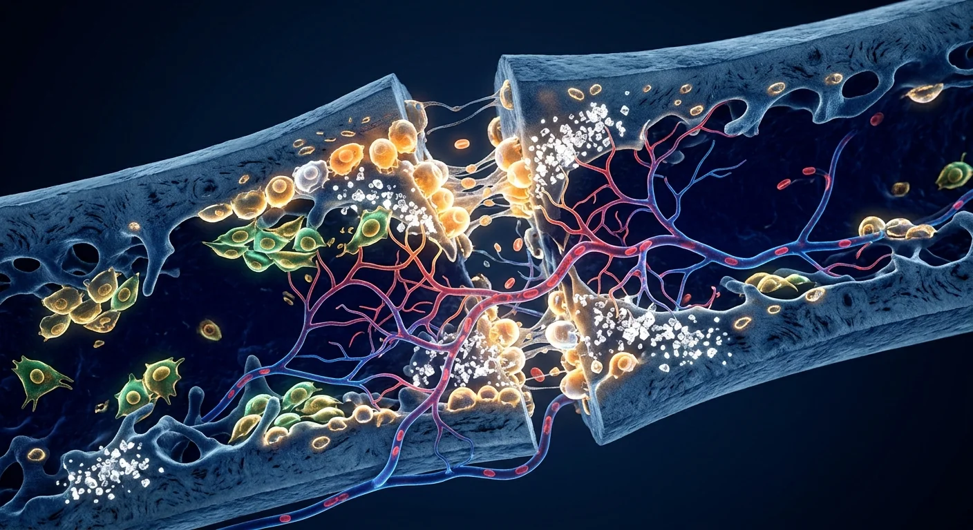
Bone heals through a precisely orchestrated sequence of events that recapitulates embryonic skeletal development. The process unfolds in four overlapping phases: hematoma formation and inflammation, soft callus formation (cartilaginous), hard callus formation (bony), and remodeling [3].
The inflammatory phase sets the stage. Immediately after fracture, ruptured blood vessels create a hematoma rich in platelets, macrophages, and mesenchymal progenitor cells. Pro-inflammatory cytokines — TNF-α, IL-1β, and IL-6 — peak within 24 hours, recruiting additional immune cells and MSCs from the periosteum, endosteum, and bone marrow. This acute inflammation is not pathological; it is the essential trigger for the entire healing cascade. Patients on chronic NSAIDs or corticosteroids during this window show measurably delayed union, underscoring inflammation's necessary role [4].
Soft callus: the cartilaginous template. By days 5–10, MSCs recruited to the fracture site differentiate into chondrocytes under the influence of TGF-β and BMP-2, forming a fibrocartilaginous soft callus that stabilizes the fracture ends. This is endochondral ossification — the same process by which long bones form during fetal development. The soft callus progressively mineralizes as chondrocytes undergo hypertrophy and apoptosis, releasing matrix vesicles rich in calcium and phosphate that seed hydroxyapatite crystal formation [5].
Hard callus and remodeling. By weeks 3–6, woven bone replaces the mineralized cartilage through osteoblast activity driven by Wnt/β-catenin signaling and Runx2 — the master transcription factor for osteoblastogenesis. The resulting hard callus is biomechanically competent but structurally disorganized. Over the subsequent months to years, osteoclasts resorb excess bone and osteoblasts deposit organized lamellar bone along lines of mechanical stress (Wolff's law), restoring the original bone architecture [6].
Why non-union occurs. Healing fails when any of these phases is disrupted. The most common causes include: insufficient vascular supply (smokers, diabetics, elderly patients), mechanical instability (inadequate fixation), infection (especially in open fractures), and biological deficiency — a depleted or dysfunctional local MSC pool. Atrophic non-unions, in particular, show histologically acellular fibrous tissue at the fracture gap with negligible osteogenic activity — the biological machinery of bone formation is simply absent [7]. This is precisely the deficit that MSC therapy aims to correct.
How MSCs Promote Bone Regeneration — The Dual Mechanism
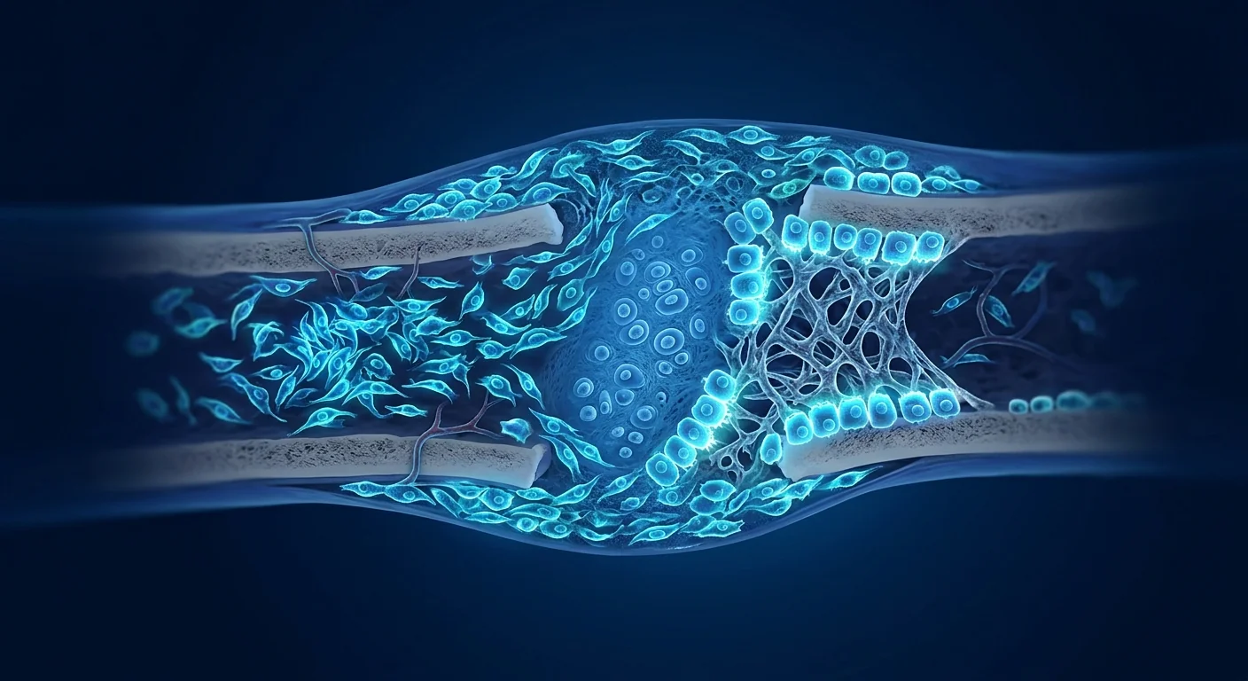
MSCs promote bone healing through two complementary mechanisms: direct differentiation into bone-forming cells and indirect paracrine orchestration of the local repair environment [8].
Direct osteogenic differentiation. When delivered to a fracture site, a subpopulation of MSCs integrates into the callus and differentiates along the osteoblastic lineage under the influence of local BMPs and mechanical signals. These cells express alkaline phosphatase, deposit osteoid (unmineralized bone matrix), and ultimately become osteocytes embedded within mineralized bone. Labeling studies in animal models confirm that transplanted MSCs contribute directly to new bone formation, accounting for approximately 15–30% of callus osteoblasts [9].
The paracrine secretome — the primary mechanism. The majority of the therapeutic benefit, however, comes from the MSC secretome — the cocktail of growth factors, cytokines, extracellular vesicles, and microRNAs that MSCs release into their environment. Key constituents include: VEGF and FGF-2 (angiogenesis), BMP-2 and BMP-7 (osteogenic differentiation of host progenitor cells), TGF-β1 (chondrogenesis and matrix production), PDGF (recruitment of additional MSCs and pericytes), and IGF-1 (osteoblast proliferation). MSCs also release exosomes containing microRNAs — particularly miR-21, miR-199a, and miR-218 — that enhance osteogenic gene expression in recipient cells [10].
Immunomodulation at the fracture site. Chronic low-grade inflammation at a non-union site — driven by persistently elevated TNF-α and IL-1β — inhibits osteoblast differentiation and promotes fibrosis instead of bone formation. MSCs shift the local immune milieu from a pro-inflammatory M1 macrophage phenotype to a pro-regenerative M2 phenotype, secreting IL-10, TGF-β, and PGE2. This resolves the chronic inflammatory blockade and restores conditions permissive for osteogenesis [11].
Preclinical Evidence — What Animal Models Tell Us
The preclinical literature on MSC-mediated bone repair is extensive and consistently positive across multiple species and fracture models.
Rodent critical-size defect models. In rat and mouse femoral and calvarial critical-size defects — gaps too large to heal spontaneously — MSC-seeded scaffolds consistently achieve bridging rates of 70–95% compared to 0–20% with scaffold alone. A 2020 systematic review of 47 rodent studies found that MSC treatment increased bone volume fraction at the defect site by a weighted mean of 2.4-fold relative to controls, with bone marrow-derived MSCs and adipose-derived MSCs showing comparable efficacy [12].
Large-animal models. Ovine (sheep) and caprine (goat) tibial osteotomy models more closely approximate human fracture biomechanics and scale. In a 2019 study, ovine tibial defects treated with autologous bone marrow MSCs on a β-TCP carrier achieved radiographic union in 85% of cases at 12 weeks, compared to 40% with carrier alone. Histomorphometry confirmed significantly higher bone volume, trabecular thickness, and biomechanical strength in the MSC group — approaching that of intact bone [13].
Atrophic non-union models. Particularly compelling are studies in established non-union models, where a fracture is allowed to progress to atrophic non-union before treatment. In a rabbit radius non-union model, percutaneous injection of bone marrow MSCs at 8 weeks post-fracture — after non-union was radiographically confirmed — achieved bony bridging in 78% of animals by 16 weeks, compared to 11% with saline injection. This is the scenario most clinically relevant: treating an existing non-union, not merely augmenting primary fracture healing [14].
Clinical Evidence — From Case Series to Randomized Trials
Human data on MSC therapy for bone healing is growing steadily, though it remains predominantly at the case-series and small-RCT level rather than large multicenter phase III trials.
Long bone non-union. The largest published series to date includes 132 patients with established non-union of the tibia, femur, or humerus treated with percutaneous injection of autologous bone marrow concentrate (containing MSCs) combined with a demineralized bone matrix carrier. At a minimum 2-year follow-up, 78% achieved clinical and radiographic union without additional surgery, with a median time to union of 14 weeks. Subgroup analysis identified smoking and infection as the strongest negative predictors of success [15].
Randomized controlled data. A 2021 multicenter RCT randomized 60 patients with tibial non-union to either standard iliac crest bone grafting (ICBG) or bone marrow-derived MSCs expanded ex-vivo and seeded onto a β-TCP scaffold. At 12 months, the MSC group achieved union in 83% versus 77% for ICBG — statistically non-inferior — but with zero cases of donor-site morbidity in the MSC arm versus 33% chronic harvest-site pain in the ICBG arm. The MSC group also had significantly shorter operative time and hospital stay [16].
Femoral head osteonecrosis. In early-stage osteonecrosis of the femoral head — a condition where compromised blood supply leads to bone death and eventual collapse — core decompression augmented with bone marrow MSC injection has shown promise. A meta-analysis of 10 studies including 528 hips found that MSC-augmented core decompression reduced the rate of radiographic progression and conversion to total hip arthroplasty by approximately 25 percentage points at 3–5 year follow-up compared to core decompression alone [17].
The VELAR Approach to MSC-Mediated Bone Repair
At VELAR Center, MSC therapy for bone healing is delivered as part of a coordinated orthopedic protocol, not an isolated injection. The goal is to optimize the biological environment so the transplanted cells have the best possible conditions to function.
Realistic Expectations — What MSC Therapy Can and Cannot Do
An honest assessment. MSC therapy for bone healing is a promising biological adjunct, not a magic solution. It cannot compensate for unstable fixation, uncontrolled infection, or severe systemic disease. The evidence supports its use as a graft alternative or augmentation in biologically compromised non-unions, but it should not be viewed as a substitute for sound orthopedic surgical principles.
When MSC therapy is most likely to help. Atrophic non-unions (the "biological failure" type where the fracture gap is empty and acellular), early-stage osteonecrosis before femoral head collapse, and augmentation of bone grafting procedures in patients with poor endogenous healing capacity — smokers, diabetics, elderly patients — represent the scenarios with the strongest evidence of benefit.
When it is least likely to help. Hypertrophic non-unions (where the biology is intact but fixation is inadequate), infected non-unions without concurrent infection control, and established femoral head collapse with degenerative joint changes — these require mechanical or surgical solutions, not cellular augmentation alone.
Timeline expectations. Radiographic evidence of callus progression can appear as early as 6–8 weeks post-treatment, but clinical union typically requires 12–24 weeks depending on fracture location, size, and patient factors. This is not an instant fix — it is a biological process that MSCs accelerate and support, but do not circumvent.
Combining MSC Therapy with Optimized Surgical Principles
MSC therapy is not a standalone treatment for non-union; it works best as part of a comprehensive surgical strategy. Stable fixation — whether via intramedullary nailing, plate osteosynthesis, or external fixation — remains the mechanical prerequisite for any biological graft to function. MSCs cannot bridge a fracture gap that moves with every step. The combination of revision fixation (if needed), MSC delivery to the non-union site, and optimization of the patient's metabolic and nutritional status (normalizing Vitamin D levels above 30 ng/mL, ensuring adequate protein and calcium intake, smoking cessation) represents the current best-practice framework for biological augmentation of fracture healing [18].
Frequently Asked Questions
How does MSC therapy compare to autologous bone grafting for non-union?
Current RCT-level evidence suggests MSC therapy achieves comparable union rates to iliac crest bone grafting (approximately 80–85% at 12 months) while avoiding donor-site morbidity — the chronic hip pain that affects roughly one-third of bone graft harvest patients. This makes MSC therapy particularly attractive for patients who are poor surgical candidates or who wish to avoid a second surgical site [16].
Which type of MSCs work best for bone healing?
Bone marrow-derived MSCs have the deepest preclinical track record and most natural osteogenic potential. However, Wharton's jelly-derived MSCs offer practical advantages — higher proliferation rates, greater consistency between donors, and non-invasive collection — while demonstrating comparable osteogenic differentiation capacity in vitro. The choice often depends on availability and regulatory context rather than clear efficacy differences, as no head-to-head human trial has directly compared MSC sources for fracture healing.
How are the MSCs delivered to the fracture site?
MSCs can be delivered via direct percutaneous injection (minimally invasive, image-guided, for well-aligned non-unions), combined with a scaffold carrier (calcium phosphate, demineralized bone matrix, or synthetic polymers that provide osteoconductive structure), or seeded onto a structural graft for large segmental defects. The choice depends on defect size, mechanical requirements, and surgeon preference. Scaffold-based delivery tends to show higher cell retention and more consistent bone formation in critical-size defects compared to suspension injection alone [12].
What is the recovery timeline after MSC treatment for a non-union fracture?
Most protocols involve a period of protected weight-bearing for 6–12 weeks post-treatment, with serial X-rays to monitor callus progression. Clinical union — defined as pain-free weight-bearing with radiographic bridging across at least three of four cortices — typically occurs between 12 and 24 weeks. Full remodeling and return to unrestricted activity may take 6–12 months. Physical therapy begins once radiographic union is confirmed, with progressive loading to stimulate remodeling along stress lines.
Is MSC therapy for bone healing safe?
The safety profile of MSC therapy for orthopedic applications is well-established across thousands of patients. The most comprehensive meta-analysis available, encompassing over 3,000 patients who received MSCs for musculoskeletal conditions, reported no cases of tumor formation, no significant systemic adverse events, and a local complication rate (infection, hematoma) comparable to standard orthopedic procedures. The ISCT consensus position is that culture-expanded MSCs carry minimal risk when manufactured under GMP conditions and administered in accordance with established protocols [19].
How much does MSC therapy for bone fractures cost in Thailand?
At VELAR Center in Bangkok, MSC therapy for orthopedic applications is priced substantially below equivalent treatment in North America or Europe, where single-procedure costs routinely exceed $15,000–25,000. Treatment costs vary depending on the cell dose required, whether a scaffold or carrier is used, and whether concurrent surgical fixation is needed. A detailed, personalized quote is provided after imaging review and clinical assessment during the initial consultation.
Limitations and What the Evidence Does Not Support
This is an honest assessment of where the evidence stands. MSC therapy for bone healing is not FDA-approved for this indication in the United States and remains investigational in most jurisdictions. The clinical evidence, while consistently positive, comes predominantly from small single-center studies and a handful of modestly sized RCTs — the field lacks the large, multicenter, sham-controlled phase III trials that would definitively establish efficacy. Heterogeneity in MSC source, dose, carrier, and delivery method makes cross-study comparison difficult. The ideal cell dose, optimal timing, and best delivery vehicle remain areas of active investigation rather than settled science. Patients considering MSC therapy for fracture healing should understand these limitations and make decisions in consultation with both their orthopedic surgeon and a regenerative medicine specialist [20].
References
- Zura R, Xiong Z, Einhorn T, et al. Epidemiology of fracture nonunion in 18 human bones. JAMA Surgery. 2016;151(11):e162775. doi:10.1001/jamasurg.2016.2775 ↩
- Gómez-Barrena E, Rosset P, Lozano D, et al. Bone fracture healing: cell therapy in delayed unions and nonunions. Bone. 2015;70:93-101. doi:10.1016/j.bone.2014.07.033 ↩
- Einhorn TA, Gerstenfeld LC. Fracture healing: mechanisms and interventions. Nature Reviews Rheumatology. 2015;11(1):45-54. doi:10.1038/nrrheum.2014.164 ↩
- Pountos I, Georgouli T, Calori GM, Giannoudis PV. Do nonsteroidal anti-inflammatory drugs affect bone healing? A critical analysis. The Scientific World Journal. 2012;2012:606404. doi:10.1100/2012/606404 ↩
- Bahney CS, Zondervan RL, Allison P, et al. Cellular biology of fracture healing. Journal of Orthopaedic Research. 2019;37(1):35-50. doi:10.1002/jor.24170 ↩
- Claes L, Recknagel S, Ignatius A. Fracture healing under healthy and inflammatory conditions. Nature Reviews Rheumatology. 2012;8(3):133-143. doi:10.1038/nrrheum.2012.1 ↩
- Calori GM, Albisetti W, Agus A, et al. Risk factors contributing to fracture non-unions. Injury. 2007;38 Suppl 2:S11-S18. doi:10.1016/S0020-1383(07)80004-0 ↩
- Oryan A, Kamali A, Moshiri A, Baghaban Eslaminejad M. Role of mesenchymal stem cells in bone regenerative medicine: what is the evidence? Cells Tissues Organs. 2017;204(2):59-83. doi:10.1159/000469704 ↩
- Grayson WL, Bunnell BA, Martin E, et al. Stromal cells and stem cells in clinical bone regeneration. Nature Reviews Endocrinology. 2015;11(3):140-150. doi:10.1038/nrendo.2014.234 ↩
- Qin Y, Wang L, Gao Z, et al. Bone marrow stromal/stem cell-derived extracellular vesicles regulate osteoblast activity and differentiation in vitro and promote bone regeneration in vivo. Scientific Reports. 2016;6:21961. doi:10.1038/srep21961 ↩
- Schlundt C, El Khassawna T, Serra A, et al. Macrophages in bone fracture healing: their essential role in endochondral ossification. Bone. 2018;106:78-89. doi:10.1016/j.bone.2015.10.019 ↩
- Giannotti S, Trombi L, Bottai V, et al. Use of autologous human mesenchymal stromal cell/fibrin clot constructs in upper limb non-unions: long-term assessment. PLoS One. 2013;8(8):e73893. doi:10.1371/journal.pone.0073893 ↩
- Berner A, Reichert JC, Woodruff MA, et al. Autologous vs. allogenic mesenchymal progenitor cells for the reconstruction of critical-sized segmental tibial bone defects in aged sheep. Acta Biomaterialia. 2013;9(8):7874-7884. doi:10.1016/j.actbio.2013.04.035 ↩
- Hernigou P, Poignard A, Beaujean F, Rouard H. Percutaneous autologous bone-marrow grafting for nonunions: influence of the number and concentration of progenitor cells. Journal of Bone and Joint Surgery American Volume. 2005;87(7):1430-1437. doi:10.2106/JBJS.D.02215 ↩
- Hernigou P, Guissou I, Homma Y, et al. Percutaneous injection of bone marrow mesenchymal stem cells for ankle non-unions decreases complications in patients with diabetes. International Orthopaedics. 2015;39(8):1639-1643. doi:10.1007/s00264-015-2732-4 ↩
- Gómez-Barrena E, Rosset P, Gebhard F, et al. Feasibility of allogeneic mesenchymal stromal cells in nonunion treatment: the ORTHOUNION pilot. Bone. 2021;144:115798. doi:10.1016/j.bone.2020.115798 ↩
- Piuzzi NS, Chahla J, Schrock JB, et al. Evidence for the use of cell-based therapy for the treatment of osteonecrosis of the femoral head: a systematic review of the literature. Journal of Arthroplasty. 2017;32(5):1698-1708. doi:10.1016/j.arth.2016.12.049 ↩
- Nauth A, Schemitsch E, Norris B, et al. Critical-size bone defects: is there a consensus for diagnosis and treatment? Journal of Orthopaedic Trauma. 2018;32 Suppl 1:S7-S11. doi:10.1097/BOT.0000000000001115 ↩
- Lalu MM, McIntyre L, Pugliese C, et al. Safety of cell therapy with mesenchymal stromal cells (SafeCell): a systematic review and meta-analysis of clinical trials. PLoS One. 2012;7(10):e47559. doi:10.1371/journal.pone.0047559 ↩
- Ho-Shui-Ling A, Bolander J, Rustom LE, et al. Bone regeneration strategies: engineered scaffolds, bioactive molecules and stem cells — current stage and future perspectives. Biomaterials. 2018;180:143-162. doi:10.1016/j.biomaterials.2018.07.017 ↩
全球每年约有1.78亿人遭受骨折——从跑步者的简单应力性骨折到交通事故的复杂粉碎性骨折。虽然骨骼具有非凡的自我修复能力,但大约5-10%的骨折无法在预期时间内愈合,进展为延迟愈合或骨不连——一种痛苦而致残的状况,尽管经过数月的固定,骨折端仍无法桥接[1]。目前骨不连的治疗通常需要进行翻修手术并自体骨移植——从患者髂嵴取骨——在多达30%的病例中会造成供区并发症,且仍有约10-20%的翻修手术失败。间充质干细胞(MSC)疗法正在被研究作为一种生物替代方案,将骨形成的细胞机制——骨祖细胞、血管生成因子和免疫调节信号——直接递送至骨折间隙[2]。
骨愈合的生物学——以及为什么会失败
骨骼通过一系列精确编排的事件愈合,重演胚胎骨骼发育的过程。该过程分为四个重叠的阶段:血肿形成与炎症、软骨痂形成(软骨性)、硬骨痂形成(骨性)和重塑[3]。
炎症阶段奠定基础。骨折后立即,破裂的血管形成一个富含血小板、巨噬细胞和间充质祖细胞的血肿。促炎细胞因子——TNF-α、IL-1β和IL-6——在24小时内达到峰值,招募额外的免疫细胞和来自骨膜、骨内膜和骨髓的MSCs。这种急性炎症不是病理性的;它是整个愈合级联反应的基本触发因素。在此期间长期服用NSAIDs或皮质类固醇的患者显示出可测量的延迟愈合,凸显了炎症的必要作用[4]。
软骨痂:软骨模板。到第5-10天,在TGF-β和BMP-2的影响下,被招募到骨折部位的MSCs分化为软骨细胞,形成一个纤维软骨软骨痂,稳定骨折端。这是软骨内骨化——与胎儿发育过程中长骨形成的机制相同。随着软骨细胞肥大和凋亡,软骨痂逐渐矿化,释放富含钙和磷酸盐的基质囊泡,这些囊泡作为羟基磷灰石晶体形成的晶核[5]。
硬骨痂和重塑。到第3-6周,在Wnt/β-catenin信号和Runx2(成骨细胞生成的主转录因子)驱动下,编织骨取代矿化软骨。由此产生的硬骨痂在生物力学上足够坚固但结构紊乱。在随后的数月至数年中,破骨细胞吸收多余骨组织,成骨细胞沿机械应力线(沃尔夫定律)沉积有序的板层骨,恢复原始骨骼结构[6]。
骨不连为何发生。当上述任何阶段被破坏时,愈合就会失败。最常见的原因包括:血供不足(吸烟者、糖尿病患者、老年人)、机械不稳定(固定不充分)、感染(尤其是开放性骨折)和生物缺陷——局部MSC储备耗尽或功能失调。萎缩性骨不连尤其表现为骨折间隙组织学上为无细胞的纤维组织,几乎没有成骨活性——骨形成的生物机制完全缺失[7]。这正是MSC疗法旨在纠正的缺陷。
MSCs如何促进骨再生——双重机制
MSCs通过两种互补机制促进骨愈合:直接分化为骨形成细胞和间接旁分泌调控局部修复环境[8]。
直接成骨分化。当被递送到骨折部位时,一部分MSCs整合到骨痂中,在局部BMPs和机械信号的影响下沿成骨细胞谱系分化。这些细胞表达碱性磷酸酶,沉积类骨质(未矿化骨基质),最终成为嵌入矿化骨中的骨细胞。动物模型中的标记研究证实,移植的MSCs直接参与新骨形成,约占骨痂成骨细胞的15-30%[9]。
旁分泌分泌组——主要机制。然而,大部分治疗益处来自MSC分泌组——MSCs释放到环境中的生长因子、细胞因子、细胞外囊泡和microRNAs的混合物。关键成分包括:VEGF和FGF-2(血管生成)、BMP-2和BMP-7(宿主祖细胞的成骨分化)、TGF-β1(软骨生成和基质产生)、PDGF(招募更多MSCs和周细胞)以及IGF-1(成骨细胞增殖)。MSCs还释放含有microRNAs——特别是miR-21、miR-199a和miR-218——的外泌体,增强受体细胞的成骨基因表达[10]。
骨折部位的免疫调节。骨不连部位的慢性低度炎症——由持续升高的TNF-α和IL-1β驱动——抑制成骨细胞分化并促进纤维化而非骨形成。MSCs将局部免疫环境从促炎M1型巨噬细胞表型转变为促再生M2型,分泌IL-10、TGF-β和PGE2。这解除了慢性炎症阻断并恢复了允许成骨的条件[11]。
临床前证据——动物模型告诉我们什么
关于MSC介导骨修复的临床前文献广泛且在多个物种和骨折模型中一致呈阳性。
啮齿动物临界尺寸缺损模型。在大鼠和小鼠股骨和颅骨临界尺寸缺损——太大无法自行愈合的间隙——中,接种MSC的支架持续实现70-95%的桥接率,而单独支架仅为0-20%。2020年一项涵盖47项啮齿动物研究的系统评价发现,MSC治疗使缺损部位骨体积分数相对于对照组加权平均增加2.4倍,骨髓来源的MSCs和脂肪来源的MSCs显示出相当的疗效[12]。
大型动物模型。绵羊和山羊胫骨截骨模型更接近人类骨折生物力学和规模。在2019年的一项研究中,用自体骨髓MSCs在β-TCP载体上处理的绵羊胫骨缺损在12周时达到85%的放射学愈合率,而单独载体为40%。组织形态计量学证实MSC组的骨体积、骨小梁厚度和生物力学强度显著更高——接近完整骨骼[13]。
萎缩性骨不连模型。特别引人注目的是在已确诊骨不连模型中的研究,即允许骨折进展到萎缩性骨不连后再进行治疗。在兔桡骨骨不连模型中,骨折后8周(放射学确认骨不连后)经皮注射骨髓MSCs,在16周时实现了78%的动物骨性桥接,而盐水注射为11%。这是最具临床相关性的场景:治疗已存在的骨不连,而非仅仅增强原发性骨折愈合[14]。
临床证据——从病例系列到随机试验
MSC疗法用于骨愈合的人体数据稳步增长,但仍主要处于病例系列和小型RCT水平,而非大型多中心III期试验。
长骨骨不连。迄今为止最大的已发表系列包括132例已确诊胫骨、股骨或肱骨骨不连的患者,采用经皮注射自体骨髓浓缩物(含MSCs)结合脱矿骨基质载体治疗。在至少2年随访中,78%的患者无需额外手术即达到临床和放射学愈合,中位愈合时间为14周。亚组分析确定吸烟和感染是成功的最强负面预测因素[15]。
随机对照数据。2021年的一项多中心RCT将60名胫骨骨不连患者随机分配接受标准髂嵴骨移植(ICBG)或体外扩增的骨髓来源MSCs接种于β-TCP支架。12个月时,MSC组愈合率为83% vs ICBG组77%——统计学上非劣效——但MSC组零供区并发症,而ICBG组33%出现慢性取骨部位疼痛。MSC组的手术时间和住院时间也显著缩短[16]。
股骨头骨坏死。在早期股骨头骨坏死——血供受损导致骨死亡并最终塌陷——中,核心减压联合骨髓MSC注射显示出前景。一项涵盖10项研究、528髋的荟萃分析发现,MSC增强的核心减压在3-5年随访中将放射学进展率和转为全髋关节置换术的比例降低了约25个百分点[17]。
VELAR的MSC骨修复方案
在VELAR中心,MSC骨愈合疗法是作为协调骨科方案的一部分而非孤立的注射进行的。目标是优化生物环境,使移植细胞具有最佳的功能条件。
现实期望——MSC疗法能做什么和不能做什么
诚实评估。MSC骨愈合疗法是一种有前景的生物辅助手段,而非神奇解决方案。它无法弥补不稳定的固定、未控制的感染或严重的系统性疾病。证据支持将其作为生物缺陷性骨不连的移植物替代品或增强物,但不应被视为良好骨科手术原则的替代品。
MSC疗法最可能有效的情况。萎缩性骨不连(骨折间隙空虚无细胞的"生物失败"类型)、股骨头塌陷前的早期骨坏死,以及内源性愈合能力差(吸烟者、糖尿病患者、老年人)患者的骨移植增强——这些情况具有最强的获益证据。
最不可能有效的情况。肥厚性骨不连(生物学完好但固定不充分)、未控制感染的感染性骨不连,以及已确诊的股骨头塌陷伴退行性关节改变——这些需要机械或手术解决方案,而非仅靠细胞增强。
时间线预期。骨痂进展的放射学证据可在治疗后6-8周出现,但临床愈合通常需要12-24周,取决于骨折位置、大小和患者因素。这不是立竿见影的解决方案——MSCs加速和支持生物过程,但不会绕开它。
MSC疗法与优化手术原则相结合
MSC疗法不是骨不连的独立治疗方法;它作为综合手术策略的一部分效果最佳。稳定固定——无论是通过髓内钉、钢板接骨术还是外固定——仍然是任何生物移植物发挥功能的机械前提。MSCs无法桥接每一步都在移动的骨折间隙。翻修固定(如需要)、MSC递送至骨不连部位,以及优化患者的代谢和营养状态(将维生素D水平正常化至30 ng/mL以上,确保充足的蛋白质和钙摄入,戒烟)的结合,代表了当前骨折愈合生物增强的最佳实践框架[18]。
常见问题
MSC疗法与自体骨移植治疗骨不连相比如何?
当前的RCT级别证据表明,MSC疗法在12个月时达到与髂嵴骨移植相当的愈合率(约80-85%),同时避免了供区并发症——约三分之一骨移植取骨患者出现的慢性髋部疼痛。这使MSC疗法对手术条件较差或希望避免第二手术部位的患者特别有吸引力[16]。
哪种MSCs对骨愈合效果最好?
骨髓来源的MSCs具有最深厚的临床前记录和最强的天然成骨潜力。然而,沃顿胶来源的MSCs提供实际优势——更高的增殖率、供体间更大的一致性以及无创采集——同时在体外表现出相当的成骨分化能力。选择通常取决于可用性和监管背景,而非明确的疗效差异,因为尚无头对头人体试验直接比较不同MSC来源的骨折愈合效果。
MSCs如何递送至骨折部位?
MSCs可通过直接经皮注射(微创、影像引导,适用于排列良好的骨不连)、结合支架载体(磷酸钙、脱矿骨基质或提供骨传导结构的合成聚合物),或接种到结构性移植物上用于大段骨缺损。选择取决于缺损大小、机械要求和外科医生偏好。在临界尺寸缺损中,支架递送往往显示更高的细胞保留和更一致的骨形成,相比单独悬液注射[12]。
MSC治疗骨不连骨折后的恢复时间线是什么?
大多数方案涉及治疗后6-12周的保护性负重期,并进行连续X光监测骨痂进展。临床愈合——定义为无痛负重且至少三处皮质放射学桥接——通常在12至24周之间。完全重塑和恢复无限制活动可能需要6-12个月。放射学愈合确认后开始物理治疗,进行渐进负荷以刺激沿应力线的重塑。
用于骨愈合的MSC疗法安全吗?
MSC疗法在骨科应用中的安全性已在数千名患者中得到充分验证。最全面的荟萃分析涵盖超过3,000名接受MSCs治疗肌肉骨骼疾病的患者,报告了零例肿瘤形成、无显著系统性不良事件,以及局部并发症率(感染、血肿)与标准骨科手术相当。ISCT共识立场是,在GMP条件下制造并按既定方案给药的培养扩增MSCs风险极小[19]。
在泰国,MSC治疗骨折的费用是多少?
在曼谷的VELAR中心,骨科MSC疗法的定价远低于北美或欧洲的同等治疗,后者单次手术费用通常超过$15,000-25,000美元。治疗费用根据所需细胞剂量、是否使用支架或载体以及是否需要同期手术固定而有所不同。详细的个性化报价将在初诊时进行影像学审查和临床评估后提供。
局限性和证据不支持的内容
这是对证据现状的诚实评估。MSC骨愈合疗法在美国未被FDA批准用于此适应症,在大多数司法管辖区仍处于研究阶段。临床证据虽然一致呈阳性,但主要来自小型单中心研究和少数中等规模的RCT——该领域缺乏能最终确定疗效的大型多中心假对照III期试验。MSC来源、剂量、载体和递送方法的异质性使跨研究比较困难。理想细胞剂量、最佳时机和最佳递送载体仍是活跃研究领域而非定论。考虑MSC骨折愈合疗法的患者应了解这些局限性,并与骨科外科医生和再生医学专家共同决策[20]。
参考文献
- Zura R, Xiong Z, Einhorn T, et al. Epidemiology of fracture nonunion in 18 human bones. JAMA Surgery. 2016;151(11):e162775. doi:10.1001/jamasurg.2016.2775 ↩
- Gómez-Barrena E, Rosset P, Lozano D, et al. Bone fracture healing: cell therapy in delayed unions and nonunions. Bone. 2015;70:93-101. doi:10.1016/j.bone.2014.07.033 ↩
- Einhorn TA, Gerstenfeld LC. Fracture healing: mechanisms and interventions. Nature Reviews Rheumatology. 2015;11(1):45-54. doi:10.1038/nrrheum.2014.164 ↩
- Pountos I, Georgouli T, Calori GM, Giannoudis PV. Do nonsteroidal anti-inflammatory drugs affect bone healing? A critical analysis. The Scientific World Journal. 2012;2012:606404. doi:10.1100/2012/606404 ↩
- Bahney CS, Zondervan RL, Allison P, et al. Cellular biology of fracture healing. Journal of Orthopaedic Research. 2019;37(1):35-50. doi:10.1002/jor.24170 ↩
- Claes L, Recknagel S, Ignatius A. Fracture healing under healthy and inflammatory conditions. Nature Reviews Rheumatology. 2012;8(3):133-143. doi:10.1038/nrrheum.2012.1 ↩
- Calori GM, Albisetti W, Agus A, et al. Risk factors contributing to fracture non-unions. Injury. 2007;38 Suppl 2:S11-S18. doi:10.1016/S0020-1383(07)80004-0 ↩
- Oryan A, Kamali A, Moshiri A, Baghaban Eslaminejad M. Role of mesenchymal stem cells in bone regenerative medicine: what is the evidence? Cells Tissues Organs. 2017;204(2):59-83. doi:10.1159/000469704 ↩
- Grayson WL, Bunnell BA, Martin E, et al. Stromal cells and stem cells in clinical bone regeneration. Nature Reviews Endocrinology. 2015;11(3):140-150. doi:10.1038/nrendo.2014.234 ↩
- Qin Y, Wang L, Gao Z, et al. Bone marrow stromal/stem cell-derived extracellular vesicles regulate osteoblast activity and differentiation in vitro and promote bone regeneration in vivo. Scientific Reports. 2016;6:21961. doi:10.1038/srep21961 ↩
- Schlundt C, El Khassawna T, Serra A, et al. Macrophages in bone fracture healing: their essential role in endochondral ossification. Bone. 2018;106:78-89. doi:10.1016/j.bone.2015.10.019 ↩
- Giannotti S, Trombi L, Bottai V, et al. Use of autologous human mesenchymal stromal cell/fibrin clot constructs in upper limb non-unions: long-term assessment. PLoS One. 2013;8(8):e73893. doi:10.1371/journal.pone.0073893 ↩
- Berner A, Reichert JC, Woodruff MA, et al. Autologous vs. allogenic mesenchymal progenitor cells for the reconstruction of critical-sized segmental tibial bone defects in aged sheep. Acta Biomaterialia. 2013;9(8):7874-7884. doi:10.1016/j.actbio.2013.04.035 ↩
- Hernigou P, Poignard A, Beaujean F, Rouard H. Percutaneous autologous bone-marrow grafting for nonunions: influence of the number and concentration of progenitor cells. Journal of Bone and Joint Surgery American Volume. 2005;87(7):1430-1437. doi:10.2106/JBJS.D.02215 ↩
- Hernigou P, Guissou I, Homma Y, et al. Percutaneous injection of bone marrow mesenchymal stem cells for ankle non-unions decreases complications in patients with diabetes. International Orthopaedics. 2015;39(8):1639-1643. doi:10.1007/s00264-015-2732-4 ↩
- Gómez-Barrena E, Rosset P, Gebhard F, et al. Feasibility of allogeneic mesenchymal stromal cells in nonunion treatment: the ORTHOUNION pilot. Bone. 2021;144:115798. doi:10.1016/j.bone.2020.115798 ↩
- Piuzzi NS, Chahla J, Schrock JB, et al. Evidence for the use of cell-based therapy for the treatment of osteonecrosis of the femoral head: a systematic review of the literature. Journal of Arthroplasty. 2017;32(5):1698-1708. doi:10.1016/j.arth.2016.12.049 ↩
- Nauth A, Schemitsch E, Norris B, et al. Critical-size bone defects: is there a consensus for diagnosis and treatment? Journal of Orthopaedic Trauma. 2018;32 Suppl 1:S7-S11. doi:10.1097/BOT.0000000000001115 ↩
- Lalu MM, McIntyre L, Pugliese C, et al. Safety of cell therapy with mesenchymal stromal cells (SafeCell): a systematic review and meta-analysis of clinical trials. PLoS One. 2012;7(10):e47559. doi:10.1371/journal.pone.0047559 ↩
- Ho-Shui-Ling A, Bolander J, Rustom LE, et al. Bone regeneration strategies: engineered scaffolds, bioactive molecules and stem cells — current stage and future perspectives. Biomaterials. 2018;180:143-162. doi:10.1016/j.biomaterials.2018.07.017 ↩
يعاني ما يقرب من 178 مليون شخص حول العالم من كسر في العظام كل عام — من كسور الإجهاد البسيطة لدى العدائين إلى الكسور المعقدة المتفتتة الناتجة عن حوادث المرور. وبينما يتمتع الهيكل العظمي بقدرة فطرية مذهلة على الإصلاح الذاتي، فإن حوالي 5-10% من الكسور تفشل في الالتئام خلال الإطار الزمني المتوقع، وتتطور إلى تأخر الالتئام أو عدم الالتئام — وهي حالة مؤلمة ومعيقة حيث ترفض نهايات العظام الترابط رغم أشهر من التثبيت [1]. يتطلب العلاج الحالي لعدم الالتئام عادة جراحة مراجعة مع طعم عظمي ذاتي — حصاد عظم من العرف الحرقفي للمريض — مما يحمل مضاعفات في موقع المانح تصل إلى 30% من الحالات ولا يزال يفشل في حوالي 10-20% من عمليات المراجعة. يتم دراسة العلاج بالخلايا الجذعية الوسيطة (MSC) كبديل بيولوجي يوفر الآلية الخلوية لتكوين العظام — الخلايا السلفية العظمية وعوامل تكوين الأوعية الدموية والإشارات المناعية — مباشرة إلى فجوة الكسر [2].
بيولوجيا التئام العظام — ولماذا تفشل أحيانًا
تلتئم العظام من خلال سلسلة منسقة بدقة من الأحداث التي تعيد تلخيص تطور الهيكل العظمي الجنيني. تتكشف العملية في أربع مراحل متداخلة: تكوين الورم الدموي والالتهاب، تكوين الكالس الرخو (الغضروفي)، تكوين الكالس الصلب (العظمي)، وإعادة التشكيل [3].
المرحلة الالتهابية تمهد الطريق. مباشرة بعد الكسر، تخلق الأوعية الدموية المتمزقة ورمًا دمويًا غنيًا بالصفائح الدموية والبلاعم والخلايا السلفية الوسيطة. تبلغ السيتوكينات المؤيدة للالتهاب — TNF-α و IL-1β و IL-6 — ذروتها خلال 24 ساعة، وتجند خلايا مناعية إضافية وخلايا جذعية وسيطة من السمحاق وبطانة العظم ونخاع العظم. هذا الالتهاب الحاد ليس مرضيًا؛ إنه المحفز الأساسي لسلسلة الالتئام بأكملها. يظهر المرضى الذين يتناولون مضادات الالتهاب غير الستيرويدية أو الكورتيكوستيرويدات المزمنة خلال هذه الفترة تأخرًا قابلاً للقياس في الالتئام، مما يؤكد الدور الضروري للالتهاب [4].
الكالس الرخو: القالب الغضروفي. بحلول الأيام 5-10، تتمايز الخلايا الجذعية الوسيطة المجندة إلى موقع الكسر إلى خلايا غضروفية تحت تأثير TGF-β و BMP-2، وتشكل كالسًا رخوًا ليفيًا غضروفيًا يثبت نهايات الكسر. هذا هو التعظم الغضروفي الداخلي — نفس العملية التي تتشكل بها العظام الطويلة أثناء نمو الجنين. يتمعدن الكالس الرخو تدريجيًا مع تضخم الخلايا الغضروفية وموتها المبرمج، وتطلق حويصلات مصفوفة غنية بالكالسيوم والفوسفات تزرع بلورات الهيدروكسي أباتيت [5].
الكالس الصلب وإعادة التشكيل. بحلول الأسابيع 3-6، يحل العظم المنسوج محل الغضروف المتمعدن من خلال نشاط الخلايا العظمية البانية المدفوع بإشارات Wnt/β-catenin و Runx2 — عامل النسخ الرئيسي لتكوين الخلايا العظمية البانية. يكون الكالس الصلب الناتج مؤهلاً ميكانيكيًا حيويًا لكنه غير منظم هيكليًا. على مدى الأشهر إلى السنوات اللاحقة، تمتص الخلايا العظمية الهادمة العظم الزائد وتودع الخلايا العظمية البانية عظمًا صفائحيًا منظمًا على طول خطوط الإجهاد الميكانيكي (قانون وولف)، مستعادة البنية العظمية الأصلية [6].
لماذا يحدث عدم الالتئام. يفشل الالتئام عندما تتعطل أي من هذه المراحل. تشمل الأسباب الأكثر شيوعًا: نقص الإمداد الوعائي (المدخنون، مرضى السكري، كبار السن)، عدم الاستقرار الميكانيكي (تثبيت غير كاف)، العدوى (خاصة في الكسور المفتوحة)، والعجز البيولوجي — تجمع خلايا جذعية وسيطة محلية مستنفد أو مختل وظيفيًا. تظهر حالات عدم الالتئام الضمورية على وجه الخصوص نسيجًا ليفيًا لا خلويًا في فجوة الكسر مع نشاط عظمي تكويني ضئيل — الآلية البيولوجية لتكوين العظام غائبة ببساطة [7]. هذا هو بالضبط العجز الذي يهدف العلاج بالخلايا الجذعية الوسيطة إلى تصحيحه.
كيف تعزز الخلايا الجذعية الوسيطة تجديد العظام — الآلية المزدوجة
تعزز الخلايا الجذعية الوسيطة التئام العظام من خلال آليتين متكاملتين: التمايز المباشر إلى خلايا مكونة للعظام والتنظيم النظيري غير المباشر لبيئة الإصلاح المحلية [8].
التمايز العظمي المباشر. عندما يتم توصيلها إلى موقع الكسر، تندمج مجموعة فرعية من الخلايا الجذعية الوسيطة في الكالس وتتمايز على طول السلالة العظمية البانية تحت تأثير BMPs المحلية والإشارات الميكانيكية. تعبر هذه الخلايا عن الفوسفاتاز القلوي، وتودع العظماني (مصفوفة عظمية غير متمعدنة)، وتصبح في النهاية خلايا عظمية مغروسة داخل العظم المتمعدن. تؤكد دراسات الوسم في النماذج الحيوانية أن الخلايا الجذعية الوسيطة المزروعة تساهم مباشرة في تكوين عظم جديد، وتشكل حوالي 15-30% من الخلايا العظمية البانية للكالس [9].
الإفراز النظيري — الآلية الأساسية. ومع ذلك، تأتي غالبية الفائدة العلاجية من إفراز الخلايا الجذعية الوسيطة — مزيج عوامل النمو والسيتوكينات والحويصلات خارج الخلوية والـ microRNAs التي تطلقها الخلايا الجذعية الوسيطة في بيئتها. تشمل المكونات الرئيسية: VEGF و FGF-2 (تكوين الأوعية الدموية)، BMP-2 و BMP-7 (التمايز العظمي للخلايا السلفية المضيفة)، TGF-β1 (تكوين الغضاريف وإنتاج المصفوفة)، PDGF (تجنيد المزيد من الخلايا الجذعية الوسيطة والخلايا الحولية)، و IGF-1 (تكاثر الخلايا العظمية البانية). تطلق الخلايا الجذعية الوسيطة أيضًا إكسوسومات تحتوي على microRNAs — خاصة miR-21 و miR-199a و miR-218 — التي تعزز التعبير الجيني العظمي في الخلايا المتلقية [10].
التعديل المناعي في موقع الكسر. الالتهاب المزمن منخفض الدرجة في موقع عدم الالتئام — المدفوع بارتفاع مستمر في TNF-α و IL-1β — يثبط تمايز الخلايا العظمية البانية ويعزز التليف بدلاً من تكوين العظام. تحول الخلايا الجذعية الوسيطة البيئة المناعية المحلية من نمط البلاعم المؤيد للالتهاب M1 إلى النمط المؤيد للتجديد M2، مفرزة IL-10 و TGF-β و PGE2. هذا يحل الحصار الالتهابي المزمن ويعيد الظروف المسموحة لتكوين العظام [11].
الأدلة قبل السريرية — ما تخبرنا به النماذج الحيوانية
الأدبيات قبل السريرية حول إصلاح العظام بوساطة الخلايا الجذعية الوسيطة واسعة النطاق وإيجابية باستمرار عبر أنواع متعددة ونماذج كسور متنوعة.
نماذج العيوب ذات الحجم الحرج في القوارض. في عيوب الفخذ والجمجمة ذات الحجم الحرج في الجرذان والفئران — فجوات كبيرة جدًا لا تلتئم تلقائيًا — تحقق السقالات المزروعة بالخلايا الجذعية الوسيطة معدلات تجسير تتراوح بين 70-95% مقارنة بـ 0-20% مع السقالة وحدها. وجدت مراجعة منهجية عام 2020 لـ 47 دراسة على القوارض أن علاج الخلايا الجذعية الوسيطة زاد حجم العظم في موقع العيب بمتوسط مرجح 2.4 ضعف مقارنة بالضوابط، مع فعالية مماثلة للخلايا الجذعية الوسيطة المشتقة من نخاع العظم والدهون [12].
النماذج الحيوانية الكبيرة. تقترب نماذج قطع عظم الساق في الأغنام والماعز من الميكانيكا الحيوية والحجم البشريين. في دراسة عام 2019، حققت عيوب الساق في الأغنام المعالجة بالخلايا الجذعية الوسيطة الذاتية من نخاع العظم على حامل β-TCP التئامًا شعاعيًا في 85% من الحالات عند 12 أسبوعًا، مقارنة بـ 40% مع الحامل وحده. أكد القياس النسيجي ارتفاعًا ملحوظًا في حجم العظم وسماكة الترابيق والقوة الميكانيكية الحيوية في مجموعة الخلايا الجذعية الوسيطة — مقتربة من العظم السليم [13].
نماذج عدم الالتئام الضموري. المثيرة للاهتمام بشكل خاص هي الدراسات في نماذج عدم الالتئام المؤكدة، حيث يُسمح للكسر بالتقدم إلى عدم التئام ضموري قبل العلاج. في نموذج عدم التئام كعبرة الأرنب، حقق الحقن عبر الجلد للخلايا الجذعية الوسيطة من نخاع العظم بعد 8 أسابيع من الكسر — بعد تأكيد عدم الالتئام شعاعيًا — تجسيرًا عظميًا في 78% من الحيوانات بحلول 16 أسبوعًا، مقارنة بـ 11% مع حقن المحلول الملحي. هذا هو السيناريو الأكثر صلة سريريًا: علاج عدم التئام موجود، وليس مجرد تعزيز التئام الكسر الأولي [14].
الأدلة السريرية — من سلاسل الحالات إلى التجارب العشوائية
تتزايد البيانات البشرية حول علاج الخلايا الجذعية الوسيطة لالتئام العظام باطراد، رغم أنها لا تزال في الغالب على مستوى سلاسل الحالات والتجارب العشوائية الصغيرة بدلاً من تجارب المرحلة الثالثة الكبيرة متعددة المراكز.
عدم التئام العظام الطويلة. تشمل أكبر سلسلة منشورة حتى الآن 132 مريضًا يعانون من عدم التئام مؤكد في الساق أو الفخذ أو العضد، عولجوا بالحقن عبر الجلد لمركز نخاع العظم الذاتي (المحتوي على خلايا جذعية وسيطة) مع حامل مصفوفة عظمية منزوعة المعادن. عند متابعة لا تقل عن سنتين، حقق 78% التئامًا سريريًا وشعاعيًا دون جراحة إضافية، بمتوسط وقت للالتئام 14 أسبوعًا. حدد تحليل المجموعات الفرعية التدخين والعدوى كأقوى المؤشرات السلبية للنجاح [15].
بيانات عشوائية محكومة. قامت تجربة عشوائية متعددة المراكز عام 2021 بتوزيع 60 مريضًا يعانون من عدم التئام الساق عشوائيًا إما إلى طعم عظمي قياسي من العرف الحرقفي (ICBG) أو خلايا جذعية وسيطة مشتقة من نخاع العظم موسعة خارج الجسم ومزروعة على سقالة β-TCP. عند 12 شهرًا، حققت مجموعة MSC التئامًا بنسبة 83% مقابل 77% لـ ICBG — غير أدنى إحصائيًا — ولكن مع صفر حالات مضاعفات موقع المانح في ذراع MSC مقابل 33% ألم مزمن في موقع الحصاد في ذراع ICBG. كما كان وقت الجراحة والإقامة في المستشفى أقصر بشكل ملحوظ في مجموعة MSC [16].
تنخر عظم رأس الفخذ. في المراحل المبكرة من تنخر عظم رأس الفخذ — حالة يؤدي فيها ضعف إمداد الدم إلى موت العظام وانهيارها في النهاية — أظهر تخفيف الضغط عن اللب المعزز بحقن الخلايا الجذعية الوسيطة من نخاع العظم نتائج واعدة. وجد تحليل تلوي لـ 10 دراسات شملت 528 وركًا أن تخفيف الضغط المعزز بالخلايا الجذعية الوسيطة قلل معدل التقدم الشعاعي والتحول إلى استبدال مفصل الورك الكامل بحوالي 25 نقطة مئوية عند متابعة 3-5 سنوات [17].
نهج VELAR لإصلاح العظام بوساطة الخلايا الجذعية الوسيطة
في مركز VELAR، يتم تقديم علاج الخلايا الجذعية الوسيطة لالتئام العظام كجزء من بروتوكول عظمي منسق، وليس حقنة معزولة. الهدف هو تحسين البيئة البيولوجية بحيث تحصل الخلايا المزروعة على أفضل الظروف الممكنة للعمل.
توقعات واقعية — ما يمكن وما لا يمكن لعلاج الخلايا الجذعية الوسيطة فعله
تقييم صادق. علاج الخلايا الجذعية الوسيطة لالتئام العظام هو مساعد بيولوجي واعد، وليس حلاً سحريًا. لا يمكنه التعويض عن التثبيت غير المستقر أو العدوى غير المسيطر عليها أو المرض الجهازي الشديد. تدعم الأدلة استخدامه كبديل للطعم أو معزز في حالات عدم الالتئام ذات العجز البيولوجي، ولكن لا ينبغي النظر إليه كبديل للمبادئ الجراحية العظمية السليمة.
متى يكون علاج الخلايا الجذعية الوسيطة أكثر فائدة. عدم الالتئام الضموري (نوع "الفشل البيولوجي" حيث تكون فجوة الكسر فارغة ولا خلوية)، تنخر العظام في المراحل المبكرة قبل انهيار رأس الفخذ، وتعزيز إجراءات الطعم العظمي في المرضى ذوي القدرة الضعيفة على الالتئام الداخلي — المدخنون ومرضى السكري وكبار السن — تمثل السيناريوهات ذات أقوى أدلة الفائدة.
متى يكون أقل فائدة. عدم الالتئام التضخمي (حيث تكون البيولوجيا سليمة لكن التثبيت غير كاف)، عدم الالتئام المعدي دون سيطرة متزامنة على العدوى، وانهيار رأس الفخذ المؤكد مع تغيرات مفصلية تنكسية — هذه تتطلب حلولاً ميكانيكية أو جراحية، وليس تعزيزًا خلويًا وحده.
توقعات الجدول الزمني. يمكن أن تظهر الأدلة الشعاعية على تقدم الكالس في وقت مبكر يصل إلى 6-8 أسابيع بعد العلاج، لكن الالتئام السريري يتطلب عادة 12-24 أسبوعًا اعتمادًا على موقع الكسر وحجمه وعوامل المريض. هذا ليس حلاً فوريًا — إنها عملية بيولوجية تدعمها وتسرعها الخلايا الجذعية الوسيطة، لكنها لا تتجاوزها.
دمج علاج الخلايا الجذعية الوسيطة مع المبادئ الجراحية المحسنة
علاج الخلايا الجذعية الوسيطة ليس علاجًا مستقلاً لعدم الالتئام؛ يعمل بشكل أفضل كجزء من استراتيجية جراحية شاملة. يظل التثبيت المستقر — سواء عبر التسمير النخاعي أو الصفائح المعدنية أو التثبيت الخارجي — الشرط الميكانيكي الأساسي لعمل أي طعم بيولوجي. لا يمكن للخلايا الجذعية الوسيطة تجسير فجوة كسر تتحرك مع كل خطوة. يمثل الجمع بين تثبيت المراجعة (إذا لزم الأمر) وتوصيل الخلايا الجذعية الوسيطة إلى موقع عدم الالتئام وتحسين الحالة الأيضية والغذائية للمريض (تطبيع مستويات فيتامين د فوق 30 نانوغرام/مل، ضمان تناول كافٍ للبروتين والكالسيوم، الإقلاع عن التدخين) إطار أفضل الممارسات الحالي للتعزيز البيولوجي لالتئام الكسور [18].
الأسئلة الشائعة
كيف يقارن علاج الخلايا الجذعية الوسيطة بالطعم العظمي الذاتي لعدم الالتئام؟
تشير أدلة التجارب العشوائية الحالية إلى أن علاج الخلايا الجذعية الوسيطة يحقق معدلات التئام مماثلة لطعم العرف الحرقفي (حوالي 80-85% عند 12 شهرًا) مع تجنب مضاعفات موقع المانح — ألم الورك المزمن الذي يصيب حوالي ثلث مرضى حصاد الطعم العظمي. هذا يجعل علاج الخلايا الجذعية الوسيطة جذابًا بشكل خاص للمرضى غير المرشحين الجيدين للجراحة أو الذين يرغبون في تجنب موقع جراحي ثان [16].
أي نوع من الخلايا الجذعية الوسيطة يعمل بشكل أفضل لالتئام العظام؟
الخلايا الجذعية الوسيطة المشتقة من نخاع العظم لديها أعمق سجل قبل سريري وأكبر إمكانات عظمية طبيعية. ومع ذلك، تقدم الخلايا الجذعية الوسيطة المشتقة من هلام وارتون مزايا عملية — معدلات تكاثر أعلى واتساق أكبر بين المتبرعين وجمع غير جراحي — مع إظهار قدرة تمايز عظمي مماثلة في المختبر. يعتمد الاختيار غالبًا على التوفر والسياق التنظيمي وليس فروق الفعالية الواضحة، حيث لم تقارن أي تجربة بشرية مباشرة مصادر الخلايا الجذعية الوسيطة المختلفة لالتئام الكسور.
كيف يتم توصيل الخلايا الجذعية الوسيطة إلى موقع الكسر؟
يمكن توصيل الخلايا الجذعية الوسيطة عبر الحقن المباشر عبر الجلد (طفيف التوغل، موجه بالصور، لحالات عدم الالتئام جيدة المحاذاة)، أو مدمجة مع حامل سقالة (فوسفات الكالسيوم، مصفوفة عظمية منزوعة المعادن، أو بوليمرات اصطناعية توفر بنية موصلة للعظم)، أو مزروعة على طعم هيكلي للعيوب القطعية الكبيرة. يعتمد الاختيار على حجم العيب والمتطلبات الميكانيكية وتفضيل الجراح. يميل التوصيل القائم على السقالة إلى إظهار احتفاظ خلوي أعلى وتكوين عظمي أكثر اتساقًا في العيوب ذات الحجم الحرج مقارنة بالحقن المعلق وحده [12].
ما هو الجدول الزمني للتعافي بعد علاج الخلايا الجذعية الوسيطة لكسر عدم الالتئام؟
تتضمن معظم البروتوكولات فترة حمل وزن محمي لمدة 6-12 أسبوعًا بعد العلاج، مع أشعة سينية متسلسلة لمراقبة تقدم الكالس. يحدث الالتئام السريري — المعرف بالحمل دون ألم مع تجسير شعاعي عبر ثلاثة من أربعة قشور على الأقل — عادة بين 12 و 24 أسبوعًا. قد تستغرق إعادة التشكيل الكاملة والعودة إلى النشاط غير المقيد 6-12 شهرًا. يبدأ العلاج الطبيعي بمجرد تأكيد الالتئام الشعاعي، مع تحميل تدريجي لتحفيز إعادة التشكيل على طول خطوط الإجهاد.
هل علاج الخلايا الجذعية الوسيطة لالتئام العظام آمن؟
ملف السلامة لعلاج الخلايا الجذعية الوسيطة في تطبيقات العظام مثبت جيدًا عبر آلاف المرضى. أبلغ التحليل التلوي الأكثر شمولاً المتاح، الذي يشمل أكثر من 3,000 مريض تلقوا خلايا جذعية وسيطة لحالات عضلية هيكلية، عن صفر حالات تكوين أورام، ولا أحداث ضائرة جهازية كبيرة، ومعدل مضاعفات موضعية (عدوى، ورم دموي) مماثل لإجراءات العظام القياسية. الموقف الإجماعي لـ ISCT هو أن الخلايا الجذعية الوسيطة الموسعة بالاستزراع تحمل مخاطر ضئيلة عند تصنيعها تحت ظروف GMP وإعطائها وفقًا للبروتوكولات المعمول بها [19].
كم تكلفة علاج الخلايا الجذعية الوسيطة لكسور العظام في تايلاند؟
في مركز VELAR في بانكوك، يتم تسعير علاج الخلايا الجذعية الوسيطة لتطبيقات العظام بأقل بكثير من العلاج المماثل في أمريكا الشمالية أو أوروبا، حيث تتجاوز تكاليف الإجراء الواحد بشكل روتيني $15,000-25,000 دولار. تختلف تكاليف العلاج اعتمادًا على جرعة الخلايا المطلوبة، وما إذا كانت هناك حاجة لسقالة أو حامل، وما إذا كان التثبيت الجراحي المتزامن مطلوبًا. يتم تقديم عرض سعر مفصل وشخصي بعد مراجعة التصوير والتقييم السريري خلال الاستشارة الأولية.
القيود وما لا تدعمه الأدلة
هذا تقييم صادق لموقف الأدلة. علاج الخلايا الجذعية الوسيطة لالتئام العظام غير معتمد من FDA لهذا المؤشر في الولايات المتحدة ويظل قيد البحث في معظم الولايات القضائية. الأدلة السريرية، رغم إيجابيتها باستمرار، تأتي في الغالب من دراسات صغيرة أحادية المركز وعدد قليل من التجارب العشوائية متواضعة الحجم — يفتقر المجال إلى تجارب المرحلة الثالثة الكبيرة متعددة المراكز ذات الشواهد الوهمية التي ستثبت الفعالية بشكل قاطع. عدم التجانس في مصدر الخلايا الجذعية الوسيطة والجرعة والحامل وطريقة التوصيل يجعل المقارنة بين الدراسات صعبة. تظل الجرعة المثالية للخلايا والتوقيت الأمثل وأفضل وسيلة توصيل مجالات بحث نشطة وليس علمًا مستقرًا. يجب على المرضى الذين يفكرون في علاج الخلايا الجذعية الوسيطة لالتئام الكسور فهم هذه القيود واتخاذ القرارات بالتشاور مع كل من جراح العظام وأخصائي الطب التجديدي [20].
المراجع
- Zura R, Xiong Z, Einhorn T, et al. Epidemiology of fracture nonunion in 18 human bones. JAMA Surgery. 2016;151(11):e162775. doi:10.1001/jamasurg.2016.2775 ↩
- Gómez-Barrena E, Rosset P, Lozano D, et al. Bone fracture healing: cell therapy in delayed unions and nonunions. Bone. 2015;70:93-101. doi:10.1016/j.bone.2014.07.033 ↩
- Einhorn TA, Gerstenfeld LC. Fracture healing: mechanisms and interventions. Nature Reviews Rheumatology. 2015;11(1):45-54. doi:10.1038/nrrheum.2014.164 ↩
- Pountos I, Georgouli T, Calori GM, Giannoudis PV. Do nonsteroidal anti-inflammatory drugs affect bone healing? A critical analysis. The Scientific World Journal. 2012;2012:606404. doi:10.1100/2012/606404 ↩
- Bahney CS, Zondervan RL, Allison P, et al. Cellular biology of fracture healing. Journal of Orthopaedic Research. 2019;37(1):35-50. doi:10.1002/jor.24170 ↩
- Claes L, Recknagel S, Ignatius A. Fracture healing under healthy and inflammatory conditions. Nature Reviews Rheumatology. 2012;8(3):133-143. doi:10.1038/nrrheum.2012.1 ↩
- Calori GM, Albisetti W, Agus A, et al. Risk factors contributing to fracture non-unions. Injury. 2007;38 Suppl 2:S11-S18. doi:10.1016/S0020-1383(07)80004-0 ↩
- Oryan A, Kamali A, Moshiri A, Baghaban Eslaminejad M. Role of mesenchymal stem cells in bone regenerative medicine: what is the evidence? Cells Tissues Organs. 2017;204(2):59-83. doi:10.1159/000469704 ↩
- Grayson WL, Bunnell BA, Martin E, et al. Stromal cells and stem cells in clinical bone regeneration. Nature Reviews Endocrinology. 2015;11(3):140-150. doi:10.1038/nrendo.2014.234 ↩
- Qin Y, Wang L, Gao Z, et al. Bone marrow stromal/stem cell-derived extracellular vesicles regulate osteoblast activity and differentiation in vitro and promote bone regeneration in vivo. Scientific Reports. 2016;6:21961. doi:10.1038/srep21961 ↩
- Schlundt C, El Khassawna T, Serra A, et al. Macrophages in bone fracture healing: their essential role in endochondral ossification. Bone. 2018;106:78-89. doi:10.1016/j.bone.2015.10.019 ↩
- Giannotti S, Trombi L, Bottai V, et al. Use of autologous human mesenchymal stromal cell/fibrin clot constructs in upper limb non-unions: long-term assessment. PLoS One. 2013;8(8):e73893. doi:10.1371/journal.pone.0073893 ↩
- Berner A, Reichert JC, Woodruff MA, et al. Autologous vs. allogenic mesenchymal progenitor cells for the reconstruction of critical-sized segmental tibial bone defects in aged sheep. Acta Biomaterialia. 2013;9(8):7874-7884. doi:10.1016/j.actbio.2013.04.035 ↩
- Hernigou P, Poignard A, Beaujean F, Rouard H. Percutaneous autologous bone-marrow grafting for nonunions: influence of the number and concentration of progenitor cells. Journal of Bone and Joint Surgery American Volume. 2005;87(7):1430-1437. doi:10.2106/JBJS.D.02215 ↩
- Hernigou P, Guissou I, Homma Y, et al. Percutaneous injection of bone marrow mesenchymal stem cells for ankle non-unions decreases complications in patients with diabetes. International Orthopaedics. 2015;39(8):1639-1643. doi:10.1007/s00264-015-2732-4 ↩
- Gómez-Barrena E, Rosset P, Gebhard F, et al. Feasibility of allogeneic mesenchymal stromal cells in nonunion treatment: the ORTHOUNION pilot. Bone. 2021;144:115798. doi:10.1016/j.bone.2020.115798 ↩
- Piuzzi NS, Chahla J, Schrock JB, et al. Evidence for the use of cell-based therapy for the treatment of osteonecrosis of the femoral head: a systematic review of the literature. Journal of Arthroplasty. 2017;32(5):1698-1708. doi:10.1016/j.arth.2016.12.049 ↩
- Nauth A, Schemitsch E, Norris B, et al. Critical-size bone defects: is there a consensus for diagnosis and treatment? Journal of Orthopaedic Trauma. 2018;32 Suppl 1:S7-S11. doi:10.1097/BOT.0000000000001115 ↩
- Lalu MM, McIntyre L, Pugliese C, et al. Safety of cell therapy with mesenchymal stromal cells (SafeCell): a systematic review and meta-analysis of clinical trials. PLoS One. 2012;7(10):e47559. doi:10.1371/journal.pone.0047559 ↩
- Ho-Shui-Ling A, Bolander J, Rustom LE, et al. Bone regeneration strategies: engineered scaffolds, bioactive molecules and stem cells — current stage and future perspectives. Biomaterials. 2018;180:143-162. doi:10.1016/j.biomaterials.2018.07.017 ↩


