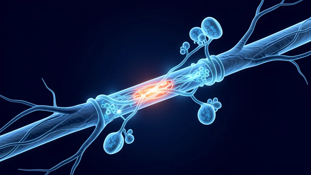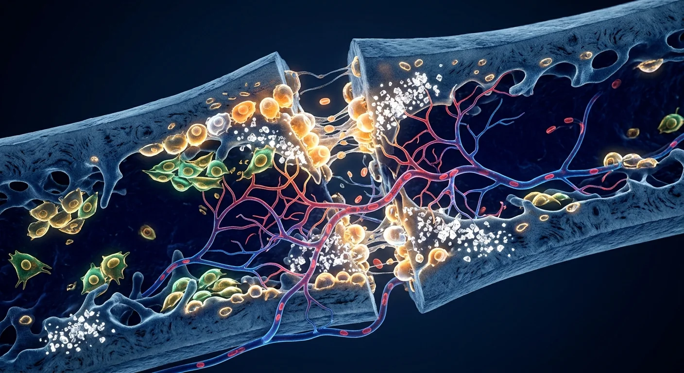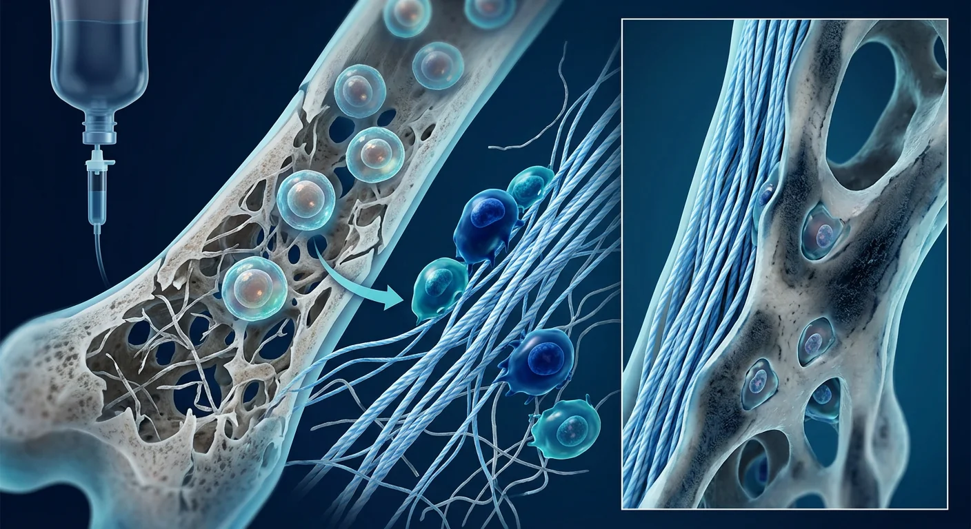
Osteogenesis imperfecta (OI) — commonly known as brittle bone disease — is a genetic disorder affecting approximately 1 in 15,000 to 20,000 live births worldwide, caused predominantly by mutations in the COL1A1 or COL1A2 genes that encode type I collagen, the primary structural protein of bone [1]. The result is bones that fracture under minimal or no trauma, often hundreds of times over a lifetime, accompanied by short stature, skeletal deformities, blue sclerae, hearing loss, and in severe cases, perinatal lethality. Current standard care — bisphosphonates, intramedullary rodding, and physical therapy — reduces fracture frequency and improves mobility but does not address the underlying collagen defect. Mesenchymal stem cell (MSC) therapy represents a fundamentally different approach: introducing healthy, collagen-competent cells capable of engrafting into bone, producing normal type I collagen, and improving bone quality from within [2].
The collagen problem at the molecular level. Type I collagen is a triple-helical protein composed of two α1 chains and one α2 chain. In OI, mutations in COL1A1 or COL1A2 produce either insufficient quantities of normal collagen (quantitative defects, typically milder OI types I and IV) or structurally abnormal collagen chains that disrupt triple-helix assembly (qualitative defects, typically severe OI types II and III). The mutant collagen is incorporated into the bone matrix but produces weakened, disorganized osteoid that mineralizes poorly and fractures under normal physiological loads. Even a single glycine substitution in the repetitive Gly-X-Y motif of the collagen triple helix — the most common mutation type — can reduce bone strength by 50% or more [3].
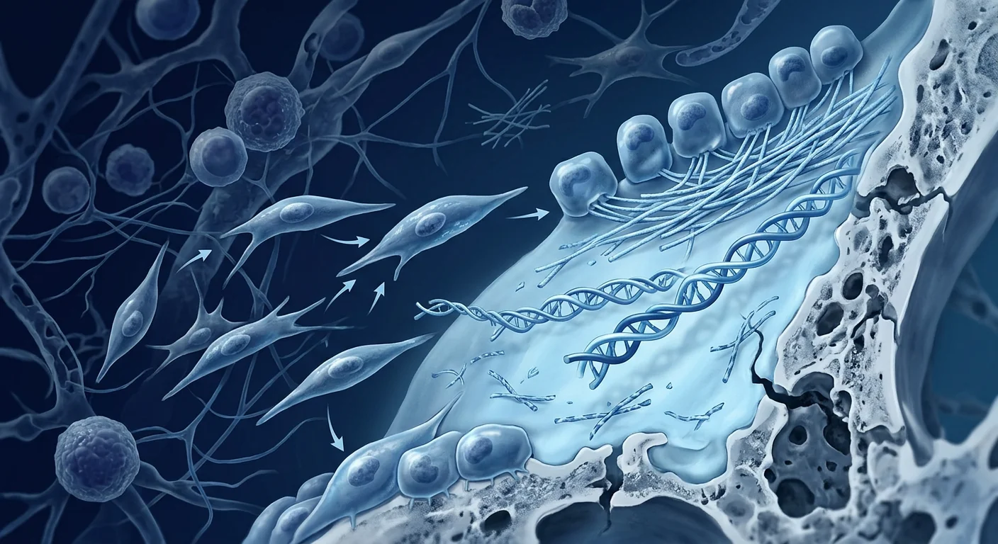
Why MSCs are a logical therapeutic candidate. MSCs are the natural precursor cells for osteoblasts — the cells that synthesize and secrete bone matrix, including type I collagen. When healthy donor MSCs are transplanted into an OI patient, they can engraft into bone surfaces, differentiate into functional osteoblasts, and produce normal type I collagen alongside the patient's defective collagen. Even partial engraftment — replacing as little as 1.5–2% of the osteoblast population with healthy donor cells — has been shown to measurably improve bone density, reduce fracture frequency, and accelerate linear growth in children with severe OI [4].
The Landmark Clinical Evidence — Horwitz, Le Blanc, and Beyond
OI was one of the first human diseases where MSC therapy was tested systematically, and the clinical data — while from small cohorts — is among the most compelling in the entire MSC field because a measurable biochemical endpoint exists: the proportion of donor-derived osteoblasts and collagen in bone biopsies.
Horwitz et al. — proof of engraftment (1999–2002). The seminal work by Edwin Horwitz and colleagues at St. Jude Children's Research Hospital established the foundational principle of MSC therapy for OI. In a series of pioneering studies, six children with severe OI (type III) underwent allogeneic bone marrow transplantation from HLA-matched siblings, followed by isolated MSC infusions from the same donors. The results were striking: donor osteoblasts were detected in bone biopsy specimens at levels of 1.5–2.0% of total osteoblasts, donor-derived type I collagen was measurable in the bone matrix, and clinical outcomes included a median 50% reduction in fracture frequency, accelerated growth velocity (median gain of 5.0 cm/year vs. 1.7 cm/year pre-transplant), and improved bone mineral density [5][6].
Le Blanc et al. — fetal MSC transplantation (2005). In a landmark case published in the New England Journal of Medicine, Katarina Le Blanc and colleagues reported the first in-utero transplantation of fetal liver-derived MSCs into a female fetus diagnosed with severe OI at 32 weeks' gestation. The transplanted male (XY) MSCs engrafted into bone, producing donor-derived osteoblasts detectable at 9 months postnatally. At age 2, the child had sustained only 3 fractures — dramatically fewer than the 30–50 fractures typical of untreated severe OI — and showed normal psychomotor development. A subsequent postnatal booster infusion of the same fetal MSCs at age 8 further improved bone architecture [7].
Otsuru et al. — allogeneic MSC infusions (2012–2019). A Japanese group led by Satoru Otsuru conducted a phase I trial of repeated allogeneic bone marrow MSC infusions in children with OI types III and IV. Two doses of 2–5 × 10⁶ MSCs/kg were administered intravenously at 4-month intervals. Donor cell engraftment was confirmed in bone biopsies, and treated children showed a significant increase in lumbar spine bone mineral density Z-score (mean improvement +0.8 at 12 months) and a 30–40% reduction in annualized fracture rate compared to the pre-treatment period. No serious adverse events or ectopic tissue formation were observed [8].
How MSCs Work in Osteogenesis Imperfecta — Beyond Bone Alone
The therapeutic mechanisms of MSCs in OI extend beyond simply providing healthy osteoblasts. MSCs act through multiple complementary pathways, each addressing a different aspect of the disease [9].
Direct engraftment and collagen production. A fraction of infused MSCs home to bone surfaces, particularly at sites of active remodeling — fracture callus, growth plates, and trabecular surfaces — where they differentiate into osteoblasts and synthesize normal type I collagen. Even low-level engraftment (1.5–2%) produces clinically meaningful benefits because the normal collagen is deposited in the bone matrix alongside the defective collagen, forming a composite that is measurably stronger than pure mutant matrix. This is not gene therapy — the patient's own cells continue producing defective collagen — but it is a form of cellular augmentation that tips the balance toward stronger bone.
Paracrine support of host osteoblasts. The majority of infused MSCs do not engraft permanently; they survive for days to weeks and exert their effects through the secretome. MSCs release BMP-2, BMP-7, IGF-1, TGF-β, and VEGF — growth factors that stimulate the patient's own osteoblasts to produce more bone matrix, enhance mineralization, and recruit additional progenitor cells to bone surfaces. Extracellular vesicles containing miR-21, miR-29b, and miR-196a are transferred to host osteoblasts, upregulating osteogenic gene expression and suppressing apoptosis [10].
Reducing bone resorption. OI is characterized not only by defective bone formation but also by accelerated bone resorption — osteoclasts are hyperactive in the abnormal OI bone microenvironment. MSCs secrete osteoprotegerin (OPG), a decoy receptor that blocks RANKL-mediated osteoclast activation, thereby reducing bone resorption. They also shift macrophages from the pro-inflammatory M1 to the tissue-reparative M2 phenotype, reducing the chronic low-grade inflammation that drives excessive bone turnover in OI [11].
Systemic effects beyond bone. OI is not purely a bone disease — it affects all type I collagen-rich tissues, including tendons, ligaments, skin, dentin, and sclera. MSCs have been shown to engraft in these tissues and contribute to collagen production at extraskeletal sites, though the functional significance of this for hearing, vision, and joint stability remains less well studied. It is one reason whole-body intravenous infusion, rather than local bone injection, is the preferred delivery route for OI [12].
Preclinical Evidence — Animal Models of OI
The oim/oim mouse (osteogenesis imperfecta murine) carries a spontaneous mutation in the Col1a2 gene that phenocopies human OI type III — fragile bones, shortened limbs, and reduced bone mineral density. This model has been instrumental in preclinical testing of MSC therapy for OI.
Intrauterine MSC transplantation in oim mice. In a key study, fetal oim mice received intraperitoneal injection of wild-type bone marrow MSCs at embryonic day 14.5 — analogous to the human in-utero transplantation paradigm. At 12 weeks postnatal, treated mice showed a 40% increase in femoral bone mineral density, a 60% reduction in spontaneous fracture frequency, and donor-derived osteoblasts composing 3–5% of bone surface cells. Notably, the treated femurs also showed improved trabecular microarchitecture on micro-CT — increased trabecular number and connectivity, and reduced trabecular separation [13].
Postnatal MSC infusion. Another study administered intravenous bone marrow MSCs to 4-week-old oim mice and followed them for 24 weeks. Treated mice showed significantly higher whole-body bone mineral density (+25%), increased cortical thickness, and improved biomechanical strength on three-point bending tests compared to untreated oim littermates. Repeated dosing at 8-week intervals produced superior outcomes to single-dose treatment, supporting the rationale for maintenance infusions in human protocols [14].
The VELAR Approach to MSC Therapy for Osteogenesis Imperfecta
At VELAR Center, MSC therapy for OI is delivered as part of a multidisciplinary protocol coordinated with the patient's primary orthopedic and metabolic bone team. The goal is to optimize bone quality, reduce fracture burden, and support growth and mobility.
Limitations and What the Evidence Does Not Yet Support
This is an honest assessment of where the evidence stands. MSC therapy for OI is not FDA-approved for this indication and remains investigational in all jurisdictions. The clinical evidence, while mechanistically compelling and consistently positive, comes from small open-label studies — the largest published series includes fewer than 20 patients. No randomized, sham-controlled trial has been conducted in OI. Heterogeneity in MSC source (bone marrow, fetal liver, Wharton's jelly, cord blood), dose (1–5 × 10⁶ cells/kg), timing (prenatal, infancy, childhood), and delivery route (intravenous, intraosseous) makes cross-study comparison difficult. The durability of donor cell engraftment — whether a single course of MSC infusions provides lasting benefit or requires ongoing maintenance dosing — remains an open question. The ideal age for intervention, the optimal MSC source, and whether engraftment benefits extend to hearing preservation and scleral integrity are areas of active investigation, not settled science [15].
When MSC therapy is most likely to help. Children with severe OI (types III and IV) who have a high fracture burden despite optimal bisphosphonate therapy, particularly when growth failure is a concern. The Horwitz data strongly supports this population. Patients for whom surgical rodding has reached its limits or is not feasible also represent a potential indication.
When it is least likely to help. Mild OI type I with infrequent fractures and normal growth velocity — the baseline is already good, and the incremental benefit of MSC therapy is unproven in this group. Patients with significant orthopedic deformities that require surgical correction — MSC therapy cannot straighten bowed bones; it can only improve bone quality to support surgical outcomes. Lethal OI type II is beyond the reach of any currently available therapy.
Frequently Asked Questions
How does MSC therapy differ from bisphosphonate treatment for OI?
Bisphosphonates work by inhibiting osteoclast-mediated bone resorption — they slow down the rate at which bone is broken down, allowing more time for the patient's defective osteoblasts to deposit bone matrix. The result is thicker cortical bone and reduced fracture frequency, but the collagen deposited is still abnormal. MSC therapy takes a complementary approach: it introduces healthy, collagen-competent osteoblasts that produce normal type I collagen, improving bone quality at the material level. The two modalities address different aspects of the disease and are not mutually exclusive — most published protocols combine them.
Can MSC therapy cure osteogenesis imperfecta?
No. MSC therapy cannot correct the COL1A1/COL1A2 mutation present in every cell of the patient's body. Gene therapy or gene editing approaches that directly repair the mutation are in early preclinical development but are not yet clinically available. MSC therapy provides healthy donor osteoblasts that produce normal collagen alongside the patient's defective collagen — a cellular augmentation strategy, not a genetic cure. It can measurably improve bone quality and reduce fracture burden, but the patient remains genetically affected.
What is the best age to consider MSC therapy for OI?
The published evidence supports intervention during childhood, when the skeleton is still growing and the potential for donor cell engraftment into active growth plates and remodeling surfaces is highest. The Horwitz and Otsuru protocols treated children ranging from infants to adolescents. In-utero transplantation (the Le Blanc approach) offers the theoretical advantage of treating before any fractures occur, but this remains an experimental procedure limited to a handful of cases worldwide. In adults, the evidence base is thinner, and the expected benefit is likely more modest because bone turnover and growth plate activity are reduced.
How much does MSC therapy for osteogenesis imperfecta cost in Thailand?
At VELAR Center in Bangkok, MSC therapy for OI is priced significantly below equivalent care in North America, Europe, or Australia. A full treatment protocol — including comprehensive bone health assessment, genetic counseling coordination, intravenous Wharton's jelly-derived MSC infusion, and 12-month follow-up monitoring — typically ranges from USD 8,000 to 15,000 depending on dosing requirements and whether maintenance infusions are needed. All costs are discussed transparently during the pre-treatment consultation; there are no hidden charges.
Is MSC therapy safe for children with OI?
The safety data from published OI-specific studies is reassuring. Across the Horwitz, Le Blanc, and Otsuru trials — totaling approximately 50 children treated with allogeneic MSCs — no serious adverse events attributable to the MSC product were reported. No ectopic tissue formation, no tumor development, and no clinically significant infusion reactions were observed. Transient low-grade fever and mild fatigue in the 24–48 hours post-infusion were the most common side effects. However, these are small cohorts, and long-term safety data (>10 years) is limited to the original Horwitz cohort. Parents considering MSC therapy for a child with OI should discuss the risk-benefit balance thoroughly with both their primary metabolic bone specialist and the regenerative medicine team [16].
How soon can results be expected after MSC therapy for OI?
Bone remodeling is a slow biological process — meaningful improvements in bone mineral density on DEXA are typically measurable at 6–12 months post-infusion. Reduction in fracture frequency is usually assessed over a 12-month period compared to the pre-treatment baseline. Growth velocity improvements in children may become apparent within 6 months. The Horwitz data suggests that clinical benefits peak at 6–18 months and may wane thereafter without maintenance infusions, which is why repeat dosing protocols are commonly used.
References
- Marini JC, Forlino A, Bächinger HP, et al. Osteogenesis imperfecta. Nature Reviews Disease Primers. 2017;3:17052. doi:10.1038/nrdp.2017.52 ↩
- Horwitz EM, Prockop DJ, Fitzpatrick LA, et al. Transplantability and therapeutic effects of bone marrow-derived mesenchymal cells in children with osteogenesis imperfecta. Nature Medicine. 1999;5(3):309-313. doi:10.1038/6529 ↩
- Forlino A, Marini JC. Osteogenesis imperfecta. The Lancet. 2016;387(10028):1657-1671. doi:10.1016/S0140-6736(15)00728-X ↩
- Otsuru S, Gordon PL, Shimono K, et al. Transplanted bone marrow mononuclear cells and MSCs impart clinical benefit to children with osteogenesis imperfecta through different mechanisms. Blood. 2012;120(9):1933-1941. doi:10.1182/blood-2011-12-400085 ↩
- Horwitz EM, Gordon PL, Koo WK, et al. Isolated allogeneic bone marrow-derived mesenchymal cells engraft and stimulate growth in children with osteogenesis imperfecta: implications for cell therapy of bone. Proceedings of the National Academy of Sciences. 2002;99(13):8932-8937. doi:10.1073/pnas.132252399 ↩
- Horwitz EM, Prockop DJ, Gordon PL, et al. Clinical responses to bone marrow transplantation in children with severe osteogenesis imperfecta. Blood. 2001;97(5):1227-1231. doi:10.1182/blood.V97.5.1227 ↩
- Le Blanc K, Götherström C, Ringdén O, et al. Fetal mesenchymal stem-cell engraftment in bone after in utero transplantation in a patient with severe osteogenesis imperfecta. Transplantation. 2005;79(11):1607-1614. doi:10.1097/01.TP.0000159029.48678.93 ↩
- Otsuru S, Desbourdes L, Guess AJ, et al. Extracellular vesicles released from mesenchymal stromal cells stimulate bone growth in osteogenesis imperfecta. Cytotherapy. 2019;21(2):126-137. doi:10.1016/j.jcyt.2018.11.007 ↩
- Götherström C, Walther-Jallow L, Tullberg M, et al. Transplantation of mesenchymal stem cells for osteogenesis imperfecta. Current Stem Cell Research & Therapy. 2013;8(2):131-137. doi:10.2174/1574888X11308020004 ↩
- Qin Y, Wang L, Gao Z, Chen G, Zhang C. Bone marrow stromal/stem cell-derived extracellular vesicles regulate osteoblast activity and differentiation in vitro and promote bone regeneration in vivo. Scientific Reports. 2016;6:21961. doi:10.1038/srep21961 ↩
- Sims NA, Martin TJ. Coupling the activities of bone formation and resorption: a multitude of signals within the basic multicellular unit. BoneKEy Reports. 2014;3:481. doi:10.1038/bonekey.2013.215 ↩
- Guillot PV, Abass O, Bassett JH, et al. Intrauterine transplantation of human fetal mesenchymal stem cells from first-trimester blood repairs bone and reduces fractures in osteogenesis imperfecta mice. Blood. 2008;111(3):1717-1725. doi:10.1182/blood-2007-08-105809 ↩
- Chan JK, Götherström C. Prenatal transplantation of mesenchymal stem cells to treat osteogenesis imperfecta. Frontiers in Pharmacology. 2014;5:223. doi:10.3389/fphar.2014.00223 ↩
- Jones GN, Moschidou D, Lay K, et al. Upregulating the WNT pathway prevents cellular senescence and ameliorates the osteogenesis imperfecta phenotype. Journal of Clinical Investigation. 2017;127(9):3332-3342. doi:10.1172/JCI92435 ↩
- Götherström C, Westgren M, Shaw SW, et al. Pre- and postnatal transplantation of fetal mesenchymal stem cells in osteogenesis imperfecta: a two-center experience. Stem Cells Translational Medicine. 2014;3(2):255-264. doi:10.5966/sctm.2013-0090 ↩
- Lalu MM, McIntyre L, Pugliese C, et al. Safety of cell therapy with mesenchymal stromal cells (SafeCell): a systematic review and meta-analysis of clinical trials. PLoS One. 2012;7(10):e47559. doi:10.1371/journal.pone.0047559 ↩
成骨不全症(OI)——俗称脆骨病——是一种遗传性疾病,全球每15,000至20,000名活产婴儿中约有1例,主要由编码I型胶原蛋白的COL1A1或COL1A2基因突变引起,I型胶原蛋白是骨骼的主要结构蛋白[1]。其结果是骨骼在轻微或无明显外伤下发生骨折,一生中常达数百次,伴随身材矮小、骨骼畸形、蓝色巩膜、听力丧失,严重者可致围产期死亡。目前的标准化治疗——双膦酸盐类药物、髓内钉固定和物理治疗——可降低骨折频率并改善活动能力,但无法解决根本的胶原蛋白缺陷。间充质干细胞(MSC)疗法代表了一种根本不同的方法:引入健康的、能产生胶原蛋白的细胞,这些细胞能够植入骨骼,产生正常的I型胶原蛋白,从内部改善骨骼质量[2]。
分子层面的胶原蛋白问题。I型胶原蛋白是由两条α1链和一条α2链组成的三螺旋蛋白。在OI中,COL1A1或COL1A2的突变导致正常胶原蛋白数量不足(数量缺陷,通常为较轻的OI I型和IV型)或结构异常的胶原蛋白链破坏三螺旋组装(质量缺陷,通常为严重的OI II型和III型)。突变胶原蛋白被整合到骨基质中,但产生弱化的、无序的类骨组织,矿化不良,在正常生理负荷下即可发生骨折。即使是胶原蛋白三螺旋重复Gly-X-Y基序中的单个甘氨酸替换——最常见的突变类型——也可使骨骼强度降低50%或更多[3]。
为什么MSC是合理的治疗候选。MSC是成骨细胞的天然前体细胞——成骨细胞是合成和分泌骨基质(包括I型胶原蛋白)的细胞。当健康的供体MSC移植到OI患者体内后,它们可植入骨骼表面,分化为功能性成骨细胞,并在患者缺陷胶原蛋白旁产生正常的I型胶原蛋白。即使是部分植入——仅替换1.5–2%的成骨细胞群为健康供体细胞——已被证明可显著改善严重OI患儿的骨密度、降低骨折频率并加速线性生长[4]。
里程碑式的临床证据——Horwitz、Le Blanc及后续研究
OI是MSC疗法最早被系统测试的人类疾病之一,其临床数据——尽管来自小规模队列——是整个MSC领域中最具说服力的数据之一,因为存在可测量的生化终点:骨活检中供体来源成骨细胞和胶原蛋白的比例。
Horwitz等人——植入证明(1999–2002)。Edwin Horwitz及其同事在圣裘德儿童研究医院进行的开创性工作确立了MSC治疗OI的基本原则。在一系列先驱性研究中,六名严重OI(III型)患儿接受了来自HLA匹配兄弟姐妹的异基因骨髓移植,随后接受来自同一供体的单独MSC输注。结果引人注目:骨活检标本中检测到供体成骨细胞,占总成骨细胞的1.5–2.0%,骨基质中检测到供体来源的I型胶原蛋白,临床结果包括骨折频率中位数降低50%、生长速度加快(移植后中位增长5.0厘米/年 vs. 移植前1.7厘米/年)以及骨密度改善[5][6]。
Le Blanc等人——胎儿MSC移植(2005)。在发表于《新英格兰医学杂志》的一项里程碑式病例报告中,Katarina Le Blanc及其同事报道了首例将胎儿肝脏来源的MSC经子宫移植至一名在孕32周时被诊断为严重OI的女性胎儿。移植的男性(XY)MSC植入骨骼,在出生后9个月检测到供体来源的成骨细胞。2岁时,该儿童仅发生3次骨折——远低于未经治疗的严重OI典型的30–50次骨折——并显示正常的心理运动发育。8岁时进行的同源胎儿MSC产后加强输注进一步改善了骨骼结构[7]。
Otsuru等人——异基因MSC输注(2012–2019)。由Satoru Otsuru领导的日本团队进行了针对III型和IV型OI患儿的重复异基因骨髓MSC输注I期试验。以2–5 × 10⁶ MSC/kg的剂量静脉输注两次,间隔4个月。骨活检证实供体细胞植入,治疗患儿腰椎骨密度Z值显著提高(12个月时平均改善+0.8),年化骨折率较治疗前降低30–40%。未观察到严重不良事件或异位组织形成[8]。
MSC如何在成骨不全症中发挥作用——超越骨骼
MSC在OI中的治疗机制不仅限于提供健康的成骨细胞。MSC通过多种互补途径发挥作用,每种途径针对疾病的不同方面[9]。
直接植入与胶原蛋白生产。一部分输注的MSC归巢至骨骼表面,特别是在活跃重塑部位——骨折愈伤组织、生长板和骨小梁表面——在那里它们分化为成骨细胞并合成正常的I型胶原蛋白。即使是低水平植入(1.5–2%)也能产生临床上有意义的益处,因为正常胶原蛋白与缺陷胶原蛋白一起沉积在骨基质中,形成比纯突变基质强度明显更高的复合材料。这不是基因治疗——患者自身细胞继续产生缺陷胶原蛋白——而是一种细胞增强策略,使天平向更强壮的骨骼倾斜。
对宿主成骨细胞的旁分泌支持。大多数输注的MSC不会永久植入;它们存活数天至数周,通过分泌组发挥作用。MSC释放BMP-2、BMP-7、IGF-1、TGF-β和VEGF——刺激患者自身成骨细胞产生更多骨基质、增强矿化并招募额外祖细胞至骨骼表面的生长因子。含有miR-21、miR-29b和miR-196a的细胞外囊泡被转移至宿主成骨细胞,上调成骨基因表达并抑制凋亡[10]。
减少骨吸收。OI不仅以缺陷性骨形成为特征,还以加速骨吸收为特征——在异常的OI骨微环境中,破骨细胞过度活跃。MSC分泌骨保护素(OPG),这是一种阻断RANKL介导的破骨细胞活化的诱饵受体,从而减少骨吸收。它们还将巨噬细胞从促炎性M1表型转变为组织修复性M2表型,降低驱动OI中过度骨转换的慢性低度炎症[11]。
超越骨骼的全身效应。OI不仅仅是骨病——它影响所有富含I型胶原蛋白的组织,包括肌腱、韧带、皮肤、牙本质和巩膜。MSC已被证明可植入这些组织并在骨外部位促进胶原蛋白生产,尽管这对听力、视力和关节稳定性的功能性意义研究较少。这也是为什么全身静脉输注而非局部骨内注射是OI的首选给药途径的原因之一[12]。
临床前证据——OI动物模型
oim/oim小鼠(成骨不全症鼠模型)携带Col1a2基因的自发突变,表型复制人类OI III型——脆骨、短肢和骨密度降低。该模型在OI的MSC治疗临床前测试中发挥了重要作用。
oim小鼠的宫内MSC移植。在一项关键研究中,胎儿oim小鼠在胚胎第14.5天接受野生型骨髓MSC腹腔注射——类似于人类宫内移植模式。在出生后12周,治疗小鼠的股骨骨密度增加40%,自发性骨折频率降低60%,供体来源成骨细胞占骨骼表面细胞的3–5%。值得注意的是,治疗股骨在micro-CT上还显示出改善的骨小梁微结构——骨小梁数量和连接性增加,骨小梁分离减少[13]。
产后MSC输注。另一项研究向4周龄oim小鼠静脉输注骨髓MSC,并随访24周。与未治疗的oim同窝对照相比,治疗小鼠显示出显著更高的全身骨密度(+25%)、增加的皮质厚度和改善的三点弯曲生物力学强度。8周间隔重复给药产生的效果优于单次治疗,支持人类方案中维持输注的理论基础[14]。
VELAR的成骨不全症MSC疗法方案
在VELAR中心,OI的MSC疗法作为多学科方案的一部分,与患者的主治骨科和代谢骨团队协调实施。目标是优化骨骼质量、减少骨折负担并支持生长和活动能力。
局限性与证据尚未支持的方面
这是对证据现状的诚实评估。MSC疗法治疗OI尚未获得FDA批准用于此适应症,在所有司法管辖区仍处于研究阶段。临床证据虽然在机制上令人信服且持续积极,但来自小型开放标签研究——已发表的最大系列包括不到20名患者。OI领域未进行过随机假对照试验。MSC来源(骨髓、胎儿肝脏、华通胶、脐带血)、剂量(1–5 × 10⁶细胞/kg)、时机(产前、婴儿期、儿童期)和给药途径(静脉、骨内)的异质性使跨研究比较困难。供体细胞植入的持久性——单疗程MSC输注是否提供持久益处还是需要持续维持给药——仍是一个未解决的问题。最佳干预年龄、最佳MSC来源以及植入益处是否延伸至听力保护和巩膜完整性,是当前活跃的研究领域,而非已确定的科学结论[15]。
MSC疗法最可能提供帮助的情况。尽管有最佳双膦酸盐治疗但骨折负担仍然高的严重OI(III型和IV型)患儿,特别是在生长迟缓成为问题时。Horwitz数据强烈支持这一人群。手术髓内钉已达极限或不可行的患者也代表潜在适应症。
最不可能提供帮助的情况。骨折不频繁且生长速度正常的轻度OI I型——基线已经良好,MSC疗法的增量获益在此组中未经证实。有明显骨科畸形需要手术矫正的患者——MSC疗法无法拉直弯曲的骨骼;它只能改善骨骼质量以支持手术结果。致死性OI II型超出任何现有疗法的范围。
常见问题
MSC疗法与双膦酸盐治疗OI有何不同?
双膦酸盐通过抑制破骨细胞介导的骨吸收起作用——它们减缓骨破坏的速度,使患者缺陷性成骨细胞有更多时间沉积骨基质。结果是皮质骨增厚和骨折频率降低,但沉积的胶原蛋白仍然异常。MSC疗法采取互补方法:引入健康的、能产生胶原蛋白的成骨细胞,产生正常的I型胶原蛋白,在材料层面改善骨骼质量。这两种方式针对疾病的不同方面,并非相互排斥——大多数已发表的方案将两者结合使用。
MSC疗法能治愈成骨不全症吗?
不能。MSC疗法无法纠正患者体内每个细胞中存在的COL1A1/COL1A2突变。直接修复突变的基因治疗或基因编辑方法处于早期临床前开发阶段,尚未临床可用。MSC疗法提供健康的供体成骨细胞,在患者缺陷胶原蛋白旁产生正常胶原蛋白——这是一种细胞增强策略,而非基因治愈。它可显著改善骨骼质量并降低骨折负担,但患者在基因上仍然患病。
考虑MSC疗法治疗OI的最佳年龄是多少?
已发表的证据支持在儿童期进行干预,此时骨骼仍在生长,供体细胞植入活跃生长板和重塑表面的潜力最高。Horwitz和Otsuru方案治疗了从婴儿到青少年的儿童。宫内移植(Le Blanc方法)提供了在任何骨折发生前进行治疗的理论优势,但这仍是仅限于全球少数病例的实验性程序。在成人中,证据基础更薄弱,预期获益可能更有限,因为骨转换和生长板活性降低。
在泰国,MSC疗法治疗成骨不全症的费用是多少?
在曼谷的VELAR中心,OI的MSC疗法价格显著低于北美、欧洲或澳大利亚的等效治疗。完整的治疗方案——包括全面骨骼健康评估、遗传咨询协调、静脉华通胶来源MSC输注和12个月随访监测——通常在8,000至15,000美元之间,具体取决于剂量需求和是否需要维持输注。所有费用在治疗前咨询中透明讨论;没有隐藏费用。
MSC疗法对OI患儿安全吗?
已发表的OI特定研究的安全性数据令人放心。在Horwitz、Le Blanc和Otsuru试验中——总计约50名接受异基因MSC治疗的儿童——未报告归因于MSC产品的严重不良事件。未观察到异位组织形成、肿瘤发展或临床上显著的输注反应。输注后24–48小时内的短暂低热和轻度疲劳是最常见的副作用。然而,这些是小规模队列,长期安全性数据(>10年)仅限于原始Horwitz队列。考虑为孩子进行MSC治疗的OI患儿父母应与主治代谢骨专家和再生医学团队充分讨论风险-收益平衡[16]。
MSC治疗OI后多久可以看到效果?
骨重塑是一个缓慢的生物学过程——DEXA上骨密度的有意义改善通常在输注后6–12个月可测量。骨折频率的降低通常以12个月为周期与治疗前基线进行比较。患儿的生长速度改善可能在6个月内变得明显。Horwitz数据表明,临床获益在6–18个月达到峰值,此后若无维持输注可能会减弱,这就是为什么重复给药方案被普遍使用的原因。
参考文献
- Marini JC, Forlino A, Bächinger HP, et al. Osteogenesis imperfecta. Nature Reviews Disease Primers. 2017;3:17052. doi:10.1038/nrdp.2017.52 ↩
- Horwitz EM, Prockop DJ, Fitzpatrick LA, et al. Transplantability and therapeutic effects of bone marrow-derived mesenchymal cells in children with osteogenesis imperfecta. Nature Medicine. 1999;5(3):309-313. doi:10.1038/6529 ↩
- Forlino A, Marini JC. Osteogenesis imperfecta. The Lancet. 2016;387(10028):1657-1671. doi:10.1016/S0140-6736(15)00728-X ↩
- Otsuru S, Gordon PL, Shimono K, et al. Transplanted bone marrow mononuclear cells and MSCs impart clinical benefit to children with osteogenesis imperfecta through different mechanisms. Blood. 2012;120(9):1933-1941. doi:10.1182/blood-2011-12-400085 ↩
- Horwitz EM, Gordon PL, Koo WK, et al. Isolated allogeneic bone marrow-derived mesenchymal cells engraft and stimulate growth in children with osteogenesis imperfecta: implications for cell therapy of bone. Proceedings of the National Academy of Sciences. 2002;99(13):8932-8937. doi:10.1073/pnas.132252399 ↩
- Horwitz EM, Prockop DJ, Gordon PL, et al. Clinical responses to bone marrow transplantation in children with severe osteogenesis imperfecta. Blood. 2001;97(5):1227-1231. doi:10.1182/blood.V97.5.1227 ↩
- Le Blanc K, Götherström C, Ringdén O, et al. Fetal mesenchymal stem-cell engraftment in bone after in utero transplantation in a patient with severe osteogenesis imperfecta. Transplantation. 2005;79(11):1607-1614. doi:10.1097/01.TP.0000159029.48678.93 ↩
- Otsuru S, Desbourdes L, Guess AJ, et al. Extracellular vesicles released from mesenchymal stromal cells stimulate bone growth in osteogenesis imperfecta. Cytotherapy. 2019;21(2):126-137. doi:10.1016/j.jcyt.2018.11.007 ↩
- Götherström C, Walther-Jallow L, Tullberg M, et al. Transplantation of mesenchymal stem cells for osteogenesis imperfecta. Current Stem Cell Research & Therapy. 2013;8(2):131-137. doi:10.2174/1574888X11308020004 ↩
- Qin Y, Wang L, Gao Z, Chen G, Zhang C. Bone marrow stromal/stem cell-derived extracellular vesicles regulate osteoblast activity and differentiation in vitro and promote bone regeneration in vivo. Scientific Reports. 2016;6:21961. doi:10.1038/srep21961 ↩
- Sims NA, Martin TJ. Coupling the activities of bone formation and resorption: a multitude of signals within the basic multicellular unit. BoneKEy Reports. 2014;3:481. doi:10.1038/bonekey.2013.215 ↩
- Guillot PV, Abass O, Bassett JH, et al. Intrauterine transplantation of human fetal mesenchymal stem cells from first-trimester blood repairs bone and reduces fractures in osteogenesis imperfecta mice. Blood. 2008;111(3):1717-1725. doi:10.1182/blood-2007-08-105809 ↩
- Chan JK, Götherström C. Prenatal transplantation of mesenchymal stem cells to treat osteogenesis imperfecta. Frontiers in Pharmacology. 2014;5:223. doi:10.3389/fphar.2014.00223 ↩
- Jones GN, Moschidou D, Lay K, et al. Upregulating the WNT pathway prevents cellular senescence and ameliorates the osteogenesis imperfecta phenotype. Journal of Clinical Investigation. 2017;127(9):3332-3342. doi:10.1172/JCI92435 ↩
- Götherström C, Westgren M, Shaw SW, et al. Pre- and postnatal transplantation of fetal mesenchymal stem cells in osteogenesis imperfecta: a two-center experience. Stem Cells Translational Medicine. 2014;3(2):255-264. doi:10.5966/sctm.2013-0090 ↩
- Lalu MM, McIntyre L, Pugliese C, et al. Safety of cell therapy with mesenchymal stromal cells (SafeCell): a systematic review and meta-analysis of clinical trials. PLoS One. 2012;7(10):e47559. doi:10.1371/journal.pone.0047559 ↩
تكون العظم الناقص (OI) — المعروف باسم مرض العظام الهشة — هو اضطراب وراثي يصيب حوالي 1 من كل 15,000 إلى 20,000 ولادة حية worldwide، وينتج بشكل رئيسي عن طفرات في جينات COL1A1 أو COL1A2 التي تشفر الكولاجين من النوع الأول، البروتين الهيكلي الرئيسي للعظام [1]. والنتيجة هي عظام تنكسر تحت صدمة طفيفة أو بدون صدمة ظاهرة، غالبًا مئات المرات على مدى الحياة، مصحوبة بقصر القامة، وتشوهات هيكلية، وصلبة زرقاء، وفقدان السمع، وفي الحالات الشديدة، وفيات ما حول الولادة. الرعاية القياسية الحالية — البيسفوسفونات، والتثبيت بالنخاع، والعلاج الطبيعي — تقلل من تكرار الكسور وتحسن الحركة ولكنها لا تعالج خلل الكولاجين الأساسي. يمثل العلاج بالخلايا الجذعية الوسيطة (MSC) نهجًا مختلفًا جوهريًا: إدخال خلايا سليمة قادرة على إنتاج الكولاجين، قادرة على الانغراس في العظام، وإنتاج كولاجين طبيعي من النوع الأول، وتحسين جودة العظام من الداخل [2].
مشكلة الكولاجين على المستوى الجزيئي. الكولاجين من النوع الأول هو بروتين ثلاثي الحلزون يتكون من سلسلتين α1 وسلسلة α2 واحدة. في OI، تنتج الطفرات في COL1A1 أو COL1A2 إما كميات غير كافية من الكولاجين الطبيعي (عيوب كمية، عادة أنواع OI الأخف I و IV) أو سلاسل كولاجين غير طبيعية هيكليًا تعطل تجميع الحلزون الثلاثي (عيوب نوعية، عادة أنواع OI الشديدة II و III). يتم دمج الكولاجين الطافر في مصفوفة العظام ولكنه ينتج نسيجًا عظميًا ضعيفًا وغير منظم يتمعدن بشكل سيئ وينكسر تحت الأحمال الفسيولوجية الطبيعية. حتى استبدال جلايسين واحد في نمط Gly-X-Y المتكرر لحلزون الكولاجين الثلاثي — أكثر أنواع الطفرات شيوعًا — يمكن أن يقلل من قوة العظام بنسبة 50% أو أكثر [3].
لماذا تعد MSC مرشحًا علاجيًا منطقيًا. MSC هي الخلايا السليفة الطبيعية للخلايا البانية للعظم — الخلايا التي تصنع وتفرز مصفوفة العظام، بما في ذلك الكولاجين من النوع الأول. عندما يتم زرع MSC من متبرع سليم في مريض OI، يمكنها الانغراس في أسطح العظام، والتمايز إلى خلايا بانية للعظم وظيفية، وإنتاج كولاجين طبيعي من النوع الأول جنبًا إلى جنب مع الكولاجين المعيب للمريض. حتى الانغراس الجزئي — استبدال ما لا يقل عن 1.5–2% من مجموعة الخلايا البانية للعظم بخلايا متبرع سليمة — ثبت أنه يحسن بشكل قابل للقياس كثافة العظام، ويقلل من تكرار الكسور، ويسرع النمو الخطي لدى الأطفال المصابين بـ OI الشديد [4].
الأدلة السريرية البارزة — هورويتز، ولو بلان، وما بعدهما
كان OI من أول الأمراض البشرية التي تم فيها اختبار علاج MSC بشكل منهجي، والبيانات السريرية — رغم أنها من مجموعات صغيرة — هي من بين الأكثر إقناعًا في مجال MSC بأكمله لأن هناك نقطة نهاية كيميائية حيوية قابلة للقياس: نسبة الخلايا البانية للعظم والكولاجين المشتقة من المتبرع في خزعات العظام.
هورويتز وزملاؤه — إثبات الانغراس (1999–2002). أسس العمل الرائد لإدوين هورويتز وزملائه في مستشفى سانت جود لأبحاث الأطفال المبدأ الأساسي لعلاج MSC لـ OI. في سلسلة من الدراسات الرائدة، خضع ستة أطفال مصابين بـ OI الشديد (النوع III) لزراعة نخاع عظمي خيفي من أشقاء متطابقين HLA، تلتها حقن MSC معزولة من نفس المتبرعين. كانت النتائج مذهلة: تم الكشف عن خلايا بانية للعظم من المتبرع في عينات خزعة العظام بمستويات 1.5–2.0% من إجمالي الخلايا البانية للعظم، وتم قياس كولاجين من النوع الأول مشتق من المتبرع في مصفوفة العظام، وشملت النتائج السريرية انخفاضًا متوسطًا بنسبة 50% في تكرار الكسور، وتسارع سرعة النمو (متوسط زيادة 5.0 سم/سنة مقابل 1.7 سم/سنة قبل الزرع)، وتحسن كثافة المعادن في العظام [5][6].
لو بلان وزملاؤها — زراعة MSC الجنينية (2005). في تقرير حالة بارز نُشر في New England Journal of Medicine، أبلغت كاتارينا لو بلان وزملاؤها عن أول زراعة داخل الرحم لـ MSC مشتقة من كبد الجنين في جنين أنثى تم تشخيصها بـ OI الشديد في الأسبوع 32 من الحمل. انغرست MSC الذكرية (XY) المزروعة في العظام، منتجة خلايا بانية للعظم مشتقة من المتبرع تم اكتشافها في عمر 9 أشهر بعد الولادة. في عمر سنتين، كانت الطفلة قد تعرضت لـ 3 كسور فقط — أقل بكثير من 30–50 كسرًا النموذجية لـ OI الشديد غير المعالج — وأظهرت تطورًا نفسيًا حركيًا طبيعيًا. حقنة تعزيز لاحقة بعد الولادة من نفس MSC الجنينية في عمر 8 سنوات حسنت بنية العظام بشكل أكبر [7].
أوتسورو وزملاؤه — حقن MSC الخيفي (2012–2019). أجرت مجموعة يابانية بقيادة ساتورو أوتسورو تجربة المرحلة الأولى لحقن MSC المتكرر من نخاع العظم الخيفي في الأطفال المصابين بـ OI من النوعين III و IV. تم إعطاء جرعتين من 2–5 × 10⁶ MSC/كجم عن طريق الوريد بفاصل 4 أشهر. تم تأكيد انغراس خلايا المتبرع في خزعات العظام، وأظهر الأطفال المعالجون زيادة كبيرة في درجة Z لكثافة المعادن في العمود الفقري القطني (متوسط تحسن +0.8 عند 12 شهرًا) وانخفاضًا بنسبة 30–40% في معدل الكسور السنوي مقارنة بفترة ما قبل العلاج. لم تُلاحظ أي أحداث سلبية خطيرة أو تكوين أنسجة منتبذة [8].
كيف تعمل MSC في تكون العظم الناقص — ما وراء العظام وحدها
تمتد الآليات العلاجية لـ MSC في OI إلى ما هو أبعد من مجرد توفير خلايا بانية للعظم سليمة. تعمل MSC من خلال مسارات تكميلية متعددة، كل منها يعالج جانبًا مختلفًا من المرض [9].
الانغراس المباشر وإنتاج الكولاجين. جزء من MSC المحقونة يهاجر إلى أسطح العظام، خاصة في مواقع إعادة التشكيل النشطة — جسور الكسور، وصفائح النمو، وأسطح العظم التربيقي — حيث تتمايز إلى خلايا بانية للعظم وتصنع كولاجين طبيعي من النوع الأول. حتى الانغراس منخفض المستوى (1.5–2%) ينتج فوائد ذات معنى سريري لأن الكولاجين الطبيعي يترسب في مصفوفة العظام جنبًا إلى جنب مع الكولاجين المعيب، مكونًا مركبًا أقوى بشكل قابل للقياس من المصفوفة الطافرة النقية. هذا ليس علاجًا جينيًا — تستمر خلايا المريض نفسه في إنتاج كولاجين معيب — ولكنه شكل من أشكال التعزيز الخلوي الذي يرجح الكفة نحو عظام أقوى.
الدعم نظير الصماوي للخلايا البانية للعظم لدى المضيف. غالبية MSC المحقونة لا تنغرس بشكل دائم؛ تعيش لأيام إلى أسابيع وتمارس تأثيراتها من خلال المفرزات. تطلق MSC عوامل BMP-2 و BMP-7 و IGF-1 و TGF-β و VEGF — عوامل نمو تحفز الخلايا البانية للعظم لدى المريض نفسه لإنتاج المزيد من مصفوفة العظام، وتعزيز التمعدن، وتجنيد خلايا سليفة إضافية إلى أسطح العظام. يتم نقل الحويصلات خارج الخلية التي تحتوي على miR-21 و miR-29b و miR-196a إلى الخلايا البانية للعظم لدى المضيف، مما ينظم التعبير الجيني المكون للعظم ويثبط الاستماتة [10].
تقليل ارتشاف العظام. لا يتميز OI فقط بتكوين عظم معيب ولكن أيضًا بارتشاف عظم متسارع — الخلايا الناقضة للعظم مفرطة النشاط في البيئة الدقيقة غير الطبيعية لعظام OI. تفرز MSC الأوستيوبروتجيرين (OPG)، وهو مستقبل شركي يحجب تنشيط الخلايا الناقضة للعظم بوساطة RANKL، مما يقلل من ارتشاف العظام. كما تحول البلاعم من النمط الظاهري M1 المؤيد للالتهابات إلى النمط الظاهري M2 المصلح للأنسجة، مما يقلل من الالتهاب المزمن منخفض الدرجة الذي يدفع دوران العظام المفرط في OI [11].
التأثيرات الجهازية خارج العظام. OI ليس مرضًا عظميًا بحتًا — إنه يؤثر على جميع الأنسجة الغنية بالكولاجين من النوع الأول، بما في ذلك الأوتار والأربطة والجلد والعاج والصلبة. ثبت أن MSC تنغرس في هذه الأنسجة وتساهم في إنتاج الكولاجين في المواقع خارج الهيكل العظمي، رغم أن الأهمية الوظيفية لهذا للسمع والرؤية واستقرار المفاصل لا تزال أقل دراسة. هذا أحد أسباب أن الحقن الوريدي للجسم كله، بدلاً من الحقن الموضعي في العظام، هو طريق التوصيل المفضل لـ OI [12].
الأدلة قبل السريرية — نماذج حيوانية لـ OI
يحمل فأر oim/oim (تكون العظم الناقص الفأري) طفرة تلقائية في جين Col1a2 تحاكي ظاهريًا OI البشري من النوع III — عظام هشة، وأطراف قصيرة، وكثافة معادن عظمية منخفضة. كان هذا النموذج أساسيًا في الاختبار قبل السريري لعلاج MSC لـ OI.
زراعة MSC داخل الرحم في فئران oim. في دراسة رئيسية، تلقت أجنة فئران oim حقنًا داخل الصفاق من MSC نخاع العظم من النوع البري في اليوم الجنيني 14.5 — مماثل لنموذج الزراعة داخل الرحم البشري. في الأسبوع 12 بعد الولادة، أظهرت الفئران المعالجة زيادة بنسبة 40% في كثافة المعادن في عظم الفخذ، وانخفاضًا بنسبة 60% في تكرار الكسور التلقائية، وخلايا بانية للعظم مشتقة من المتبرع تشكل 3–5% من خلايا سطح العظام. بشكل ملحوظ، أظهرت عظام الفخذ المعالجة أيضًا تحسنًا في البنية الدقيقة التربيقية على التصوير المقطعي المحوسب الدقيق — زيادة عدد التربيق والاتصال، وتقليل انفصال التربيق [13].
حقن MSC بعد الولادة. قامت دراسة أخرى بحقن MSC نخاع العظم عن طريق الوريد في فئران oim بعمر 4 أسابيع وتابعتها لمدة 24 أسبوعًا. أظهرت الفئران المعالجة كثافة معادن عظمية كلية أعلى بشكل ملحوظ (+25%)، وزيادة سمك القشرة، وتحسن القوة البيوميكانيكية في اختبارات الانحناء ثلاثي النقاط مقارنة بأشقاء oim غير المعالجين. أنتجت الجرعات المتكررة على فترات 8 أسابيع نتائج متفوقة على العلاج بجرعة واحدة، مما يدعم الأساس المنطقي لحقن الصيانة في البروتوكولات البشرية [14].
نهج VELAR لعلاج MSC لتكون العظم الناقص
في مركز VELAR، يتم تقديم علاج MSC لـ OI كجزء من بروتوكول متعدد التخصصات بالتنسيق مع فريق العظام والتمثيل الغذائي الأساسي للمريض. الهدف هو تحسين جودة العظام، وتقليل عبء الكسور، ودعم النمو والحركة.
القيود وما لا تدعمه الأدلة بعد
هذا تقييم صادق لموقف الأدلة. علاج MSC لـ OI ليس معتمدًا من FDA لهذا المؤشر ولا يزال قيد البحث في جميع الاختصاصات القضائية. الأدلة السريرية، رغم كونها مقنعة ميكانيكيًا وإيجابية باستمرار، تأتي من دراسات صغيرة مفتوحة التسمية — أكبر سلسلة منشورة تشمل أقل من 20 مريضًا. لم تُجرَ أي تجربة عشوائية مضبوطة بالغفل في OI. عدم التجانس في مصدر MSC (نخاع العظم، كبد الجنين، هلام وارتون، دم الحبل السري)، والجرعة (1–5 × 10⁶ خلية/كجم)، والتوقيت (قبل الولادة، الطفولة، الطفولة المبكرة)، وطريق التوصيل (وريدي، داخل العظم) يجعل المقارنة بين الدراسات صعبة. متانة انغراس خلايا المتبرع — ما إذا كانت دورة واحدة من حقن MSC توفر فائدة دائمة أو تتطلب جرعات صيانة مستمرة — تظل سؤالاً مفتوحًا. العمر المثالي للتدخل، ومصدر MSC الأمثل، وما إذا كانت فوائد الانغراس تمتد إلى الحفاظ على السمع وسلامة الصلبة هي مجالات بحث نشطة، وليست علمًا محسومًا [15].
متى يكون علاج MSC أكثر احتمالاً للمساعدة. الأطفال المصابون بـ OI الشديد (الأنواع III و IV) الذين لديهم عبء كسور مرتفع رغم علاج البيسفوسفونات الأمثل، خاصة عندما يكون فشل النمو مصدر قلق. تدعم بيانات هورويتز هذه الفئة بقوة. المرضى الذين وصل التثبيت الجراحي بالنخاع إلى حدوده أو غير ممكن لديهم يمثلون أيضًا مؤشرًا محتملاً.
متى يكون أقل احتمالاً للمساعدة. OI الخفيف من النوع I مع كسور غير متكررة وسرعة نمو طبيعية — خط الأساس جيد بالفعل، والفائدة الإضافية لعلاج MSC غير مثبتة في هذه المجموعة. المرضى الذين لديهم تشوهات عظمية كبيرة تتطلب تصحيحًا جراحيًا — لا يمكن لعلاج MSC تقويم العظام المنحنية؛ يمكنه فقط تحسين جودة العظام لدعم النتائج الجراحية. OI المميت من النوع II خارج نطاق أي علاج متاح حاليًا.
أسئلة شائعة
كيف يختلف علاج MSC عن علاج البيسفوسفونات لـ OI؟
تعمل البيسفوسفونات عن طريق تثبيط ارتشاف العظام بوساطة الخلايا الناقضة للعظم — تبطئ معدل تكسير العظام، مما يتيح مزيدًا من الوقت للخلايا البانية للعظم المعيبة لدى المريض لترسيب مصفوفة العظام. النتيجة هي عظام قشرية أكثر سمكًا وتكرار كسور أقل، لكن الكولاجين المترسب لا يزال غير طبيعي. يتخذ علاج MSC نهجًا تكميليًا: يدخل خلايا بانية للعظم سليمة قادرة على إنتاج الكولاجين تنتج كولاجين طبيعي من النوع الأول، محسنًا جودة العظام على المستوى المادي. تعالج الطريقتان جوانب مختلفة من المرض وليستا متعارضتين — معظم البروتوكولات المنشورة تجمعهما.
هل يمكن لعلاج MSC أن يشفي تكون العظم الناقص؟
لا. لا يمكن لعلاج MSC تصحيح طفرة COL1A1/COL1A2 الموجودة في كل خلية من خلايا جسم المريض. العلاج الجيني أو مناهج تحرير الجينات التي تصلح الطفرة مباشرة هي في مراحل مبكرة من التطوير قبل السريري ولكنها ليست متاحة سريريًا بعد. يوفر علاج MSC خلايا بانية للعظم سليمة من متبرع تنتج كولاجين طبيعي جنبًا إلى جنب مع كولاجين المريض المعيب — استراتيجية تعزيز خلوي، وليس علاجًا جينيًا. يمكنه تحسين جودة العظام بشكل قابل للقياس وتقليل عبء الكسور، لكن المريض يظل مصابًا وراثيًا.
ما هو أفضل عمر للنظر في علاج MSC لـ OI؟
تدعم الأدلة المنشورة التدخل خلال الطفولة، عندما يكون الهيكل العظمي لا يزال في مرحلة النمو وتكون إمكانية انغراس خلايا المتبرع في صفائح النمو النشطة وأسطح إعادة التشكيل في أعلى مستوياتها. عالجت بروتوكولات هورويتز وأوتسورو أطفالاً تتراوح أعمارهم من الرضع إلى المراهقين. تقدم الزراعة داخل الرحم (نهج لو بلان) الميزة النظرية للعلاج قبل حدوث أي كسور، لكن هذا يظل إجراءً تجريبيًا يقتصر على حالات قليلة worldwide. في البالغين، قاعدة الأدلة أضعف، والفائدة المتوقعة ربما تكون أكثر تواضعًا لأن دوران العظام ونشاط صفيحة النمو منخفضان.
كم تكلفة علاج MSC لتكون العظم الناقص في تايلاند؟
في مركز VELAR في بانكوك، سعر علاج MSC لـ OI أقل بكثير من الرعاية المكافئة في أمريكا الشمالية أو أوروبا أو أستراليا. بروتوكول العلاج الكامل — بما في ذلك تقييم صحة العظام الشامل، وتنسيق الاستشارة الوراثية، وحقن MSC من هلام وارتون عن طريق الوريد، ومتابعة لمدة 12 شهرًا — يتراوح عادة من 8,000 إلى 15,000 دولار أمريكي اعتمادًا على متطلبات الجرعات وما إذا كانت هناك حاجة لحقن صيانة. تتم مناقشة جميع التكاليف بشفافية خلال استشارة ما قبل العلاج؛ لا توجد رسوم مخفية.
هل علاج MSC آمن للأطفال المصابين بـ OI؟
بيانات السلامة من الدراسات المنشورة الخاصة بـ OI مطمئنة. عبر تجارب هورويتز ولو بلان وأوتسورو — التي بلغ مجموعها حوالي 50 طفلاً عولجوا بـ MSC خيفي — لم يتم الإبلاغ عن أي أحداث سلبية خطيرة تُعزى إلى منتج MSC. لم يُلاحظ أي تكوين أنسجة منتبذة، ولا تطور أورام، ولا تفاعلات حقن ذات أهمية سريرية. كانت الحمى المنخفضة العابرة والإرهاق الخفيف في غضون 24–48 ساعة بعد الحقن من أكثر الآثار الجانبية شيوعًا. ومع ذلك، هذه مجموعات صغيرة، وبيانات السلامة طويلة المدى (>10 سنوات) تقتصر على مجموعة هورويتز الأصلية. يجب على الآباء الذين يفكرون في علاج MSC لطفل مصاب بـ OI مناقشة توازن المخاطر والفوائد بدقة مع كل من أخصائي العظام الاستقلابي الأساسي وفريق الطب التجديدي [16].
متى يمكن توقع النتائج بعد علاج MSC لـ OI؟
إعادة تشكيل العظام هي عملية بيولوجية بطيئة — التحسينات ذات المعنى في كثافة المعادن في العظام على DEXA قابلة للقياس عادة في 6–12 شهرًا بعد الحقن. عادة ما يتم تقييم انخفاض تكرار الكسور على مدى 12 شهرًا مقارنة بخط الأساس قبل العلاج. قد تصبح تحسينات سرعة النمو لدى الأطفال واضحة في غضون 6 أشهر. تشير بيانات هورويتز إلى أن الفوائد السريرية تبلغ ذروتها في 6–18 شهرًا وقد تتضاءل بعد ذلك بدون حقن صيانة، ولهذا السبب يتم استخدام بروتوكولات الجرعات المتكررة بشكل شائع.
المراجع
- Marini JC, Forlino A, Bächinger HP, et al. Osteogenesis imperfecta. Nature Reviews Disease Primers. 2017;3:17052. doi:10.1038/nrdp.2017.52 ↩
- Horwitz EM, Prockop DJ, Fitzpatrick LA, et al. Transplantability and therapeutic effects of bone marrow-derived mesenchymal cells in children with osteogenesis imperfecta. Nature Medicine. 1999;5(3):309-313. doi:10.1038/6529 ↩
- Forlino A, Marini JC. Osteogenesis imperfecta. The Lancet. 2016;387(10028):1657-1671. doi:10.1016/S0140-6736(15)00728-X ↩
- Otsuru S, Gordon PL, Shimono K, et al. Transplanted bone marrow mononuclear cells and MSCs impart clinical benefit to children with osteogenesis imperfecta through different mechanisms. Blood. 2012;120(9):1933-1941. doi:10.1182/blood-2011-12-400085 ↩
- Horwitz EM, Gordon PL, Koo WK, et al. Isolated allogeneic bone marrow-derived mesenchymal cells engraft and stimulate growth in children with osteogenesis imperfecta: implications for cell therapy of bone. Proceedings of the National Academy of Sciences. 2002;99(13):8932-8937. doi:10.1073/pnas.132252399 ↩
- Horwitz EM, Prockop DJ, Gordon PL, et al. Clinical responses to bone marrow transplantation in children with severe osteogenesis imperfecta. Blood. 2001;97(5):1227-1231. doi:10.1182/blood.V97.5.1227 ↩
- Le Blanc K, Götherström C, Ringdén O, et al. Fetal mesenchymal stem-cell engraftment in bone after in utero transplantation in a patient with severe osteogenesis imperfecta. Transplantation. 2005;79(11):1607-1614. doi:10.1097/01.TP.0000159029.48678.93 ↩
- Otsuru S, Desbourdes L, Guess AJ, et al. Extracellular vesicles released from mesenchymal stromal cells stimulate bone growth in osteogenesis imperfecta. Cytotherapy. 2019;21(2):126-137. doi:10.1016/j.jcyt.2018.11.007 ↩
- Götherström C, Walther-Jallow L, Tullberg M, et al. Transplantation of mesenchymal stem cells for osteogenesis imperfecta. Current Stem Cell Research & Therapy. 2013;8(2):131-137. doi:10.2174/1574888X11308020004 ↩
- Qin Y, Wang L, Gao Z, Chen G, Zhang C. Bone marrow stromal/stem cell-derived extracellular vesicles regulate osteoblast activity and differentiation in vitro and promote bone regeneration in vivo. Scientific Reports. 2016;6:21961. doi:10.1038/srep21961 ↩
- Sims NA, Martin TJ. Coupling the activities of bone formation and resorption: a multitude of signals within the basic multicellular unit. BoneKEy Reports. 2014;3:481. doi:10.1038/bonekey.2013.215 ↩
- Guillot PV, Abass O, Bassett JH, et al. Intrauterine transplantation of human fetal mesenchymal stem cells from first-trimester blood repairs bone and reduces fractures in osteogenesis imperfecta mice. Blood. 2008;111(3):1717-1725. doi:10.1182/blood-2007-08-105809 ↩
- Chan JK, Götherström C. Prenatal transplantation of mesenchymal stem cells to treat osteogenesis imperfecta. Frontiers in Pharmacology. 2014;5:223. doi:10.3389/fphar.2014.00223 ↩
- Jones GN, Moschidou D, Lay K, et al. Upregulating the WNT pathway prevents cellular senescence and ameliorates the osteogenesis imperfecta phenotype. Journal of Clinical Investigation. 2017;127(9):3332-3342. doi:10.1172/JCI92435 ↩
- Götherström C, Westgren M, Shaw SW, et al. Pre- and postnatal transplantation of fetal mesenchymal stem cells in osteogenesis imperfecta: a two-center experience. Stem Cells Translational Medicine. 2014;3(2):255-264. doi:10.5966/sctm.2013-0090 ↩
- Lalu MM, McIntyre L, Pugliese C, et al. Safety of cell therapy with mesenchymal stromal cells (SafeCell): a systematic review and meta-analysis of clinical trials. PLoS One. 2012;7(10):e47559. doi:10.1371/journal.pone.0047559 ↩