Cerebral palsy (CP) is the most common motor disability in childhood, affecting approximately 2–3 per 1,000 live births worldwide. It is not a single disease but a group of permanent disorders of movement and posture caused by non-progressive damage to the developing brain — occurring before, during, or shortly after birth. For decades, the therapeutic landscape has been largely supportive: physical therapy, occupational therapy, orthotics, anti-spasticity medications, and in some cases, orthopedic or neurosurgical intervention. These approaches are essential for managing symptoms and maximizing function, but they do not address the underlying brain injury. Mesenchymal Stem Cell therapy represents a new frontier — one that aims to support neural repair, reduce neuroinflammation, and improve functional outcomes by targeting the biological processes that drive disability in CP.
Understanding what is happening in the brain in cerebral palsy
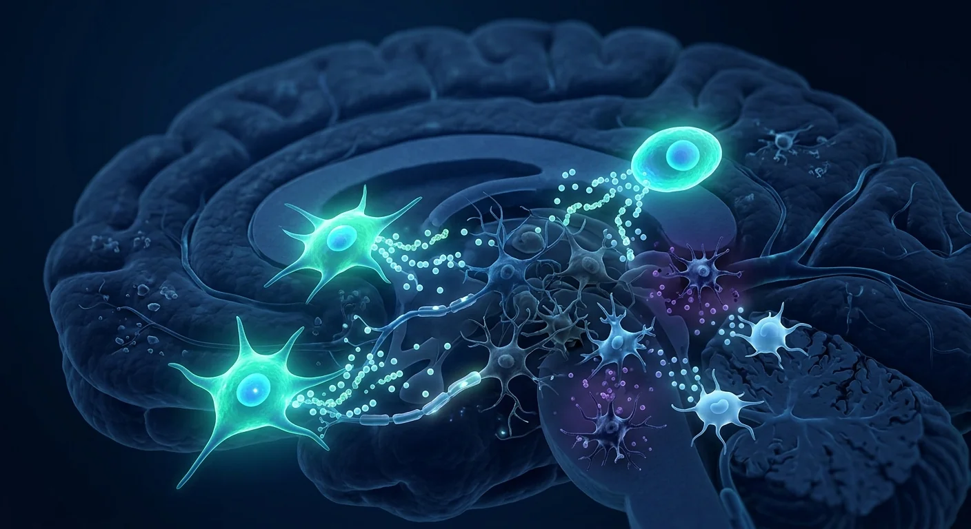
Cerebral palsy arises from injury to the developing brain — most commonly periventricular leukomalacia (white matter injury in preterm infants), hypoxic-ischemic encephalopathy, intraventricular hemorrhage, or perinatal stroke. The common thread is loss or dysfunction of oligodendrocytes, the cells responsible for producing myelin — the insulating sheath around nerve fibers that enables rapid, coordinated signal transmission. When myelin is damaged or underdeveloped, the motor signals traveling from the brain's motor cortex to the muscles are disrupted, resulting in the spasticity, dyskinesia, ataxia, and muscle weakness characteristic of CP.
Critically, the injury is not purely structural. There is a chronic neuroinflammatory component: activated microglia and astrocytes persist in the damaged brain regions, releasing pro-inflammatory cytokines (IL-1β, IL-6, TNF-α) and reactive oxygen species that perpetuate tissue damage and inhibit endogenous repair mechanisms long after the initial insult. This ongoing inflammation contributes to the plateau in function that many children with CP experience — and it is precisely this inflammatory environment that MSC therapy is designed to modulate.[1]
How MSC therapy works in the brain
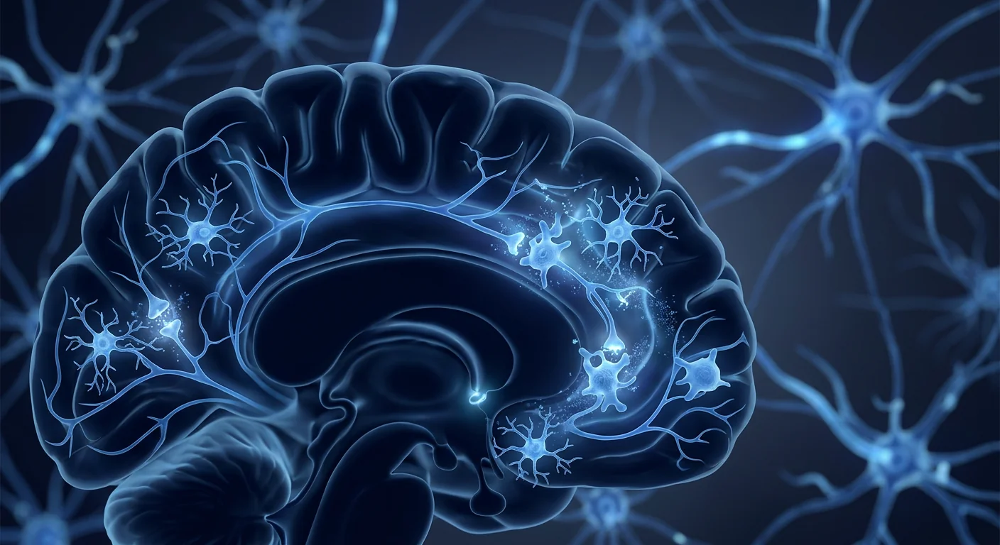
When clinical-grade Mesenchymal Stem Cells are delivered intravenously or intrathecally, they do not need to engraft permanently or replace neurons to exert therapeutic effects. Instead, they work through a coordinated set of paracrine mechanisms — releasing bioactive molecules that signal the brain's own repair systems:
1. Neuroinflammation modulation
MSCs respond to the inflammatory signals in the injured brain by secreting powerful anti-inflammatory mediators — TSG-6, PGE2, IL-10, and TGF-β. These molecules shift microglia from a pro-inflammatory (M1-like) to a neuroprotective (M2-like) phenotype, reducing the chronic inflammatory state that suppresses neural repair. This is arguably the most important mechanism by which MSCs improve function in CP.[2]
2. Neurotrophic support
MSCs secrete a rich cocktail of neurotrophic factors — BDNF (brain-derived neurotrophic factor), NGF (nerve growth factor), GDNF (glial-derived neurotrophic factor), and IGF-1. These molecules support the survival of existing neurons, promote synaptic plasticity, and create a permissive environment for neural circuit reorganization — the biological basis for functional improvement.[3]
3. Oligodendrocyte protection and myelination support
MSC-derived factors protect oligodendrocyte precursor cells from apoptosis and encourage their differentiation into mature, myelin-producing oligodendrocytes. In animal models of neonatal brain injury, MSC treatment has been shown to increase myelination and improve motor outcomes — a finding that has driven clinical interest in CP.[4]
4. Angiogenesis and perfusion
MSCs secrete VEGF and other angiogenic factors that support the formation of new blood vessels in damaged brain regions, improving oxygen and nutrient delivery to recovering neural tissue.
5. Anti-apoptotic signaling
MSC paracrine factors activate survival pathways (PI3K/Akt, MAPK/ERK) in threatened neurons and glial cells, reducing programmed cell death in the penumbra of the original injury.
What MSC therapy does NOT do
It is important to set honest expectations. MSC therapy does not cure cerebral palsy. It does not regenerate large areas of lost brain tissue or reverse fixed structural damage. What it does — based on a growing body of clinical evidence — is reduce neuroinflammation, support neural plasticity, improve motor function, reduce spasticity, and in many cases, enhance quality of life in ways that conventional therapies alone cannot achieve. The goal is functional improvement, not a cure — and the most meaningful outcomes are seen when MSC therapy is combined with intensive rehabilitation.[5]
What the clinical evidence says
The clinical evidence base for MSC therapy in cerebral palsy has grown substantially over the past decade, with multiple published trials — including randomized controlled studies — reporting measurable functional improvements:[6]
- Gross Motor Function: Multiple studies using the GMFM-66 (Gross Motor Function Measure) have reported statistically significant improvements in motor function scores at 3, 6, and 12 months post-treatment, with benefits sustained in longer follow-up periods.
- Spasticity Reduction: Reductions in the Modified Ashworth Scale — the standard clinical measure of spasticity — have been consistently reported across trials, often within 4–8 weeks of treatment.
- Neuroimaging Correlates: Some studies have reported increased fractional anisotropy on diffusion tensor imaging (DTI) — a measure of white matter tract integrity — following MSC treatment, suggesting structural improvement in myelination pathways.
- Functional Independence: Improvements in daily living activities, communication, and social interaction have been reported, particularly when MSC therapy is combined with structured rehabilitation programs.
It is important to note that while the overall direction of evidence is positive, individual responses vary significantly — influenced by age at treatment, type and severity of CP, MSC source and dose, delivery route, and the intensity of concurrent rehabilitation. The strongest outcomes are typically seen in younger children with greater baseline neuroplasticity.[7]

Who is a good candidate?
The evidence suggests that a broad range of CP patients may benefit from MSC therapy, but certain characteristics are associated with stronger outcomes:[8]
- Younger age (greater neuroplasticity — typically under 12 years, though adults have also shown benefit)
- GMFCS levels I–IV (patients at all functional levels have been studied; GMFCS V patients may benefit but expectations should be adjusted)
- Absence of uncontrolled seizure disorder
- Good nutritional status and general health
- Availability of and commitment to post-treatment rehabilitation
- Realistic expectations from family and caregivers
Patients with severe, fixed joint contractures, uncontrolled epilepsy, or progressive neurological conditions (which would suggest a diagnosis other than CP) require careful evaluation. A reputable clinic will conduct thorough neurological assessment, review brain imaging, and provide honest guidance about expected outcomes before proceeding.[9]
What the treatment process looks like
At a clinical-grade centre, a typical CP-focused MSC therapy journey follows a structured, multidisciplinary path:
Step 1: Comprehensive assessment
Neurological examination, review of brain MRI, GMFCS classification, functional assessment (typically GMFM-66), spasticity evaluation (Modified Ashworth Scale), and review of medical history, including seizure control and current medications.
Step 2: Protocol design
The clinical team determines cell dose (typically weight-based), delivery route (IV infusion, intrathecal injection, or combined), session frequency (single vs. multiple sessions over 3–6 months), and the rehabilitation plan that will follow each treatment session.
Step 3: Treatment delivery
IV infusion is the most common route — typically 60–90 minutes, outpatient, with monitoring during and after administration. Intrathecal delivery may be used in specific cases for more direct CNS access. Sessions are generally well-tolerated, with most children returning to normal activities within 24 hours.
Step 4: Intensive rehabilitation
This is a critical component. The neuroplasticity window following MSC treatment is optimized by structured physical therapy, occupational therapy, and where appropriate, speech therapy. The strongest outcomes come when MSC therapy and rehabilitation are tightly integrated — not delivered sequentially, but as a combined protocol.
Step 5: Outcome monitoring
Follow-up at 1, 3, 6, and 12 months tracks motor function (GMFM-66), spasticity (Modified Ashworth), functional independence (WeeFIM or PEDI), and in some cases, repeat neuroimaging. Maintenance sessions may be recommended based on individual response patterns.
Realistic timelines: what to expect when
Cellular therapy for a neurodevelopmental condition follows a different trajectory than for orthopedic or inflammatory conditions. The brain's repair processes are slower but potentially more transformative:[10]
It is essential to understand that progress in CP is typically incremental — not dramatic overnight transformations, but meaningful, cumulative gains in motor control, spasticity reduction, and functional independence. The most important predictor of outcome is the combination of quality MSC therapy with consistent, intensive rehabilitation. Families who commit to both see the strongest results.[11]
Safety and what every family should know
When delivered with clinical-grade cells, appropriate dosing, and medical supervision, MSC therapy has demonstrated a strong safety profile in pediatric populations. The most common side effects are transient: mild fever (typically within 24 hours of infusion), temporary fatigue, or mild headache — all self-limiting. Serious adverse events are rare in published pediatric CP trials.[12]
The greatest preventable risk in this field is unregulated providers using non-clinical-grade cells with unverified sterility, identity, or potency — particularly concerning in pediatric patients. Families should always confirm: MSC source (Wharton's jelly, bone marrow, or adipose), laboratory certifications (GMP, ISO), cell characterization (ISCT criteria: ≥95% CD73/CD90/CD105 positive), viability (>95% at delivery), and sterility testing. A Certificate of Analysis for the specific dose being administered should be available upon request.[13]
For families living with cerebral palsy, MSC therapy is not about unrealistic promises — it is about expanding the window of neuroplasticity and giving rehabilitation a biological partner. The gains are measured in small, meaningful increments that, over time, can change the trajectory of a child's development.
— VELAR Clinical Team
The VELAR approach to cerebral palsy
CP protocols at VELAR Center begin with comprehensive neurological assessment, brain MRI review, GMFCS classification, and functional evaluation to confirm candidacy and personalize dosing. Each protocol uses clinical-grade Wharton's jelly–derived MSCs (≥95% MSC marker expression, >95% viability at delivery), delivered via IV infusion with intrathecal options where clinically indicated. Every session is physician-led, paired with structured rehabilitation, and monitored across the 1, 3, 6, and 12-month milestones. The VELAR team works closely with the child's existing therapy team to ensure continuity of care and integrated rehabilitation.[14]
If you are considering regenerative therapy for a child with cerebral palsy, the most important first step is an honest, thorough assessment of candidacy — and a clear conversation about what realistic outcomes look like for your child's specific type and severity of CP.
References
- Graham HK, Rosenbaum P, Paneth N, et al. Cerebral palsy. Nature Reviews Disease Primers. 2016;2:15082. doi:10.1038/nrdp.2015.82 ↩
- Ahn SY, Chang YS, Sung DK, et al. Mesenchymal stem cells prevent hydrocephalus after severe intraventricular hemorrhage. Stroke. 2013;44(2):497-504. doi:10.1161/STROKEAHA.112.679092 ↩
- Park WS, Ahn SY, Sung SI, Ahn JY, Chang YS. Strategies to enhance paracrine potency of transplanted mesenchymal stem cells in intractable neonatal disorders. Pediatric Research. 2018;83(1-2):214-222. doi:10.1038/pr.2017.249 ↩
- van Velthoven CT, Kavelaars A, van Bel F, Heijnen CJ. Mesenchymal stem cell treatment after neonatal hypoxic-ischemic brain injury improves behavioral outcome and induces neuronal and oligodendrocyte regeneration. Brain, Behavior, and Immunity. 2010;24(3):387-393. doi:10.1016/j.bbi.2009.10.017 ↩
- Novak I, Walker K, Hunt RW, Wallace EM, Fahey M, Badawi N. Concise review: stem cell interventions for people with cerebral palsy: systematic review with meta-analysis. Stem Cells Translational Medicine. 2016;5(8):1014-1025. doi:10.5966/sctm.2015-0372 ↩
- Min K, Song J, Kang JY, et al. Umbilical cord blood therapy potentiated by erythropoietin for children with cerebral palsy: a double-blind, randomized, placebo-controlled trial. Stem Cells. 2013;31(3):581-591. doi:10.1002/stem.1304 ↩
- Sun JM, Song AW, Case LE, et al. Effect of autologous cord blood infusion on motor function and brain connectivity in young children with cerebral palsy: a randomized, placebo-controlled trial. Stem Cells Translational Medicine. 2017;6(12):2071-2078. doi:10.1002/sctm.17-0102 ↩
- Rah WJ, Lee YH, Moon JH, et al. Neuroregenerative potential of intravenous G-CSF and autologous peripheral blood mononuclear cells in children with cerebral palsy. Journal of Translational Medicine. 2017;15(1):28. doi:10.1186/s12967-017-1128-5 ↩
- Feng M, Lu A, Gao H, et al. Safety of allogeneic umbilical cord blood stem cells therapy in patients with severe cerebral palsy: a retrospective study. Stem Cells International. 2015;2015:325652. doi:10.1155/2015/325652 ↩
- Jensen A, Hamelmann E. First autologous cell therapy of cerebral palsy caused by hypoxic-ischemic brain damage in a child after cardiac arrest — individual treatment with cord blood. Case Reports in Transplantation. 2013;2013:951827. doi:10.1155/2013/951827 ↩
- Boruczkowski D, Zdolinska-Malinowska I. Wharton's jelly mesenchymal stem cell administration improves quality of life and self-sufficiency in children with cerebral palsy: results from a retrospective study. Stem Cells International. 2019;2019:7402151. doi:10.1155/2019/7402151 ↩
- Lv ZY, Li Y, Liu J. Progress in clinical trials of stem cell therapy for cerebral palsy. Neural Regeneration Research. 2021;16(7):1377-1382. doi:10.4103/1673-5374.300979 ↩
- Dominici M, Le Blanc K, Mueller I, et al. Minimal criteria for defining multipotent mesenchymal stromal cells. The International Society for Cellular Therapy position statement. Cytotherapy. 2006;8(4):315-317. doi:10.1080/14653240600855905 ↩
- Gu J, Huang L, Zhang C, et al. Therapeutic evidence of umbilical cord-derived mesenchymal stem cell transplantation for cerebral palsy: a randomized, controlled trial. Stem Cell Research & Therapy. 2020;11(1):43. doi:10.1186/s13287-019-1545-x ↩
脑瘫(Cerebral Palsy,CP)是儿童期最常见的运动障碍,全球约每1,000例活产中有2–3例。它不是单一疾病,而是一组由发育中大脑的非进行性损伤引起的永久性运动与姿势障碍——发生于出生前、出生时或出生后不久。数十年来,治疗领域主要是支持性的:物理治疗、作业治疗、矫形器、抗痉挛药物以及某些情况下的骨科或神经外科干预。这些方法对于管理症状和最大化功能至关重要,但它们并未解决潜在的脑损伤。间充质干细胞疗法代表了一个新的前沿——旨在通过针对驱动CP残疾的生物过程,支持神经修复、减少神经炎症并改善功能预后。
了解脑瘫中大脑正在发生什么
脑瘫源于发育中大脑的损伤——最常见的是脑室周围白质软化(早产儿的白质损伤)、缺氧缺血性脑病、脑室内出血或围产期卒中。共同点是少突胶质细胞的丧失或功能障碍——这些细胞负责产生髓鞘,即神经纤维周围的绝缘鞘,使快速协调的信号传输成为可能。当髓鞘受损或发育不良时,从大脑运动皮层传向肌肉的运动信号被中断,导致CP特征性的痉挛、运动障碍、共济失调和肌肉无力。
关键的是,损伤不仅仅是结构性的。还存在慢性神经炎症成分:活化的微胶质细胞和星形胶质细胞持续存在于受损脑区,释放促炎细胞因子(IL-1β、IL-6、TNF-α)和活性氧,在初始损伤后很久仍延续组织损伤并抑制内源性修复机制。这种持续的炎症导致许多CP儿童经历的功能平台期——而MSC疗法正是设计来调节这种炎症环境的。[1]
MSC疗法如何在大脑中运作
当临床级间充质干细胞通过静脉或鞘内递送时,它们不需要永久植入或替换神经元即可发挥治疗效果。相反,它们通过一套协调的旁分泌机制运作——释放生物活性分子,信号激活大脑自身的修复系统:
1. 神经炎症调节
MSC通过分泌强效抗炎介质——TSG-6、PGE2、IL-10和TGF-β——对受损大脑中的炎症信号做出反应。这些分子将微胶质细胞从促炎(M1样)表型转变为神经保护(M2样)表型,减少抑制神经修复的慢性炎症状态。这可以说是MSC改善CP功能的最重要机制。[2]
2. 神经营养支持
MSC分泌丰富的神经营养因子混合物——BDNF(脑源性神经营养因子)、NGF(神经生长因子)、GDNF(胶质源性神经营养因子)和IGF-1。这些分子支持现有神经元的存活,促进突触可塑性,并为神经回路重组创造有利环境——这是功能改善的生物学基础。[3]
3. 少突胶质细胞保护与髓鞘化支持
MSC衍生因子保护少突胶质前体细胞免于凋亡,并促进其分化为成熟、产生髓鞘的少突胶质细胞。在新生儿脑损伤动物模型中,MSC治疗已被证明可增加髓鞘化并改善运动预后——这一发现推动了CP领域的临床兴趣。[4]
4. 血管生成与灌注
MSC分泌VEGF和其他血管生成因子,支持受损脑区新血管的形成,改善对恢复中神经组织的氧气和营养输送。
5. 抗凋亡信号
MSC旁分泌因子激活受威胁神经元和胶质细胞中的存活通路(PI3K/Akt、MAPK/ERK),减少原始损伤周围半暗带的程序性细胞死亡。
MSC疗法所不能做的事
设定诚实的预期至关重要。MSC疗法不能治愈脑瘫。它不能再生大面积丧失的脑组织或逆转固定的结构损伤。它能做的——基于日益增长的临床证据——是减少神经炎症、支持神经可塑性、改善运动功能、减少痉挛,并在许多情况下以传统疗法无法做到的方式提高生活质量。目标是功能改善,而不是治愈——并且当MSC疗法与强化康复相结合时,能看到最有意义的疗效。[5]
临床证据说明了什么
过去十年中,MSC疗法治疗脑瘫的临床证据基础大幅增长,多项已发表的试验——包括随机对照研究——报告了可量测的功能改善:[6]
- 粗大运动功能:使用GMFM-66(粗大运动功能测量)的多项研究报告治疗后3、6和12个月的运动功能评分有统计学显著改善,并在更长的随访期持续受益。
- 痉挛减轻:改良Ashworth量表——标准的痉挛临床测量——在试验中一致报告减少,通常在治疗后4–8周内。
- 神经影像关联:一些研究报告MSC治疗后扩散张量成像(DTI)的各向异性分数增加——白质束完整性的测量——表明髓鞘通路的改善。
- 功能独立性:日常生活活动、沟通和社交互动的改善已被报告,特别是当MSC疗法与结构化康复项目相结合时。
需要注意的是,虽然整体证据方向是积极的,但个体反应差异显著——受治疗年龄、CP类型与严重程度、MSC来源和剂量、递送途径以及同期康复强度的影响。最强的疗效通常见于具有更大基线神经可塑性的年幼儿童。[7]
谁是合适的候选者?
证据表明,广泛的CP患者可能从MSC疗法中受益,但某些特征与更强的疗效相关:[8]
- 较年幼(更大的神经可塑性——通常12岁以下,但成人也显示获益)
- GMFCS I–IV级(所有功能水平的患者均已被研究;GMFCS V级患者可能受益但应调整预期)
- 无未控制的癫痫发作障碍
- 良好的营养状况和总体健康
- 治疗后康复的可及性和承诺
- 家庭和照顾者的现实预期
具有严重固定关节挛缩、未控制癫痫或进行性神经系统疾病(可能提示CP以外的诊断)的患者需要仔细评估。信誉良好的诊所将进行全面的神经系统评估,审阅脑部影像,并在进行前提供诚实的预期疗效指导。[9]
治疗过程是什么样子
在临床级中心,典型的CP导向MSC疗法旅程遵循结构化、多学科的路径:
第一步:全面评估
神经系统检查、脑部MRI审阅、GMFCS分类、功能评估(通常为GMFM-66)、痉挛评估(改良Ashworth量表),以及病史回顾,包括癫痫控制和当前用药。
第二步:方案设计
临床团队确定细胞剂量(通常基于体重)、递送途径(IV输注、鞘内注射或联合)、会话频率(单次与会话系列持续3–6个月),以及每次治疗会话后续的康复计划。
第三步:治疗递送
IV输注是最常见的途径——通常60–90分钟,门诊,给药期间和之后进行监测。鞘内递送可在特定情况下用于更直接的中枢神经系统接入。会话通常耐受良好,大多数儿童在24小时内恢复正常活动。
第四步:强化康复
这是关键组成部分。MSC治疗后神经可塑性窗口通过结构化物理治疗、作业治疗以及适当时的言语治疗得到优化。最强的疗效来自MSC疗法与康复的紧密结合——不是顺序递送,而是作为一个组合方案。
第五步:疗效监测
1、3、6和12个月的随访追踪运动功能(GMFM-66)、痉挛(改良Ashworth)、功能独立(WeeFIM或PEDI),以及在部分病例中重复神经影像。可根据个体反应模式推荐维持会话。
实际时间表:何时期待什么
神经发育状况的细胞疗法遵循与骨科或炎症状况不同的轨迹。大脑的修复过程较慢但可能更具转化性:[10]
必须理解的是,CP的进展通常是渐进性的——不是戏剧性的一夜转变,而是在运动控制、痉挛减轻和功能独立方面有意义的累积收获。疗效的最重要预测因素是将优质MSC疗法与持续强化康复相结合。同时承诺两者的家庭看到最强的效果。[11]
安全性与每个家庭应知道的
当以临床级细胞、适当剂量和医疗监督递送时,MSC疗法在儿科人群中展示了强大的安全特性。最常见的副作用是暂时的:轻度发热(通常在输注后24小时内)、暂时疲劳或轻度头痛——均为自限性。已发表的儿科CP试验中严重不良事件少见。[12]
该领域最大的可预防风险是未受监管的提供者使用未经验证无菌性、身份或效力的非临床级细胞——在儿科患者中尤为令人担忧。家庭应始终确认:MSC来源(华通氏胶、骨髓或脂肪)、实验室认证(GMP、ISO)、细胞特征(ISCT标准:≥95% CD73/CD90/CD105阳性)、活性(交付时>95%)和无菌测试。所给予具体剂量的分析证书应可应要求提供。[13]
对于与脑瘫共同生活的家庭来说,MSC疗法不是关于不切实际的承诺——而是关于扩大神经可塑性的窗口,为康复提供一个生物学伙伴。收获是以小的、有意义的增量来衡量的,这些增量随着时间的推移可以改变儿童发育的轨迹。
——VELAR 临床团队
VELAR对脑瘫的治疗方法
VELAR Center的CP方案从全面的神经系统评估、脑部MRI审阅、GMFCS分类和功能评估开始,以确认候选资格并个性化剂量。每个方案使用临床级华通氏胶来源MSC(MSC标志物表达≥95%、交付时活性>95%),通过IV输注递送,并在临床指示时有鞘内选项。每次会话由医生主导,配合结构化康复,并在1、3、6和12个月里程碑进行监测。VELAR团队与儿童现有治疗团队紧密合作,确保护理的连续性和康复的整合。[14]
如果您正在考虑为脑瘫儿童进行再生疗法,最重要的第一步是对候选资格的诚实、全面评估——以及对您孩子特定CP类型和严重程度的实际疗效的清晰对话。
参考文献
- Graham HK, Rosenbaum P, Paneth N, et al. Cerebral palsy. Nature Reviews Disease Primers. 2016;2:15082. doi:10.1038/nrdp.2015.82 ↩
- Ahn SY, Chang YS, Sung DK, et al. Mesenchymal stem cells prevent hydrocephalus after severe intraventricular hemorrhage. Stroke. 2013;44(2):497-504. doi:10.1161/STROKEAHA.112.679092 ↩
- Park WS, Ahn SY, Sung SI, Ahn JY, Chang YS. Strategies to enhance paracrine potency of transplanted mesenchymal stem cells in intractable neonatal disorders. Pediatric Research. 2018;83(1-2):214-222. doi:10.1038/pr.2017.249 ↩
- van Velthoven CT, Kavelaars A, van Bel F, Heijnen CJ. Mesenchymal stem cell treatment after neonatal hypoxic-ischemic brain injury improves behavioral outcome and induces neuronal and oligodendrocyte regeneration. Brain, Behavior, and Immunity. 2010;24(3):387-393. doi:10.1016/j.bbi.2009.10.017 ↩
- Novak I, Walker K, Hunt RW, Wallace EM, Fahey M, Badawi N. Concise review: stem cell interventions for people with cerebral palsy: systematic review with meta-analysis. Stem Cells Translational Medicine. 2016;5(8):1014-1025. doi:10.5966/sctm.2015-0372 ↩
- Min K, Song J, Kang JY, et al. Umbilical cord blood therapy potentiated by erythropoietin for children with cerebral palsy: a double-blind, randomized, placebo-controlled trial. Stem Cells. 2013;31(3):581-591. doi:10.1002/stem.1304 ↩
- Sun JM, Song AW, Case LE, et al. Effect of autologous cord blood infusion on motor function and brain connectivity in young children with cerebral palsy: a randomized, placebo-controlled trial. Stem Cells Translational Medicine. 2017;6(12):2071-2078. doi:10.1002/sctm.17-0102 ↩
- Rah WJ, Lee YH, Moon JH, et al. Neuroregenerative potential of intravenous G-CSF and autologous peripheral blood mononuclear cells in children with cerebral palsy. Journal of Translational Medicine. 2017;15(1):28. doi:10.1186/s12967-017-1128-5 ↩
- Feng M, Lu A, Gao H, et al. Safety of allogeneic umbilical cord blood stem cells therapy in patients with severe cerebral palsy: a retrospective study. Stem Cells International. 2015;2015:325652. doi:10.1155/2015/325652 ↩
- Jensen A, Hamelmann E. First autologous cell therapy of cerebral palsy caused by hypoxic-ischemic brain damage in a child after cardiac arrest — individual treatment with cord blood. Case Reports in Transplantation. 2013;2013:951827. doi:10.1155/2013/951827 ↩
- Boruczkowski D, Zdolinska-Malinowska I. Wharton's jelly mesenchymal stem cell administration improves quality of life and self-sufficiency in children with cerebral palsy: results from a retrospective study. Stem Cells International. 2019;2019:7402151. doi:10.1155/2019/7402151 ↩
- Lv ZY, Li Y, Liu J. Progress in clinical trials of stem cell therapy for cerebral palsy. Neural Regeneration Research. 2021;16(7):1377-1382. doi:10.4103/1673-5374.300979 ↩
- Dominici M, Le Blanc K, Mueller I, et al. Minimal criteria for defining multipotent mesenchymal stromal cells. The International Society for Cellular Therapy position statement. Cytotherapy. 2006;8(4):315-317. doi:10.1080/14653240600855905 ↩
- Gu J, Huang L, Zhang C, et al. Therapeutic evidence of umbilical cord-derived mesenchymal stem cell transplantation for cerebral palsy: a randomized, controlled trial. Stem Cell Research & Therapy. 2020;11(1):43. doi:10.1186/s13287-019-1545-x ↩
الشلل الدماغي (CP) هو أكثر إعاقات الحركة شيوعاً في الطفولة، حيث يصيب حوالي 2–3 من كل 1,000 ولادة حية حول العالم. إنه ليس مرضاً واحداً بل مجموعة من الاضطرابات الدائمة في الحركة والوضعية ناتجة عن ضرر غير متقدم في الدماغ النامي — يحدث قبل الولادة أو أثناءها أو بعدها بفترة قصيرة. لعقود، كان المشهد العلاجي داعماً إلى حد كبير: العلاج الطبيعي، العلاج الوظيفي، الأجهزة التقويمية، أدوية مضادة للتشنج، وفي بعض الحالات، تدخل جراحي عظمي أو عصبي. هذه الأساليب أساسية لإدارة الأعراض وتعظيم الوظيفة، لكنها لا تعالج إصابة الدماغ الأساسية. يمثل العلاج بالخلايا الجذعية الوسيطة حدوداً جديدة — تهدف إلى دعم الإصلاح العصبي، تقليل الالتهاب العصبي، وتحسين النتائج الوظيفية من خلال استهداف العمليات البيولوجية التي تدفع الإعاقة في الشلل الدماغي.
فهم ما يحدث في الدماغ في الشلل الدماغي
ينشأ الشلل الدماغي من إصابة في الدماغ النامي — الأكثر شيوعاً تلين المادة البيضاء حول البطينات (إصابة المادة البيضاء في الخدج)، الاعتلال الدماغي بنقص التأكسج الإقفاري، النزف داخل البطينات، أو السكتة الدماغية حول الولادة. القاسم المشترك هو فقدان أو خلل في الخلايا قليلة التغصن، الخلايا المسؤولة عن إنتاج المايلين — الغمد العازل حول الألياف العصبية الذي يمكّن من نقل الإشارات السريع والمنسق. عندما يتلف المايلين أو يكون غير مكتمل النمو، تتعطل الإشارات الحركية المسافرة من القشرة الحركية للدماغ إلى العضلات، مما ينتج عنه التشنج وخلل الحركة والرنح وضعف العضلات المميزة للشلل الدماغي.
بشكل حاسم، الإصابة ليست هيكلية بحتة. هناك مكون التهابي عصبي مزمن: تستمر الخلايا الدبقية الصغيرة والخلايا النجمية المنشطة في مناطق الدماغ المتضررة، مطلقةً سيتوكينات التهابية (IL-1β وIL-6 وTNF-α) وأنواع الأكسجين التفاعلية التي تديم الضرر النسيجي وتثبط آليات الإصلاح الذاتية بعد مدة طويلة من الإصابة الأولية. يساهم هذا الالتهاب المستمر في استقرار الوظيفة الذي يختبره العديد من الأطفال المصابين بالشلل الدماغي — وهذه البيئة الالتهابية بالتحديد هي ما صُمم علاج MSC لتعديله.[1]
كيف يعمل علاج MSC في الدماغ
عندما تُسلَّم الخلايا الجذعية الوسيطة ذات الجودة السريرية عن طريق الوريد أو داخل القراب، لا تحتاج إلى الانغراس بشكل دائم أو استبدال الخلايا العصبية لممارسة التأثيرات العلاجية. بدلاً من ذلك، تعمل من خلال مجموعة منسقة من آليات التأثير المجاور للإفراز — مطلقةً جزيئات نشطة بيولوجياً تُشير إلى أنظمة الإصلاح الذاتية في الدماغ:
1. تعديل الالتهاب العصبي
تستجيب MSC لإشارات الالتهاب في الدماغ المصاب بإفراز وسطاء أقوياء مضادين للالتهاب — TSG-6 وPGE2 وIL-10 وTGF-β. تنقل هذه الجزيئات الخلايا الدبقية الصغيرة من نمط التهابي (شبيه بـ M1) إلى نمط واقٍ للأعصاب (شبيه بـ M2)، مما يقلل من حالة الالتهاب المزمنة التي تثبط الإصلاح العصبي. يمكن القول إن هذه هي الآلية الأكثر أهمية التي تحسن بها MSC الوظيفة في الشلل الدماغي.[2]
2. الدعم العصبي التغذوي
تفرز MSC مزيجاً غنياً من العوامل العصبية التغذوية — BDNF (عامل التغذية العصبية المشتق من الدماغ)، NGF (عامل نمو الأعصاب)، GDNF (عامل التغذية العصبية المشتق من الخلايا الدبقية)، وIGF-1. تدعم هذه الجزيئات بقاء الخلايا العصبية الموجودة، وتعزز اللدونة المشبكية، وتخلق بيئة مسموحة لإعادة تنظيم الدوائر العصبية — الأساس البيولوجي للتحسن الوظيفي.[3]
3. حماية الخلايا قليلة التغصن ودعم الميالين
تحمي العوامل المشتقة من MSC الخلايا السليفة قليلة التغصن من الاستماتة وتشجع تمايزها إلى خلايا قليلة التغصن ناضجة منتجة للمايلين. في نماذج حيوانية لإصابة الدماغ الوليدية، أظهر علاج MSC زيادة في الميالين وتحسين النتائج الحركية — وهي نتيجة دفعت الاهتمام السريري في الشلل الدماغي.[4]
4. تولد الأوعية والتروية
تفرز MSC عامل VEGF وعوامل مولدة للأوعية أخرى تدعم تشكيل أوعية دموية جديدة في مناطق الدماغ المتضررة، محسنةً توصيل الأكسجين والمغذيات إلى النسيج العصبي المتعافي.
5. الإشارات المضادة للاستماتة
تنشط عوامل MSC المجاورة للإفراز مسارات البقاء (PI3K/Akt وMAPK/ERK) في الخلايا العصبية والدبقية المهددة، مما يقلل من موت الخلايا المبرمج في منطقة شبه الظل للإصابة الأصلية.
ما لا يفعله علاج MSC
من المهم وضع توقعات صادقة. علاج MSC لا يعالج الشلل الدماغي. لا يُعيد توليد مناطق كبيرة من نسيج الدماغ المفقود أو يعكس الضرر الهيكلي الثابت. ما يفعله — استناداً إلى مجموعة متزايدة من الأدلة السريرية — هو تقليل الالتهاب العصبي، دعم اللدونة العصبية، تحسين الوظيفة الحركية، تقليل التشنج، وفي كثير من الحالات، تحسين جودة الحياة بطرق لا تستطيع العلاجات التقليدية وحدها تحقيقها. الهدف هو التحسن الوظيفي، وليس الشفاء — وتُرى النتائج الأكثر معنىً عندما يُدمج علاج MSC مع إعادة التأهيل المكثف.[5]
ماذا تقول الأدلة السريرية
نمت قاعدة الأدلة السريرية لعلاج MSC في الشلل الدماغي بشكل كبير خلال العقد الماضي، مع تجارب منشورة متعددة — بما في ذلك دراسات معشاة مضبوطة — أبلغت عن تحسنات وظيفية قابلة للقياس:[6]
- الوظيفة الحركية الكبرى: دراسات متعددة باستخدام مقياس GMFM-66 (مقياس الوظيفة الحركية الكبرى) أبلغت عن تحسنات ذات دلالة إحصائية في درجات الوظيفة الحركية عند 3 و6 و12 شهراً بعد العلاج، مع فوائد مستدامة في فترات متابعة أطول.
- تقليل التشنج: تم الإبلاغ عن انخفاضات في مقياس Ashworth المعدل — المقياس السريري المعياري للتشنج — بشكل متسق عبر التجارب، غالباً خلال 4–8 أسابيع من العلاج.
- الارتباطات التصويرية العصبية: أبلغت بعض الدراسات عن زيادة في التباين الجزئي في تصوير الموتر الانتشار (DTI) — مقياس لسلامة مسالك المادة البيضاء — بعد علاج MSC، مما يشير إلى تحسن هيكلي في مسارات الميالين.
- الاستقلال الوظيفي: تم الإبلاغ عن تحسنات في أنشطة الحياة اليومية والتواصل والتفاعل الاجتماعي، خاصة عندما يُدمج علاج MSC مع برامج إعادة تأهيل منظمة.
من المهم ملاحظة أنه بينما الاتجاه العام للأدلة إيجابي، تختلف الاستجابات الفردية بشكل كبير — متأثرةً بالعمر عند العلاج، نوع وشدة الشلل الدماغي، مصدر وجرعة MSC، طريقة التوصيل، وشدة إعادة التأهيل المتزامنة. تُرى أقوى النتائج عادةً في الأطفال الأصغر سناً ذوي اللدونة العصبية الأساسية الأكبر.[7]
من هو المرشح المناسب؟
تشير الأدلة إلى أن مجموعة واسعة من مرضى الشلل الدماغي قد يستفيدون من علاج MSC، لكن بعض الخصائص مرتبطة بنتائج أقوى:[8]
- العمر الأصغر (لدونة عصبية أكبر — عادةً تحت 12 سنة، لكن البالغين أظهروا فائدة أيضاً)
- مستويات GMFCS I–IV (تمت دراسة المرضى في جميع المستويات الوظيفية؛ قد يستفيد مرضى GMFCS V لكن يجب تعديل التوقعات)
- غياب اضطراب النوبات غير المسيطر عليه
- حالة تغذوية جيدة وصحة عامة
- توفر والتزام بإعادة التأهيل بعد العلاج
- توقعات واقعية من الأسرة ومقدمي الرعاية
المرضى الذين يعانون من تقلصات مفصلية ثابتة شديدة، صرع غير مسيطر عليه، أو حالات عصبية تقدمية (مما قد يشير إلى تشخيص غير الشلل الدماغي) يحتاجون إلى تقييم دقيق. ستجري العيادة ذات السمعة الجيدة تقييماً عصبياً شاملاً، ومراجعة تصوير الدماغ، وتقديم توجيه صادق حول النتائج المتوقعة قبل المتابعة.[9]
كيف تبدو عملية العلاج
في مركز ذي جودة سريرية، تتبع رحلة علاج MSC النموذجية المتمحورة حول الشلل الدماغي مساراً منظماً ومتعدد التخصصات:
الخطوة 1: التقييم الشامل
الفحص العصبي، مراجعة تصوير الدماغ بالرنين المغناطيسي، تصنيف GMFCS، التقييم الوظيفي (عادةً GMFM-66)، تقييم التشنج (مقياس Ashworth المعدل)، ومراجعة التاريخ الطبي، بما في ذلك السيطرة على النوبات والأدوية الحالية.
الخطوة 2: تصميم البروتوكول
يحدد الفريق السريري جرعة الخلايا (عادةً بناءً على الوزن)، طريقة التوصيل (تسريب وريدي، حقن داخل القراب، أو مجتمع)، تكرار الجلسات (جلسة واحدة مقابل جلسات متعددة على مدى 3–6 أشهر)، وخطة إعادة التأهيل التي ستلي كل جلسة علاج.
الخطوة 3: تسليم العلاج
التسريب الوريدي هو الطريق الأكثر شيوعاً — عادة 60–90 دقيقة، خارجي، مع مراقبة أثناء وبعد الإعطاء. قد يُستخدم التوصيل داخل القراب في حالات محددة للوصول المباشر الأكبر للجهاز العصبي المركزي. الجلسات جيدة التحمل عموماً، ويعود معظم الأطفال إلى أنشطتهم الطبيعية خلال 24 ساعة.
الخطوة 4: إعادة التأهيل المكثف
هذا مكون حاسم. تُحسَّن نافذة اللدونة العصبية بعد علاج MSC من خلال العلاج الطبيعي المنظم، العلاج الوظيفي، وحيثما كان مناسباً، علاج النطق. تأتي أقوى النتائج عندما يُدمج علاج MSC وإعادة التأهيل بشكل محكم — ليس بالتسليم المتسلسل، بل كبروتوكول مشترك.
الخطوة 5: مراقبة النتائج
المتابعة في 1 و3 و6 و12 شهراً تتتبع الوظيفة الحركية (GMFM-66)، التشنج (Ashworth المعدل)، الاستقلال الوظيفي (WeeFIM أو PEDI)، وفي بعض الحالات، إعادة التصوير العصبي. قد تُوصى جلسات صيانة بناءً على أنماط الاستجابة الفردية.
جداول زمنية واقعية: ماذا تتوقع ومتى
يتبع العلاج الخلوي لحالة نمائية عصبية مساراً مختلفاً عن الحالات العظمية أو الالتهابية. عمليات إصلاح الدماغ أبطأ لكنها قد تكون أكثر تحويلاً:[10]
من الضروري أن نفهم أن التقدم في الشلل الدماغي يكون عادةً تدريجياً — ليس تحولات دراماتيكية بين ليلة وضحاها، بل مكاسب تراكمية ذات معنى في التحكم الحركي، تقليل التشنج، والاستقلال الوظيفي. أهم مؤشر للناتج هو الجمع بين علاج MSC عالي الجودة وإعادة التأهيل المكثف المستمر. الأسر التي تلتزم بكليهما ترى أقوى النتائج.[11]
السلامة وما يجب أن تعرفه كل أسرة
عند التسليم بخلايا ذات جودة سريرية، جرعات مناسبة، وإشراف طبي، أظهر علاج MSC ملف أمان قوي في فئات الأطفال. الآثار الجانبية الأكثر شيوعاً عابرة: حمى خفيفة (عادةً خلال 24 ساعة من التسريب)، تعب مؤقت، أو صداع خفيف — كلها محددة ذاتياً. الأحداث السلبية الخطيرة نادرة في تجارب الشلل الدماغي المنشورة لدى الأطفال.[12]
أكبر خطر يمكن منعه في هذا المجال هو المزودون غير المنظمين الذين يستخدمون خلايا غير سريرية الجودة بعقامة أو هوية أو فعالية غير متحقق منها — وهو أمر مقلق بشكل خاص في المرضى الأطفال. يجب على الأسر دائماً تأكيد: مصدر MSC (هلام وارتون، نخاع العظم، أو الدهون)، اعتمادات المختبر (GMP، ISO)، توصيف الخلايا (معايير ISCT: ≥95% إيجابية لـ CD73/CD90/CD105)، الحيوية (>95% عند التسليم)، واختبار العقامة. يجب أن تكون شهادة تحليل للجرعة المحددة المُعطاة متاحة عند الطلب.[13]
بالنسبة للأسر التي تعيش مع الشلل الدماغي، علاج MSC ليس حول وعود غير واقعية — بل حول توسيع نافذة اللدونة العصبية وإعطاء إعادة التأهيل شريكاً بيولوجياً. تُقاس المكاسب بزيادات صغيرة ذات معنى يمكنها، بمرور الوقت، أن تغير مسار نمو الطفل.
— فريق VELAR السريري
منهج VELAR للشلل الدماغي
تبدأ بروتوكولات الشلل الدماغي في VELAR Center بتقييم عصبي شامل، مراجعة تصوير الدماغ بالرنين المغناطيسي، تصنيف GMFCS، وتقييم وظيفي لتأكيد الأهلية وتخصيص الجرعات. يستخدم كل بروتوكول MSC ذات الجودة السريرية المشتقة من هلام وارتون (تعبير علامات MSC ≥95%، حيوية عند التسليم >95%)، تُسلَّم عبر التسريب الوريدي مع خيارات داخل القراب حيثما يُشار سريرياً. تُقاد كل جلسة من قبل طبيب، مقترنةً بإعادة تأهيل منظمة، ومُراقَبة عبر علامات 1 و3 و6 و12 شهراً. يعمل فريق VELAR عن كثب مع فريق العلاج الحالي للطفل لضمان استمرارية الرعاية وإعادة التأهيل المتكاملة.[14]
إذا كنت تفكر في العلاج التجديدي لطفل مصاب بالشلل الدماغي، فإن أهم خطوة أولى هي تقييم صادق وشامل للأهلية — ومحادثة واضحة حول كيف تبدو النتائج الواقعية لنوع وشدة الشلل الدماغي المحدد لطفلك.
المراجع
- Graham HK, Rosenbaum P, Paneth N, et al. Cerebral palsy. Nature Reviews Disease Primers. 2016;2:15082. doi:10.1038/nrdp.2015.82 ↩
- Ahn SY, Chang YS, Sung DK, et al. Mesenchymal stem cells prevent hydrocephalus after severe intraventricular hemorrhage. Stroke. 2013;44(2):497-504. doi:10.1161/STROKEAHA.112.679092 ↩
- Park WS, Ahn SY, Sung SI, Ahn JY, Chang YS. Strategies to enhance paracrine potency of transplanted mesenchymal stem cells in intractable neonatal disorders. Pediatric Research. 2018;83(1-2):214-222. doi:10.1038/pr.2017.249 ↩
- van Velthoven CT, Kavelaars A, van Bel F, Heijnen CJ. Mesenchymal stem cell treatment after neonatal hypoxic-ischemic brain injury improves behavioral outcome and induces neuronal and oligodendrocyte regeneration. Brain, Behavior, and Immunity. 2010;24(3):387-393. doi:10.1016/j.bbi.2009.10.017 ↩
- Novak I, Walker K, Hunt RW, Wallace EM, Fahey M, Badawi N. Concise review: stem cell interventions for people with cerebral palsy: systematic review with meta-analysis. Stem Cells Translational Medicine. 2016;5(8):1014-1025. doi:10.5966/sctm.2015-0372 ↩
- Min K, Song J, Kang JY, et al. Umbilical cord blood therapy potentiated by erythropoietin for children with cerebral palsy: a double-blind, randomized, placebo-controlled trial. Stem Cells. 2013;31(3):581-591. doi:10.1002/stem.1304 ↩
- Sun JM, Song AW, Case LE, et al. Effect of autologous cord blood infusion on motor function and brain connectivity in young children with cerebral palsy: a randomized, placebo-controlled trial. Stem Cells Translational Medicine. 2017;6(12):2071-2078. doi:10.1002/sctm.17-0102 ↩
- Rah WJ, Lee YH, Moon JH, et al. Neuroregenerative potential of intravenous G-CSF and autologous peripheral blood mononuclear cells in children with cerebral palsy. Journal of Translational Medicine. 2017;15(1):28. doi:10.1186/s12967-017-1128-5 ↩
- Feng M, Lu A, Gao H, et al. Safety of allogeneic umbilical cord blood stem cells therapy in patients with severe cerebral palsy: a retrospective study. Stem Cells International. 2015;2015:325652. doi:10.1155/2015/325652 ↩
- Jensen A, Hamelmann E. First autologous cell therapy of cerebral palsy caused by hypoxic-ischemic brain damage in a child after cardiac arrest — individual treatment with cord blood. Case Reports in Transplantation. 2013;2013:951827. doi:10.1155/2013/951827 ↩
- Boruczkowski D, Zdolinska-Malinowska I. Wharton's jelly mesenchymal stem cell administration improves quality of life and self-sufficiency in children with cerebral palsy: results from a retrospective study. Stem Cells International. 2019;2019:7402151. doi:10.1155/2019/7402151 ↩
- Lv ZY, Li Y, Liu J. Progress in clinical trials of stem cell therapy for cerebral palsy. Neural Regeneration Research. 2021;16(7):1377-1382. doi:10.4103/1673-5374.300979 ↩
- Dominici M, Le Blanc K, Mueller I, et al. Minimal criteria for defining multipotent mesenchymal stromal cells. The International Society for Cellular Therapy position statement. Cytotherapy. 2006;8(4):315-317. doi:10.1080/14653240600855905 ↩
- Gu J, Huang L, Zhang C, et al. Therapeutic evidence of umbilical cord-derived mesenchymal stem cell transplantation for cerebral palsy: a randomized, controlled trial. Stem Cell Research & Therapy. 2020;11(1):43. doi:10.1186/s13287-019-1545-x ↩