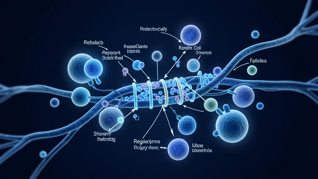
Optic neuritis is an inflammatory demyelinating condition of the optic nerve that can cause sudden, often frightening vision loss — typically in one eye, accompanied by pain on eye movement, reduced colour perception, and visual field defects. For roughly half of patients it is the first clinical episode of multiple sclerosis; for others it occurs in the context of neuromyelitis optica spectrum disorder (NMOSD), myelin oligodendrocyte glycoprotein antibody-associated disease (MOGAD), or as an isolated idiopathic event. Whatever the underlying driver, the shared mechanism is immune-mediated attack on the myelin sheath that insulates optic nerve axons — and if inflammation is severe or prolonged, axonal loss follows, leaving permanent visual deficit. [1][2]
Where conventional treatment falls short. High-dose intravenous corticosteroids remain the first-line acute therapy, and they do accelerate visual recovery in the short term. But the landmark Optic Neuritis Treatment Trial (ONTT) showed that steroids improve the speed of recovery — not the final visual outcome at six months or one year. They suppress inflammation but do not actively repair myelin or protect axons from degeneration. For patients with recurrent episodes or poor recovery, the gap between symptomatic control and tissue-level repair is real and consequential.
The deeper problem is neurodegenerative. Acute demyelination triggers a cascade: inflammatory cytokines, oxidative stress, and glutamate excitotoxicity damage oligodendrocytes — the cells that produce and maintain myelin — while also injuring retinal ganglion cells and their axons directly. Over time this axonal loss is what drives permanent visual impairment. A therapy that only quiets inflammation without supporting remyelination and neuroprotection leaves the underlying tissue damage unaddressed. [3][4]
MSC therapy targets the biology beneath the inflammation. Rather than simply suppressing the immune attack, mesenchymal stem cells are being studied for their capacity to simultaneously modulate autoimmunity, secrete neurotrophic factors that support retinal ganglion cell survival, and create a microenvironment conducive to remyelination by endogenous oligodendrocyte precursor cells. This three-pronged mechanism — immunomodulation, neuroprotection, and remyelination support — is what makes MSCs a conceptually compelling candidate for optic neuritis and other optic neuropathies. [5]
What Is Optic Neuritis — and Why the Optic Nerve Is Vulnerable
Optic neuritis is acute inflammation of the optic nerve. The optic nerve is not a true peripheral nerve — it is a white-matter tract of the central nervous system, myelinated by oligodendrocytes, and therefore susceptible to the same autoimmune demyelinating processes that affect the brain and spinal cord in multiple sclerosis. When autoreactive T-cells cross the blood–optic nerve barrier, they trigger a focal inflammatory lesion that strips myelin, slows or blocks signal conduction, and — if unchecked — leads to irreversible axonal degeneration. [6]
Clinically, the typical presentation is subacute monocular visual loss evolving over hours to days, with pain exacerbated by eye movement (92% of cases), reduced colour saturation, and a central or centrocecal scotoma. MRI typically shows T2 hyperintensity with gadolinium enhancement in the affected optic nerve. Optical coherence tomography (OCT) reveals thinning of the retinal nerve fibre layer (RNFL) over subsequent months — a direct measure of axonal loss that correlates with permanent visual deficit. The RNFL thinning that follows an acute episode is a stark reminder that inflammation leaves lasting structural damage, even when visual acuity recovers on a Snellen chart.
How MSCs Address Optic Nerve Pathology — the Three Mechanisms
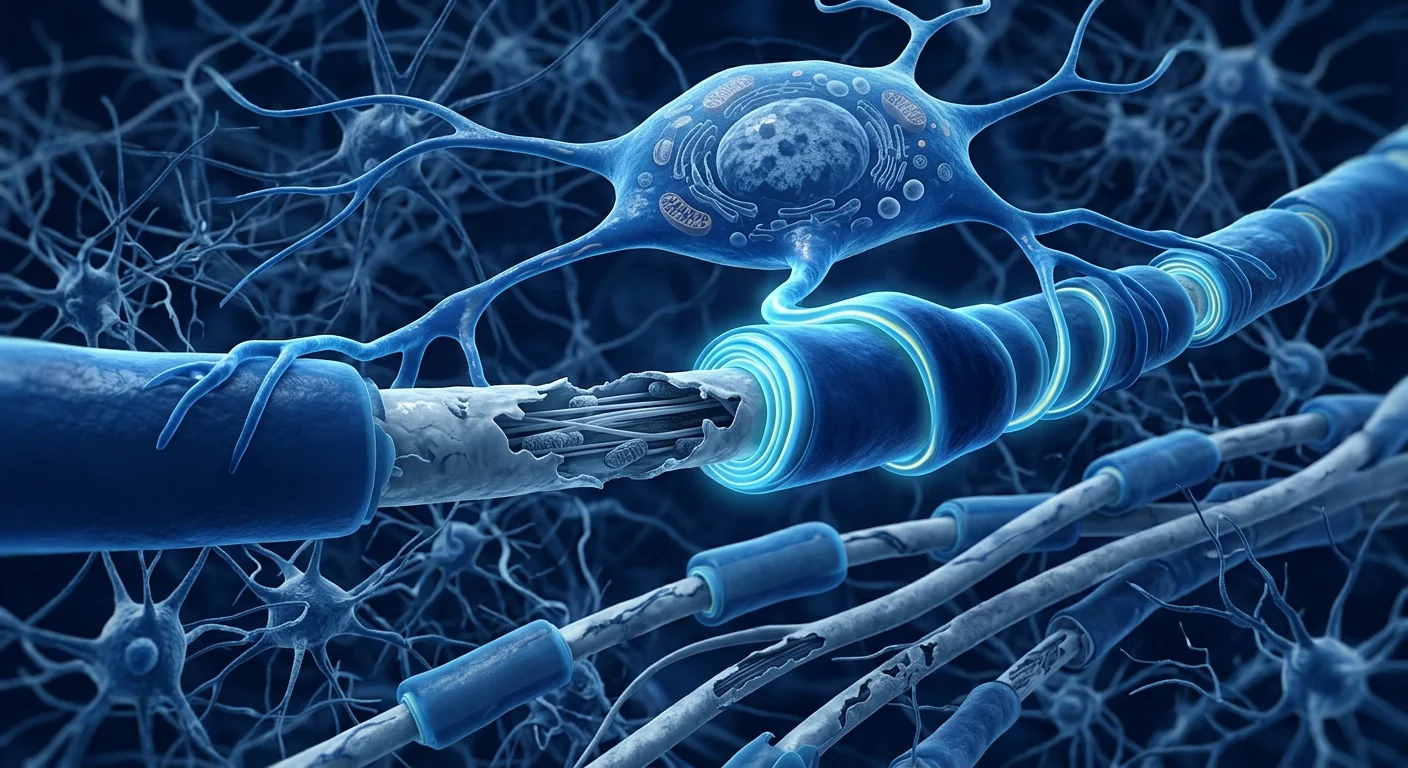
MSCs work through paracrine signalling, not cell replacement. When delivered intravenously or locally, MSCs home to sites of inflammation and injury, but they do not become new optic nerve cells or oligodendrocytes. Instead, they secrete a rich cocktail of bioactive molecules — cytokines, growth factors, extracellular vesicles, and microRNAs — that collectively shift the local environment from pro-inflammatory and degenerative toward anti-inflammatory and regenerative. Three distinct mechanisms are relevant to optic neuritis.
1. Immunomodulation — Calming the Autoimmune Attack
MSCs suppress the autoreactive T-cell and B-cell responses that drive optic nerve demyelination. They do this through multiple pathways: secretion of prostaglandin E2 (PGE2), indoleamine 2,3-dioxygenase (IDO), transforming growth factor-beta (TGF-β), and interleukin-10 (IL-10); induction of regulatory T-cells (Tregs); inhibition of Th1 and Th17 effector responses; and polarization of macrophages from the pro-inflammatory M1 phenotype to the anti-inflammatory M2 phenotype. In the context of optic neuritis, this means dampening the immune attack at its source — directly addressing the mechanism that strips myelin from the nerve. [7][8]
2. Neuroprotection — Saving Retinal Ganglion Cells and Axons
MSCs secrete a panel of neurotrophic factors — brain-derived neurotrophic factor (BDNF), ciliary neurotrophic factor (CNTF), nerve growth factor (NGF), and glial cell line-derived neurotrophic factor (GDNF) among them — that directly support the survival of retinal ganglion cells and their axons under inflammatory and oxidative stress. These factors activate pro-survival signalling cascades (PI3K/Akt, MAPK/ERK) within neurons, reduce caspase-3-mediated apoptosis, and upregulate anti-oxidant defences. In animal models of optic nerve injury, intravitreal or systemic MSC administration consistently reduces retinal ganglion cell loss and preserves RNFL thickness — outcomes that translate to measurable preservation of visual function. [9][10]
3. Remyelination Support — Rebuilding the Insulation
Perhaps the most therapeutically attractive mechanism is the ability of MSCs to promote remyelination. MSC-derived factors — especially hepatocyte growth factor (HGF), insulin-like growth factor-1 (IGF-1), and exosomes carrying pro-myelinating microRNAs — stimulate the proliferation, migration, and differentiation of oligodendrocyte precursor cells (OPCs) into mature, myelin-producing oligodendrocytes. In the experimental autoimmune encephalomyelitis (EAE) model — the standard animal model for MS and optic neuritis — MSC infusion has been shown to increase remyelination of demyelinated lesions in the optic nerve and spinal cord. The result is not merely less inflammation but actual structural repair. [11][12]
The three-way advantage
What distinguishes MSCs from corticosteroids — and from monoclonal antibody disease-modifying therapies — is the combination of actions. Steroids suppress inflammation acutely but contribute nothing to neuroprotection or myelin repair. DMTs reduce relapse rate over time but do not actively reverse established damage. MSCs, at least in preclinical models, address all three dimensions simultaneously: calming the immune attack, supporting neuronal survival, and creating conditions for remyelination. This is why the preclinical optic neuritis literature is genuinely encouraging — even though clinical translation is still in its early phases.
Preclinical Evidence: What Animal Models Show
The preclinical evidence for MSC therapy in optic neuritis and optic nerve injury is substantial and consistent. Multiple independent laboratories, using different MSC sources (bone marrow, adipose, umbilical cord), different delivery routes (intravenous, intravitreal, retrobulbar), and different animal models (EAE, optic nerve crush, NMDA-induced excitotoxicity) have reported converging findings:
- Reduced retinal ganglion cell (RGC) apoptosis. MSC-treated animals consistently show higher RGC survival counts — typically 40–60% greater than untreated controls — as measured by retrograde labelling and histological quantification. [13]
- Preserved RNFL thickness. OCT measurements in MSC-treated animals show significantly less thinning of the retinal nerve fibre layer following optic neuritis episodes, directly correlating with functional visual preservation measured by visual evoked potentials (VEPs).
- Enhanced remyelination. Electron microscopy of optic nerves from MSC-treated EAE mice reveals a higher proportion of remyelinated axons and thicker myelin sheaths compared to vehicle-treated controls.
- Reduced inflammatory infiltrate. Histological analysis shows fewer CD3+ T-cells and Mac-3+ macrophages in the optic nerves of MSC-treated animals, confirming that the immunomodulatory effects observed systemically also reach the target tissue.
- Functional visual improvement. Optokinetic tracking and VEP measurements show faster and more complete recovery of visual function in MSC-treated animals — outcomes that directly parallel what would matter most to a human patient.
It is important to note that preclinical success does not guarantee clinical translation — the history of neurology is filled with therapies that worked beautifully in EAE mice and failed in humans. But the reproducibility, mechanistic coherence, and dose-response relationships in the optic neuritis-specific preclinical literature provide a stronger-than-average rationale for proceeding to well-designed human trials.

Clinical Evidence: Where Human Data Stand
The human evidence is early but not absent. No large, randomized, placebo-controlled trial has yet been completed specifically for MSC therapy in acute optic neuritis. However, several lines of indirect clinical evidence and small pilot studies inform the current picture:
- MS trials with visual endpoints. Several Phase I/II MSC trials in multiple sclerosis have included visual outcomes as secondary endpoints. A 2015 study by Connick et al. reported improved visual acuity and VEP latency in MS patients receiving intravenous MSCs. A 2018 Phase II trial by Uccelli et al. noted trends toward preserved RNFL thickness in the MSC-treated group. These were not optic neuritis trials, but they demonstrate biological activity of MSCs on the visual pathway in a related demyelinating disease. [14]
- Small case series in NMOSD. A handful of published case reports and small series describe MSC administration in patients with neuromyelitis optica spectrum disorder — a condition in which optic neuritis is often more severe and more likely to leave permanent visual impairment than in MS. These reports describe stabilization or modest improvement of visual function in some patients refractory to conventional immunotherapy, but the numbers are too small and the designs too uncontrolled to draw firm conclusions. [15]
- Ongoing and planned trials. As of mid-2026, at least two registered clinical trials are evaluating MSC therapy specifically for optic nerve disorders — one for acute non-arteritic anterior ischemic optic neuropathy (NAION) using intravitreal MSCs, and one for optic neuritis in the context of MS using intravenous umbilical cord-derived MSCs. Results are pending, and until they report, the only honest position is that MSC therapy for optic neuritis remains investigational.
How MSC Therapy Compares to Conventional Optic Neuritis Treatment
| Aspect | IV Corticosteroids | Disease-Modifying Therapies | MSC Therapy (Investigational) |
|---|---|---|---|
| Mechanism | Broad immunosuppression | Targeted immune modulation | Immunomodulation + neuroprotection + remyelination support |
| Speed of visual recovery | Accelerates (ONTT evidence) | No acute effect | Unknown — preclinical suggests delayed but sustained benefit |
| Final visual outcome | Does not improve (ONTT) | Reduces relapse rate; uncertain effect per episode | Preclinical suggests improved RGC survival; human data pending |
| Axonal protection | No | Indirect (via relapse prevention) | Direct — neurotrophic factor secretion |
| Remyelination | No | Limited indirect evidence | Direct — OPC recruitment and differentiation |
| Safety profile | Well characterized; short-term use generally well tolerated | Variable; risk of infection, autoimmunity | Generally favourable in trials; infusion reactions, no major safety signals |
| Evidence level | Established (ONTT, Cochrane) | Established (multiple Phase III) | Investigational — preclinical + early clinical |
Who Might Be a Candidate — and Who Is Not
No patient with acute optic neuritis should forgo intravenous methylprednisolone in favour of an investigational cell therapy. Steroids are the evidence-based first-line treatment, and delaying them risks avoidable axonal loss. The question of MSC therapy arises in a different context:
- Patients with recurrent optic neuritis who accumulate visual deficit with each episode despite disease-modifying therapy.
- Patients with steroid-resistant or steroid-dependent optic neuritis where conventional acute management is insufficient.
- Patients with NMOSD- or MOGAD-associated optic neuritis who have incomplete recovery and are exploring adjunctive regenerative strategies — understanding that the evidence is still at the pilot-study level.
- Patients with established optic atrophy from prior episodes, where the goal shifts from acute inflammation control to neuroprotection and possible remyelination — a setting where MSC therapy is theoretically attractive but not yet proven.
MSC therapy is not appropriate as first-line treatment for acute optic neuritis, nor is it a replacement for disease-modifying therapy in MS or NMOSD. It is an investigational adjunct being studied for what it may add — not for what it replaces.
Frequently Asked Questions
Can stem cells restore vision lost from optic neuritis?
MSC therapy has shown the ability to preserve retinal ganglion cells and promote remyelination in animal models, but whether this translates to meaningful vision restoration in humans is not yet established. Current evidence supports the concept — preclinical data are strong — but clinical proof for vision restoration specifically in optic neuritis is still pending from ongoing trials.
How soon after an optic neuritis episode should MSC therapy be considered?
In the acute phase, the priority is intravenous corticosteroids to control inflammation. The window for neuroprotection is thought to be days to weeks — the period during which retinal ganglion cells are stressed but not yet lost. Most preclinical protocols administer MSCs within the first week post-injury. However, no clinical protocol has been validated for timing, and treatment timing is one of the key questions that future trials must answer.
Is MSC therapy safe for patients who also have multiple sclerosis?
MSC therapy has been studied in multiple Phase I/II MS trials and has demonstrated a favourable safety profile — no significant increase in MS relapses, no increased MRI lesion activity, and no serious MSC-attributable adverse events. This safety record in MS patients is directly relevant to optic neuritis, since roughly half of optic neuritis patients have or will develop MS.
What is the cost of stem cell therapy for optic neuritis in Bangkok?
MSC therapy at GMP-compliant centres in Bangkok typically ranges from USD 8,000–18,000 per infusion protocol, depending on cell source, dose, and number of infusions. Since MSC therapy for optic neuritis is investigational, it is generally not covered by insurance. A detailed cost breakdown and payment schedule is provided during the clinical consultation at VELAR.
How does MSC therapy compare to optic nerve sheath fenestration or other surgical options?
MSC therapy is a biological intervention — it aims to modulate the immune environment and support endogenous repair. Surgical options like optic nerve sheath fenestration address elevated intracranial pressure (papilloedema), not inflammatory demyelination. They are not alternatives to each other; they apply to entirely different pathologies.
Limitations and Honest Assessment
What we know — and what we do not
What the evidence supports: Preclinical data consistently show that MSCs reduce RGC apoptosis, preserve RNFL thickness, promote remyelination, and improve functional visual outcomes in animal models of optic neuritis and optic nerve injury. Early-phase MS trials that included visual endpoints have shown signals of biological activity on the visual pathway. The safety profile of MSC therapy in neuro-inflammatory conditions is favourable.
What the evidence does not yet support: No Phase III trial has demonstrated that MSC therapy improves final visual outcome in human optic neuritis. The optimal cell source, dose, route, and timing are unknown. The durability of any neuroprotective or remyelinating effect — and whether it translates to clinically meaningful vision preservation — has not been established. MSC therapy for optic neuritis should be described as investigational and offered only in the context of an honest, evidence-based clinical discussion.
Every patient should understand: Optic neuritis has a natural history of recovery in most patients — with or without steroids, roughly 90% recover to 20/40 or better within one year. This high spontaneous recovery rate makes it especially difficult to attribute visual improvement to any investigational therapy without a controlled trial. Any patient considering MSC therapy for optic neuritis deserves a candid conversation about this natural history and the distinction between recovery that would have happened anyway and recovery that can be attributed to treatment.
References
- Toosy AT, Mason DF, Miller DH. Optic neuritis. The Lancet Neurology. 2014;13(1):83-99. doi:10.1016/S1474-4422(13)70259-X ↩
- Beck RW, Cleary PA, Anderson MM, et al. A randomized, controlled trial of corticosteroids in the treatment of acute optic neuritis. New England Journal of Medicine. 1992;326(9):581-588. doi:10.1056/NEJM199202273260901 ↩
- Petzold A, de Boer JF, Schippling S, et al. Optical coherence tomography in multiple sclerosis: a systematic review and meta-analysis. The Lancet Neurology. 2010;9(9):921-932. doi:10.1016/S1474-4422(10)70168-X ↩
- Costello F, Coupland S, Hodge W, et al. Quantifying axonal loss after optic neuritis with optical coherence tomography. Annals of Neurology. 2006;59(6):963-969. doi:10.1002/ana.20851 ↩
- Uccelli A, Moretta L, Pistoia V. Mesenchymal stem cells in health and disease. Nature Reviews Immunology. 2008;8(9):726-736. doi:10.1038/nri2395 ↩
- Balcer LJ. Optic neuritis. New England Journal of Medicine. 2006;354(12):1273-1280. doi:10.1056/NEJMcp053247 ↩
- Bernardo ME, Fibbe WE. Mesenchymal stromal cells: sensors and switchers of inflammation. Cell Stem Cell. 2013;13(4):392-402. doi:10.1016/j.stem.2013.09.006 ↩
- Glenn JD, Whartenby KA. Mesenchymal stem cells: emerging mechanisms of immunomodulation and therapy. World Journal of Stem Cells. 2014;6(5):526-539. doi:10.4252/wjsc.v6.i5.526 ↩
- Johnson TV, Bull ND, Hunt DP, et al. Neuroprotective effects of intravitreal mesenchymal stem cell transplantation in experimental glaucoma. Investigative Ophthalmology & Visual Science. 2010;51(4):2051-2059. doi:10.1167/iovs.09-4509 ↩
- Mead B, Berry M, Logan A, et al. Stem cell treatment of degenerative eye disease. Stem Cell Research. 2015;14(3):243-257. doi:10.1016/j.scr.2015.02.003 ↩
- Bai L, Lennon DP, Eaton V, et al. Human bone marrow-derived mesenchymal stem cells induce Th2-polarized immune response and promote endogenous repair in animal models of multiple sclerosis. Glia. 2009;57(11):1192-1203. doi:10.1002/glia.20841 ↩
- Zhang J, Li Y, Chen J, et al. Human bone marrow stromal cell treatment improves neurological functional recovery in EAE mice. Experimental Neurology. 2005;195(1):16-26. doi:10.1016/j.expneurol.2005.03.018 ↩
- Li N, Li XR, Yuan JQ. Effects of bone-marrow mesenchymal stem cells transplanted into vitreous cavity of rat injured by ischemia/reperfusion. Graefe's Archive for Clinical and Experimental Ophthalmology. 2009;247(4):503-514. doi:10.1007/s00417-008-1009-y ↩
- Connick P, Kolappan M, Crawley C, et al. Autologous mesenchymal stem cells for the treatment of secondary progressive multiple sclerosis: an open-label phase 2a proof-of-concept study. The Lancet Neurology. 2012;11(2):150-156. doi:10.1016/S1474-4422(11)70305-2 ↩
- Fu Y, Yan Y, Qi Y, et al. Impact of autologous mesenchymal stem cell infusion on neuromyelitis optica spectrum disorder: a pilot study. Cell Transplantation. 2016;25(12):2239-2247. doi:10.3727/096368916X692032 ↩
视神经炎的干细胞疗法:基于MSC的神经保护与髓鞘再生
视神经炎是一种视神经的炎性脱髓鞘疾病,可导致突发性视力丧失——通常为单眼,伴眼球运动痛、色觉减退和视野缺损。大约半数患者以视神经炎作为多发性硬化的首发临床表现;其他患者则发生在视神经脊髓炎谱系疾病(NMOSD)、髓鞘少突胶质细胞糖蛋白抗体相关疾病(MOGAD)中,或作为孤立性特发性事件。无论潜在病因如何,共同机制均为免疫介导的对视神经轴突髓鞘的攻击——若炎症严重或持续时间长,随之而来的轴突丧失将导致永久性视力缺损。[1][2]
传统治疗的局限性。大剂量静脉糖皮质激素仍为一线急性治疗,确实能在短期内加速视力恢复。但里程碑式的视神经炎治疗试验(ONTT)表明,激素可加速恢复速度——而非改善六个月或一年后的最终视力结局。激素抑制炎症,但不会主动修复髓鞘或保护轴突免于变性。对于反复发作或恢复不良的患者,症状控制与组织水平修复之间的差距是真实且后果严重的。
更深层的问题是神经变性。急性脱髓鞘触发级联反应:炎症细胞因子、氧化应激和谷氨酸兴奋性毒性损伤少突胶质细胞——即产生和维持髓鞘的细胞——同时直接损伤视网膜神经节细胞及其轴突。随时间推移,轴突丧失是导致永久性视力损害的原因。仅抑制炎症而不支持髓鞘再生和神经保护的疗法无法解决潜在的组织损伤。[3][4]
间充质干细胞(MSC)疗法靶向炎症背后的生物学。MSC不仅仅是抑制免疫攻击,其潜在价值在于同时调节自身免疫、分泌支持视网膜神经节细胞存活的神经营养因子,以及创造有利于内源性少突胶质前体细胞进行髓鞘再生的微环境。这三重机制——免疫调节、神经保护和髓鞘再生支持——使MSC成为视神经炎和其他视神经病变具有理论吸引力的候选方案。[5]
什么是视神经炎——为什么视神经如此脆弱
视神经炎是视神经的急性炎症。视神经并非真正的周围神经——它是中枢神经系统的白质束,由少突胶质细胞形成髓鞘,因此易受与多发性硬化中脑和脊髓相同的自身免疫脱髓鞘过程影响。当自身反应性T细胞穿过血-视神经屏障时,触发局灶性炎症病变,剥离髓鞘,减缓或阻断信号传导——若不加以控制,将导致不可逆的轴突变性。[6]
临床上,典型表现为亚急性单眼视力丧失,在数小时至数天内进展,伴眼球运动痛(92%的病例)、色觉饱和度下降及中心性或中心盲点暗点。MRI通常显示受累视神经的T2高信号伴钆增强。光学相干断层扫描(OCT)在随后数月内显示视网膜神经纤维层(RNFL)变薄——这是与永久性视力缺损相关的轴突丧失的直接测量。急性发作后的RNFL变薄明确提醒我们,即使Snellen视力表上视力恢复,炎症仍留下持久的结构性损伤。
MSC如何应对视神经病理——三重机制
MSC通过旁分泌信号发挥作用,而非细胞替代。当静脉或局部给药时,MSC归巢至炎症和损伤部位,但它们并不变成新的视神经细胞或少突胶质细胞。相反,它们分泌丰富的生物活性分子——细胞因子、生长因子、细胞外囊泡和微小RNA——共同将局部环境从促炎性和退行性转向抗炎性和再生性。三种不同的机制与视神经炎相关。
1. 免疫调节——平息自身免疫攻击
MSC通过多种途径抑制驱动视神经脱髓鞘的自身反应性T细胞和B细胞反应:分泌前列腺素E2(PGE2)、吲哚胺2,3-双加氧酶(IDO)、转化生长因子-β(TGF-β)和白介素-10(IL-10);诱导调节性T细胞(Tregs);抑制Th1和Th17效应反应;以及将巨噬细胞从促炎性M1表型极化为抗炎性M2表型。在视神经炎背景下,这意味着在源头抑制免疫攻击——直接针对剥离神经髓鞘的机制。[7][8]
2. 神经保护——挽救视网膜神经节细胞和轴突
MSC分泌一组神经营养因子——包括脑源性神经营养因子(BDNF)、睫状神经营养因子(CNTF)、神经生长因子(NGF)和胶质细胞源性神经营养因子(GDNF)——在炎症和氧化应激下直接支持视网膜神经节细胞及其轴突的存活。这些因子激活神经元内的促存活信号级联(PI3K/Akt、MAPK/ERK),减少caspase-3介导的凋亡,并上调抗氧化防御。在视神经损伤动物模型中,玻璃体内或全身MSC给药一致地减少视网膜神经节细胞丧失并保留RNFL厚度——这些结局转化为可测量的视功能保留。[9][10]
3. 髓鞘再生支持——重建绝缘层
也许最具治疗吸引力的机制是MSC促进髓鞘再生的能力。MSC衍生的因子——尤其是肝细胞生长因子(HGF)、胰岛素样生长因子-1(IGF-1)和携带促髓鞘微小RNA的外泌体——刺激少突胶质前体细胞(OPCs)的增殖、迁移和分化为成熟的产髓鞘少突胶质细胞。在实验性自身免疫脑脊髓炎(EAE)模型——即MS和视神经炎的标准动物模型——中,MSC输注已被证明可增加视神经和脊髓脱髓鞘病变的髓鞘再生。其结果不仅仅是减轻炎症,而是实际的结构修复。[11][12]
三重优势
MSC区别于糖皮质激素——以及单克隆抗体疾病修饰疗法——的关键在于作用的组合性。激素可急性抑制炎症,但对神经保护或髓鞘修复毫无贡献。DMTs随时间降低复发率,但不主动逆转已形成的损伤。MSC至少在临床前模型中同时解决三个维度:平息免疫攻击、支持神经元存活、为髓鞘再生创造条件。这就是临床前视神经炎文献真正令人鼓舞的原因——尽管临床转化仍处于早期阶段。
临床前证据:动物模型的发现
MSC治疗视神经炎和视神经损伤的临床前证据是大量且一致的。多个独立实验室,使用不同MSC来源(骨髓、脂肪、脐带)、不同给药途径(静脉、玻璃体内、球后)和不同动物模型(EAE、视神经钳夹伤、NMDA诱导的兴奋性毒性),报告了趋同的发现:
- 减少视网膜神经节细胞(RGC)凋亡。MSC处理动物一致显示更高的RGC存活率——通常比未处理对照组高40–60%——通过逆行标记和组织学定量测量。[13]
- 保留RNFL厚度。MSC处理动物的OCT测量显示视神经炎发作后视网膜神经纤维层变薄显著减少,与视觉诱发电位(VEPs)测量的功能性视力保留直接相关。
- 增强髓鞘再生。MSC处理的EAE小鼠视神经电子显微镜显示更高比例的髓鞘再生轴突和更厚的髓鞘。
- 减少炎症浸润。组织学分析显示MSC处理动物视神经中CD3+ T细胞和Mac-3+巨噬细胞减少,确认系统性免疫调节效应也到达靶组织。
- 功能性视力改善。光运动追踪和VEP测量显示MSC处理动物更快更完全的视功能恢复——这些结局直接对应人类患者最关心的结果。
需注意临床前成功不能保证临床转化——神经病学的历史充满了在EAE小鼠中效果良好却在人类中失败的疗法。但视神经炎特异性临床前文献中的可重复性、机制一致性和剂量-反应关系为推进到设计良好的人体试验提供了高于平均水平的理论依据。
临床证据:人体数据的现状
人体证据尚早但并非空白。尚未有针对急性视神经炎MSC疗法完成的大规模随机安慰剂对照试验。然而,几条间接临床证据和小型初步研究构成了当前的图景:
- 包含视觉终点的MS试验。多项MSC治疗多发性硬化的I/II期试验已将视觉结局作为次要终点纳入。Connick等人2015年研究报告接受静脉MSC的MS患者视力和VEP潜伏期改善。Uccelli等人2018年的II期试验注意到MSC组RNFL厚度保留的趋势。这些不是视神经炎试验,但证明了MSC在相关脱髓鞘疾病中对视觉通路的生物活性。[14]
- NMOSD的小病例系列。少数已发表的病例报告和小系列描述了在视神经脊髓炎谱系疾病患者中使用MSC——该病中视神经炎通常比MS更严重且更可能留下永久性视力损害。这些报告描述了部分对常规免疫治疗无效患者的视功能稳定或适度改善,但病例数太少且设计缺乏对照,无法得出明确结论。[15]
- 进行中和计划中的试验。截至2026年中,至少有两项注册临床试验正在评估MSC疗法专门用于视神经疾病——一项使用玻璃体内MSC治疗急性非动脉炎性前部缺血性视神经病变(NAION),另一项使用静脉脐带来源MSC治疗MS背景下的视神经炎。结果尚未公布,在结果出来之前,唯一诚实的立场是MSC治疗视神经炎仍处于研究阶段。
常见问题
干细胞能否恢复视神经炎丧失的视力?
MSC疗法在动物模型中显示能保留视网膜神经节细胞并促进髓鞘再生,但这是否能转化为人类有意义的视力恢复尚未确定。当前证据支持这一概念——临床前数据有力——但专门针对视神经炎视力恢复的临床证据仍有待进行中试验的结果。
视神经炎发作后多久应考虑MSC治疗?
急性期优先使用静脉糖皮质激素控制炎症。神经保护的时间窗口被认为是数天至数周——即视网膜神经节细胞受到应激但尚未丧失的时期。大多数临床前方案在损伤后第一周内给予MSC。然而,尚无经过验证的临床给药时机方案,治疗时机是未来试验必须回答的关键问题之一。
局限性与诚实评估
我们知道的——和我们不知道的
证据支持的内容:临床前数据一致显示MSC可在视神经炎和视神经损伤动物模型中减少RGC凋亡、保留RNFL厚度、促进髓鞘再生和改善功能性视力结局。纳入视觉终点的早期MS试验已显示MSC对视神经通路有生物活性的信号。MSC疗法在神经炎症性疾病中的安全性良好。
证据尚不支持的内容:尚无III期试验证明MSC疗法能改善人类视神经炎的最终视力结局。最佳细胞来源、剂量、途径和时机尚不明确。任何神经保护或髓鞘再生效应的持久性——以及是否能转化为有临床意义的视力保留——尚未确定。MSC治疗视神经炎应被描述为研究性的,仅在诚实、循证的临床讨论背景下提供。
参考文献
- Toosy AT, Mason DF, Miller DH. Optic neuritis. The Lancet Neurology. 2014;13(1):83-99. doi:10.1016/S1474-4422(13)70259-X ↩
- Beck RW, Cleary PA, Anderson MM, et al. A randomized, controlled trial of corticosteroids in the treatment of acute optic neuritis. New England Journal of Medicine. 1992;326(9):581-588. doi:10.1056/NEJM199202273260901 ↩
- Petzold A, de Boer JF, Schippling S, et al. Optical coherence tomography in multiple sclerosis: a systematic review and meta-analysis. The Lancet Neurology. 2010;9(9):921-932. doi:10.1016/S1474-4422(10)70168-X ↩
- Costello F, Coupland S, Hodge W, et al. Quantifying axonal loss after optic neuritis with optical coherence tomography. Annals of Neurology. 2006;59(6):963-969. doi:10.1002/ana.20851 ↩
- Uccelli A, Moretta L, Pistoia V. Mesenchymal stem cells in health and disease. Nature Reviews Immunology. 2008;8(9):726-736. doi:10.1038/nri2395 ↩
- Balcer LJ. Optic neuritis. New England Journal of Medicine. 2006;354(12):1273-1280. doi:10.1056/NEJMcp053247 ↩
- Bernardo ME, Fibbe WE. Mesenchymal stromal cells: sensors and switchers of inflammation. Cell Stem Cell. 2013;13(4):392-402. doi:10.1016/j.stem.2013.09.006 ↩
- Glenn JD, Whartenby KA. Mesenchymal stem cells: emerging mechanisms of immunomodulation and therapy. World Journal of Stem Cells. 2014;6(5):526-539. doi:10.4252/wjsc.v6.i5.526 ↩
- Johnson TV, Bull ND, Hunt DP, et al. Neuroprotective effects of intravitreal mesenchymal stem cell transplantation in experimental glaucoma. Investigative Ophthalmology & Visual Science. 2010;51(4):2051-2059. doi:10.1167/iovs.09-4509 ↩
- Mead B, Berry M, Logan A, et al. Stem cell treatment of degenerative eye disease. Stem Cell Research. 2015;14(3):243-257. doi:10.1016/j.scr.2015.02.003 ↩
- Bai L, Lennon DP, Eaton V, et al. Human bone marrow-derived mesenchymal stem cells induce Th2-polarized immune response and promote endogenous repair in animal models of multiple sclerosis. Glia. 2009;57(11):1192-1203. doi:10.1002/glia.20841 ↩
- Zhang J, Li Y, Chen J, et al. Human bone marrow stromal cell treatment improves neurological functional recovery in EAE mice. Experimental Neurology. 2005;195(1):16-26. doi:10.1016/j.expneurol.2005.03.018 ↩
- Li N, Li XR, Yuan JQ. Effects of bone-marrow mesenchymal stem cells transplanted into vitreous cavity of rat injured by ischemia/reperfusion. Graefe's Archive for Clinical and Experimental Ophthalmology. 2009;247(4):503-514. doi:10.1007/s00417-008-1009-y ↩
- Connick P, Kolappan M, Crawley C, et al. Autologous mesenchymal stem cells for the treatment of secondary progressive multiple sclerosis: an open-label phase 2a proof-of-concept study. The Lancet Neurology. 2012;11(2):150-156. doi:10.1016/S1474-4422(11)70305-2 ↩
- Fu Y, Yan Y, Qi Y, et al. Impact of autologous mesenchymal stem cell infusion on neuromyelitis optica spectrum disorder: a pilot study. Cell Transplantation. 2016;25(12):2239-2247. doi:10.3727/096368916X692032 ↩
العلاج بالخلايا الجذعية لالتهاب العصب البصري: الحماية العصبية وإعادة بناء الميالين باستخدام الخلايا الجذعية الوسيطة
التهاب العصب البصري هو حالة التهابية مزيلة للميالين تصيب العصب البصري ويمكن أن تسبب فقدانًا مفاجئًا ومخيفًا للبصر — عادة في عين واحدة، مصحوبًا بألم عند حركة العين، وانخفاض في إدراك الألوان، وعيوب في المجال البصري. بالنسبة لنحو نصف المرضى، يعتبر ذلك أول نوبة سريرية للتصلب المتعدد؛ وبالنسبة لآخرين، يحدث في سياق اضطراب طيف التهاب النخاع والعصب البصري (NMOSD)، أو المرض المرتبط بالأجسام المضادة للبروتين السكري للخلايا قليلة التغصن (MOGAD)، أو كحدث معزول مجهول السبب. ومهما كان المحرك الأساسي، فإن الآلية المشتركة هي هجوم مناعي على غمد الميالين الذي يعزل محاور العصب البصري — وإذا كان الالتهاب شديدًا أو طويل الأمد، يتبعه فقدان المحاور العصبية، مما يترك عجزًا بصريًا دائمًا. [1][2]
أوجه قصور العلاج التقليدي. تبقى الكورتيكوستيرويدات الوريدية عالية الجرعة العلاج الحاد الأساسي، وهي بالفعل تسرّع التعافي البصري على المدى القصير. لكن تجربة علاج التهاب العصب البصري (ONTT) المرجعية أظهرت أن الستيرويدات تحسن سرعة التعافي — وليس النتيجة البصرية النهائية بعد ستة أشهر أو سنة. إنها تكبت الالتهاب لكنها لا تصلح الميالين بنشاط ولا تحمي المحاور العصبية من التنكس. بالنسبة للمرضى الذين يعانون من نوبات متكررة أو تعافٍ ضعيف، فإن الفجوة بين السيطرة على الأعراض والإصلاح على مستوى الأنسجة حقيقية وذات عواقب.
المشكلة الأعمق هي التنكس العصبي. تؤدي إزالة الميالين الحادة إلى سلسلة متتالية: السيتوكينات الالتهابية، والإجهاد التأكسدي، وسمية الغلوتامات الاستثارية تلحق الضرر بالخلايا قليلة التغصن — وهي الخلايا التي تنتج الميالين وتحافظ عليه — بينما تؤذي أيضًا خلايا العقدة الشبكية ومحاورها مباشرة. وبمرور الوقت، فإن فقدان المحاور هو ما يسبب ضعف البصر الدائم. العلاج الذي يهدئ الالتهاب فقط دون دعم إعادة بناء الميالين والحماية العصبية يترك الضرر الأساسي للأنسجة دون معالجة. [3][4]
يستهدف العلاج بالخلايا الجذعية الوسيطة (MSC) البيولوجيا الكامنة وراء الالتهاب. بدلاً من مجرد كبت الهجوم المناعي، تتم دراسة الخلايا الجذعية الوسيطة لقدرتها على تعديل المناعة الذاتية في آنٍ واحد، وإفراز العوامل المغذية للأعصاب التي تدعم بقاء خلايا العقدة الشبكية، وخلق بيئة مكروية مواتية لإعادة بناء الميالين بواسطة الخلايا السليفة قليلة التغصن الذاتية. هذه الآلية ثلاثية المحاور — تعديل المناعة، والحماية العصبية، ودعم إعادة بناء الميالين — هي ما يجعل الخلايا الجذعية الوسيطة مرشحًا مقنعًا من الناحية المفاهيمية لالتهاب العصب البصري واعتلالات العصب البصري الأخرى. [5]
ما هو التهاب العصب البصري — ولماذا يكون العصب البصري عرضة للإصابة
التهاب العصب البصري هو التهاب حاد في العصب البصري. العصب البصري ليس عصبًا محيطيًا حقيقيًا — إنه حزمة من المادة البيضاء في الجهاز العصبي المركزي، مغطاة بالميالين بواسطة الخلايا قليلة التغصن، وبالتالي فهو عرضة لنفس عمليات إزالة الميالين المناعية الذاتية التي تؤثر على الدماغ والحبل الشوكي في التصلب المتعدد. عندما تعبر الخلايا التائية ذاتية التفاعل حاجز الدم-العصب البصري، فإنها تثير آفة التهابية بؤرية تجرد الميالين، وتبطئ أو تمنع توصيل الإشارات — وإذا لم يتم التحكم فيها، تؤدي إلى تنكس محواري لا رجعة فيه. [6]
سريريًا، يتمثل العرض النموذجي في فقدان بصري أحادي تحت حاد يتطور على مدى ساعات إلى أيام، مع ألم يتفاقم بحركة العين (92% من الحالات)، وانخفاض تشبع الألوان، وعتمة مركزية أو مركزية-عورية. يظهر التصوير بالرنين المغناطيسي عادةً فرط كثافة T2 مع تعزيز بالغادولينيوم في العصب البصري المصاب. يكشف التصوير المقطعي البصري (OCT) عن ترقق طبقة الألياف العصبية الشبكية (RNFL) خلال الأشهر اللاحقة — وهو مقياس مباشر لفقدان المحاور العصبية الذي يرتبط بالعجز البصري الدائم. إن ترقق RNFL الذي يلي النوبة الحادة هو تذكير صارخ بأن الالتهاب يترك ضررًا هيكليًا دائمًا، حتى عندما تتعافى حدة البصر على مخطط سنيلين.
كيف تعالج الخلايا الجذعية الوسيطة أمراض العصب البصري — الآليات الثلاث
تعمل الخلايا الجذعية الوسيطة من خلال الإشارات الباراكرينية، وليس استبدال الخلايا. عند إعطائها وريديًا أو موضعيًا، تتوجه الخلايا الجذعية الوسيطة إلى مواقع الالتهاب والإصابة، لكنها لا تصبح خلايا عصب بصري جديدة أو خلايا قليلة التغصن. بدلاً من ذلك، تفرز مزيجًا غنيًا من الجزيئات النشطة بيولوجيًا — السيتوكينات، وعوامل النمو، والحويصلات خارج الخلية، والحمض النووي الريبي الدقيق — التي تحول مجتمعة البيئة المحلية من مؤيدة للالتهاب وتنكسية إلى مضادة للالتهاب وتجديدية. هناك ثلاث آليات متميزة ذات صلة بالتهاب العصب البصري.
١. تعديل المناعة — تهدئة الهجوم المناعي الذاتي
تكبت الخلايا الجذعية الوسيطة استجابات الخلايا التائية والبائية ذاتية التفاعل التي تدفع إزالة الميالين في العصب البصري. تفعل ذلك من خلال مسارات متعددة: إفراز البروستاغلاندين E2 (PGE2)، وإنزيم إندوليامين 3,2-ديوكسيجيناز (IDO)، وعامل النمو المحول بيتا (TGF-β)، والإنترلوكين-10 (IL-10)؛ وتحفيز الخلايا التائية التنظيمية (Tregs)؛ وتثبيط استجابات Th1 وTh17 المستفعلة؛ واستقطاب البلاعم من النمط الظاهري M1 المؤيد للالتهاب إلى النمط الظاهري M2 المضاد للالتهاب. في سياق التهاب العصب البصري، يعني هذا تثبيط الهجوم المناعي عند مصدره — استهداف الآلية التي تجرد العصب من الميالين مباشرة. [7][8]
٢. الحماية العصبية — إنقاذ خلايا العقدة الشبكية والمحاور
تفرز الخلايا الجذعية الوسيطة مجموعة من العوامل المغذية للأعصاب — عامل التغذية العصبية المشتق من الدماغ (BDNF)، وعامل التغذية العصبية الهدبية (CNTF)، وعامل نمو الأعصاب (NGF)، وعامل التغذية العصبية المشتق من الخلايا الدبقية (GDNF) من بينها — التي تدعم مباشرة بقاء خلايا العقدة الشبكية ومحاورها تحت الضغط الالتهابي والتأكسدي. تنشط هذه العوامل شلالات إشارات البقاء (PI3K/Akt، MAPK/ERK) داخل الخلايا العصبية، وتقلل من موت الخلايا المبرمج بوساطة كاسباس-3، وترفع الدفاعات المضادة للأكسدة. في نماذج إصابة العصب البصري الحيوانية، يقلل إعطاء الخلايا الجذعية الوسيطة داخل الجسم الزجاجي أو الجهازي باستمرار من فقدان خلايا العقدة الشبكية ويحافظ على سمك RNFL — وهي نتائج تترجم إلى حفاظ قابل للقياس على الوظيفة البصرية. [9][10]
٣. دعم إعادة بناء الميالين — إعادة بناء العزل
لعل أكثر آلية جاذبية من الناحية العلاجية هي قدرة الخلايا الجذعية الوسيطة على تعزيز إعادة بناء الميالين. العوامل المشتقة من الخلايا الجذعية الوسيطة — خاصة عامل نمو الخلايا الكبدية (HGF)، وعامل النمو شبيه الأنسولين-1 (IGF-1)، والحويصلات الخارجية الحاملة للحمض النووي الريبي الدقيق المحفز لإنتاج الميالين — تحفز تكاثر وهجرة وتمايز الخلايا السليفة قليلة التغصن (OPCs) إلى خلايا قليلة التغصن ناضجة منتجة للميالين. في نموذج التهاب الدماغ والنخاع المناعي الذاتي التجريبي (EAE) — النموذج الحيواني القياسي للتصلب المتعدد والتهاب العصب البصري — ثبت أن حقن الخلايا الجذعية الوسيطة يزيد من إعادة بناء الميالين للآفات منزوعة الميالين في العصب البصري والحبل الشوكي. والنتيجة ليست مجرد التهاب أقل بل إصلاح هيكلي فعلي. [11][12]
الميزة ثلاثية الأبعاد
ما يميز الخلايا الجذعية الوسيطة عن الكورتيكوستيرويدات — وعن العلاجات المعدلة للمرض بالأجسام المضادة أحادية النسيلة — هو الجمع بين الإجراءات. الستيرويدات تكبت الالتهاب بشكل حاد لكنها لا تساهم في الحماية العصبية أو إصلاح الميالين. العلاجات المعدلة للمرض تقلل معدل الانتكاس بمرور الوقت لكنها لا تعكس بنشاط الضرر القائم. الخلايا الجذعية الوسيطة، على الأقل في النماذج قبل السريرية، تعالج الأبعاد الثلاثة في آن واحد: تهدئة الهجوم المناعي، ودعم بقاء الخلايا العصبية، وخلق ظروف لإعادة بناء الميالين. هذا هو السبب في أن الأدبيات قبل السريرية حول التهاب العصب البصري مشجعة حقًا — على الرغم من أن الترجمة السريرية لا تزال في مراحلها المبكرة.
الأدلة قبل السريرية: ما تظهره النماذج الحيوانية
الأدلة قبل السريرية للعلاج بالخلايا الجذعية الوسيطة في التهاب العصب البصري وإصابة العصب البصري كبيرة ومتسقة. أبلغت مختبرات مستقلة متعددة، باستخدام مصادر مختلفة للخلايا الجذعية الوسيطة (نخاع العظم، الدهون، الحبل السري)، وطرق إعطاء مختلفة (وريدي، داخل الجسم الزجاجي، خلف المقلة)، ونماذج حيوانية مختلفة (EAE، سحق العصب البصري، السمية الاستثارية المحفزة بـ NMDA)، عن نتائج متقاربة:
- انخفاض موت خلايا العقدة الشبكية (RGC) المبرمج. تظهر الحيوانات المعالجة بالخلايا الجذعية الوسيطة باستمرار معدلات بقاء أعلى لخلايا RGC — عادة 40-60% أعلى من المجموعات الضابطة غير المعالجة — كما تم قياسه بالوسم الرجعي والتقدير الكمي النسيجي.
- الحفاظ على سمك RNFL. تظهر قياسات OCT في الحيوانات المعالجة بالخلايا الجذعية الوسيطة ترققًا أقل بكثير لطبقة الألياف العصبية الشبكية بعد نوبات التهاب العصب البصري، ويرتبط مباشرة بالحفاظ الوظيفي على البصر المقاس بجهود الاستثارة البصرية (VEPs).
- تعزيز إعادة بناء الميالين. يكشف المجهر الإلكتروني للأعصاب البصرية من فئران EAE المعالجة بالخلايا الجذعية الوسيطة عن نسبة أعلى من المحاور المعاد بناؤها بالميالين وأغماد ميالين أكثر سمكًا مقارنة بالمجموعات الضابطة.
- انخفاض الارتشاح الالتهابي. يظهر التحليل النسيجي عددًا أقل من الخلايا التائية CD3+ والبلاعم Mac-3+ في الأعصاب البصرية للحيوانات المعالجة بالخلايا الجذعية الوسيطة، مؤكدًا أن التأثيرات المعدلة للمناعة التي لوحظت جهازيًا تصل أيضًا إلى النسيج المستهدف.
- تحسن وظيفي بصري. تظهر قياسات التتبع الحركي البصري وجهود الاستثارة البصرية تعافيًا أسرع وأكثر اكتمالاً للوظيفة البصرية في الحيوانات المعالجة بالخلايا الجذعية الوسيطة — وهي نتائج تتوازى مباشرة مع ما يهم المريض البشري أكثر من غيره.
من المهم ملاحظة أن النجاح قبل السريري لا يضمن الترجمة السريرية — تاريخ طب الأعصاب مليء بالعلاجات التي نجحت بشكل جميل في فئران EAE وفشلت في البشر. لكن قابلية التكرار، والتماسك الآلي، وعلاقات الجرعة-الاستجابة في الأدبيات قبل السريرية الخاصة بالتهاب العصب البصري توفر مبررًا أقوى من المتوسط للمضي قدمًا إلى تجارب بشرية مصممة جيدًا.
الأدلة السريرية: حيث تقف البيانات البشرية
الأدلة البشرية مبكرة لكنها ليست غائبة. لم تكتمل بعد أي تجربة كبيرة عشوائية مضبوطة بالعلاج الوهمي خصيصًا للعلاج بالخلايا الجذعية الوسيطة في التهاب العصب البصري الحاد. ومع ذلك، فإن عدة خطوط من الأدلة السريرية غير المباشرة والدراسات الأولية الصغيرة تشكل الصورة الحالية:
- تجارب التصلب المتعدد ذات النقاط النهائية البصرية. أدرجت العديد من تجارب المرحلة الأولى/الثانية للخلايا الجذعية الوسيطة في التصلب المتعدد النتائج البصرية كنقاط نهائية ثانوية. أبلغت دراسة كونيك وآخرون عام 2015 عن تحسن حدة البصر وزمن استجابة VEP لدى مرضى التصلب المتعدد الذين تلقوا خلايا جذعية وسيطة وريدية. لاحظت تجربة المرحلة الثانية لأوتشيلي وآخرون عام 2018 اتجاهات نحو الحفاظ على سمك RNFL في مجموعة الخلايا الجذعية الوسيطة. لم تكن هذه تجارب التهاب العصب البصري، لكنها تثبت النشاط البيولوجي للخلايا الجذعية الوسيطة على المسار البصري في مرض مزيل للميالين ذي صلة. [14]
- سلاسل حالات صغيرة في NMOSD. تصف حفنة من تقارير الحالات والسلاسل الصغيرة المنشورة إعطاء الخلايا الجذعية الوسيطة لمرضى اضطراب طيف التهاب النخاع والعصب البصري — وهي حالة يكون فيها التهاب العصب البصري غالبًا أكثر شدة وأكثر احتمالاً لترك ضعف بصري دائم مقارنة بالتصلب المتعدد. تصف هذه التقارير استقرارًا أو تحسنًا متواضعًا في الوظيفة البصرية لدى بعض المرضى المقاومين للعلاج المناعي التقليدي، لكن الأعداد صغيرة جدًا والتصاميم غير مضبوطة بشكل كافٍ لاستخلاص استنتاجات قاطعة. [15]
الأسئلة الشائعة
هل يمكن للخلايا الجذعية استعادة البصر المفقود بسبب التهاب العصب البصري؟
أظهر العلاج بالخلايا الجذعية الوسيطة القدرة على الحفاظ على خلايا العقدة الشبكية وتعزيز إعادة بناء الميالين في النماذج الحيوانية، لكن ما إذا كان هذا يترجم إلى استعادة بصر ذات معنى لدى البشر لم يثبت بعد. الأدلة الحالية تدعم المفهوم — البيانات قبل السريرية قوية — لكن الدليل السريري لاستعادة البصر تحديدًا في التهاب العصب البصري لا يزال معلقًا بانتظار نتائج التجارب الجارية.
متى يجب التفكير في العلاج بالخلايا الجذعية الوسيطة بعد نوبة التهاب العصب البصري؟
في المرحلة الحادة، الأولوية هي الكورتيكوستيرويدات الوريدية للسيطرة على الالتهاب. يُعتقد أن نافذة الحماية العصبية هي أيام إلى أسابيع — الفترة التي تكون فيها خلايا العقدة الشبكية تحت الضغط لكنها لم تفقد بعد. معظم البروتوكولات قبل السريرية تعطي الخلايا الجذعية الوسيطة خلال الأسبوع الأول بعد الإصابة. ومع ذلك، لم يتم التحقق من صحة أي بروتوكول سريري للتوقيت، وتوقيت العلاج هو أحد الأسئلة الرئيسية التي يجب أن تجيب عنها التجارب المستقبلية.
القيود والتقييم الصادق
ما نعرفه — وما لا نعرفه
ما تدعمه الأدلة: تظهر البيانات قبل السريرية باستمرار أن الخلايا الجذعية الوسيطة تقلل من موت خلايا RGC المبرمج، وتحافظ على سمك RNFL، وتعزز إعادة بناء الميالين، وتحسن النتائج البصرية الوظيفية في النماذج الحيوانية لالتهاب العصب البصري وإصابة العصب البصري. أظهرت تجارب التصلب المتعدد في المراحل المبكرة التي تضمنت نقاطًا نهائية بصرية إشارات على النشاط البيولوجي على المسار البصري. ملف السلامة للعلاج بالخلايا الجذعية الوسيطة في الحالات الالتهابية العصبية مواتٍ.
ما لا تدعمه الأدلة بعد: لم تثبت أي تجربة من المرحلة الثالثة أن العلاج بالخلايا الجذعية الوسيطة يحسن النتيجة البصرية النهائية في التهاب العصب البصري البشري. المصدر الأمثل للخلايا، والجرعة، والطريق، والتوقيت غير معروفين. لم يتم تحديد استدامة أي تأثير للحماية العصبية أو إعادة بناء الميالين — وما إذا كان يترجم إلى حفظ بصر ذي معنى سريري. يجب وصف العلاج بالخلايا الجذعية الوسيطة لالتهاب العصب البصري بأنه قيد البحث ولا يقدم إلا في سياق مناقشة سريرية صادقة قائمة على الأدلة.
المراجع
- Toosy AT, Mason DF, Miller DH. Optic neuritis. The Lancet Neurology. 2014;13(1):83-99. doi:10.1016/S1474-4422(13)70259-X ↩
- Beck RW, Cleary PA, Anderson MM, et al. A randomized, controlled trial of corticosteroids in the treatment of acute optic neuritis. New England Journal of Medicine. 1992;326(9):581-588. doi:10.1056/NEJM199202273260901 ↩
- Petzold A, de Boer JF, Schippling S, et al. Optical coherence tomography in multiple sclerosis: a systematic review and meta-analysis. The Lancet Neurology. 2010;9(9):921-932. doi:10.1016/S1474-4422(10)70168-X ↩
- Costello F, Coupland S, Hodge W, et al. Quantifying axonal loss after optic neuritis with optical coherence tomography. Annals of Neurology. 2006;59(6):963-969. doi:10.1002/ana.20851 ↩
- Uccelli A, Moretta L, Pistoia V. Mesenchymal stem cells in health and disease. Nature Reviews Immunology. 2008;8(9):726-736. doi:10.1038/nri2395 ↩
- Balcer LJ. Optic neuritis. New England Journal of Medicine. 2006;354(12):1273-1280. doi:10.1056/NEJMcp053247 ↩
- Bernardo ME, Fibbe WE. Mesenchymal stromal cells: sensors and switchers of inflammation. Cell Stem Cell. 2013;13(4):392-402. doi:10.1016/j.stem.2013.09.006 ↩
- Glenn JD, Whartenby KA. Mesenchymal stem cells: emerging mechanisms of immunomodulation and therapy. World Journal of Stem Cells. 2014;6(5):526-539. doi:10.4252/wjsc.v6.i5.526 ↩
- Johnson TV, Bull ND, Hunt DP, et al. Neuroprotective effects of intravitreal mesenchymal stem cell transplantation in experimental glaucoma. Investigative Ophthalmology & Visual Science. 2010;51(4):2051-2059. doi:10.1167/iovs.09-4509 ↩
- Mead B, Berry M, Logan A, et al. Stem cell treatment of degenerative eye disease. Stem Cell Research. 2015;14(3):243-257. doi:10.1016/j.scr.2015.02.003 ↩
- Bai L, Lennon DP, Eaton V, et al. Human bone marrow-derived mesenchymal stem cells induce Th2-polarized immune response and promote endogenous repair in animal models of multiple sclerosis. Glia. 2009;57(11):1192-1203. doi:10.1002/glia.20841 ↩
- Zhang J, Li Y, Chen J, et al. Human bone marrow stromal cell treatment improves neurological functional recovery in EAE mice. Experimental Neurology. 2005;195(1):16-26. doi:10.1016/j.expneurol.2005.03.018 ↩
- Li N, Li XR, Yuan JQ. Effects of bone-marrow mesenchymal stem cells transplanted into vitreous cavity of rat injured by ischemia/reperfusion. Graefe's Archive for Clinical and Experimental Ophthalmology. 2009;247(4):503-514. doi:10.1007/s00417-008-1009-y ↩
- Connick P, Kolappan M, Crawley C, et al. Autologous mesenchymal stem cells for the treatment of secondary progressive multiple sclerosis: an open-label phase 2a proof-of-concept study. The Lancet Neurology. 2012;11(2):150-156. doi:10.1016/S1474-4422(11)70305-2 ↩
- Fu Y, Yan Y, Qi Y, et al. Impact of autologous mesenchymal stem cell infusion on neuromyelitis optica spectrum disorder: a pilot study. Cell Transplantation. 2016;25(12):2239-2247. doi:10.3727/096368916X692032 ↩