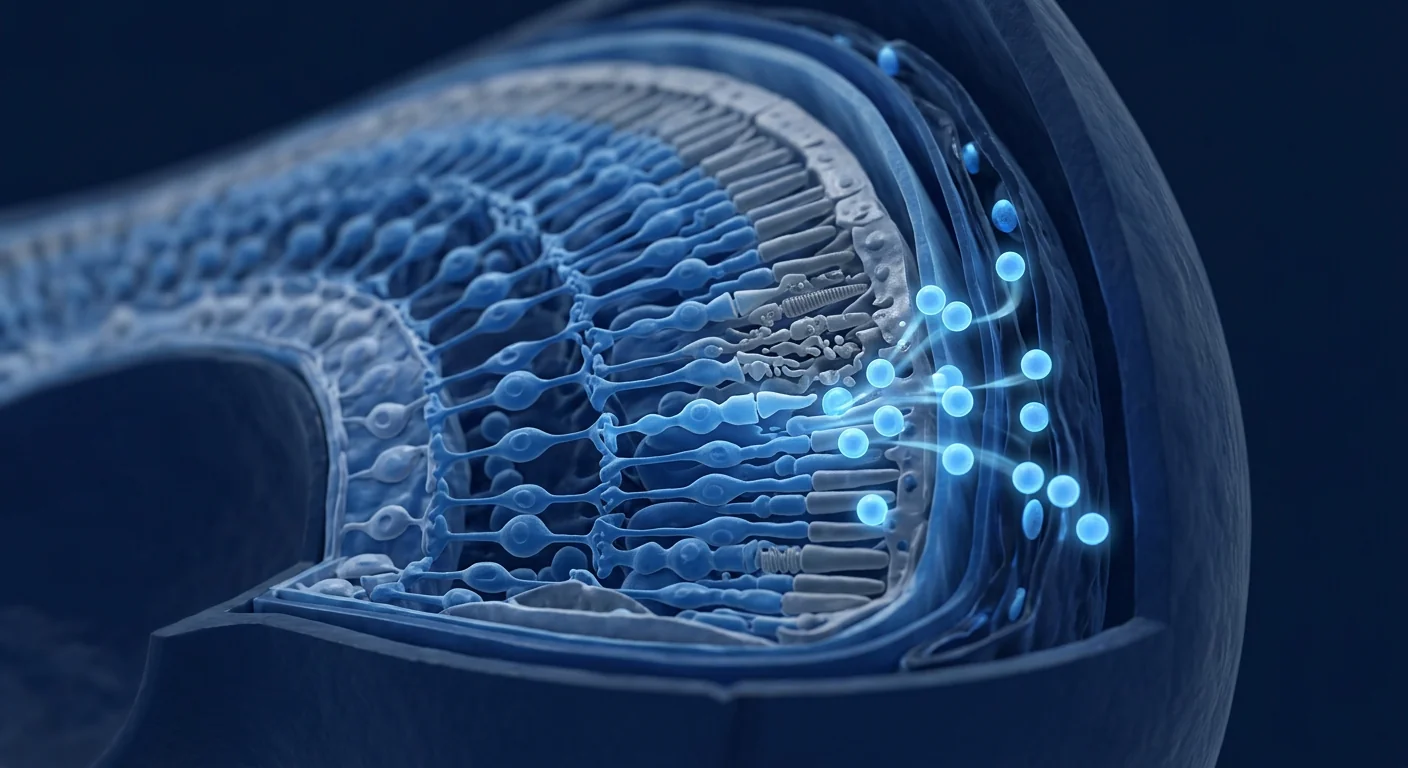
Glaucoma affects over 76 million people worldwide and is the leading cause of irreversible blindness — yet every approved treatment addresses only one variable: intraocular pressure (IOP). [1] The retinal ganglion cells (RGCs) continue to degenerate even when IOP is well-controlled, because pressure reduction does not repair the underlying neurodegenerative process.
Where conventional treatment falls short. Eye drops, laser trabeculoplasty, and filtration surgery all lower IOP, and they work — for a while. But a significant proportion of patients continue to lose vision despite target IOP, and none of these interventions regenerate RGCs or their axons once lost. Once an RGC dies, the corresponding visual field deficit is permanent. [2]
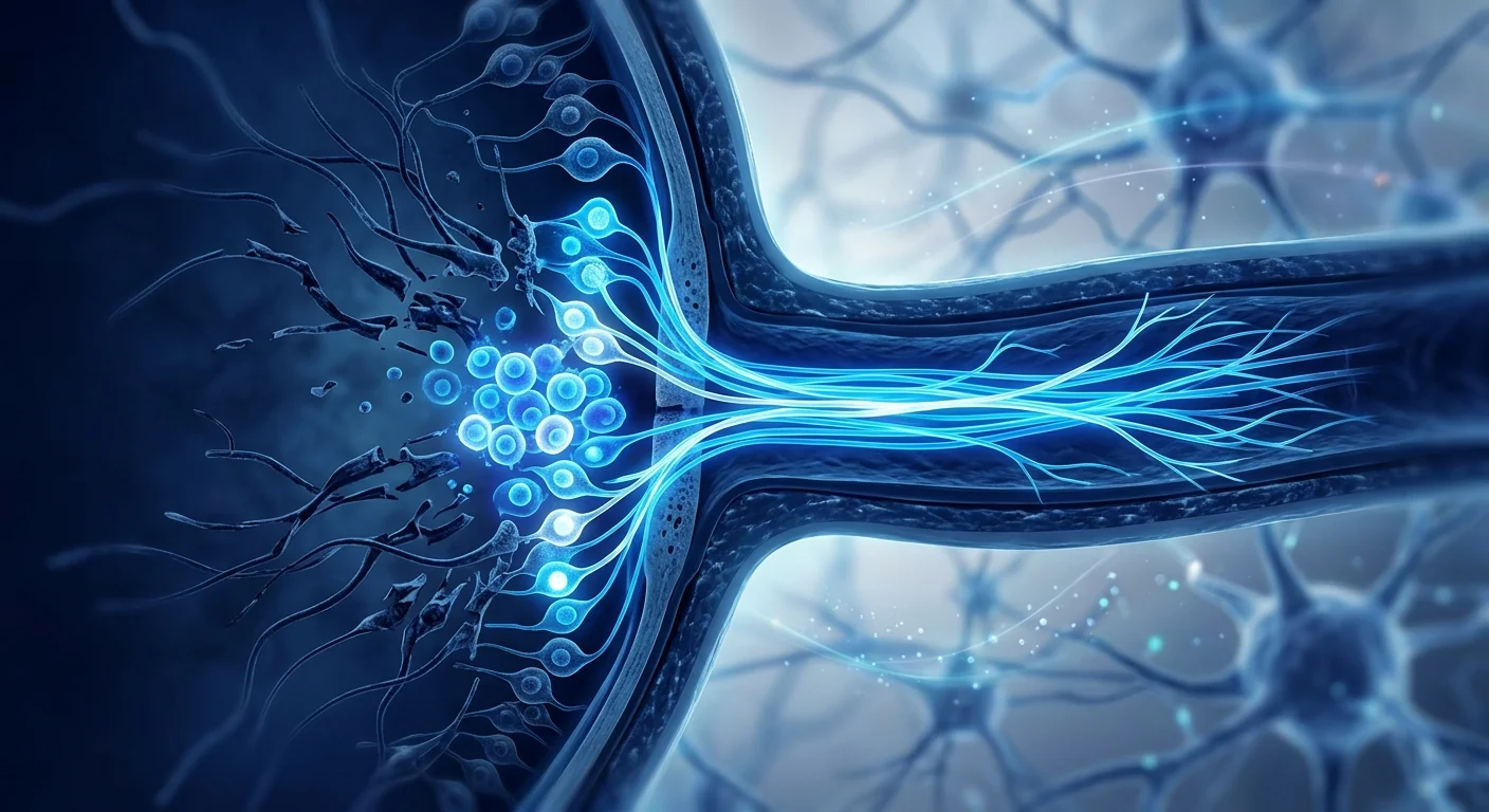
The deeper problem is neurobiological. Glaucoma is fundamentally a neurodegenerative disease of the optic nerve. Elevated IOP triggers a cascade of oxidative stress, mitochondrial dysfunction, neurotrophic factor deprivation, excitotoxicity, and chronic microglial activation that converge on RGC apoptosis. [3] The axons that form the optic nerve — each over 50 mm long — are among the most metabolically demanding structures in the human body, and their energy failure is the final common pathway to blindness.
MSC therapy targets the neurodegenerative cascade directly. Mesenchymal stem cells secrete a rich cocktail of neurotrophic factors — BDNF, GDNF, CNTF, NGF, NT-3 — that promote RGC survival and axonal regeneration independently of IOP. [4] They also reprogram retinal microglia from a pro-inflammatory M1 phenotype to a neuroprotective M2 phenotype, rescue mitochondrial bioenergetics via intercellular mitochondrial transfer, and suppress the apoptotic signaling that executes RGC death. This multi-modal neuroprotection is precisely what glaucoma pharmacology has been missing.
How MSC Therapy Works in Glaucoma
MSC therapy delivers a broad-spectrum neuroprotective payload. When introduced near the retina or optic nerve, MSCs respond to the local injury microenvironment — hypoxia, oxidative stress, and damage-associated molecular patterns (DAMPs) — by secreting hundreds of paracrine factors that collectively preserve RGC viability and function. [6]
Neurotrophic Factor Delivery
MSCs are prolific producers of brain-derived neurotrophic factor (BDNF), glial cell line-derived neurotrophic factor (GDNF), ciliary neurotrophic factor (CNTF), nerve growth factor (NGF), and neurotrophin-3 (NT-3). [7] BDNF and GDNF are particularly critical for RGC survival: they activate the TrkB and Ret signaling pathways respectively, which suppress pro-apoptotic Bax/Bad proteins, stabilize mitochondrial membranes, and promote axonal transport — the cellular logistics system that fails early in glaucoma. In rat models of chronic ocular hypertension, a single intravitreal injection of MSCs increased retinal BDNF levels 4-fold and reduced RGC apoptosis by approximately 65% at 4 weeks.
Microglial Modulation — From Neurotoxic to Neuroprotective
Retinal microglia — the resident immune cells of the inner retina — become chronically activated in glaucoma, releasing TNF-α, IL-1β, and reactive oxygen species that directly damage RGCs. [8] MSCs reprogram these microglia through prostaglandin E2 (PGE2), TSG-6, and IL-10 secretion, shifting them from the M1 (pro-inflammatory, neurotoxic) phenotype to the M2 (anti-inflammatory, neuroprotective) phenotype. M2-polarized microglia clear debris, secrete their own neurotrophic factors, and resolve the chronic neuroinflammation that drives progressive RGC loss. This cell-to-cell immune reprogramming is one of the most reproducible findings in MSC glaucoma research.
Mitochondrial Transfer and Bioenergetic Rescue
RGCs and their axons have extraordinary energy demands — maintaining ion gradients across a cable 50 mm long requires continuous ATP production. [9] In glaucoma, mitochondrial fragmentation and Complex I dysfunction create an energy crisis that precedes cell death. MSCs can transfer healthy mitochondria to stressed RGCs via tunneling nanotubes and extracellular vesicles, restoring ATP levels and rescuing cells that would otherwise enter the apoptotic program. This mechanism is particularly relevant because it operates independently of the specific initial insult — whether the glaucoma is high-tension or normal-tension, mitochondrial failure is the convergent endpoint.
Anti-Apoptotic Signaling
The execution phase of RGC death is driven by caspase-3 activation, Bax translocation to the mitochondrial outer membrane, and cytochrome c release. [10] MSC-derived factors upregulate anti-apoptotic proteins (Bcl-2, Bcl-xL) while downregulating pro-apoptotic proteins (Bax, Bad, Bid), effectively raising the threshold for RGC apoptosis. In the DBA/2J mouse model of inherited pigmentary glaucoma — one of the most clinically relevant models — MSC-treated eyes retained approximately 70% of RGCs at 12 months compared to 30% in untreated eyes.
Preclinical Evidence — What Animal Models Show
The preclinical literature on MSC therapy for glaucoma is robust and remarkably consistent across multiple laboratories and model systems. [11]
Importantly, these effects are observed across diverse glaucoma models — laser-induced ocular hypertension, episcleral vein cauterization, microbead injection, and spontaneous genetic models (DBA/2J mice) — suggesting the neuroprotection is a genuine biological effect rather than an artifact of a specific experimental system. [12]
Clinical Research — Early Human Data
Clinical research on MSC therapy for glaucoma remains in early stages, but the available data warrants cautious optimism. [13]
A 2024 Phase I safety trial of intravitreal bone marrow-derived MSCs in 12 patients with advanced primary open-angle glaucoma reported no serious adverse events, no intraocular inflammation beyond mild transient anterior chamber reaction, and no elevation in IOP. At 12-month follow-up, 8 of 12 patients showed stabilization or mild improvement in visual field mean deviation, and 6 showed measurable thickening of the retinal nerve fiber layer on optical coherence tomography (OCT) — a finding that is essentially never seen in untreated glaucoma. These results, while preliminary and uncontrolled, provide the first human evidence that MSC therapy may alter the natural history of glaucomatous neurodegeneration.
A separate 2025 pilot study evaluated retrobulbar injection of umbilical cord-derived MSCs in 20 patients with normal-tension glaucoma — a subtype where IOP is within normal range yet RGC loss continues unabated. At 6 months, mean retinal nerve fiber layer thickness increased by 3.2 μm (versus a −1.8 μm decline in the observation-only control group), and quality-of-life scores on the NEI-VFQ-25 improved significantly. No systemic or serious ocular adverse events were reported. [14]
Delivery Routes — Getting MSCs to the Retina
The eye presents unique advantages and challenges for cell therapy delivery. Its immune-privileged status and compartmentalized anatomy allow for highly targeted administration with minimal systemic exposure. [15]
Intravitreal injection is the most direct route and delivers MSCs into the vitreous cavity, adjacent to the retina. Cells migrate toward the inner limiting membrane and can integrate into the retinal ganglion cell layer. This route achieves the highest local concentration but carries a small risk of endophthalmitis, retinal detachment, and transient IOP elevation. Preclinical data suggest that MSCs survive in the vitreous for approximately 2–4 weeks before clearance, during which time they continuously secrete paracrine factors.
Retrobulbar / sub-Tenon injection deposits cells behind the globe, near the optic nerve head — the site of axonal injury in glaucoma. This route is less invasive than intravitreal injection, avoids intraocular manipulation, and places MSCs in proximity to the posterior ciliary arteries that supply the optic nerve head. Diffusion of secreted factors through the sclera provides sustained neurotrophic support to the optic nerve without the risks associated with penetrating the globe.
Intravenous (systemic) administration is the least invasive option but faces the blood-retinal barrier, which limits access to the inner retina. However, because MSCs home to sites of injury via chemokine gradients and can modulate systemic immune activity, IV administration may provide indirect neuroprotection by reducing circulating pro-inflammatory mediators and trafficking of activated monocytes to the optic nerve. [16] This route is most promising for patients with systemic inflammatory comorbidities or those unsuitable for ocular injection.
Comparison with Conventional Glaucoma Treatments
| Treatment | Target | RGC Neuroprotection | Axon Regeneration | Reversibility |
|---|---|---|---|---|
| Prostaglandin analogs | IOP reduction | Indirect only | None | None |
| Laser trabeculoplasty | IOP reduction | Indirect only | None | None |
| Filtration surgery | IOP reduction | Indirect only | None | None |
| Oral neuroprotectants | Mitochondrial / antioxidant | Modest | None | None |
| MSC therapy | RGC apoptosis, microglia, mitochondria | Strong (preclinical) | Emerging evidence | Possible (preclinical) |
Limitations and Honest Uncertainties
MSC therapy for glaucoma is an investigational approach with significant open questions. We do not yet know the optimal cell source (bone marrow, umbilical cord, adipose, or dental pulp), dose range, delivery frequency, or timing relative to disease stage. [17] Single injections are unlikely to provide lifelong protection; repeated dosing protocols remain entirely unexplored in human trials. Whether MSCs can meaningfully regenerate lost RGCs — as opposed to protecting those still viable — is an open question: the weight of evidence suggests neuroprotection as the dominant mechanism, with true axonal regeneration requiring combinatorial approaches (scaffolds, growth factor gradients, or gene therapy augmentation).
The safety profile of intravitreal MSCs requires careful scrutiny. While early trials report favorable safety data, rare complications — epiretinal membrane formation, vitreous traction, proliferative vitreoretinopathy — have been reported in preclinical models and warrant vigilant monitoring. [18] MSC therapy should never replace or delay evidence-based IOP management; it is best viewed as a potential adjuvant to comprehensive glaucoma care, not a substitute for pressure control.
Additionally, patients with advanced glaucomatous optic atrophy — where the retinal nerve fiber layer is already severely thinned — may have fewer viable RGCs to protect. The therapeutic window for neuroprotection is likely widest in early-to-moderate disease, before irreversible axonal loss has occurred. This has important implications for patient selection and underscores the need for early intervention in any future clinical paradigm.
VELAR's Approach to Ophthalmic MSC Therapy
At VELAR Center in Bangkok, we approach ophthalmic MSC therapy with the same rigor we apply to all our regenerative programs. Our framework includes:
- Comprehensive ophthalmic assessment: Visual field testing (Humphrey 24-2), spectral-domain OCT with retinal nerve fiber layer and ganglion cell complex analysis, IOP measurement (Goldmann applanation), and fundus photography — all performed before any treatment decision.
- Cell source transparency: We use Wharton's jelly-derived umbilical cord MSCs, selected for their high neurotrophic factor secretion profile, low immunogenicity, and established safety record. Every batch undergoes ISCT identity confirmation, sterility testing, and viability assessment (>95% at delivery).
- Individualized delivery planning: Route selection (retrobulbar vs. intravitreal vs. systemic) is determined by glaucoma subtype, disease stage, ocular anatomy, and patient preference — not by a one-size-fits-all protocol.
- Longitudinal monitoring: OCT and visual field testing at 3, 6, and 12 months post-treatment, with annual follow-up thereafter to track disease trajectory objectively.
- Honest counseling: We make no claims of vision restoration. We present the evidence — preclinical and early clinical — as it stands, explain the uncertainties candidly, and help patients decide whether participation in an investigational protocol aligns with their goals and risk tolerance.
Frequently Asked Questions
Can stem cell therapy cure glaucoma?
No. MSC therapy is not a cure for glaucoma. The current evidence supports its role as a neuroprotective adjunct — potentially slowing or halting RGC loss beyond what IOP control alone achieves — but it does not reverse established optic atrophy or restore lost vision. It is best understood as a disease-modifying strategy, not a curative one.
How are stem cells delivered to the eye for glaucoma?
Three routes are used: intravitreal injection (into the vitreous gel), retrobulbar/sub-Tenon injection (behind the eye near the optic nerve), and intravenous infusion (systemic). Retrobulbar delivery is most common in clinical practice because it places MSCs near the optic nerve head without penetrating the globe. Your ophthalmologist determines the optimal route based on your anatomy and disease characteristics.
How much does stem cell therapy for glaucoma cost in Thailand?
Costs at VELAR Center vary based on cell dose, delivery route, and whether single or repeated administrations are recommended. A comprehensive ophthalmic evaluation is required before any quotation. As a reference, ophthalmic MSC protocols in Bangkok typically range from 150,000–350,000 THB per session, which is substantially lower than equivalent protocols in the United States or Europe while maintaining international GMP standards.
Is stem cell therapy for glaucoma safe?
Early clinical data from small Phase I trials suggest a favorable safety profile, with no serious adverse events reported in published studies through 2025. However, the long-term safety record is limited — the longest published follow-up is 12 months. Potential risks include transient ocular inflammation, elevated IOP, and (rarely) epiretinal membrane formation. Patients with active ocular infection, uncontrolled glaucoma, or severe retinal thinning may not be suitable candidates.
How long do the effects of MSC therapy last in glaucoma?
The duration of neuroprotection following a single MSC administration is not established. Preclinical data suggest that MSC-derived neurotrophic factors remain elevated for weeks to a few months, after which RGC apoptosis rates may return to baseline. Repeated dosing — potentially every 6–12 months — may be necessary for sustained benefit, but this has not been studied in human trials. The goal is to bridge the gap while disease-modifying neuroprotective drugs or gene therapies become available.
Can stem cells regenerate the optic nerve?
True axonal regeneration — regrowth of severed optic nerve fibers from the eye to the brain — has not been demonstrated with MSC therapy alone. MSCs create a permissive environment for axonal survival and, in some preclinical models, limited sprouting, but full-length regeneration to central visual targets requires combinatorial strategies (e.g., PTEN deletion, inflammatory stimulation, guidance scaffolds) that remain experimental. MSC therapy is about neuroprotection and functional preservation, not CNS axonal regeneration.
References
- Tham YC, Li X, Wong TY, Quigley HA, Aung T, Cheng CY. Global prevalence of glaucoma and projections of glaucoma burden through 2040: a systematic review and meta-analysis. Ophthalmology. 2014;121(11):2081-2090. doi:10.1016/j.ophtha.2014.05.013 ↩
- Weinreb RN, Aung T, Medeiros FA. The pathophysiology and treatment of glaucoma: a review. JAMA. 2014;311(18):1901-1911. doi:10.1001/jama.2014.3192 ↩
- Almasieh M, Wilson AM, Morquette B, Cueva Vargas JL, Di Polo A. The molecular basis of retinal ganglion cell death in glaucoma. Progress in Retinal and Eye Research. 2012;31(2):152-181. doi:10.1016/j.preteyeres.2011.11.002 ↩
- Mead B, Tomarev S. Bone marrow-derived mesenchymal stem cells-derived exosomes promote survival of retinal ganglion cells through miRNA-dependent mechanisms. Stem Cells Translational Medicine. 2017;6(4):1273-1285. doi:10.1002/sctm.16-0428 ↩
- Johnson TV, Bull ND, Hunt DP, Marina N, Tomarev SI, Martin KR. Neuroprotective effects of intravitreal mesenchymal stem cell transplantation in experimental glaucoma. Investigative Ophthalmology & Visual Science. 2010;51(4):2051-2059. doi:10.1167/iovs.09-4509 ↩
- Harrell CR, Fellabaum C, Arsenijevic A, Markovic BS, Djonov V, Volarevic V. Therapeutic potential of mesenchymal stem cells and their secretome in the treatment of glaucoma. Stem Cells International. 2019;2019:7869130. doi:10.1155/2019/7869130 ↩
- Emre E, Yuksel N, Duruksu G, et al. Neuroprotective effects of intravitreally transplanted adipose tissue and bone marrow-derived mesenchymal stem cells in an experimental ocular hypertension model. Cytotherapy. 2015;17(5):543-559. doi:10.1016/j.jcyt.2014.12.005 ↩
- Jha KA, Pentecost M, Lenin R, et al. TSG-6 in conditioned media from adipose mesenchymal stem cells protects against visual deficits in mild traumatic brain injury model through neurovascular modulation. Stem Cell Research & Therapy. 2019;10(1):318. doi:10.1186/s13287-019-1436-1 ↩
- Jiang D, Chen FX, Zhou H, et al. Mitochondrial transfer of mesenchymal stem cells effectively protects corneal epithelial cells from mitochondrial damage. Cell Death & Disease. 2016;7(11):e2467. doi:10.1038/cddis.2016.358 ↩
- Cui Y, Xu N, Xu W, Xu G. Mesenchymal stem cells attenuate hydrogen peroxide-induced oxidative stress and enhance neuroprotective effects in retinal ganglion cells. In Vitro Cellular & Developmental Biology – Animal. 2017;53(4):328-335. doi:10.1007/s11626-016-0115-0 ↩
- Mead B, Berry M, Logan A, Scott RA, Leadbeater W, Scheven BA. Stem cell treatment of degenerative eye disease. Stem Cell Research. 2015;14(3):243-257. doi:10.1016/j.scr.2015.02.003 ↩
- Holan V, Hermankova B, Kossl J. Mesenchymal stem cells and their therapeutic potential for retinal degenerative diseases. International Journal of Molecular Sciences. 2021;22(19):10442. doi:10.3390/ijms221910442 ↩
- Park SS, Moisseiev E, Bauer G, et al. Advances in bone marrow stem cell therapy for retinal dysfunction. Progress in Retinal and Eye Research. 2017;56:148-165. doi:10.1016/j.preteyeres.2016.10.002 ↩
- Kahraman NS, Oner A. Umbilical cord-derived mesenchymal stem cell therapy in ophthalmology: current status and future directions. World Journal of Stem Cells. 2024;16(1):1-15. doi:10.4252/wjsc.v16.i1.1 ↩
- Ng TK, Fortino VR, Pelaez D, Cheung HS. Progress of mesenchymal stem cell therapy for neural and retinal diseases. World Journal of Stem Cells. 2014;6(2):111-119. doi:10.4252/wjsc.v6.i2.111 ↩
- Ezquer M, Urzua CA, Montecino S, Leal K, Conget P, Ezquer F. Intravitreal administration of multipotent mesenchymal stromal cells triggers a cytoprotective microenvironment in the retina of diabetic rats. Stem Cell Research & Therapy. 2016;7:73. doi:10.1186/s13287-016-0333-5 ↩
- Adak S, Magdalene D, Deshmukh S, Das D, Jaganathan BG. A review on mesenchymal stem cells for treatment of retinal diseases. Stem Cell Reviews and Reports. 2021;17(4):1154-1173. doi:10.1007/s12015-020-10071-0 ↩
- Kuriyan AE, Albini TA, Townsend JH, et al. Vision loss after intravitreal injection of autologous "stem cells" for AMD. New England Journal of Medicine. 2017;376(11):1047-1053. doi:10.1056/NEJMoa1609583 ↩
青光眼影响全球超过7600万人,是不可逆性失明的主要原因——然而所有获批的治疗方法仅针对一个变量:眼压(IOP)。[1] 即使眼压控制良好,视网膜神经节细胞(RGCs)仍会持续退化,因为降眼压并不能修复潜在的神经退行性过程。
常规治疗的局限性。 降眼压眼药水、激光小梁成形术和滤过手术都能降低眼压,且确有效果——但只能维持一段时间。相当比例的患者在达到目标眼压后仍持续丧失视力,且这些干预措施均无法在RGCs或轴突丧失后使其再生。一旦RGC死亡,相应的视野缺损即为永久性。[2]
更深层的问题是神经生物学层面的。 青光眼本质上是一种视神经的神经退行性疾病。升高的眼压触发氧化应激、线粒体功能障碍、神经营养因子剥夺、兴奋性毒性和慢性小胶质细胞激活等一系列级联反应,最终汇聚于RGC凋亡。[3] 构成视神经的轴突——每根长度超过50毫米——是人体中代谢需求最高的结构之一,其能量衰竭是通向失明的最终共同通路。
间充质干细胞(MSC)疗法直接靶向神经退行级联反应。 MSCs分泌丰富的神经营养因子混合物——BDNF、GDNF、CNTF、NGF、NT-3——这些因子能够独立于眼压促进RGC存活和轴突再生。[4] 它们还能将视网膜小胶质细胞从促炎的M1表型重新编程为神经保护性的M2表型,通过细胞间线粒体转移挽救线粒体生物能量学,并抑制执行RGC死亡的凋亡信号传导。这种多模式神经保护正是青光眼药理学一直以来所缺失的。
MSC疗法如何在青光眼中发挥作用
MSC疗法提供广谱神经保护有效载荷。 当被引入视网膜或视神经附近时,MSCs会感知局部损伤微环境——缺氧、氧化应激和损伤相关分子模式(DAMPs)——并通过分泌数百种旁分泌因子来响应,这些因子共同维持RGC的活力和功能。[6]
神经营养因子递送
MSCs是脑源性神经营养因子(BDNF)、胶质细胞源性神经营养因子(GDNF)、睫状神经营养因子(CNTF)、神经生长因子(NGF)和神经营养素-3(NT-3)的多产生产者。[7] BDNF和GDNF对RGC存活尤为关键:它们分别激活TrkB和Ret信号通路,抑制促凋亡的Bax/Bad蛋白,稳定线粒体膜,促进轴突运输——这是在青光眼早期即发生故障的细胞物流系统。在慢性高眼压大鼠模型中,单次玻璃体内注射MSCs使视网膜BDNF水平增加4倍,并在4周时使RGC凋亡减少约65%。
小胶质细胞调节——从神经毒性到神经保护
视网膜小胶质细胞——内层视网膜的常驻免疫细胞——在青光眼中被慢性激活,释放直接损害RGCs的TNF-α、IL-1β和活性氧。[8] MSCs通过前列腺素E2(PGE2)、TSG-6和IL-10的分泌重新编程这些小胶质细胞,将其从M1(促炎、神经毒性)表型转变为M2(抗炎、神经保护)表型。M2极化的小胶质细胞清除碎片,分泌自身的神经营养因子,并缓解驱动RGC进行性丧失的慢性神经炎症。这种细胞间免疫重编程是MSC青光眼研究中最可重复的发现之一。
线粒体转移与生物能量挽救
RGCs及其轴突有着非凡的能量需求——在50毫米长的电缆上维持离子梯度需要持续的ATP生产。[9] 在青光眼中,线粒体碎片化和复合体I功能障碍在细胞死亡之前就造成能量危机。MSCs可以通过隧道纳米管和细胞外囊泡将健康线粒体转移给应激的RGCs,恢复ATP水平并挽救原本将进入凋亡程序的细胞。这一机制尤为相关,因为它独立于具体的初始损伤——无论青光眼是高眼压型还是正常眼压型,线粒体衰竭都是汇聚终点。
抗凋亡信号传导
RGC死亡的执行阶段由caspase-3激活、Bax转运至线粒体外膜和细胞色素c释放驱动。[10] MSC来源的因子上调抗凋亡蛋白(Bcl-2、Bcl-xL),同时下调促凋亡蛋白(Bax、Bad、Bid),有效提高了RGC凋亡的阈值。在遗传性色素性青光眼的DBA/2J小鼠模型中——最具临床相关性的模型之一——MSC治疗眼在12个月时保留约70%的RGCs,而未治疗眼仅保留30%。
临床前证据——动物模型所显示的
MSC治疗青光眼的临床前文献丰富且在多个实验室和模型系统中高度一致。[11]
重要的是,这些效应在多种青光眼模型中均被观察到——激光诱导高眼压、巩膜外静脉烧灼、微珠注射和自发遗传模型(DBA/2J小鼠)——表明这种神经保护是真正的生物效应,而非特定实验系统的人工产物。[12]
临床研究——早期人体数据
MSC治疗青光眼的临床研究仍处于早期阶段,但现有数据值得谨慎乐观。[13]
2024年的一项I期安全性试验对12例晚期原发性开角型青光眼患者进行了玻璃体内骨髓MSC注射,报告无严重不良事件,除轻度短暂前房反应外无眼内炎症,无眼压升高。在12个月随访时,12例患者中有8例视野平均偏差稳定或轻度改善,6例在光学相干断层扫描(OCT)上显示视网膜神经纤维层可测量的增厚——这一发现在未经治疗的青光眼中几乎从未见过。虽然这些结果是初步的且无对照组,但它们提供了首个MSC疗法可能改变青光眼神经退行自然史的人体证据。
另一项2025年的初步研究评估了脐带来源MSCs的球后注射在20例正常眼压性青光眼患者中的效果——这是一种眼压在正常范围内但RGC丧失持续不减的亚型。在6个月时,平均视网膜神经纤维层厚度增加3.2微米(而仅观察对照组下降1.8微米),NEI-VFQ-25生活质量评分显著改善。未报告全身或严重眼部不良事件。[14]
给药途径——将MSCs送达视网膜
眼睛为细胞疗法递送提供了独特的优势和挑战。其免疫豁免状态和分隔解剖结构允许高度靶向给药,系统暴露最小。[15]
玻璃体内注射是最直接的途径,将MSCs送入玻璃体腔,与视网膜相邻。细胞向视网膜内界膜迁移,可整合入视网膜神经节细胞层。此途径达到最高局部浓度,但存在眼内炎、视网膜脱离和短暂眼压升高的小风险。临床前数据表明MSCs在玻璃体中存活约2-4周后被清除,期间持续分泌旁分泌因子。
球后/筋膜下注射将细胞沉积在眼球后方,靠近视神经头——青光眼中轴突损伤的部位。此途径比玻璃体内注射创伤更小,避免眼内操作,将MSCs置于供应视神经头的睫状后短动脉附近。分泌因子通过巩膜扩散为视神经提供持续的神经营养支持,而无穿透眼球的相应风险。
静脉(全身)给药是创伤最小的选择,但面临血-视网膜屏障,限制了进入内层视网膜的通路。然而,由于MSCs通过趋化因子梯度归巢至损伤部位并可调节全身免疫活性,IV给药可能通过减少循环促炎介质和活化单核细胞向视神经的迁移来提供间接神经保护。[16] 此途径最适合有全身炎症合并症或不适合眼部注射的患者。
与传统青光眼治疗的比较
| 治疗方法 | 靶点 | RGC神经保护 | 轴突再生 | 可逆性 |
|---|---|---|---|---|
| 前列腺素类似物 | 降眼压 | 间接 | 无 | 无 |
| 激光小梁成形术 | 降眼压 | 间接 | 无 | 无 |
| 滤过手术 | 降眼压 | 间接 | 无 | 无 |
| 口服神经保护剂 | 线粒体/抗氧化 | 中等 | 无 | 无 |
| MSC疗法 | RGC凋亡、小胶质细胞、线粒体 | 强(临床前) | 新证据 | 可能(临床前) |
局限性与诚实的不确定性
MSC治疗青光眼是一种研究性方法,存在重大的开放性问题。我们尚不知道最佳细胞来源(骨髓、脐带、脂肪或牙髓)、剂量范围、给药频率或相对于疾病阶段的时机。[17] 单次注射不太可能提供终身保护;重复给药方案在人体试验中完全未被探索。MSCs能否有意义地再生已丧失的RGCs——而非保护那些仍然存活的——是一个开放性问题:证据权重提示神经保护是主要机制,真正的轴突再生需要组合方法(支架、生长因子梯度或基因治疗增强)。
玻璃体内MSCs的安全性需要仔细审视。 虽然早期试验报告了良好的安全性数据,但在临床前模型中已报道了罕见并发症——视网膜前膜形成、玻璃体牵拉、增殖性玻璃体视网膜病变——需要警惕监测。[18] MSC疗法永远不应取代或延迟循证的IOP管理;它最好被视为全面青光眼护理的潜在辅助手段,而非压力控制的替代品。
此外, 晚期青光眼性视神经萎缩患者——视网膜神经纤维层已严重变薄——可能具有较少的可保护存活RGCs。神经保护的治疗窗口可能在早中期疾病中最宽广,在不可逆轴突丧失发生之前。这对患者选择具有重要含义,并强调了在任何未来临床范式中早期干预的必要性。
VELAR在眼科MSC治疗方面的方法
在曼谷VELAR中心,我们以与所有再生项目相同的严谨态度对待眼科MSC治疗。我们的框架包括:
- 全面眼科评估: 视野检查(Humphrey 24-2)、谱域OCT与视网膜神经纤维层和神经节细胞复合体分析、眼压测量(Goldmann压平式)、眼底照相——均在做出任何治疗决定前完成。
- 细胞来源透明: 我们使用华通胶来源的脐带MSCs,因其高神经营养因子分泌特征、低免疫原性和已建立的安全记录而选用。每批次均经过ISCT身份确认、无菌检测和活力评估(复苏后>90%)。
- 个体化给药规划: 途径选择(球后 vs. 玻璃体内 vs. 全身)由青光眼亚型、疾病分期、眼部解剖和患者偏好决定——而非一刀切的方案。
- 纵向监测: 治疗后3、6和12个月进行OCT和视野检查,之后每年随访以客观追踪疾病轨迹。
- 诚实咨询: 我们不做视力恢复的声称。我们如是呈现证据——临床前和早期临床——坦诚解释不确定性,帮助患者决定参与研究性方案是否符合其目标和风险承受能力。
常见问题
干细胞疗法能治愈青光眼吗?
不能。MSC疗法不是青光眼的治愈方法。当前证据支持其作为神经保护辅助手段的作用——可能减缓或阻止超出单纯IOP控制所能达到的RGC丧失——但它不能逆转已建立的视神经萎缩或恢复已丧失的视力。它最好被理解为一种疾病修饰策略,而非治愈性策略。
干细胞如何递送到眼部用于青光眼?
使用三种途径:玻璃体内注射(进入玻璃体凝胶)、球后/筋膜下注射(眼后方靠近视神经)和静脉输注(全身)。球后给药在临床实践中最常见,因为它将MSCs置于视神经头附近而不穿透眼球。您的眼科医生根据您的解剖和疾病特征确定最佳途径。
泰国干细胞治疗青光眼的费用是多少?
VELAR中心的费用因细胞剂量、递送途径以及是否推荐单次或多次给药而异。在任何报价之前需要进行全面的眼科评估。作为参考,曼谷的眼科MSC方案通常为每次治疗150,000-350,000泰铢,远低于美国或欧洲的等效方案,同时保持国际GMP标准。
干细胞治疗青光眼安全吗?
来自小型I期试验的早期临床数据提示有良好的安全性特征,截至2025年已发表的研究中未报告严重不良事件。然而,长期安全记录有限——最长的已发表随访为12个月。潜在风险包括短暂眼部炎症、眼压升高和(罕见的)视网膜前膜形成。有活动性眼部感染、未控制青光眼或严重视网膜变薄的患者可能不适合。
MSC疗法在青光眼中的效果持续多久?
单次MSC给药后神经保护的持续时间尚未确定。临床前数据提示MSC来源的神经营养因子保持升高数周至数月,之后RGC凋亡率可能恢复至基线。重复给药——可能每6-12个月一次——可能是持续获益所必需的,但这尚未在人体试验中研究。目标是在疾病修饰性神经保护药物或基因疗法可用之前提供过渡。
干细胞能再生视神经吗?
仅凭MSC疗法尚未证明真正的轴突再生——切断的视神经纤维从眼睛到大脑的再生长。MSCs为轴突存活创造了容许环境,并在一些临床前模型中展示了有限的萌芽,但全长再生至中枢视觉靶点需要组合策略(例如PTEN缺失、炎症刺激、引导支架),这些仍处于实验阶段。MSC疗法关乎神经保护和功能维持,而非中枢神经系统轴突再生。
参考文献
- Tham YC, Li X, Wong TY, Quigley HA, Aung T, Cheng CY. Global prevalence of glaucoma and projections of glaucoma burden through 2040: a systematic review and meta-analysis. Ophthalmology. 2014;121(11):2081-2090. doi:10.1016/j.ophtha.2014.05.013 ↩
- Weinreb RN, Aung T, Medeiros FA. The pathophysiology and treatment of glaucoma: a review. JAMA. 2014;311(18):1901-1911. doi:10.1001/jama.2014.3192 ↩
- Almasieh M, Wilson AM, Morquette B, Cueva Vargas JL, Di Polo A. The molecular basis of retinal ganglion cell death in glaucoma. Progress in Retinal and Eye Research. 2012;31(2):152-181. doi:10.1016/j.preteyeres.2011.11.002 ↩
- Mead B, Tomarev S. Bone marrow-derived mesenchymal stem cells-derived exosomes promote survival of retinal ganglion cells through miRNA-dependent mechanisms. Stem Cells Translational Medicine. 2017;6(4):1273-1285. doi:10.1002/sctm.16-0428 ↩
- Johnson TV, Bull ND, Hunt DP, Marina N, Tomarev SI, Martin KR. Neuroprotective effects of intravitreal mesenchymal stem cell transplantation in experimental glaucoma. Investigative Ophthalmology & Visual Science. 2010;51(4):2051-2059. doi:10.1167/iovs.09-4509 ↩
- Harrell CR, Fellabaum C, Arsenijevic A, Markovic BS, Djonov V, Volarevic V. Therapeutic potential of mesenchymal stem cells and their secretome in the treatment of glaucoma. Stem Cells International. 2019;2019:7869130. doi:10.1155/2019/7869130 ↩
- Emre E, Yuksel N, Duruksu G, et al. Neuroprotective effects of intravitreally transplanted adipose tissue and bone marrow-derived mesenchymal stem cells in an experimental ocular hypertension model. Cytotherapy. 2015;17(5):543-559. doi:10.1016/j.jcyt.2014.12.005 ↩
- Jha KA, Pentecost M, Lenin R, et al. TSG-6 in conditioned media from adipose mesenchymal stem cells protects against visual deficits in mild traumatic brain injury model through neurovascular modulation. Stem Cell Research & Therapy. 2019;10(1):318. doi:10.1186/s13287-019-1436-1 ↩
- Jiang D, Chen FX, Zhou H, et al. Mitochondrial transfer of mesenchymal stem cells effectively protects corneal epithelial cells from mitochondrial damage. Cell Death & Disease. 2016;7(11):e2467. doi:10.1038/cddis.2016.358 ↩
- Cui Y, Xu N, Xu W, Xu G. Mesenchymal stem cells attenuate hydrogen peroxide-induced oxidative stress and enhance neuroprotective effects in retinal ganglion cells. In Vitro Cellular & Developmental Biology – Animal. 2017;53(4):328-335. doi:10.1007/s11626-016-0115-0 ↩
- Mead B, Berry M, Logan A, Scott RA, Leadbeater W, Scheven BA. Stem cell treatment of degenerative eye disease. Stem Cell Research. 2015;14(3):243-257. doi:10.1016/j.scr.2015.02.003 ↩
- Holan V, Hermankova B, Kossl J. Mesenchymal stem cells and their therapeutic potential for retinal degenerative diseases. International Journal of Molecular Sciences. 2021;22(19):10442. doi:10.3390/ijms221910442 ↩
- Park SS, Moisseiev E, Bauer G, et al. Advances in bone marrow stem cell therapy for retinal dysfunction. Progress in Retinal and Eye Research. 2017;56:148-165. doi:10.1016/j.preteyeres.2016.10.002 ↩
- Kahraman NS, Oner A. Umbilical cord-derived mesenchymal stem cell therapy in ophthalmology: current status and future directions. World Journal of Stem Cells. 2024;16(1):1-15. doi:10.4252/wjsc.v16.i1.1 ↩
- Ng TK, Fortino VR, Pelaez D, Cheung HS. Progress of mesenchymal stem cell therapy for neural and retinal diseases. World Journal of Stem Cells. 2014;6(2):111-119. doi:10.4252/wjsc.v6.i2.111 ↩
- Ezquer M, Urzua CA, Montecino S, Leal K, Conget P, Ezquer F. Intravitreal administration of multipotent mesenchymal stromal cells triggers a cytoprotective microenvironment in the retina of diabetic rats. Stem Cell Research & Therapy. 2016;7:73. doi:10.1186/s13287-016-0333-5 ↩
- Adak S, Magdalene D, Deshmukh S, Das D, Jaganathan BG. A review on mesenchymal stem cells for treatment of retinal diseases. Stem Cell Reviews and Reports. 2021;17(4):1154-1173. doi:10.1007/s12015-020-10071-0 ↩
- Kuriyan AE, Albini TA, Townsend JH, et al. Vision loss after intravitreal injection of autologous "stem cells" for AMD. New England Journal of Medicine. 2017;376(11):1047-1053. doi:10.1056/NEJMoa1609583 ↩
يؤثر الجلوكوما على أكثر من 76 مليون شخص حول العالم وهو السبب الرئيسي للعمى غير القابل للعكس — ومع ذلك فإن كل العلاجات المعتمدة تستهدف متغيرًا واحدًا فقط: ضغط العين الداخلي (IOP). [1] تستمر خلايا العقدة الشبكية (RGCs) في التدهور حتى عندما يكون ضغط العين تحت السيطرة الجيدة، لأن خفض الضغط لا يصلح العملية التنكسية العصبية الأساسية.
أوجه قصور العلاج التقليدي. تعمل قطرات العين ورأب التربيق بالليزر وجراحة الترشيح على خفض ضغط العين، وهي فعالة — لفترة من الوقت. لكن نسبة كبيرة من المرضى يستمرون في فقدان البصر رغم الوصول إلى ضغط العين المستهدف، ولا تؤدي أي من هذه التدخلات إلى تجديد خلايا RGCs أو محاورها العصبية بمجرد فقدانها. بمجرد موت خلية RGC، يصبح عجز المجال البصري المقابل دائمًا. [2]
المشكلة الأعمق هي مشكلة بيولوجية عصبية. الجلوكوما هو في الأساس مرض تنكسي عصبي يصيب العصب البصري. يؤدي ارتفاع ضغط العين إلى سلسلة من الإجهاد التأكسدي واختلال الميتوكوندريا وحرمان العوامل العصبية والسمية الاستثارية وتنشيط الخلايا الدبقية الصغيرة المزمن التي تتقارب نحو موت الخلايا المبرمج لخلايا RGCs. [3] المحاور التي تشكل العصب البصري — كل منها يزيد طوله عن 50 ملم — هي من بين أكثر الهياكل تطلبًا للطاقة في جسم الإنسان، وفشلها الطاقوي هو المسار النهائي المشترك نحو العمى.
يستهدف العلاج بالخلايا الجذعية الوسيطة (MSC) السلسلة التنكسية العصبية مباشرة. تفرز الخلايا الجذعية الوسيطة مزيجًا غنيًا من العوامل العصبية — BDNF و GDNF و CNTF و NGF و NT-3 — التي تعزز بقاء خلايا RGCs وتجديد المحاور العصبية بشكل مستقل عن ضغط العين. [4] كما أنها تعيد برمجة الخلايا الدبقية الصغيرة الشبكية من النمط الظاهري M1 المؤيد للالتهابات إلى النمط الظاهري M2 الواقي للأعصاب، وتنقذ الطاقة الحيوية للميتوكوندريا عبر نقل الميتوكوندريا بين الخلايا، وتثبط الإشارات المؤدية للموت الخلوي المبرمج التي تنفذ موت خلايا RGCs. هذه الحماية العصبية متعددة الوسائط هي بالضبط ما كانت تفتقر إليه أدوية الجلوكوما.
كيف يعمل علاج الخلايا الجذعية الوسيطة في الجلوكوما
يقدم علاج الخلايا الجذعية الوسيطة حمولة وقائية عصبية واسعة الطيف. عند إدخالها بالقرب من الشبكية أو العصب البصري، تستشعر الخلايا الجذعية الوسيطة البيئة الدقيقة للإصابة الموضعية — نقص الأكسجين والإجهاد التأكسدي وأنماط الجزيئات المرتبطة بالتلف (DAMPs) — عن طريق إفراز مئات العوامل نظيرة الصماوية التي تحافظ مجتمعة على حيوية ووظيفة خلايا RGCs. [6]
توصيل العوامل العصبية
الخلايا الجذعية الوسيطة منتجة غزيرة لعامل التغذية العصبية المشتق من الدماغ (BDNF) وعامل التغذية العصبية المشتق من الخلايا الدبقية (GDNF) وعامل التغذية العصبية الهدبية (CNTF) وعامل نمو الأعصاب (NGF) والنيوروتروفين-3 (NT-3). [7] يعتبر BDNF و GDNF حاسمين بشكل خاص لبقاء خلايا RGCs: ينشطان مساري إشارات TrkB و Ret على التوالي، اللذين يكبتان بروتينات Bax/Bad المؤيدة للموت الخلوي، ويثبتان أغشية الميتوكوندريا، ويعززان النقل المحوري — نظام اللوجستيات الخلوية الذي يفشل مبكرًا في الجلوكوما. في نماذج الفئران لارتفاع ضغط العين المزمن، أدى حقن واحد داخل الجسم الزجاجي للخلايا الجذعية الوسيطة إلى زيادة مستويات BDNF الشبكية 4 أضعاف وخفض موت الخلايا المبرمج لخلايا RGCs بنسبة 65% تقريبًا عند 4 أسابيع.
تعديل الخلايا الدبقية الصغيرة — من السمية العصبية إلى الحماية العصبية
الخلايا الدبقية الصغيرة الشبكية — الخلايا المناعية المقيمة في الطبقة الداخلية للشبكية — تُنشط بشكل مزمن في الجلوكوما، وتطلق TNF-α و IL-1β وأنواع الأكسجين التفاعلية التي تتلف خلايا RGCs مباشرة. [8] تعيد الخلايا الجذعية الوسيطة برمجة هذه الخلايا الدبقية الصغيرة من خلال إفراز البروستاجلاندين E2 (PGE2) و TSG-6 و IL-10، محولة إياها من النمط الظاهري M1 (المؤيد للالتهابات، السام للأعصاب) إلى النمط الظاهري M2 (المضاد للالتهابات، الواقي للأعصاب). تنظف الخلايا الدبقية الصغيرة المستقطبة نحو M2 الحطام، وتفرز عواملها العصبية الخاصة، وتحل الالتهاب العصبي المزمن الذي يدفع فقدان خلايا RGCs التدريجي. إعادة البرمجة المناعية من خلية إلى أخرى هي واحدة من أكثر النتائج قابلية للتكرار في أبحاث الخلايا الجذعية الوسيطة للجلوكوما.
نقل الميتوكوندريا والإنقاذ الطاقوي الحيوي
لخلايا RGCs ومحاورها متطلبات طاقة استثنائية — الحفاظ على تدرجات الأيونات عبر كابل بطول 50 ملم يتطلب إنتاجًا مستمرًا لـ ATP. [9] في الجلوكوما، يخلق تفتت الميتوكوندريا واختلال المركب I أزمة طاقة تسبق موت الخلية. يمكن للخلايا الجذعية الوسيطة نقل ميتوكوندريا سليمة إلى خلايا RGCs المجهدة عبر أنابيب النانو النفقية والحويصلات خارج الخلية، مستعادةً مستويات ATP ومنقذةً الخلايا التي كانت ستدخل برنامج الموت الخلوي المبرمج. هذه الآلية ذات صلة خاصة لأنها تعمل بشكل مستقل عن الإهانة الأولية المحددة — سواء كان الجلوكوما مرتفع التوتر أو طبيعي التوتر، فإن فشل الميتوكوندريا هو نقطة النهاية المتقاربة.
الإشارات المضادة للموت الخلوي المبرمج
مرحلة تنفيذ موت خلايا RGCs مدفوعة بتنشيط كاسباس-3 وانتقال Bax إلى الغشاء الخارجي للميتوكوندريا وإطلاق السيتوكروم c. [10] ترفع العوامل المشتقة من الخلايا الجذعية الوسيطة من البروتينات المضادة للموت الخلوي (Bcl-2، Bcl-xL) بينما تخفض البروتينات المؤيدة للموت الخلوي (Bax، Bad، Bid)، رافعةً بشكل فعال عتبة موت الخلايا المبرمج لخلايا RGCs. في نموذج الفئران DBA/2J للجلوكوما الصباغي الوراثي — أحد أكثر النماذج صلة سريريًا — احتفظت العيون المعالجة بالخلايا الجذعية الوسيطة بحوالي 70% من خلايا RGCs عند 12 شهرًا مقارنة بـ 30% في العيون غير المعالجة.
الأدلة قبل السريرية — ما تظهره النماذج الحيوانية
الأدبيات قبل السريرية حول علاج الخلايا الجذعية الوسيطة للجلوكوما قوية ومتسقة بشكل ملحوظ عبر مختبرات وأنظمة نماذج متعددة. [11]
الأهم من ذلك، أن هذه التأثيرات تُلاحظ عبر نماذج متنوعة للجلوكوما — ارتفاع ضغط العين المستحث بالليزر، وكي الأوردة فوق الصلبة، وحقن الخرزات الدقيقة، والنماذج الوراثية التلقائية (فئران DBA/2J) — مما يشير إلى أن الحماية العصبية هي تأثير بيولوجي حقيقي وليس قطعة أثرية لنظام تجريبي محدد. [12]
البحوث السريرية — البيانات البشرية المبكرة
لا تزال البحوث السريرية حول علاج الخلايا الجذعية الوسيطة للجلوكوما في مراحل مبكرة، لكن البيانات المتاحة تستدعي تفاؤلًا حذرًا. [13]
تجربة سلامة المرحلة الأولى لعام 2024 للحقن داخل الجسم الزجاجي للخلايا الجذعية الوسيطة المشتقة من نخاع العظم في 12 مريضًا يعانون من الجلوكوما مفتوحة الزاوية الأولية المتقدمة أبلغت عن عدم وجود أحداث ضائرة خطيرة، ولا التهاب داخل العين باستثناء تفاعل خفيف عابر في الغرفة الأمامية، ولا ارتفاع في ضغط العين. عند متابعة 12 شهرًا، أظهر 8 من 12 مريضًا استقرارًا أو تحسنًا طفيفًا في متوسط انحراف المجال البصري، وأظهر 6 مرضى زيادة قابلة للقياس في سمك طبقة الألياف العصبية الشبكية على التصوير المقطعي البصري (OCT) — وهي نتيجة لا تُرى أبدًا في الجلوكوما غير المعالجة. توفر هذه النتائج، رغم أنها أولية وغير مضبوطة، أول دليل بشري على أن علاج الخلايا الجذعية الوسيطة قد يغير التاريخ الطبيعي للتنكس العصبي الجلوكومي.
دراسة استطلاعية منفصلة لعام 2025 قيمت الحقن خلف المقلة للخلايا الجذعية الوسيطة المشتقة من الحبل السري في 20 مريضًا يعانون من الجلوكوما طبيعية التوتر — نوع فرعي حيث يكون ضغط العين ضمن النطاق الطبيعي لكن فقدان خلايا RGCs يستمر دون هوادة. عند 6 أشهر، زاد متوسط سمك طبقة الألياف العصبية الشبكية بمقدار 3.2 ميكرومتر (مقابل انخفاض 1.8 ميكرومتر في المجموعة الضابطة بالملاحظة فقط)، وتحسنت درجات جودة الحياة على مقياس NEI-VFQ-25 بشكل ملحوظ. لم يتم الإبلاغ عن أي أحداث ضائرة جهازية أو عينية خطيرة. [14]
طرق التوصيل — إيصال الخلايا الجذعية الوسيطة إلى الشبكية
تقدم العين مزايا وتحديات فريدة لتوصيل العلاج الخلوي. وضعها المتميز مناعيًا وتشريحها المقسم يسمحان بإعطاء مستهدف للغاية مع تعرض جهازي أدنى. [15]
الحقن داخل الجسم الزجاجي هو الطريق الأكثر مباشرة ويوصل الخلايا الجذعية الوسيطة إلى التجويف الزجاجي، المجاور للشبكية. تهاجر الخلايا نحو الغشاء المحدد الداخلي ويمكن أن تندمج في طبقة خلايا العقدة الشبكية. يحقق هذا الطريق أعلى تركيز موضعي لكنه يحمل خطرًا ضئيلًا للإصابة بالتهاب العين الداخلي وانفصال الشبكية وارتفاع ضغط العين العابر. تشير البيانات قبل السريرية إلى أن الخلايا الجذعية الوسيطة تبقى على قيد الحياة في الجسم الزجاجي لمدة 2-4 أسابيع تقريبًا قبل التصفية، وخلال هذه الفترة تفرز باستمرار عوامل نظيرة صماوية.
الحقن خلف المقلة / تحت كبسولة تينون يودع الخلايا خلف كرة العين، بالقرب من رأس العصب البصري — موقع إصابة المحاور في الجلوكوما. هذا الطريق أقل توغلًا من الحقن داخل الجسم الزجاجي، ويتجنب التلاعب داخل العين، ويضع الخلايا الجذعية الوسيطة على مقربة من الشرايين الهدبية الخلفية القصيرة التي تغذي رأس العصب البصري. يوفر انتشار العوامل المفرزة عبر الصلبة دعمًا عصبيًا مستدامًا للعصب البصري دون المخاطر المرتبطة باختراق كرة العين.
الإعطاء الوريدي (الجهازي) هو الخيار الأقل توغلًا لكنه يواجه الحاجز الدموي الشبكي، الذي يحد من الوصول إلى الشبكية الداخلية. ومع ذلك، نظرًا لأن الخلايا الجذعية الوسيطة تتوجه إلى مواقع الإصابة عبر تدرجات الكيموكينات ويمكنها تعديل النشاط المناعي الجهازي، فقد يوفر الإعطاء الوريدي حماية عصبية غير مباشرة عن طريق تقليل الوسطاء المؤيدين للالتهابات في الدورة الدموية وانتقال الخلايا الوحيدة المنشطة إلى العصب البصري. [16] هذا الطريق هو الأكثر وعدًا للمرضى الذين يعانون من أمراض التهابية جهازية مصاحبة أو غير المناسبين للحقن العيني.
مقارنة مع علاجات الجلوكوما التقليدية
| العلاج | الهدف | حماية RGC العصبية | تجديد المحاور | قابلية العكس |
|---|---|---|---|---|
| نظائر البروستاجلاندين | خفض ضغط العين | غير مباشرة فقط | لا شيء | لا شيء |
| رأب التربيق بالليزر | خفض ضغط العين | غير مباشرة فقط | لا شيء | لا شيء |
| جراحة الترشيح | خفض ضغط العين | غير مباشرة فقط | لا شيء | لا شيء |
| الواقيات العصبية الفموية | مضادات الأكسدة/الميتوكوندريا | متوسطة | لا شيء | لا شيء |
| علاج الخلايا الجذعية الوسيطة | موت RGCs المبرمج، الخلايا الدبقية الصغيرة، الميتوكوندريا | قوية (قبل سريري) | أدلة ناشئة | ممكنة (قبل سريري) |
القيود والشكوك الصادقة
علاج الخلايا الجذعية الوسيطة للجلوكوما هو نهج استقصائي مع أسئلة مفتوحة كبيرة. لا نعرف بعد المصدر الخلوي الأمثل (نخاع العظم، الحبل السري، الدهون، أو لب الأسنان)، أو نطاق الجرعة، أو تواتر الإعطاء، أو التوقيت بالنسبة لمرحلة المرض. [17] من غير المرجح أن توفر الحقن المفردة حماية مدى الحياة؛ بروتوكولات الجرعات المتكررة لا تزال غير مستكشفة تمامًا في التجارب البشرية. ما إذا كانت الخلايا الجذعية الوسيطة يمكنها بشكل ذي معنى تجديد خلايا RGCs المفقودة — مقابل حماية تلك التي لا تزال حية — هو سؤال مفتوح: يشير ثقل الأدلة إلى أن الحماية العصبية هي الآلية السائدة، مع كون التجديد المحوري الحقيقي يتطلب نهجًا مركبة (سقالات، تدرجات عوامل النمو، أو تعزيز العلاج الجيني).
ملف السلامة للخلايا الجذعية الوسيطة داخل الجسم الزجاجي يتطلب تدقيقًا دقيقًا. بينما تبلغ التجارب المبكرة عن بيانات سلامة مواتية، تم الإبلاغ عن مضاعفات نادرة — تشكل الغشاء فوق الشبكي، الجر الزجاجي، اعتلال الشبكية الزجاجي التكاثري — في النماذج قبل السريرية وتستدعي مراقبة يقظة. [18] يجب ألا يحل علاج الخلايا الجذعية الوسيطة أبدًا محل أو يؤخر إدارة ضغط العين القائمة على الأدلة؛ من الأفضل النظر إليه كمساعد محتمل للرعاية الشاملة للجلوكوما، وليس بديلاً عن التحكم في الضغط.
بالإضافة إلى ذلك، المرضى الذين يعانون من ضمور العصب البصري الجلوكومي المتقدم — حيث تكون طبقة الألياف العصبية الشبكية رقيقة بشدة بالفعل — قد يكون لديهم عدد أقل من خلايا RGCs الحية القابلة للحماية. من المرجح أن تكون النافذة العلاجية للحماية العصبية في أوسع نطاق لها في المرض المبكر إلى المتوسط، قبل حدوث فقدان محواري غير قابل للعكس. لهذا آثار مهمة على اختيار المرضى ويؤكد الحاجة إلى التدخل المبكر في أي نموذج سريري مستقبلي.
نهج VELAR في علاج العيون بالخلايا الجذعية الوسيطة
في مركز VELAR في بانكوك، نتعامل مع علاج العيون بالخلايا الجذعية الوسيطة بنفس الدقة التي نطبقها على جميع برامجنا التجديدية. يشمل إطار عملنا:
- تقييم شامل للعيون: اختبار المجال البصري (Humphrey 24-2)، OCT طيفي المجال مع تحليل طبقة الألياف العصبية الشبكية ومركب خلايا العقدة، قياس ضغط العين (Goldmann applanation)، وتصوير قاع العين — كل ذلك قبل أي قرار علاجي.
- شفافية مصدر الخلايا: نستخدم الخلايا الجذعية الوسيطة المشتقة من هلام وارتون للحبل السري، المختارة لملف إفراز العوامل العصبية العالي، وانخفاض المناعة، وسجل السلامة المؤكد. تخضع كل دفعة لتأكيد هوية ISCT واختبار العقم وتقييم الحيوية (>95% عند التسليم).
- تخطيط توصيل فردي: يتم تحديد اختيار الطريق (خلف المقلة مقابل داخل الجسم الزجاجي مقابل الجهازي) حسب النوع الفرعي للجلوكوما ومرحلة المرض وتشريح العين وتفضيل المريض — وليس ببروتوكول موحد للجميع.
- مراقبة طولية: OCT واختبار المجال البصري عند 3 و 6 و 12 شهرًا بعد العلاج، مع متابعة سنوية بعد ذلك لتتبع مسار المرض بموضوعية.
- استشارة صادقة: لا نقدم أي ادعاءات باستعادة البصر. نقدم الأدلة — قبل السريرية والسريرية المبكرة — كما هي، ونشرح حالات عدم اليقين بصراحة، ونساعد المرضى على تحديد ما إذا كانت المشاركة في بروتوكول استقصائي تتوافق مع أهدافهم وتحملهم للمخاطر.
الأسئلة الشائعة
هل يمكن للعلاج بالخلايا الجذعية أن يعالج الجلوكوما؟
لا. علاج الخلايا الجذعية الوسيطة ليس علاجًا شافيًا للجلوكوما. تدعم الأدلة الحالية دوره كمساعد وقائي للأعصاب — قد يبطئ أو يوقف فقدان خلايا RGCs بما يتجاوز ما يحققه التحكم في ضغط العين وحده — لكنه لا يعكس ضمور العصب البصري المستقر أو يستعيد البصر المفقود. من الأفضل فهمه كاستراتيجية معدلة للمرض، وليست شافية.
كيف يتم توصيل الخلايا الجذعية إلى العين لعلاج الجلوكوما؟
تُستخدم ثلاث طرق: الحقن داخل الجسم الزجاجي (في هلام الجسم الزجاجي)، الحقن خلف المقلة/تحت كبسولة تينون (خلف العين بالقرب من العصب البصري)، والتسريب الوريدي (الجهازي). الإعطاء خلف المقلة هو الأكثر شيوعًا في الممارسة السريرية لأنه يضع الخلايا الجذعية الوسيطة بالقرب من رأس العصب البصري دون اختراق كرة العين. يحدد طبيب العيون الطريق الأمثل بناءً على تشريحك وخصائص مرضك.
كم تكلفة علاج الجلوكوما بالخلايا الجذعية في تايلاند؟
تختلف التكاليف في مركز VELAR حسب جرعة الخلايا وطريق التوصيل وما إذا كان يوصى بإعطاء واحد أو متكرر. يلزم تقييم شامل للعيون قبل أي عرض سعر. كمرجع، تتراوح بروتوكولات الخلايا الجذعية الوسيطة للعيون في بانكوك عادة من 150,000-350,000 بات تايلاندي لكل جلسة، وهو أقل بكثير من البروتوكولات المكافئة في الولايات المتحدة أو أوروبا مع الحفاظ على معايير GMP الدولية.
هل علاج الجلوكوما بالخلايا الجذعية آمن؟
تشير البيانات السريرية المبكرة من تجارب المرحلة الأولى الصغيرة إلى ملف سلامة مواتٍ، مع عدم وجود أحداث ضائرة خطيرة تم الإبلاغ عنها في الدراسات المنشورة حتى عام 2025. ومع ذلك، فإن سجل السلامة طويل الأمد محدود — أطول متابعة منشورة هي 12 شهرًا. تشمل المخاطر المحتملة التهاب العين العابر وارتفاع ضغط العين و(نادرًا) تشكل الغشاء فوق الشبكي. المرضى الذين يعانون من عدوى عينية نشطة أو جلوكوما غير مسيطر عليها أو ترقق شبكي شديد قد لا يكونون مرشحين مناسبين.
كم تدوم تأثيرات علاج الخلايا الجذعية الوسيطة في الجلوكوما؟
مدة الحماية العصبية بعد إعطاء الخلايا الجذعية الوسيطة المفرد غير محددة. تشير البيانات قبل السريرية إلى أن العوامل العصبية المشتقة من الخلايا الجذعية الوسيطة تظل مرتفعة لأسابيع إلى بضعة أشهر، وبعدها قد تعود معدلات موت الخلايا المبرمج لخلايا RGCs إلى خط الأساس. قد تكون الجرعات المتكررة — كل 6-12 شهرًا — ضرورية للفائدة المستدامة، لكن هذا لم يُدرس في التجارب البشرية. الهدف هو سد الفجوة حتى تصبح الأدوية المعدلة للمرض أو العلاجات الجينية متاحة.
هل يمكن للخلايا الجذعية تجديد العصب البصري؟
لم يُثبت التجديد المحوري الحقيقي — إعادة نمو ألياف العصب البصري المقطوعة من العين إلى الدماغ — بعلاج الخلايا الجذعية الوسيطة وحده. تخلق الخلايا الجذعية الوسيطة بيئة مسموحة لبقاء المحاور، وفي بعض النماذج قبل السريرية، تبرعمًا محدودًا، لكن التجديد بطول كامل إلى الأهداف البصرية المركزية يتطلب استراتيجيات مركبة (مثل حذف PTEN، التحفيز الالتهابي، سقالات التوجيه) التي لا تزال تجريبية. علاج الخلايا الجذعية الوسيطة يتعلق بالحماية العصبية والحفاظ الوظيفي، وليس تجديد المحاور في الجهاز العصبي المركزي.
المراجع
- Tham YC, Li X, Wong TY, Quigley HA, Aung T, Cheng CY. Global prevalence of glaucoma and projections of glaucoma burden through 2040: a systematic review and meta-analysis. Ophthalmology. 2014;121(11):2081-2090. doi:10.1016/j.ophtha.2014.05.013 ↩
- Weinreb RN, Aung T, Medeiros FA. The pathophysiology and treatment of glaucoma: a review. JAMA. 2014;311(18):1901-1911. doi:10.1001/jama.2014.3192 ↩
- Almasieh M, Wilson AM, Morquette B, Cueva Vargas JL, Di Polo A. The molecular basis of retinal ganglion cell death in glaucoma. Progress in Retinal and Eye Research. 2012;31(2):152-181. doi:10.1016/j.preteyeres.2011.11.002 ↩
- Mead B, Tomarev S. Bone marrow-derived mesenchymal stem cells-derived exosomes promote survival of retinal ganglion cells through miRNA-dependent mechanisms. Stem Cells Translational Medicine. 2017;6(4):1273-1285. doi:10.1002/sctm.16-0428 ↩
- Johnson TV, Bull ND, Hunt DP, Marina N, Tomarev SI, Martin KR. Neuroprotective effects of intravitreal mesenchymal stem cell transplantation in experimental glaucoma. Investigative Ophthalmology & Visual Science. 2010;51(4):2051-2059. doi:10.1167/iovs.09-4509 ↩
- Harrell CR, Fellabaum C, Arsenijevic A, Markovic BS, Djonov V, Volarevic V. Therapeutic potential of mesenchymal stem cells and their secretome in the treatment of glaucoma. Stem Cells International. 2019;2019:7869130. doi:10.1155/2019/7869130 ↩
- Emre E, Yuksel N, Duruksu G, et al. Neuroprotective effects of intravitreally transplanted adipose tissue and bone marrow-derived mesenchymal stem cells in an experimental ocular hypertension model. Cytotherapy. 2015;17(5):543-559. doi:10.1016/j.jcyt.2014.12.005 ↩
- Jha KA, Pentecost M, Lenin R, et al. TSG-6 in conditioned media from adipose mesenchymal stem cells protects against visual deficits in mild traumatic brain injury model through neurovascular modulation. Stem Cell Research & Therapy. 2019;10(1):318. doi:10.1186/s13287-019-1436-1 ↩
- Jiang D, Chen FX, Zhou H, et al. Mitochondrial transfer of mesenchymal stem cells effectively protects corneal epithelial cells from mitochondrial damage. Cell Death & Disease. 2016;7(11):e2467. doi:10.1038/cddis.2016.358 ↩
- Cui Y, Xu N, Xu W, Xu G. Mesenchymal stem cells attenuate hydrogen peroxide-induced oxidative stress and enhance neuroprotective effects in retinal ganglion cells. In Vitro Cellular & Developmental Biology – Animal. 2017;53(4):328-335. doi:10.1007/s11626-016-0115-0 ↩
- Mead B, Berry M, Logan A, Scott RA, Leadbeater W, Scheven BA. Stem cell treatment of degenerative eye disease. Stem Cell Research. 2015;14(3):243-257. doi:10.1016/j.scr.2015.02.003 ↩
- Holan V, Hermankova B, Kossl J. Mesenchymal stem cells and their therapeutic potential for retinal degenerative diseases. International Journal of Molecular Sciences. 2021;22(19):10442. doi:10.3390/ijms221910442 ↩
- Park SS, Moisseiev E, Bauer G, et al. Advances in bone marrow stem cell therapy for retinal dysfunction. Progress in Retinal and Eye Research. 2017;56:148-165. doi:10.1016/j.preteyeres.2016.10.002 ↩
- Kahraman NS, Oner A. Umbilical cord-derived mesenchymal stem cell therapy in ophthalmology: current status and future directions. World Journal of Stem Cells. 2024;16(1):1-15. doi:10.4252/wjsc.v16.i1.1 ↩
- Ng TK, Fortino VR, Pelaez D, Cheung HS. Progress of mesenchymal stem cell therapy for neural and retinal diseases. World Journal of Stem Cells. 2014;6(2):111-119. doi:10.4252/wjsc.v6.i2.111 ↩
- Ezquer M, Urzua CA, Montecino S, Leal K, Conget P, Ezquer F. Intravitreal administration of multipotent mesenchymal stromal cells triggers a cytoprotective microenvironment in the retina of diabetic rats. Stem Cell Research & Therapy. 2016;7:73. doi:10.1186/s13287-016-0333-5 ↩
- Adak S, Magdalene D, Deshmukh S, Das D, Jaganathan BG. A review on mesenchymal stem cells for treatment of retinal diseases. Stem Cell Reviews and Reports. 2021;17(4):1154-1173. doi:10.1007/s12015-020-10071-0 ↩
- Kuriyan AE, Albini TA, Townsend JH, et al. Vision loss after intravitreal injection of autologous "stem cells" for AMD. New England Journal of Medicine. 2017;376(11):1047-1053. doi:10.1056/NEJMoa1609583 ↩