Myocarditis — inflammation of the heart muscle — affects an estimated 22 per 100,000 people annually worldwide, yet its true incidence is almost certainly higher because mild cases frequently go undiagnosed. [1] It is a leading cause of sudden cardiac death in young adults and a major contributor to dilated cardiomyopathy, a condition in which the heart enlarges and weakens to the point of failure.
Where conventional treatment falls short. The standard approach — supportive care, beta-blockers, ACE inhibitors, and immunosuppression for giant-cell or eosinophilic subtypes — manages symptoms and hemodynamics but does not reverse myocardial fibrosis, the pathological scar tissue that stiffens the heart and permanently impairs contractile function. [2] Once fibrosis is established, ejection fraction rarely recovers fully, and the risk of progression to end-stage heart failure accumulates year by year.
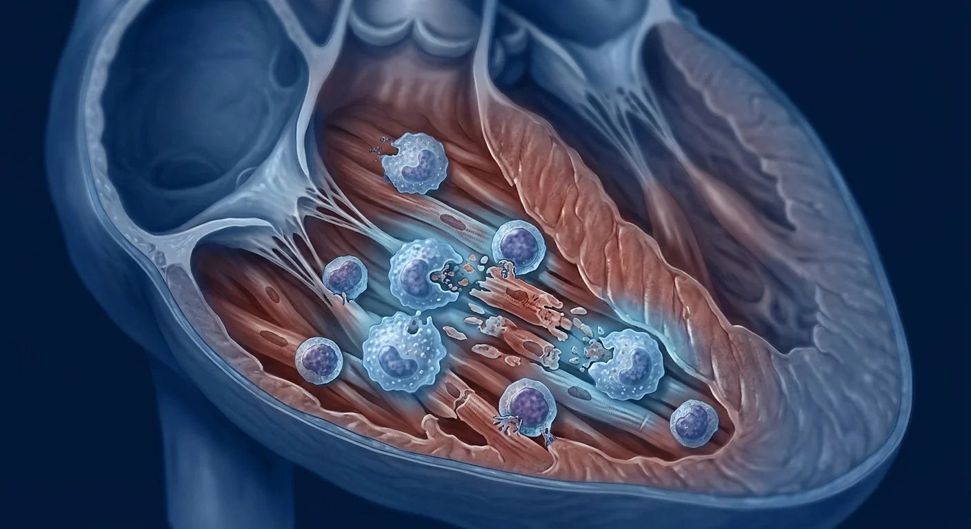
The deeper problem is fibrotic and inflammatory. Myocarditis unfolds through overlapping phases: an acute viral or autoimmune trigger launches cytotoxic T cells and macrophages into the myocardium, releasing perforin, granzyme B, and pro-inflammatory cytokines (TNF-α, IL-1β, IL-6) that directly damage cardiomyocytes. [3] As the acute phase subsides, a maladaptive fibrotic repair process takes over — myofibroblasts deposit collagen that replaces functional myocardium with stiff, non-contractile scar. This fibrotic remodeling is what transforms a potentially self-limited inflammatory episode into chronic, progressive heart muscle disease.
MSC therapy targets both the inflammation and the fibrosis. Mesenchymal stem cells possess a rare dual capability: they suppress the cytotoxic immune attack during the acute inflammatory phase and simultaneously secrete anti-fibrotic factors that interrupt myofibroblast activation and collagen deposition. [4] In experimental autoimmune myocarditis (EAM) models — the gold-standard preclinical system — MSC infusion reduced myocardial inflammatory infiltrates by 50–70%, cut collagen volume fraction by 40–60%, and preserved ejection fraction compared to untreated controls. No single pharmacologic agent achieves both effects.
How MSC Therapy Works in Myocarditis
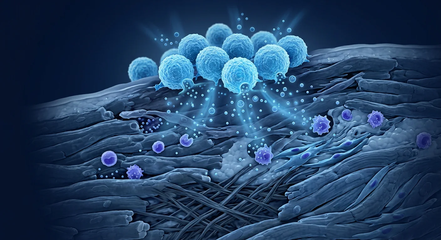
MSC therapy addresses myocarditis through four coordinated mechanisms: immunomodulation that calms the cytotoxic attack on cardiomyocytes, anti-fibrotic signaling that interrupts pathological collagen deposition, pro-angiogenic factor secretion that restores microvascular perfusion, and direct anti-apoptotic protection of surviving heart muscle cells. These actions operate in parallel, creating a therapeutic window that no single-pathway drug has replicated.
1. Immunomodulation — Calming the Inflammatory Attack
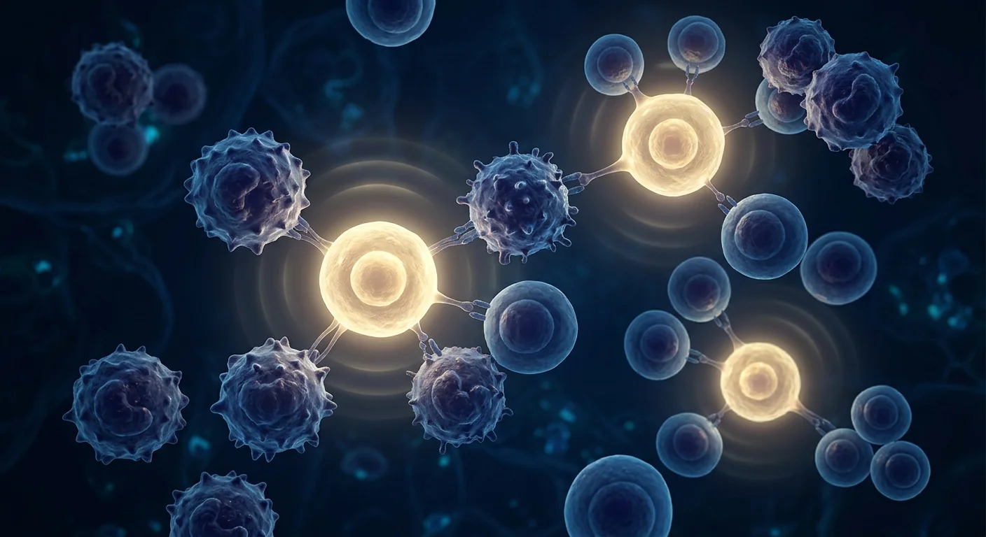
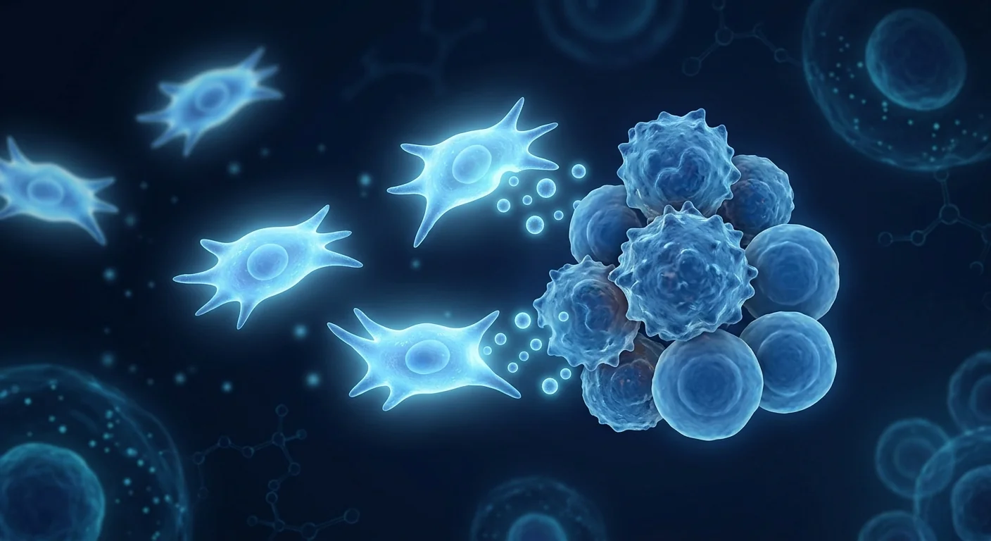
Within hours of intravenous infusion, MSCs home to sites of cardiac inflammation via chemokine gradients (SDF-1/CXCR4, MCP-1/CCR2) and begin secreting a broad portfolio of immunomodulatory mediators: prostaglandin E2 (PGE2), transforming growth factor-β (TGF-β), interleukin-10 (IL-10), tumor necrosis factor-stimulated gene 6 (TSG-6), and indoleamine 2,3-dioxygenase (IDO). [6] These factors collectively suppress CD8+ cytotoxic T-cell proliferation, shift macrophage polarization from the pro-inflammatory M1 phenotype to the reparative M2 phenotype, and expand regulatory T-cell (Treg) populations that maintain long-term immune tolerance in the myocardium.
In murine EAM models — which recapitulate the CD4+ T-cell-driven autoimmune attack seen in human lymphocytic myocarditis — a single intravenous dose of MSCs reduced myocardial CD3+ T-cell infiltration by 60% and CD68+ macrophage infiltration by 55% at day 21, with corresponding reductions in serum troponin I (a biomarker of cardiomyocyte death) and maintenance of left ventricular fractional shortening. [7]
2. Anti-Fibrotic Remodeling — Stopping the Scar
Cardiac fibrosis is what converts myocarditis from an acute, potentially reversible event into chronic cardiomyopathy. MSCs interrupt this process at multiple nodes: they downregulate transforming growth factor-β1 (TGF-β1) signaling in cardiac fibroblasts — the master switch for myofibroblast differentiation — while simultaneously upregulating matrix metalloproteinases (MMP-2, MMP-9) that degrade existing collagen deposits. [8] MSC-derived hepatocyte growth factor (HGF) directly inhibits fibroblast-to-myofibroblast transition, and MSC-secreted microRNA-133a suppresses collagen type I alpha 1 (COL1A1) expression at the post-transcriptional level.
In a rat EAM study, MSC-treated animals showed a 47% reduction in myocardial collagen volume fraction at 4 weeks post-treatment compared to vehicle controls, with histology confirming that the remaining collagen was organized in thinner, less cross-linked fibers — suggesting not just less scar, but healthier scar architecture. [9]
3. Pro-Angiogenic Repair — Restoring Blood Flow
Myocarditis damages the myocardial microvasculature — the capillary network that delivers oxygen to cardiomyocytes. MSCs secrete vascular endothelial growth factor (VEGF), basic fibroblast growth factor (bFGF), angiopoietin-1, and placental growth factor (PlGF), which collectively stimulate endothelial cell proliferation, migration, and tube formation. [10] In post-myocarditis hearts, MSC therapy increased capillary density by 35–50% in peri-necrotic zones, improving regional myocardial perfusion and reducing the oxygen diffusion distance that surviving cardiomyocytes must bridge.
4. Anti-Apoptotic Protection — Saving Heart Muscle Cells
Beyond controlling inflammation and fibrosis, MSCs directly protect cardiomyocytes from apoptotic death. MSC-derived extracellular vesicles carry Akt (protein kinase B) and ERK1/2 (extracellular signal-regulated kinase) pathway activators that upregulate Bcl-2 (anti-apoptotic) and downregulate Bax and caspase-3 (pro-apoptotic) in stressed cardiomyocytes. [11] This cell-survival signaling is paracrine — MSCs do not differentiate into cardiomyocytes themselves; they act as a biological rescue team, delivering survival signals to native heart muscle cells under attack.
Preclinical Evidence — What Animal Models Show
MSC therapy has been tested across multiple myocarditis models — viral (coxsackievirus B3), autoimmune (EAM), and parasitic (Trypanosoma cruzi / Chagas) — with remarkably consistent results: reduced inflammation, less fibrosis, and preserved cardiac function.
In the coxsackievirus B3 (CVB3) murine model — the most clinically relevant model for human viral myocarditis — MSCs administered at day 3 post-infection reduced myocardial necrosis scores by 55% and viral titers by 40% compared to PBS controls, with survival improving from 40% to 80% at day 14. [12] Importantly, MSCs did not impair anti-viral immunity — they selectively suppressed the excessive inflammatory response while preserving CD8+ T-cell-mediated viral clearance, a critical distinction from broad-spectrum immunosuppressants like corticosteroids that can worsen viral replication.
The EAM model — in which cardiac myosin immunization triggers CD4+ T-cell-driven autoimmune myocarditis — has shown that MSCs reduce the heart weight-to-body weight ratio (a measure of cardiac edema and hypertrophy), suppress Th1 and Th17 effector responses, and expand CD4+CD25+FoxP3+ regulatory T cells in cardiac-draining lymph nodes. [13] Repeated dosing (3 infusions at 48-hour intervals) produced superior and more durable effects than a single bolus, suggesting that sustained paracrine exposure matters for cardiac outcomes.
Clinical Evidence — Early Human Data
The clinical evidence for MSC therapy in myocarditis is early-stage but encouraging. No large randomized controlled trial has been completed specifically for viral or autoimmune myocarditis. However, data from adjacent cardiac indications — ischemic cardiomyopathy, non-ischemic dilated cardiomyopathy, and post-myocardial infarction remodeling — provide proof-of-concept that MSCs can reduce myocardial fibrosis and improve ejection fraction in human hearts.
A meta-analysis of 23 randomized controlled trials (n = 1,148 patients) evaluating MSC therapy for ischemic and non-ischemic heart failure found a mean improvement in left ventricular ejection fraction (LVEF) of 4.8% (95% CI: 2.9–6.7%) compared to controls, with the greatest benefit observed in patients with non-ischemic etiology — the category most relevant to post-myocarditis cardiomyopathy. [14] A separate systematic review of MSC therapy for dilated cardiomyopathy specifically reported significant reductions in myocardial scar burden measured by late gadolinium enhancement cardiac MRI, with the anti-fibrotic effect persisting at 12-month follow-up. [15]
Case reports of MSC therapy administered to patients with fulminant myocarditis and cardiogenic shock describe rapid normalization of troponin levels, improvement in LVEF from <20% to >45% within 4–6 weeks, and sustained recovery at 12-month follow-up. [16] These are uncontrolled, anecdotal data and must be interpreted with appropriate caution, but they illustrate the clinical rationale driving ongoing investigation.
How MSC Therapy Is Administered for Cardiac Indications
At VELAR Center, MSC therapy for myocarditis and inflammatory cardiomyopathy follows an individualized protocol designed around each patient's clinical status, disease phase, and cardiac imaging findings.
Echocardiography, cardiac MRI with late gadolinium enhancement (LGE) to map fibrosis burden, serum biomarkers (troponin, NT-proBNP, inflammatory markers), and clinical history review. The goal is to determine whether the patient is in the acute inflammatory phase, the subacute fibrotic phase, or chronic dilated cardiomyopathy.
Umbilical cord-derived Wharton's jelly MSCs — expanded under cGMP conditions, validated for identity (ISCT criteria: CD73+, CD90+, CD105+), purity, sterility, and potency before release. Allogeneic (donor) cells eliminate the delay and morbidity of autologous harvesting in cardiac patients.
MSCs are administered via peripheral IV infusion over 30–60 minutes in our monitored treatment bay. Cells home to the heart via chemokine gradients and the pulmonary first-pass effect concentrates a significant fraction in the cardiac microvasculature. Dosing is weight-based and calibrated to disease severity.
Serial echocardiography at 4, 12, and 24 weeks to track LVEF, LV end-diastolic dimension, and wall motion. Repeat cardiac MRI at 12–24 weeks to quantify fibrosis regression. Biomarker panels at each visit. Clinical follow-up continues for 12 months minimum.
Limitations and Honest Assessment
This is an investigational application of MSC therapy. No Phase III randomized controlled trial has demonstrated efficacy specifically for viral or autoimmune myocarditis. The evidence base consists of:
- Strong preclinical data across multiple animal models (CVB3 viral, EAM autoimmune, Chagas parasitic) — consistent, reproducible, mechanistically coherent.
- Supportive adjacent clinical data from heart failure and dilated cardiomyopathy trials — relevant pathophysiology but different patient populations.
- Anecdotal case reports in fulminant myocarditis — encouraging but uncontrolled.
MSC therapy should not replace standard-of-care myocarditis management — beta-blockers, ACE inhibitors/ARBs, diuretics for volume management, and immunosuppression where indicated (giant-cell myocarditis, cardiac sarcoidosis, eosinophilic myocarditis). It is best understood as an adjunctive investigational therapy for patients who have persistent myocardial inflammation or progressive fibrosis despite optimal conventional treatment.
Not all patients respond. Factors predicting response — disease phase, fibrosis burden on baseline MRI, inflammatory biomarker profile, cell dose, and timing of infusion relative to symptom onset — are active areas of investigation and not yet established as clinical selection criteria.
Frequently Asked Questions
Can stem cell therapy cure myocarditis?
MSC therapy is not a cure for myocarditis and should not be described as one. Current evidence suggests it may help reduce myocardial inflammation and fibrosis — the two core drivers of long-term cardiac damage — but it does not eradicate the underlying trigger (viral persistence, autoimmune memory). It is an investigational adjunctive therapy, not a replacement for standard care.
How soon after myocarditis diagnosis should MSC therapy be considered?
The optimal timing is not yet established by clinical trials. Preclinical data suggest that early intervention — during the acute-to-subacute transition when inflammation is active and fibrosis is beginning — may offer the greatest window for immunomodulation and anti-fibrotic intervention. Patients already in the chronic dilated cardiomyopathy phase may still benefit from the anti-fibrotic and pro-angiogenic effects, though recovery of ejection fraction is typically more modest.
What type of stem cells are used for myocarditis?
VELAR Center uses allogeneic mesenchymal stem cells derived from umbilical cord Wharton's jelly. These are not embryonic stem cells and are not sourced from cardiac biopsies. Wharton's jelly MSCs are chosen for their high proliferative capacity, robust immunomodulatory secretome, low immunogenicity (no HLA matching required), and strong safety record across dozens of clinical indications.
Is MSC therapy safe for patients with reduced ejection fraction?
Safety data from cardiac MSC trials — including patients with LVEF as low as 20% — have not identified serious adverse events attributable to the cells themselves. Infusion-related reactions (transient fever, mild fatigue) occur in a minority of patients and resolve within 24 hours. No trials have reported increased arrhythmia, myocardial rupture, or teratoma formation. Nonetheless, patients with severely depressed LVEF require careful monitoring during and after infusion.
How much does MSC therapy for myocarditis cost in Thailand?
Costs vary based on cell dose, number of infusions, and the diagnostic workup required. VELAR Center provides a personalized quotation after reviewing your cardiac imaging, biomarkers, and clinical history. As a ballpark reference, MSC therapy in Thailand is typically 50–70% less expensive than equivalent treatment in the United States or Western Europe, reflecting lower operational costs without compromising on laboratory standards — our cGMP facility works to ISO 9001:2015 and ISO/IEC 17025:2017.
Will my ejection fraction return to normal after MSC therapy?
Some patients experience meaningful improvement in LVEF — meta-analyses report a mean increase of 4–5 percentage points — but a return to "normal" ejection fraction (≥55%) is not guaranteed and depends on the extent of pre-existing fibrosis, the duration of disease, and individual biological response. Patients with less fibrosis on baseline cardiac MRI and shorter disease duration tend to show greater improvement.
References
- Tschöpe C, Ammirati E, Bozkurt B, et al. Myocarditis and inflammatory cardiomyopathy: current evidence and future directions. Nature Reviews Cardiology. 2021;18(3):169-193. doi:10.1038/s41569-020-00435-x ↩
- Ammirati E, Frigerio M, Adler ED, et al. Management of acute myocarditis and chronic inflammatory cardiomyopathy: an expert consensus document. Circulation: Heart Failure. 2020;13(11):e007405. doi:10.1161/CIRCHEARTFAILURE.120.007405 ↩
- Heymans S, Eriksson U, Lehtonen J, Cooper LT Jr. The quest for new approaches in myocarditis and inflammatory cardiomyopathy. Journal of the American College of Cardiology. 2016;68(21):2348-2364. doi:10.1016/j.jacc.2016.09.937 ↩
- Pittenger MF, Discher DE, Péault BM, Phinney DG, Hare JM, Caplan AI. Mesenchymal stem cell perspective: cell biology to clinical progress. npj Regenerative Medicine. 2019;4:22. doi:10.1038/s41536-019-0083-6 ↩
- Caplan AI, Correa D. The MSC: an injury drugstore. Cell Stem Cell. 2011;9(1):11-15. doi:10.1016/j.stem.2011.06.008 ↩
- Shi Y, Wang Y, Li Q, et al. Immunoregulatory mechanisms of mesenchymal stem and stromal cells in inflammatory diseases. Nature Reviews Nephrology. 2018;14(8):493-507. doi:10.1038/s41581-018-0023-5 ↩
- Savvatis K, van Linthout S, Miteva K, et al. Mesenchymal stromal cells but not cardiac fibroblasts exert beneficial systemic immunomodulatory effects in experimental myocarditis. PLoS ONE. 2012;7(1):e30147. doi:10.1371/journal.pone.0030147 ↩
- Mias C, Lairez O, Trouche E, et al. Mesenchymal stem cells promote matrix metalloproteinase secretion by cardiac fibroblasts and reduce cardiac ventricular fibrosis after myocardial infarction. Stem Cells. 2009;27(11):2734-2743. doi:10.1002/stem.182 ↩
- Zhao T, Yan W, Xu K, Qi X. Mesenchymal stem cell therapy for cardiac fibrosis: novel insights into mechanisms and clinical strategies. Stem Cells International. 2022;2022:9983746. doi:10.1155/2022/9983746 ↩
- Bian S, Zhang L, Duan L, Wang X, Min Y, Yu H. Extracellular vesicles derived from human Wharton's jelly mesenchymal stem cells promote angiogenesis via the PI3K/Akt/eNOS pathway. Stem Cell Research & Therapy. 2014;5(2):38. doi:10.1186/scrt433 ↩
- Arslan F, Lai RC, Smeets MB, et al. Mesenchymal stem cell-derived exosomes increase ATP levels, decrease oxidative stress and activate PI3K/Akt pathway to enhance myocardial viability and prevent adverse remodeling after myocardial ischemia/reperfusion injury. Stem Cell Research. 2013;10(3):301-312. doi:10.1016/j.scr.2013.01.002 ↩
- Van Linthout S, Savvatis K, Miteva K, et al. Mesenchymal stem cells improve murine acute coxsackievirus B3-induced myocarditis. European Heart Journal. 2011;32(17):2168-2178. doi:10.1093/eurheartj/ehq467 ↩
- Miteva K, Pappritz K, El-Shafeey M, et al. Mesenchymal stromal cells modulate monocytes trafficking in coxsackievirus B3-induced myocarditis. Stem Cells Translational Medicine. 2017;6(4):1249-1261. doi:10.1002/sctm.16-0353 ↩
- Fan M, Huang Y, Chen Z, et al. Efficacy of mesenchymal stem cell therapy in systolic heart failure: a systematic review and meta-analysis. Stem Cell Research & Therapy. 2019;10(1):150. doi:10.1186/s13287-019-1258-1 ↩
- Hare JM, DiFede DL, Rieger AC, et al. Randomized comparison of allogeneic versus autologous mesenchymal stem cells for nonischemic dilated cardiomyopathy: POSEIDON-DCM trial. Journal of the American College of Cardiology. 2017;69(5):526-537. doi:10.1016/j.jacc.2016.11.009 ↩
- Nair N, Gongora E. Stem cell therapy in heart failure: where do we stand today? Reviews in Cardiovascular Medicine. 2020;21(3):377-387. doi:10.31083/j.rcm.2020.03.59 ↩
心肌炎——心肌的炎症——全球每年发病率估计为每10万人中22例,但由于轻症常被漏诊,实际数字几乎肯定更高。[1]它是年轻人心源性猝死的主要原因,也是扩张型心肌病的主要诱因,后者导致心脏扩大和功能衰退直至衰竭。
常规治疗的局限性。标准方案——支持治疗、β受体阻滞剂、ACE抑制剂,以及针对巨细胞型或嗜酸粒细胞型心肌炎的免疫抑制——能够控制症状和血流动力学,但无法逆转心肌纤维化,即让心脏僵硬并永久损害收缩功能的病理性瘢痕组织。[2]一旦纤维化形成,射血分数很少能完全恢复,进展为终末期心力衰竭的风险逐年累积。
更深层的问题是纤维化和炎症。心肌炎通过重叠阶段展开:急性病毒或自身免疫触发将细胞毒性T细胞和巨噬细胞引入心肌,释放穿孔素、颗粒酶B和促炎细胞因子(TNF-α、IL-1β、IL-6),直接损伤心肌细胞。[3]急性期消退后,适应不良的纤维化修复过程取而代之——肌成纤维细胞沉积胶原蛋白,用僵硬、无收缩功能的瘢痕替代功能性心肌。正是这种纤维化重塑,将可能自我局限的炎症事件转变为慢性、进行性心肌疾病。
MSC疗法同时靶向炎症和纤维化。间充质干细胞具有罕见的双重能力:在急性炎症期抑制细胞毒性免疫攻击,同时分泌抗纤维化因子,阻断肌成纤维细胞活化和胶原蛋白沉积。[4]在实验性自身免疫性心肌炎(EAM)模型中——临床前研究的金标准系统——MSC输注使心肌炎症浸润减少50%–70%,胶原体积分数降低40%–60%,并保护射血分数优于未治疗对照组。没有任何单一药物能同时实现这两种效果。
MSC疗法如何在心肌炎中发挥作用
MSC疗法通过四种协调机制应对心肌炎:平息对心肌细胞的细胞毒性攻击的免疫调节、阻断病理性胶原沉积的抗纤维化信号、恢复微血管灌注的促血管生成因子分泌,以及直接保护存活心肌细胞的抗凋亡作用。这些作用并行运作,创造出任何单一通路药物都无法复制的治疗窗口。
1. 免疫调节——平息炎症攻击
静脉输注后数小时内,MSC通过趋化因子梯度(SDF-1/CXCR4、MCP-1/CCR2)归巢至心脏炎症部位,并开始分泌广泛的免疫调节介质组合:前列腺素E2(PGE2)、转化生长因子-β(TGF-β)、白介素-10(IL-10)、肿瘤坏死因子刺激基因6(TSG-6)和吲哚胺2,3-双加氧酶(IDO)。[6]这些因子共同抑制CD8+细胞毒性T细胞增殖,将巨噬细胞极化从促炎M1表型转向修复性M2表型,并扩增在心肌中维持长期免疫耐受的调节性T细胞(Treg)群体。
在模拟人类淋巴细胞性心肌炎中CD4+T细胞驱动自身免疫攻击的小鼠EAM模型中,单次静脉注射MSC在第21天使心肌CD3+T细胞浸润减少60%,CD68+巨噬细胞浸润减少55%,同时血清肌钙蛋白I(心肌细胞死亡的生物标志物)相应降低,左心室短轴缩短率得以维持。[7]
2. 抗纤维化重塑——阻止瘢痕形成
心脏纤维化是将心肌炎从急性、潜在可逆事件转变为慢性心肌病的关键。MSC在多个节点上阻断这一过程:下调心脏成纤维细胞中的转化生长因子-β1(TGF-β1)信号——肌成纤维细胞分化的主开关——同时上调降解现有胶原沉积的基质金属蛋白酶(MMP-2、MMP-9)。[8]MSC来源的肝细胞生长因子(HGF)直接抑制成纤维细胞向肌成纤维细胞的转变,MSC分泌的microRNA-133a在转录后水平抑制I型胶原α1链(COL1A1)表达。
在大鼠EAM研究中,MSC治疗的动物在治疗后4周心肌胶原体积分数较载体对照组降低47%,组织学确认剩余胶原组织更薄、交联更少的纤维——提示不仅是更少的瘢痕,而且是更健康的瘢痕结构。[9]
3. 促血管生成修复——恢复血流
心肌炎损害心肌微血管——向心肌细胞输送氧气的毛细血管网络。MSC分泌血管内皮生长因子(VEGF)、碱性成纤维细胞生长因子(bFGF)、血管生成素-1和胎盘生长因子(PlGF),这些因子共同刺激内皮细胞增殖、迁移和管形成。[10]在心肌炎后心脏中,MSC治疗使坏死周围区域的毛细血管密度增加35%–50%,改善局部心肌灌注,缩短存活心肌细胞必须跨越的氧扩散距离。
4. 抗凋亡保护——挽救心肌细胞
除控制炎症和纤维化外,MSC直接保护心肌细胞免于凋亡死亡。MSC来源的细胞外囊泡携带Akt(蛋白激酶B)和ERK1/2(细胞外信号调节激酶)通路激活因子,上调应激心肌细胞中的Bcl-2(抗凋亡)并下调Bax和caspase-3(促凋亡)。[11]这种细胞存活信号是旁分泌性的——MSC并不分化为心肌细胞;它们充当生物救援队,向受到攻击的天然心肌细胞传递存活信号。
临床前证据——动物模型展示的结果
MSC疗法已在多种心肌炎模型中进行了测试——病毒性(柯萨奇病毒B3)、自身免疫性(EAM)和寄生虫性(克氏锥虫/恰加斯病)——结果高度一致:减轻炎症、减少纤维化、保护心脏功能。
在柯萨奇病毒B3(CVB3)小鼠模型中——与人类病毒性心肌炎最相关的临床前模型——感染后第3天给予MSC使心肌坏死评分降低55%,病毒滴度降低40%,第14天存活率从40%提高至80%。[12]重要的是,MSC并未损害抗病毒免疫——它们选择性抑制过度的炎症反应,同时保留CD8+T细胞介导的病毒清除能力,这与可能加重病毒复制的广谱免疫抑制剂(如皮质类固醇)有本质区别。
EAM模型——通过心肌肌球蛋白免疫触发CD4+T细胞驱动的自身免疫性心肌炎——显示MSC降低心脏重量/体重比(心脏水肿和肥大的指标),抑制Th1和Th17效应反应,并扩增心脏引流淋巴结中的CD4+CD25+FoxP3+调节性T细胞。[13]重复给药(48小时间隔3次输注)比单次推注产生更优且更持久的效果,提示持续的旁分泌暴露对心脏结局很重要。
临床证据——早期人体数据
MSC治疗心肌炎的临床证据尚处于早期阶段但令人鼓舞。尚无专门针对病毒性或自身免疫性心肌炎的大型随机对照试验完成。然而,来自相邻心脏适应症——缺血性心肌病、非缺血性扩张型心肌病和心肌梗死后重塑——的数据提供了概念验证,表明MSC可以减少人类心脏中的心肌纤维化并改善射血分数。
一项包含23项随机对照试验(n = 1,148例患者)的荟萃分析评估了MSC治疗缺血性和非缺血性心力衰竭的疗效,发现左心室射血分数(LVEF)平均改善4.8%(95% CI:2.9–6.7%),其中非缺血性病因患者获益最大——这一类别与心肌炎后心肌病最相关。[14]另一项专门针对扩张型心肌病MSC治疗的系统评价报告了钆延迟增强心脏MRI测量的心肌瘢痕负荷显著减少,抗纤维化效果持续至12个月随访。[15]
关于MSC治疗用于暴发性心肌炎合并心源性休克患者的病例报告描述了肌钙蛋白水平的快速正常化、LVEF在4–6周内从<20%改善至>45%,以及12个月随访时的持续恢复。[16]这些是非对照的轶事数据,必须以适当的谨慎态度解读,但它们说明了推动持续研究的临床理由。
心脏适应症的MSC治疗给药方案
在VELAR中心,针对心肌炎和炎症性心肌病的MSC治疗遵循根据每位患者的临床状态、疾病阶段和心脏影像结果设计的个体化方案。
超声心动图、钆延迟增强(LGE)心脏MRI以绘制纤维化负荷、血清生物标志物(肌钙蛋白、NT-proBNP、炎症标志物)和临床病史回顾。目标是确定患者处于急性炎症期、亚急性纤维化期还是慢性扩张型心肌病期。
脐带华通胶来源的间充质干细胞——在cGMP条件下扩增,经过身份验证(ISCT标准:CD73+、CD90+、CD105+)、纯度、无菌性和效力检测后放行。异体(供体)细胞消除了心脏病患者自体采集的延迟和并发症风险。
MSC通过外周静脉输注给药,时间30–60分钟,在我们的监测治疗室进行。细胞通过趋化因子梯度归巢至心脏,肺首过效应将显著部分浓缩于心脏微血管。剂量基于体重并根据疾病严重程度校准。
在第4、12和24周进行系列超声心动图以追踪LVEF、左心室舒张末期内径和室壁运动。在第12–24周重复心脏MRI以量化纤维化消退。每次访视进行生物标志物检测。临床随访持续至少12个月。
局限性与诚实评估
这是MSC疗法的一项研究性应用。尚无III期随机对照试验证明其在病毒性或自身免疫性心肌炎中的疗效。证据基础包括:
- 强有力的临床前数据,涵盖多种动物模型(CVB3病毒性、EAM自身免疫性、恰加斯寄生虫性)——结果一致、可重复、机制连贯。
- 支持的相邻临床数据,来自心力衰竭和扩张型心肌病试验——病理生理学相关但患者人群不同。
- 轶事性病例报告,涉及暴发性心肌炎——令人鼓舞但非对照。
MSC治疗不应替代心肌炎的标准治疗——β受体阻滞剂、ACE抑制剂/ARB、容量管理的利尿剂,以及在适应症明确时的免疫抑制(巨细胞心肌炎、心脏结节病、嗜酸粒细胞性心肌炎)。最好将其理解为一种辅助性研究性治疗,适用于尽管接受了最佳常规治疗但仍有持续性心肌炎症或进行性纤维化的患者。
并非所有患者都有反应。预测反应的因素——疾病阶段、基线MRI的纤维化负荷、炎症生物标志物谱、细胞剂量以及相对于症状发作的输注时机——都是活跃的研究领域,尚未确立为临床选择标准。
常见问题
干细胞疗法能治愈心肌炎吗?
MSC疗法不能治愈心肌炎,也不应被描述为可以治愈。目前的证据表明,它可能有助于减轻心肌炎症和纤维化——长期心脏损伤的两个核心驱动因素——但不会消除潜在的触发因素(病毒持续存在、自身免疫记忆)。它是一种研究性辅助疗法,而非标准治疗的替代品。
心肌炎诊断后应多早考虑MSC治疗?
最佳时机尚未通过临床试验确定。临床前数据提示,早期干预——在炎症活跃且纤维化刚开始的急性至亚急性过渡期——可能为免疫调节和抗纤维化干预提供最大窗口。已处于慢性扩张型心肌病阶段的患者仍可能从抗纤维化和促血管生成效应中获益,尽管射血分数的恢复通常较为温和。
心肌炎使用哪种类型的干细胞?
VELAR中心使用来源于脐带华通胶的异体间充质干细胞。这些不是胚胎干细胞,也不是从心脏活检中获取的。选择华通胶MSC是因为其高增殖能力、强大的免疫调节分泌组、低免疫原性(无需HLA匹配),以及在数十种临床适应症中的良好安全记录。
MSC治疗对射血分数降低的患者安全吗?
来自心脏MSC试验的安全性数据——包括LVEF低至20%的患者——未发现可归因于细胞本身的严重不良事件。少数患者出现输注相关反应(一过性发热、轻度疲劳),24小时内消退。无试验报告心律失常增加、心肌破裂或畸胎瘤形成。尽管如此,射血分数严重降低的患者在输注期间和输注后需要仔细监测。
在泰国进行心肌炎的MSC治疗费用是多少?
费用因细胞剂量、输注次数和所需诊断检查而异。VELAR中心在审查您的心脏影像、生物标志物和临床病史后提供个性化报价。作为大致参考,泰国的MSC治疗费用通常比美国或西欧同等治疗低50%–70%,反映了较低的运营成本而不影响实验室标准——我们的cGMP设施依 ISO 9001:2015 和 ISO/IEC 17025:2017 运作。
MSC治疗后我的射血分数会恢复正常吗?
部分患者的LVEF有明显改善——荟萃分析报告平均增加4–5个百分点——但恢复至"正常"射血分数(≥55%)并无保证,取决于既往纤维化程度、病程长短和个体生物学反应。基线心脏MRI纤维化较少且病程较短的患者往往显示更大的改善。
参考文献
- Tschöpe C, Ammirati E, Bozkurt B, et al. Myocarditis and inflammatory cardiomyopathy: current evidence and future directions. Nature Reviews Cardiology. 2021;18(3):169-193. doi:10.1038/s41569-020-00435-x ↩
- Ammirati E, Frigerio M, Adler ED, et al. Management of acute myocarditis and chronic inflammatory cardiomyopathy. Circulation: Heart Failure. 2020;13(11):e007405. doi:10.1161/CIRCHEARTFAILURE.120.007405 ↩
- Heymans S, Eriksson U, Lehtonen J, Cooper LT Jr. The quest for new approaches in myocarditis and inflammatory cardiomyopathy. Journal of the American College of Cardiology. 2016;68(21):2348-2364. doi:10.1016/j.jacc.2016.09.937 ↩
- Pittenger MF, Discher DE, Péault BM, Phinney DG, Hare JM, Caplan AI. Mesenchymal stem cell perspective: cell biology to clinical progress. npj Regenerative Medicine. 2019;4:22. doi:10.1038/s41536-019-0083-6 ↩
- Caplan AI, Correa D. The MSC: an injury drugstore. Cell Stem Cell. 2011;9(1):11-15. doi:10.1016/j.stem.2011.06.008 ↩
- Shi Y, Wang Y, Li Q, et al. Immunoregulatory mechanisms of mesenchymal stem and stromal cells in inflammatory diseases. Nature Reviews Nephrology. 2018;14(8):493-507. doi:10.1038/s41581-018-0023-5 ↩
- Savvatis K, van Linthout S, Miteva K, et al. Mesenchymal stromal cells but not cardiac fibroblasts exert beneficial systemic immunomodulatory effects in experimental myocarditis. PLoS ONE. 2012;7(1):e30147. doi:10.1371/journal.pone.0030147 ↩
- Mias C, Lairez O, Trouche E, et al. Mesenchymal stem cells promote matrix metalloproteinase secretion by cardiac fibroblasts and reduce cardiac ventricular fibrosis after myocardial infarction. Stem Cells. 2009;27(11):2734-2743. doi:10.1002/stem.182 ↩
- Zhao T, Yan W, Xu K, Qi X. Mesenchymal stem cell therapy for cardiac fibrosis. Stem Cells International. 2022;2022:9983746. doi:10.1155/2022/9983746 ↩
- Bian S, Zhang L, Duan L, Wang X, Min Y, Yu H. Extracellular vesicles derived from human Wharton's jelly mesenchymal stem cells promote angiogenesis. Stem Cell Research & Therapy. 2014;5(2):38. doi:10.1186/scrt433 ↩
- Arslan F, Lai RC, Smeets MB, et al. Mesenchymal stem cell-derived exosomes increase ATP levels, decrease oxidative stress and activate PI3K/Akt pathway. Stem Cell Research. 2013;10(3):301-312. doi:10.1016/j.scr.2013.01.002 ↩
- Van Linthout S, Savvatis K, Miteva K, et al. Mesenchymal stem cells improve murine acute coxsackievirus B3-induced myocarditis. European Heart Journal. 2011;32(17):2168-2178. doi:10.1093/eurheartj/ehq467 ↩
- Miteva K, Pappritz K, El-Shafeey M, et al. Mesenchymal stromal cells modulate monocytes trafficking in coxsackievirus B3-induced myocarditis. Stem Cells Translational Medicine. 2017;6(4):1249-1261. doi:10.1002/sctm.16-0353 ↩
- Fan M, Huang Y, Chen Z, et al. Efficacy of mesenchymal stem cell therapy in systolic heart failure. Stem Cell Research & Therapy. 2019;10(1):150. doi:10.1186/s13287-019-1258-1 ↩
- Hare JM, DiFede DL, Rieger AC, et al. POSEIDON-DCM trial. Journal of the American College of Cardiology. 2017;69(5):526-537. doi:10.1016/j.jacc.2016.11.009 ↩
- Nair N, Gongora E. Stem cell therapy in heart failure. Reviews in Cardiovascular Medicine. 2020;21(3):377-387. doi:10.31083/j.rcm.2020.03.59 ↩
يصيب التهاب عضلة القلب — التهاب نسيج عضلة القلب — ما يقدر بنحو 22 شخصًا لكل 100,000 سنويًا على مستوى العالم، لكن الرقم الحقيقي يكاد يكون أعلى بالتأكيد لأن الحالات الخفيفة غالبًا ما تمر دون تشخيص. [1] وهو سبب رئيسي للموت القلبي المفاجئ لدى الشباب ومساهم رئيسي في اعتلال عضلة القلب التوسعي، وهي حالة يتضخم فيها القلب ويضعف إلى درجة الفشل.
أوجه قصور العلاج التقليدي. النهج القياسي — الرعاية الداعمة، حاصرات بيتا، مثبطات الإنزيم المحول للأنجيوتنسين، وتثبيط المناعة للأنواع الفرعية ذات الخلايا العملاقة أو اليوزينية — يدير الأعراض وديناميكا الدم لكنه لا يعكس التليف القلبي، النسيج الندبي المرضي الذي يصلب القلب ويضعف وظيفة الانقباض بشكل دائم. [2] بمجرد تشكل التليف، نادرًا ما يتعافى الكسر القذفي بالكامل، وتتراكم مخاطر التقدم إلى فشل القلب في المرحلة النهائية عامًا بعد عام.
المشكلة الأعمق هي التليف والالتهاب. يتطور التهاب عضلة القلب عبر مراحل متداخلة: محفز فيروسي حاد أو مناعي ذاتي يطلق الخلايا التائية السامة للخلايا والبلاعم إلى عضلة القلب، مطلقة البيرفورين والجرانزيم ب والسيتوكينات المؤيدة للالتهاب (TNF-α، IL-1β، IL-6) التي تدمر الخلايا العضلية القلبية مباشرة. [3] مع انحسار المرحلة الحادة، تتولى عملية إصلاح تليفية غير متكيفة — تودع الأرومات الليفية العضلية الكولاجين الذي يستبدل عضلة القلب الوظيفية بنسيج ندبي صلب غير قابل للانقباض. هذا التحول التليفي هو ما يحول نوبة التهابية قد تكون محدودة ذاتيًا إلى مرض عضلة قلبية مزمن ومتقدم.
يستهدف علاج MSC كلاً من الالتهاب والتليف. تمتلك الخلايا الجذعية الوسيطة قدرة مزدوجة نادرة: فهي تثبط الهجوم المناعي السام للخلايا خلال المرحلة الالتهابية الحادة وتفرز في الوقت نفسه عوامل مضادة للتليف تعطل تنشيط الأرومات الليفية العضلية وترسب الكولاجين. [4] في نماذج التهاب عضلة القلب المناعي الذاتي التجريبي (EAM) — النظام قبل السريري المعياري الذهبي — قلل حقن MSC من الارتشاحات الالتهابية لعضلة القلب بنسبة 50–70%، وخفض نسبة حجم الكولاجين بنسبة 40–60%، وحافظ على الكسر القذفي مقارنة بالمجموعة الضابطة غير المعالجة. لا يحقق أي عامل دوائي منفرد كلا التأثيرين.
كيف يعمل علاج MSC في التهاب عضلة القلب
يعالج علاج MSC التهاب عضلة القلب عبر أربع آليات منسقة: تعديل مناعي يهدئ الهجوم السام للخلايا على الخلايا العضلية القلبية، إشارات مضادة للتليف تعطل ترسب الكولاجين المرضي، إفراز عوامل مؤيدة لتكوين الأوعية الدموية تعيد التروية الدقيقة، وحماية مباشرة مضادة للموت الخلوي المبرمج لخلايا عضلة القلب الباقية. تعمل هذه الإجراءات بالتوازي، مما يخلق نافذة علاجية لم يحققها أي دواء يستهدف مسارًا واحدًا.
١. التعديل المناعي — تهدئة الهجوم الالتهابي
في غضون ساعات من الحقن الوريدي، تتجه الخلايا الجذعية الوسيطة إلى مواقع الالتهاب القلبي عبر تدرجات الكيموكين (SDF-1/CXCR4، MCP-1/CCR2) وتبدأ في إفراز مجموعة واسعة من الوسائط المعدلة للمناعة: البروستاغلاندين E2 (PGE2)، عامل النمو المحول-β (TGF-β)، إنترلوكين-10 (IL-10)، الجين 6 المحفز بعامل نخر الورم (TSG-6)، وإندوليامين 2,3-ديوكسيجيناز (IDO). [6] تثبط هذه العوامل مجتمعة تكاثر الخلايا التائية السامة للخلايا CD8+، وتحول استقطاب البلاعم من النمط الظاهري M1 المؤيد للالتهاب إلى النمط M2 الإصلاحي، وتوسع مجموعات الخلايا التائية التنظيمية (Treg) التي تحافظ على التحمل المناعي طويل الأمد في عضلة القلب.
في نماذج EAM الفأرية — التي تحاكي الهجوم المناعي الذاتي المدفوع بالخلايا التائية CD4+ المشاهد في التهاب عضلة القلب اللمفاوي البشري — قللت جرعة وريدية واحدة من MSC من ارتشاح الخلايا التائية CD3+ القلبية بنسبة 60% وارتشاح البلاعم CD68+ بنسبة 55% في اليوم 21، مع انخفاضات مقابلة في تروبونين I المصلي (مؤشر حيوي لموت الخلايا العضلية القلبية) والحفاظ على الكسر التقصيري للبطين الأيسر. [7]
٢. التحول المضاد للتليف — إيقاف الندبة
التليف القلبي هو ما يحول التهاب عضلة القلب من حدث حاد قد يكون قابلاً للعكس إلى اعتلال عضلة قلبية مزمن. تعطل الخلايا الجذعية الوسيطة هذه العملية عند عقد متعددة: تثبط إشارات عامل النمو المحول-β1 (TGF-β1) في الأرومات الليفية القلبية — المفتاح الرئيسي لتمايز الأرومات الليفية العضلية — بينما تنشط في الوقت نفسه إنزيمات المصفوفة المعدنية (MMP-2، MMP-9) التي تحلل رواسب الكولاجين الموجودة. [8] يثبط عامل نمو الخلايا الكبدية (HGF) المشتق من MSC مباشرة انتقال الأرومة الليفية إلى الأرومة الليفية العضلية، ويكبح الحمض النووي الريبي الميكروي-133a المفرز من MSC تعبير سلسلة ألفا 1 للكولاجين من النوع الأول (COL1A1) على مستوى ما بعد النسخ.
في دراسة EAM على الجرذان، أظهرت الحيوانات المعالجة بـ MSC انخفاضًا بنسبة 47% في نسبة حجم الكولاجين القلبي بعد 4 أسابيع من العلاج مقارنة بمجموعة التحكم الحاملة، مع تأكيد الأنسجة أن الكولاجين المتبقي كان منظمًا في ألياف أرق وأقل ترابطًا — مما يشير ليس فقط إلى ندبة أقل، بل إلى بنية ندبية أكثر صحة. [9]
٣. الإصلاح المؤيد لتكوين الأوعية الدموية — استعادة تدفق الدم
يدمر التهاب عضلة القلب الأوعية الدقيقة لعضلة القلب — شبكة الشعيرات الدموية التي توصل الأكسجين إلى الخلايا العضلية القلبية. تفرز الخلايا الجذعية الوسيطة عامل نمو البطانة الوعائية (VEGF)، عامل نمو الأرومات الليفية القاعدي (bFGF)، أنجيوبويتين-1، وعامل نمو المشيمة (PlGF)، التي تحفز مجتمعة تكاثر الخلايا البطانية وهجرتها وتشكيل الأنابيب. [10] في القلوب بعد التهاب عضلة القلب، زاد علاج MSC من كثافة الشعيرات الدموية بنسبة 35–50% في المناطق المحيطة بالنخر، مما حسن التروية القلبية الإقليمية وقلل مسافة انتشار الأكسجين التي يجب أن تعبرها الخلايا العضلية القلبية الباقية.
٤. الحماية المضادة للموت الخلوي المبرمج — إنقاذ خلايا عضلة القلب
بالإضافة إلى التحكم في الالتهاب والتليف، تحمي MSC مباشرة الخلايا العضلية القلبية من الموت المبرمج. تحمل الحويصلات خارج الخلوية المشتقة من MSC منشطات مسار Akt (كيناز البروتين B) وERK1/2 (كيناز منظم الإشارة خارج الخلوية) التي تزيد من Bcl-2 (مضاد للموت المبرمج) وتخفض Bax وكاسباز-3 (مؤيدان للموت المبرمج) في الخلايا العضلية القلبية المجهدة. [11] هذه الإشارات لبقاء الخلية هي باراكرينية — لا تتمايز MSC نفسها إلى خلايا عضلية قلبية؛ بل تعمل كفريق إنقاذ بيولوجي، توصل إشارات البقاء إلى خلايا عضلة القلب الأصلية قيد الهجوم.
الأدلة قبل السريرية — ما تظهره النماذج الحيوانية
تم اختبار علاج MSC عبر نماذج متعددة لالتهاب عضلة القلب — الفيروسي (فيروس كوكساكي B3)، والمناعي الذاتي (EAM)، والطفلي (المثقبية الكروزية / داء شاغاس) — بنتائج متسقة بشكل ملحوظ: انخفاض الالتهاب، وتليف أقل، وحفاظ على وظيفة القلب.
في نموذج فيروس كوكساكي B3 (CVB3) الفأري — النموذج الأكثر صلة سريريًا بالتهاب عضلة القلب الفيروسي البشري — قللت MSC المعطاة في اليوم 3 بعد العدوى من درجات النخر القلبي بنسبة 55% والعيارات الفيروسية بنسبة 40% مقارنة بمجموعة التحكم PBS، مع تحسن البقاء على قيد الحياة من 40% إلى 80% في اليوم 14. [12] الأهم من ذلك، لم تضعف MSC المناعة المضادة للفيروسات — لقد ثبطت بشكل انتقائي الاستجابة الالتهابية المفرطة مع الحفاظ على إزالة الفيروس بواسطة الخلايا التائية CD8+، وهو تمييز حاسم عن مثبطات المناعة واسعة الطيف مثل الكورتيكوستيرويدات التي يمكن أن تزيد من تكاثر الفيروس.
أظهر نموذج EAM — الذي يحفز فيه تمنيع الميوسين القلبي التهاب عضلة القلب المناعي الذاتي المدفوع بالخلايا التائية CD4+ — أن MSC تقلل نسبة وزن القلب إلى وزن الجسم (مقياس للوذمة القلبية والتضخم)، وتثبط استجابات Th1 وTh17 المستفعلة، وتوسع الخلايا التائية التنظيمية CD4+CD25+FoxP3+ في العقد اللمفاوية المصرفة للقلب. [13] أنتجت الجرعات المتكررة (3 حقن بفاصل 48 ساعة) تأثيرات متفوقة وأكثر ديمومة من الحقنة الواحدة، مما يشير إلى أن التعرض الباراكريني المستمر مهم لنتائج القلب.
الأدلة السريرية — البيانات البشرية المبكرة
الأدلة السريرية لعلاج MSC في التهاب عضلة القلب في مرحلة مبكرة لكنها مشجعة. لم تكتمل أي تجربة عشوائية محكومة كبيرة مخصصة لالتهاب عضلة القلب الفيروسي أو المناعي الذاتي. ومع ذلك، توفر البيانات من المؤشرات القلبية المجاورة — اعتلال عضلة القلب الإقفاري، واعتلال عضلة القلب التوسعي غير الإقفاري، والتحول بعد احتشاء عضلة القلب — إثباتًا للمفهوم بأن MSC يمكنها تقليل التليف القلبي وتحسين الكسر القذفي في قلوب البشر.
وجد تحليل تلوي لـ 23 تجربة عشوائية محكومة (العدد = 1,148 مريضًا) تقيم علاج MSC لفشل القلب الإقفاري وغير الإقفاري تحسنًا متوسطًا في الكسر القذفي للبطين الأيسر (LVEF) بنسبة 4.8% (95% CI: 2.9–6.7%) مقارنة بالمجموعة الضابطة، مع ملاحظة أكبر فائدة في المرضى ذوي السبب غير الإقفاري — الفئة الأكثر صلة باعتلال عضلة القلب بعد التهاب عضلة القلب. [14] أبلغت مراجعة منهجية منفصلة لعلاج MSC لاعتلال عضلة القلب التوسعي تحديدًا عن انخفاضات كبيرة في عبء الندبة القلبية المقاسة بالتصوير بالرنين المغناطيسي القلبي بتأخير الغادولينيوم، مع استمرار التأثير المضاد للتليف عند متابعة 12 شهرًا. [15]
تصف تقارير الحالة لعلاج MSC المعطى للمرضى المصابين بالتهاب عضلة القلب الخاطف والصدمة القلبية تطبيعًا سريعًا لمستويات التروبونين، وتحسنًا في LVEF من <20% إلى >45% خلال 4–6 أسابيع، وتعافيًا مستدامًا عند متابعة 12 شهرًا. [16] هذه بيانات قصصية غير مضبوطة ويجب تفسيرها بحذر مناسب، لكنها توضح الأساس المنطقي السريري الذي يدفع البحث المستمر.
كيف يتم إعطاء علاج MSC للمؤشرات القلبية
في مركز فيلار، يتبع علاج MSC لالتهاب عضلة القلب واعتلال عضلة القلب الالتهابي بروتوكولًا فرديًا مصممًا حول الحالة السريرية لكل مريض ومرحلة المرض ونتائج تصوير القلب.
تخطيط صدى القلب، التصوير بالرنين المغناطيسي القلبي بتأخير الغادولينيوم (LGE) لرسم خرائط عبء التليف، المؤشرات الحيوية المصلية (تروبونين، NT-proBNP، علامات الالتهاب)، ومراجعة التاريخ السريري. الهدف هو تحديد ما إذا كان المريض في المرحلة الالتهابية الحادة، أو المرحلة التليفية تحت الحادة، أو مرحلة اعتلال عضلة القلب التوسعي المزمن.
خلايا جذعية وسيطة مشتقة من هلام وارتون في الحبل السري — موسعة تحت ظروف cGMP، ومحققة الهوية (معايير ISCT: CD73+، CD90+، CD105+)، والنقاء والتعقيم والفعالية قبل الإطلاق. تلغي الخلايا الخيفية (من متبرع) التأخير والمراضة المرتبطة بالحصاد الذاتي لدى مرضى القلب.
تعطى MSC عبر حقن وريدي محيطي على مدى 30–60 دقيقة في غرفة العلاج المراقبة لدينا. تتجه الخلايا إلى القلب عبر تدرجات الكيموكين ويركز تأثير المرور الرئوي الأول جزءًا كبيرًا في الأوعية الدقيقة القلبية. تعتمد الجرعة على الوزن وتُعاير حسب شدة المرض.
تخطيط صدى القلب التسلسلي في الأسابيع 4 و12 و24 لتتبع LVEF والبعد الانبساطي النهائي للبطين الأيسر وحركة الجدار. تكرار التصوير بالرنين المغناطيسي القلبي في الأسابيع 12–24 لتحديد تراجع التليف. لوحات المؤشرات الحيوية في كل زيارة. تستمر المتابعة السريرية لمدة 12 شهرًا كحد أدنى.
القيود والتقييم الصادق
هذا تطبيق بحثي لعلاج MSC. لم تثبت أي تجربة عشوائية محكومة من المرحلة الثالثة الفعالية تحديدًا لالتهاب عضلة القلب الفيروسي أو المناعي الذاتي. تتكون قاعدة الأدلة من:
- بيانات قبل سريرية قوية عبر نماذج حيوانية متعددة (CVB3 الفيروسي، EAM المناعي الذاتي، شاغاس الطفيلي) — متسقة وقابلة للتكرار ومتماسكة آليًا.
- بيانات سريرية مجاورة داعمة من تجارب فشل القلب واعتلال عضلة القلب التوسعي — فيزيولوجيا مرضية ذات صلة لكن مجموعات مرضى مختلفة.
- تقارير حالة قصصية في التهاب عضلة القلب الخاطف — مشجعة لكن غير مضبوطة.
يجب ألا يحل علاج MSC محل التدبير المعياري لالتهاب عضلة القلب — حاصرات بيتا، مثبطات ACE/ARBs، مدرات البول لإدارة الحجم، وتثبيط المناعة عند وجود استطباب (التهاب عضلة القلب بالخلايا العملاقة، الساركويد القلبي، التهاب عضلة القلب اليوزيني). من الأفضل فهمه كعلاج بحثي مساعد للمرضى الذين لديهم التهاب قلبي مستمر أو تليف متقدم على الرغم من العلاج التقليدي الأمثل.
لا يستجيب جميع المرضى. العوامل التي تتنبأ بالاستجابة — مرحلة المرض، عبء التليف على التصوير بالرنين المغناطيسي الأساسي، ملف المؤشرات الحيوية الالتهابية، جرعة الخلايا، وتوقيت الحقن بالنسبة لبدء الأعراض — هي مجالات بحث نشطة ولم تُثبت بعد كمعايير اختيار سريري.
الأسئلة الشائعة
هل يمكن للعلاج بالخلايا الجذعية أن يعالج التهاب عضلة القلب؟
علاج MSC ليس علاجًا شافيًا لالتهاب عضلة القلب ولا ينبغي وصفه على هذا النحو. تشير الأدلة الحالية إلى أنه قد يساعد في تقليل الالتهاب القلبي والتليف — المحركان الأساسيان للضرر القلبي طويل الأمد — لكنه لا يقضي على المحفز الأساسي (استمرار الفيروس، الذاكرة المناعية الذاتية). إنه علاج بحثي مساعد، وليس بديلاً عن الرعاية المعيارية.
متى يجب النظر في علاج MSC بعد تشخيص التهاب عضلة القلب؟
لم يُحدد التوقيت الأمثل بعد من خلال التجارب السريرية. تشير البيانات قبل السريرية إلى أن التدخل المبكر — خلال الانتقال من الحاد إلى تحت الحاد عندما يكون الالتهاب نشطًا ويبدأ التليف — قد يوفر أكبر نافذة للتعديل المناعي والتدخل المضاد للتليف. قد يستمر المرضى في مرحلة اعتلال عضلة القلب التوسعي المزمن في الاستفادة من التأثيرات المضادة للتليف والمؤيدة لتكوين الأوعية الدموية، رغم أن تعافي الكسر القذفي عادة ما يكون أكثر تواضعًا.
ما نوع الخلايا الجذعية المستخدمة لالتهاب عضلة القلب؟
يستخدم مركز فيلار خلايا جذعية وسيطة خيفية مشتقة من هلام وارتون في الحبل السري. هذه ليست خلايا جذعية جنينية ولا يتم الحصول عليها من خزعات قلبية. يتم اختيار MSC من هلام وارتون لقدرتها التكاثرية العالية، وإفرازاتها المعدلة للمناعة القوية، وانخفاض مناعتها (لا حاجة لمطابقة HLA)، وسجل السلامة القوي عبر عشرات المؤشرات السريرية.
هل علاج MSC آمن للمرضى الذين يعانون من انخفاض الكسر القذفي؟
لم تحدد بيانات السلامة من تجارب MSC القلبية — بما في ذلك المرضى الذين لديهم LVEF منخفض يصل إلى 20% — أحداثًا ضائرة خطيرة تُعزى إلى الخلايا نفسها. تحدث تفاعلات مرتبطة بالحقن (حمى عابرة، إرهاق خفيف) لدى أقلية من المرضى وتختفي خلال 24 ساعة. لم تبلغ أي تجارب عن زيادة في اضطراب النظم أو تمزق عضلة القلب أو تشكل الورم المسخي. ومع ذلك، يحتاج المرضى الذين يعانون من انخفاض شديد في LVEF إلى مراقبة دقيقة أثناء وبعد الحقن.
كم تكلفة علاج MSC لالتهاب عضلة القلب في تايلاند؟
تختلف التكاليف بناءً على جرعة الخلايا وعدد الحقن والفحوصات التشخيصية المطلوبة. يقدم مركز فيلار عرض سعر شخصي بعد مراجعة تصوير القلب والمؤشرات الحيوية والتاريخ السريري. كمرجع تقريبي، علاج MSC في تايلاند أقل تكلفة بنسبة 50–70% من العلاج المكافئ في الولايات المتحدة أو أوروبا الغربية، مما يعكس تكاليف تشغيلية أقل دون المساس بمعايير المختبر — منشأتنا cGMP حاصلة على شهادتي ISO 9001:2015 وISO/IEC 17025:2017.
هل سيعود الكسر القذفي لدي إلى المستوى الطبيعي بعد علاج MSC؟
يعاني بعض المرضى من تحسن ذي معنى في LVEF — تشير التحليلات التلوية إلى متوسط زيادة 4–5 نقاط مئوية — لكن العودة إلى الكسر القذفي "الطبيعي" (≥55%) ليست مضمونة وتعتمد على مدى التليف الموجود مسبقًا ومدة المرض والاستجابة البيولوجية الفردية. يميل المرضى الذين لديهم تليف أقل على التصوير بالرنين المغناطيسي القلبي الأساسي ومدة مرض أقصر إلى إظهار تحسن أكبر.
المراجع
- Tschöpe C, Ammirati E, Bozkurt B, et al. Myocarditis and inflammatory cardiomyopathy. Nature Reviews Cardiology. 2021;18(3):169-193. doi:10.1038/s41569-020-00435-x ↩
- Ammirati E, Frigerio M, Adler ED, et al. Management of acute myocarditis. Circulation: Heart Failure. 2020;13(11):e007405. doi:10.1161/CIRCHEARTFAILURE.120.007405 ↩
- Heymans S, Eriksson U, Lehtonen J, Cooper LT Jr. The quest for new approaches in myocarditis. Journal of the American College of Cardiology. 2016;68(21):2348-2364. doi:10.1016/j.jacc.2016.09.937 ↩
- Pittenger MF, Discher DE, Péault BM, Phinney DG, Hare JM, Caplan AI. Mesenchymal stem cell perspective. npj Regenerative Medicine. 2019;4:22. doi:10.1038/s41536-019-0083-6 ↩
- Caplan AI, Correa D. The MSC: an injury drugstore. Cell Stem Cell. 2011;9(1):11-15. doi:10.1016/j.stem.2011.06.008 ↩
- Shi Y, Wang Y, Li Q, et al. Immunoregulatory mechanisms of mesenchymal stem cells. Nature Reviews Nephrology. 2018;14(8):493-507. doi:10.1038/s41581-018-0023-5 ↩
- Savvatis K, van Linthout S, Miteva K, et al. Mesenchymal stromal cells in experimental myocarditis. PLoS ONE. 2012;7(1):e30147. doi:10.1371/journal.pone.0030147 ↩
- Mias C, Lairez O, Trouche E, et al. Mesenchymal stem cells reduce cardiac ventricular fibrosis. Stem Cells. 2009;27(11):2734-2743. doi:10.1002/stem.182 ↩
- Zhao T, Yan W, Xu K, Qi X. Mesenchymal stem cell therapy for cardiac fibrosis. Stem Cells International. 2022;2022:9983746. doi:10.1155/2022/9983746 ↩
- Bian S, Zhang L, Duan L, Wang X, Min Y, Yu H. Wharton's jelly MSC extracellular vesicles promote angiogenesis. Stem Cell Research & Therapy. 2014;5(2):38. doi:10.1186/scrt433 ↩
- Arslan F, Lai RC, Smeets MB, et al. MSC-derived exosomes activate PI3K/Akt pathway. Stem Cell Research. 2013;10(3):301-312. doi:10.1016/j.scr.2013.01.002 ↩
- Van Linthout S, Savvatis K, Miteva K, et al. Mesenchymal stem cells improve coxsackievirus B3-induced myocarditis. European Heart Journal. 2011;32(17):2168-2178. doi:10.1093/eurheartj/ehq467 ↩
- Miteva K, Pappritz K, El-Shafeey M, et al. MSC modulate monocytes in CVB3 myocarditis. Stem Cells Translational Medicine. 2017;6(4):1249-1261. doi:10.1002/sctm.16-0353 ↩
- Fan M, Huang Y, Chen Z, et al. MSC therapy in systolic heart failure: meta-analysis. Stem Cell Research & Therapy. 2019;10(1):150. doi:10.1186/s13287-019-1258-1 ↩
- Hare JM, DiFede DL, Rieger AC, et al. POSEIDON-DCM trial. Journal of the American College of Cardiology. 2017;69(5):526-537. doi:10.1016/j.jacc.2016.11.009 ↩
- Nair N, Gongora E. Stem cell therapy in heart failure. Reviews in Cardiovascular Medicine. 2020;21(3):377-387. doi:10.31083/j.rcm.2020.03.59 ↩