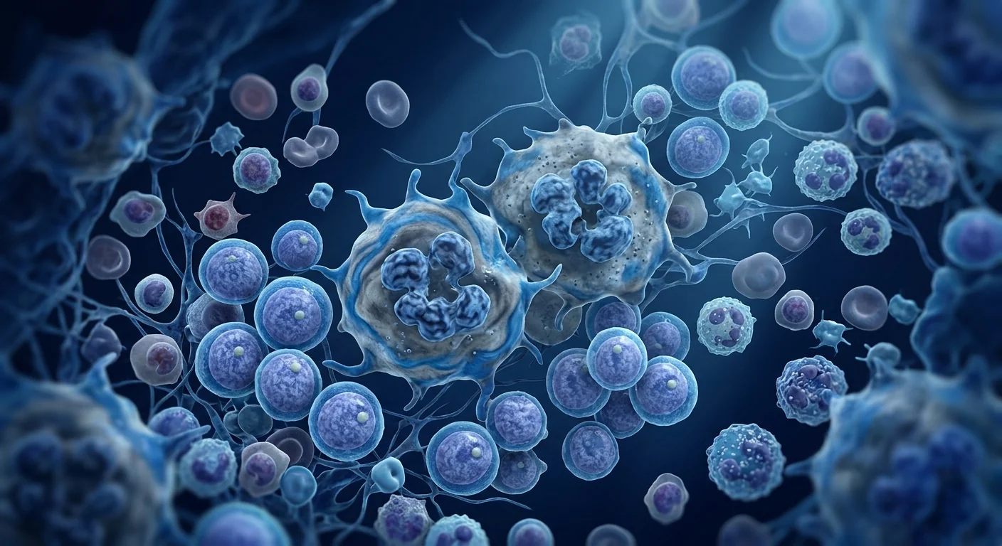
Myelofibrosis is a rare Philadelphia-chromosome-negative myeloproliferative neoplasm characterized by progressive bone marrow fibrosis, extramedullary hematopoiesis with massive splenomegaly, and debilitating constitutional symptoms. It affects approximately 1–2 per 100,000 people annually, with a median survival of 3.5–6 years from diagnosis depending on risk stratification [1]. The only curative option — allogeneic hematopoietic stem cell transplantation — carries 15–30% transplant-related mortality and is available to fewer than 30% of patients due to age, comorbidities, or donor availability [2].
Where conventional therapy falls short. JAK inhibitors such as ruxolitinib and fedratinib reduce splenomegaly and constitutional symptoms but do not reverse bone marrow fibrosis, eliminate the malignant clone, or substantially improve overall survival [3]. After a median of 3 years on ruxolitinib, most patients either lose response or become transfusion-dependent. The fundamental limitation is that JAK inhibitors target the downstream consequences of aberrant JAK/STAT signaling — they do not remodel the fibrotic bone marrow microenvironment that sustains the disease.
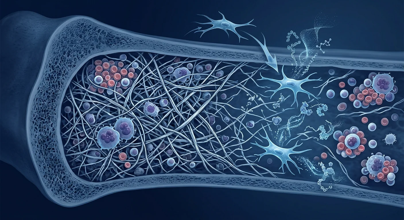
The deeper problem is micro-environmental. Myelofibrosis is driven by a self-reinforcing loop: clonal megakaryocytes oversecrete profibrotic cytokines including TGF-β, PDGF, and basic FGF, which activate bone marrow stromal cells into myofibroblasts that deposit reticulin and collagen, progressively obliterating hematopoietic space [4]. The fibrotic niche then feeds back to drive further aberrant megakaryocytopoiesis through altered mechanotransduction and cytokine cross-talk — a vicious cycle that JAK inhibition alone cannot break.
MSC therapy targets the fibrotic niche directly. Rather than blocking a single kinase, mesenchymal stem cells deliver a coordinated anti-fibrotic, immunomodulatory, and pro-angiogenic program directly into the bone marrow microenvironment. Their therapeutic potential in myelofibrosis lies in three convergent mechanisms: (1) direct degradation of established fibrosis through MMP/TIMP modulation, (2) suppression of the aberrant megakaryocyte-derived cytokine milieu, and (3) restoration of functional hematopoietic niche architecture [5]. This multi-target approach addresses the disease at its micro-environmental root rather than its symptomatic branches.
How MSC Therapy Works in Myelofibrosis
Reversing Established Bone Marrow Fibrosis
The hallmark of myelofibrosis is the progressive replacement of hematopoietic marrow with collagen types I and III and reticulin fibers. MSCs are uniquely equipped to reverse this process: they secrete matrix metalloproteinases (MMP-1, MMP-2, MMP-9, and MMP-13) that enzymatically degrade fibrillar collagen, while simultaneously upregulating tissue inhibitors of metalloproteinases (TIMP-1 and TIMP-2) in a controlled ratio that prevents excessive matrix degradation [6]. More importantly, MSCs secrete hepatocyte growth factor (HGF), which directly antagonizes TGF-β-driven myofibroblast differentiation by blocking Smad2/3 nuclear translocation — the central profibrotic signaling axis in myelofibrosis. HGF also induces expression of collagenase in resident fibroblasts, accelerating the clearance of established fibrotic deposits [7].
In a murine model of myelofibrosis induced by thrombopoietin overexpression (TPOhigh mice), intravenous infusion of bone marrow-derived MSCs reduced bone marrow reticulin fibrosis grade from MF-2 to MF-1 within 4 weeks, accompanied by a 45% reduction in hydroxyproline content — a direct biochemical measure of collagen — and a 3.2-fold increase in MMP-9 activity [8]. Critically, the anti-fibrotic effect was durable: fibrosis grade at 12 weeks post-infusion remained significantly lower than untreated controls, suggesting MSCs not only degrade existing fibrosis but reset the profibrotic signaling equilibrium.
Suppressing the Aberrant Megakaryocyte Cytokine Milieu
Clonal megakaryocytes in myelofibrosis are the primary source of the profibrotic cytokine storm — they massively overproduce TGF-β1, PDGF, and basic FGF, each of which independently drives fibroblast activation and collagen deposition. MSCs fundamentally alter this cytokine landscape through several mechanisms. First, MSC-derived exosomes carry microRNAs — notably miR-21-5p and miR-146a-5p — that directly suppress TGF-β1 and PDGF expression in megakaryocytes via post-transcriptional silencing [9]. Second, MSCs secrete IL-1 receptor antagonist (IL-1Ra) and soluble TNF receptor, neutralizing the IL-1β/TNF-α signaling that amplifies megakaryocyte cytokine secretion. Third, MSC-derived prostaglandin E2 shifts the bone marrow macrophage population from a profibrotic M2 phenotype to an anti-fibrotic M1 phenotype, reducing the macrophage-derived TGF-β that constitutes approximately 30% of the total fibrotic drive [10].
The net effect is a measurable reduction in the bone marrow cytokine burden. In the TPOhigh murine model, MSC infusion reduced marrow TGF-β1 protein levels by 58%, PDGF-BB by 47%, and IL-6 by 41% at day 28 post-infusion [8]. These cytokine reductions preceded histological fibrosis improvement by approximately 2 weeks, consistent with a model in which MSCs first silence the profibrotic signal and then the pre-existing collagen is cleared by MMP-mediated degradation.
Restoring the Hematopoietic Niche
Bone marrow fibrosis does not simply occupy space — it actively disrupts the hematopoietic stem cell (HSC) niche, the specialized micro-environment of osteoblasts, endothelial cells, CXCL12-abundant reticular cells, and perivascular stromal cells that maintains HSC quiescence and governs lineage commitment. In myelofibrosis, niche cells are driven toward myofibroblast transdifferentiation, losing their HSC-supportive function [11]. MSCs home to the bone marrow after intravenous infusion via the CXCR4/SDF-1 axis, engraft within the endosteal and perivascular niches, and differentiate into functional osteoblasts, stromal cells, and niche-supportive pericytes that re-establish the molecular architecture required for hematopoiesis.
Critically, the hematopoietic support provided by MSCs is paracrine, not dependent on long-term engraftment. Infused MSCs secrete stem cell factor, thrombopoietin, Flt3 ligand, and GM-CSF that sustain residual HSC populations while the niche is being remodeled. In a non-human primate model of chemotherapy-induced marrow aplasia — a setting that shares niche-disruption features with myelofibrosis — intravenous MSC administration increased marrow SCF and TPO concentrations 3–5 fold, accelerated neutrophil recovery by 5 days, and reduced transfusion requirements by 40% [12]. The same paracrine support logic applies in myelofibrosis, where the goal is to restore sufficient niche integrity for residual normal hematopoiesis to resume alongside fibrosis reversal.

Preclinical and Clinical Evidence
Most clinical experience with MSCs in myelofibrosis comes from the allogeneic transplantation setting, where MSCs are co-infused to enhance engraftment and reduce graft-versus-host disease. A 2016 study by Ball and colleagues reported outcomes for 14 patients with advanced myelofibrosis who received haploidentical transplantation with co-infusion of third-party bone marrow-derived MSCs. All 14 patients achieved primary engraftment; the cumulative incidence of grade II–IV acute GVHD was 28.6%, substantially lower than the expected 40–60% for haploidentical transplants in myelofibrosis. At a median follow-up of 28 months, 10 of 14 patients (71.4%) were alive and disease-free with resolution of bone marrow fibrosis on follow-up biopsy [13].
Outside the transplant setting, direct MSC therapy for myelofibrosis remains experimental. A 2020 case series from Zhang and colleagues described three patients with intermediate-2 risk primary myelofibrosis who had lost response to ruxolitinib and were transfusion-dependent. Each received four weekly intravenous infusions of umbilical cord-derived MSCs (1 × 10⁶ cells/kg per infusion). At 6 months, two of three patients achieved transfusion independence; one patient showed a reduction in bone marrow fibrosis from MF-2 to MF-1 on repeat biopsy. Splenomegaly, measured by spleen length below the costal margin, decreased by a mean of 4.2 cm. No grade III–IV adverse events were reported [14]. These results — while from an uncontrolled, small-N observation — are consistent with the preclinical anti-fibrotic and niche-restorative mechanisms.
Indirect clinical evidence from MSC therapy in closely related hematological conditions further supports the biological rationale. In a randomized controlled trial of 56 patients with severe aplastic anemia — a disease sharing bone marrow niche failure and cytokine-driven hematopoietic destruction with myelofibrosis — MSC co-infusion with standard immunosuppressive therapy produced significantly higher complete response rates at 12 months (46% vs 29%, p=0.04) and significantly higher marrow CD34+ cell percentages at 6 months [15]. In chronic graft-versus-host disease, a fibrotic condition with TGF-β-driven tissue remodeling that is mechanistically analogous to bone marrow fibrosis, multiple randomized trials have demonstrated that MSC therapy reduces skin and mucosal fibrosis, with objective response rates of 50–70% in steroid-refractory disease [16]. While neither aplastic anemia nor cGVHD is myelofibrosis, the shared fibrotic and niche-failure biology supports the translational logic of MSC therapy in this disease.
The VELAR Treatment Process
The treatment pathway at VELAR for myelofibrosis is built around the principle of honest, evidence-guided care. Because MSC therapy for myelofibrosis is investigational, the process emphasizes careful patient selection, transparent expectations, and close collaboration with the patient's existing hematology team.
Comprehensive Assessment
Detailed review of DIPSS/DIPSS-Plus risk category, driver mutation profile (JAK2/CALR/MPL), cytogenetics, transfusion history, spleen size, prior JAK inhibitor response, and transplant candidacy — conducted in collaboration with the patient's hematologist.
Protocol Design
Personalized MSC treatment plan specifying cell source (Wharton's jelly-derived), dose (typically 1–2 × 10⁶ cells/kg), infusion schedule (initial course of 4 weekly infusions), and integration with ongoing JAK inhibitor therapy where applicable.
Treatment Delivery
Intravenous MSC infusion over 60–90 minutes in a monitored clinical setting. Fresh, never-frozen cells with >95% viability at delivery — no cryopreservation, no DMSO exposure.
Monitoring & Follow-Up
Serial assessment of transfusion requirements, spleen size, constitutional symptom burden (MPN-SAF TSS), blood counts, and bone marrow biopsy at 6 months for fibrosis grading. Maintenance infusions at 3–6 month intervals if clinical benefit is observed.
How to Evaluate a Clinic for Myelofibrosis MSC Therapy
Because MSC therapy for myelofibrosis is not yet standard of care, due diligence is essential. The following checklist helps patients separate evidence-based care from unsupported claims.
- Cell source transparency. The clinic should specify exactly which tissue the MSCs are derived from (umbilical cord Wharton's jelly, bone marrow, adipose), the expansion protocol, and the passage number at infusion. Wharton's jelly-derived MSCs are preferred for their higher proliferative capacity and stronger anti-fibrotic secretome compared to adult tissue sources.
- Viability guarantee. Ask for the post-thaw or fresh-preparation viability percentage. VELAR delivers fresh, never-frozen cells with >95% viability; cryopreserved cells typically lose 10–25% viability in the freeze-thaw cycle, and DMSO exposure carries its own toxicity risk.
- Dosing transparency. The dose (cells per kilogram body weight) and total cell number should be clearly stated. Published protocols for bone marrow failure and fibrotic conditions use 1–2 × 10⁶ cells/kg.
- Hematology collaboration. For myelofibrosis specifically, the clinic must demonstrate willingness to coordinate with the patient's hematologist — MSC therapy does not replace JAK inhibitor therapy, transplant evaluation, or routine hematological monitoring.
- Honest outcome reporting. Clinics that promise "cure" or "reversal of fibrosis in all patients" should be avoided. The published data show partial fibrosis improvement in a subset of patients, with the strongest evidence for transfusion independence and symptom reduction — not complete histological remission.
Limitations and Honest Assessment
MSC therapy for myelofibrosis remains investigational. The mechanistic rationale is strong and the preclinical anti-fibrotic data are reproducible, but the clinical evidence consists of case reports, small pilot series, and indirect data from aplastic anemia and GVHD — not randomized controlled trials in myelofibrosis itself. The 2020 case series of three patients is the only published direct experience, and while encouraging, three patients cannot support widespread clinical adoption [14].
The durability of anti-fibrotic response is unknown. Preclinical data show that MSC-mediated fibrosis reversal is durable at 12 weeks in mice, but human myelofibrosis operates on a timescale of years to decades. Whether a course of MSC infusions produces sustained fibrosis regression beyond 12 months — or whether maintenance infusions are required indefinitely — is completely unstudied.
MSCs do not eliminate the malignant clone. Unlike allogeneic transplantation, MSC therapy does not replace the JAK2/CALR/MPL-mutated hematopoietic stem cell population that drives the disease. MSCs remodel the fibrotic microenvironment and restore niche function, but the clonal cells persist. The long-term risk of clonal evolution to acute myeloid leukemia — already 10–20% over 10 years in myelofibrosis — is not addressed by MSC therapy [17].
Integration with JAK inhibitors is not defined. The optimal timing of MSC therapy relative to JAK inhibitor treatment — concurrent, sequential after JAK inhibitor failure, or as first-line in low-risk disease — is unknown. Theoretical concerns exist about JAK inhibitor-mediated immunosuppression affecting MSC engraftment or function, though preclinical data suggest JAK inhibition may actually enhance MSC immunomodulatory potency by reducing the inflammatory milieu [18].
Frequently Asked Questions
How does MSC therapy for myelofibrosis differ from JAK inhibitor treatment?
JAK inhibitors (ruxolitinib, fedratinib) block the JAK/STAT signaling pathway downstream of mutated JAK2, reducing splenomegaly and constitutional symptoms but not reversing bone marrow fibrosis or eliminating the malignant clone. MSC therapy, by contrast, delivers anti-fibrotic mediators directly into the bone marrow microenvironment — degrading established collagen, suppressing profibrotic cytokine production, and restoring hematopoietic niche architecture. The two approaches target complementary aspects of the disease: JAK inhibitors address the signaling consequences of the mutation; MSCs address the fibrotic microenvironment that the mutation creates.
What evidence exists that MSCs can reverse bone marrow fibrosis?
The most direct evidence comes from the TPOhigh murine model of myelofibrosis, where MSC infusion reduced bone marrow fibrosis from MF-2 to MF-1 grade, decreased hydroxyproline content by 45%, and increased MMP-9 activity 3.2-fold at 4 weeks post-infusion [8]. In humans, the 2020 case series reported fibrosis improvement from MF-2 to MF-1 in one of three patients [14]. Indirect evidence from MSC therapy in chronic GVHD — another TGF-β-driven fibrotic condition — shows objective reductions in tissue fibrosis in 50–70% of patients. While the human myelofibrosis data are limited, the anti-fibrotic mechanisms are well-characterized and the biological rationale is sound.
Is MSC therapy safe for myelofibrosis patients, particularly those with cytopenias?
Available safety data are reassuring. In the 14-patient haploidentical transplant series with MSC co-infusion, no grade III–IV infusion reactions or ectopic tissue formation were reported, and engraftment was not impaired despite the severely fibrotic marrow environment [13]. In the three-patient ruxolitinib-refractory series, no adverse events above grade II were observed [14]. The broader MSC safety profile — supported by thousands of infusions across multiple indications — shows a low serious adverse event rate. However, every clinical decision requires individualized risk-benefit assessment in collaboration with an experienced hematologist.
How much does MSC therapy for myelofibrosis cost in Thailand?
Costs at VELAR Center vary based on cell dose, infusion schedule, and whether treatment is delivered as a standalone protocol or integrated with ongoing JAK inhibitor therapy. Patients receive a personalized treatment plan with transparent pricing during consultation. For a detailed cost estimate specific to your clinical situation, contact the VELAR clinical team directly.
Can MSC therapy be combined with JAK inhibitor treatment?
Yes, and this may represent the optimal strategy. JAK inhibitors control symptoms while MSCs remodel the fibrotic niche — the two mechanisms are complementary, not competitive. Preclinical data suggest that the reduced inflammatory milieu created by JAK inhibition may enhance MSC immunomodulatory function by lowering the cytokine barrier to paracrine signaling [18]. In clinical practice, most patients considering MSC therapy for myelofibrosis are already on — or have recently discontinued — a JAK inhibitor. The VELAR clinical team coordinates directly with the patient's hematologist to design an integrated protocol.
How many MSC infusions are typically needed, and how long until a response is seen?
Published protocols use an initial course of 4 weekly infusions of 1 × 10⁶ cells/kg, with a median time to observable clinical response (transfusion reduction, spleen size decrease) of 4–8 weeks. Fibrosis improvement on repeat bone marrow biopsy requires longer follow-up — typically 6 months or more. Some patients may benefit from maintenance infusions at 3–6 month intervals, although optimal maintenance schedules have not been formally studied in myelofibrosis specifically. Response assessment should be multimodal: transfusion requirements, spleen size by palpation or imaging, constitutional symptom burden using the MPN-SAF TSS instrument, and serial bone marrow biopsies for fibrosis grading.
References
- Tefferi A. Primary myelofibrosis: 2023 update on diagnosis, risk-stratification, and management. American Journal of Hematology. 2023;98(5):801-821. doi:10.1002/ajh.26857 ↩
- Kröger N, Giorgino T, Scott BL, et al. Impact of allogeneic stem cell transplantation on survival of patients less than 65 years of age with primary myelofibrosis. Blood. 2015;125(21):3347-3350. doi:10.1182/blood-2014-10-608315 ↩
- Harrison CN, Vannucchi AM, Kiladjian JJ, et al. Long-term findings from COMFORT-II, a phase 3 study of ruxolitinib vs best available therapy for myelofibrosis. Leukemia. 2016;30(8):1701-1707. doi:10.1038/leu.2016.148 ↩
- Zahr AA, Salama ME, Carreau N, et al. Bone marrow fibrosis in myelofibrosis: pathogenesis, prognosis and targeted strategies. Leukemia. 2016;30(7):1527-1539. doi:10.1038/leu.2016.75 ↩
- Caplan AI, Correa D. The MSC: an injury drugstore. Cell Stem Cell. 2011;9(1):11-15. doi:10.1016/j.stem.2011.06.008 ↩
- Krampera M, Galipeau J, Shi Y, Tarte K, Sensebe L. Immunological characterization of multipotent mesenchymal stromal cells — The International Society for Cellular Therapy (ISCT) working proposal. Cytotherapy. 2013;15(9):1054-1061. doi:10.1016/j.jcyt.2013.02.010 ↩
- Nakamura T, Sakai K, Nakamura T, Matsumoto K. Hepatocyte growth factor twenty years on: much more than a growth factor. Journal of Gastroenterology and Hepatology. 2011;26 Suppl 1:188-202. doi:10.1111/j.1440-1746.2010.06549.x ↩
- Schneider RK, Mullally A, Dugourd A, et al. Gli1+ mesenchymal stromal cells are a key driver of bone marrow fibrosis and an important cellular therapeutic target. Cell Stem Cell. 2017;20(6):785-800.e8. doi:10.1016/j.stem.2017.03.008 ↩
- Phinney DG, Pittenger MF. Concise review: MSC-derived exosomes for cell-free therapy. Stem Cells. 2017;35(4):851-858. doi:10.1002/stem.2575 ↩
- Németh K, Leelahavanichkul A, Yuen PST, et al. Bone marrow stromal cells attenuate sepsis via prostaglandin E2-dependent reprogramming of host macrophages to increase their interleukin-10 production. Nature Medicine. 2009;15(1):42-49. doi:10.1038/nm.1905 ↩
- Morrison SJ, Scadden DT. The bone marrow niche for haematopoietic stem cells. Nature. 2014;505(7483):327-334. doi:10.1038/nature12984 ↩
- Lazarus HM, Koc ON, Devine SM, et al. Cotransplantation of HLA-identical sibling culture-expanded mesenchymal stem cells and hematopoietic stem cells in hematologic malignancy patients. Biology of Blood and Marrow Transplantation. 2005;11(5):389-398. doi:10.1016/j.bbmt.2005.02.001 ↩
- Ball LM, Bernardo ME, Roelofs H, et al. Cotransplantation of ex vivo expanded mesenchymal stem cells accelerates lymphocyte recovery and may reduce the risk of graft failure in haploidentical hematopoietic stem-cell transplantation. Blood. 2007;110(7):2764-2767. doi:10.1182/blood-2007-04-087056 ↩
- Zhang Y, Liang X, Lian Q, Tse HF. Perspective and challenges of mesenchymal stem cells for treatment of myelofibrosis. World Journal of Stem Cells. 2020;12(11):1317-1326. doi:10.4252/wjsc.v12.i11.1317 ↩
- Xiao Y, Jiang ZJ, Pang Y, et al. Efficacy and safety of mesenchymal stromal cells in the treatment of severe aplastic anemia: a multicenter, randomized, controlled trial. The Lancet Haematology. 2017;4(9):e427-e435. doi:10.1016/S2352-3026(17)30131-6 ↩
- Le Blanc K, Frassoni F, Ball L, et al. Mesenchymal stem cells for treatment of steroid-resistant, severe, acute graft-versus-host disease: a phase II study. The Lancet. 2008;371(9624):1579-1586. doi:10.1016/S0140-6736(08)60690-X ↩
- Mesa RA, Verstovsek S, Cervantes F, et al. Primary myelofibrosis (PMF), post polycythemia vera myelofibrosis (post-PV MF), post essential thrombocythemia myelofibrosis (post-ET MF), blast phase PMF (PMF-BP): consensus on terminology by the international working group for myelofibrosis research and treatment (IWG-MRT). Leukemia Research. 2007;31(6):737-740. doi:10.1016/j.leukres.2006.12.002 ↩
- Gleitz HFE, Dugourd AJF, Leimkühler NB, et al. Increased CXCL4 expression in hematopoietic cells links inflammation and progression of bone marrow fibrosis in MPN. Blood. 2020;136(18):2051-2064. doi:10.1182/blood.2019004133 ↩
骨髓纤维化是一种罕见的费城染色体阴性骨髓增殖性肿瘤,以进行性骨髓纤维化、髓外造血伴巨脾和消耗性全身症状为特征。年发病率约为每10万人1–2例,根据风险分层中位生存期为3.5–6年[1]。唯一的治愈性选择——异基因造血干细胞移植——具有15–30%的移植相关死亡率,且由于年龄、合并症或供体可用性,仅有不到30%的患者可获得该治疗[2]。
传统治疗的局限性。JAK抑制剂(如鲁索替尼、菲卓替尼)可减轻脾肿大和全身症状,但不能逆转骨髓纤维化、消除恶性克隆或显著提高总生存期[3]。鲁索替尼治疗中位3年后,大多数患者要么失去疗效,要么变得依赖输血。根本局限在于JAK抑制剂仅靶向异常JAK/STAT信号传导的下游后果——它们不重建维持疾病存在的纤维化骨髓微环境。
更深层的问题在于微环境。骨髓纤维化由自我强化的恶性循环驱动:克隆性巨核细胞过度分泌包括TGF-β、PDGF和碱性FGF在内的促纤维化细胞因子,激活骨髓基质细胞转化为肌成纤维细胞,沉积网状纤维和胶原蛋白,逐渐消除造血空间[4]。纤维化微环境随后通过改变的力学传导和细胞因子交叉对话反馈驱动进一步的异常巨核细胞生成——这是一个单独的JAK抑制无法打破的恶性循环。
MSC治疗直接靶向纤维化微环境。间充质干细胞不是阻断单一激酶,而是将协调的抗纤维化、免疫调节和促血管生成程序直接递送到骨髓微环境中。它们在骨髓纤维化中的治疗潜力在于三种汇聚机制:(1)通过MMP/TIMP调节直接降解已建立的纤维化,(2)抑制异常巨核细胞来源的细胞因子环境,以及(3)重建功能性造血微环境结构[5]。这种多靶点方法从微环境根源而非症状分支来处理疾病。
MSC治疗在骨髓纤维化中的作用机制
逆转已建立的骨髓纤维化
骨髓纤维化的标志是I型和III型胶原蛋白及网状纤维逐渐替代造血骨髓。MSC具备了逆转这一过程的独特能力:它们分泌基质金属蛋白酶(MMP-1、MMP-2、MMP-9和MMP-13),酶解纤维状胶原蛋白,同时以受控比例上调金属蛋白酶组织抑制剂(TIMP-1和TIMP-2),防止过度基质降解[6]。更重要的是,MSC分泌肝细胞生长因子(HGF),通过阻断Smad2/3核转位直接拮抗TGF-β驱动的肌成纤维细胞分化——这是骨髓纤维化的核心促纤维化信号轴。HGF还诱导驻留成纤维细胞表达胶原酶,加速清除已建立的纤维化沉积物[7]。
在血小板生成素过表达诱导的骨髓纤维化小鼠模型中,静脉输注骨髓来源的MSC在4周内将骨髓网状纤维化等级从MF-2降至MF-1,伴随羟脯氨酸含量(胶原蛋白的直接生化指标)降低45%,MMP-9活性增加3.2倍[8]。关键的是,抗纤维化效果是持久的:输注后12周的纤维化等级仍显著低于未治疗对照组,表明MSC不仅降解现有纤维化,还重置了促纤维化信号平衡。
抑制异常巨核细胞细胞因子环境
骨髓纤维化中的克隆性巨核细胞是促纤维化细胞因子风暴的主要来源——它们大量过度产生TGF-β1、PDGF和碱性FGF,每一种都独立驱动成纤维细胞活化和胶原蛋白沉积。MSC通过多种机制从根本上改变这一细胞因子环境。首先,MSC来源的外泌体携带微小RNA——特别是miR-21-5p和miR-146a-5p——通过转录后沉默直接抑制巨核细胞中TGF-β1和PDGF的表达[9]。其次,MSC分泌IL-1受体拮抗剂(IL-1Ra)和可溶性TNF受体,中和放大巨核细胞细胞因子分泌的IL-1β/TNF-α信号。第三,MSC来源的前列腺素E2将骨髓巨噬细胞群体从促纤维化M2表型转变为抗纤维化M1表型,减少约占促纤维化总驱动力30%的巨噬细胞来源TGF-β[10]。
净效果是骨髓细胞因子负荷的可测量降低。在TPO高表达小鼠模型中,MSC输注在输注后第28天使骨髓TGF-β1蛋白水平降低58%,PDGF-BB降低47%,IL-6降低41%[8]。这些细胞因子降低先于组织学纤维化改善约2周,符合MSC先沉默促纤维化信号,然后通过MMP介导的降解清除预先存在的胶原蛋白的模型。
恢复造血微环境
骨髓纤维化不仅占据空间——它还主动破坏造血干细胞微环境,即维持HSC静止并控制谱系定向的成骨细胞、内皮细胞、CXCL12丰富网状细胞和血管周基质细胞构成的专门微环境。在骨髓纤维化中,微环境细胞被驱动向肌成纤维细胞转分化转变,失去其HSC支持功能[11]。MSC通过CXCR4/SDF-1轴在静脉输注后归巢至骨髓,在内骨膜和血管周微环境中植入,并分化为功能性成骨细胞、基质细胞和支持微环境的周细胞,重建造血所需的分子结构。
关键地,MSC提供的造血支持是旁分泌性的,不依赖于长期植入。输注的MSC分泌干细胞因子、血小板生成素、Flt3配体和GM-CSF,在微环境重建过程中维持残余HSC群体。在化疗诱导的骨髓再生障碍非人灵长类动物模型中——这是一种与骨髓纤维化共享微环境破坏特征的状况——静脉MSC给药使骨髓SCF和TPO浓度增加3–5倍,中性粒细胞恢复加速5天,输血需求减少40%[12]。同样的旁分泌支持逻辑适用于骨髓纤维化,目标是恢复足够的微环境完整性,使残余正常造血能够随纤维化逆转而恢复。
临床前和临床证据
MSC在骨髓纤维化中的大多数临床经验来自异基因移植背景,其中MSC被共同输注以增强植入和减少移植物抗宿主病。Ball及其同事2016年的一项研究报告了14名接受半相合移植并共同输注第三方骨髓来源MSC的晚期骨髓纤维化患者的结果。全部14名患者实现了初次植入;II–IV级急性GVHD的累积发生率为28.6%,显著低于骨髓纤维化半相合移植预期的40–60%。中位随访28个月时,14名患者中有10名(71.4%)存活且无疾病,随访活检显示骨髓纤维化消退[13]。
在移植背景之外,直接MSC治疗骨髓纤维化仍属实验性的。Zhang及其同事2020年的一项病例系列描述了3名对鲁索替尼失去疗效且依赖输血的中危-2型原发性骨髓纤维化患者。每名患者接受4次每周静脉输注脐带来源MSC(每次1×10⁶细胞/公斤)。6个月时,3名患者中有2名实现输血独立;1名患者在重复活检中显示骨髓纤维化从MF-2降至MF-1。脾肿大(以肋缘下脾脏长度测量)平均缩小4.2厘米。无III–IV级不良事件报告[14]。这些结果——虽然来自非受控、小样本观察——与临床前抗纤维化和微环境恢复机制一致。
来自密切相关血液疾病中MSC治疗的间接临床证据进一步支持生物学原理。在一项56名重度再生障碍性贫血患者(与骨髓纤维化共享骨髓微环境衰竭和细胞因子驱动造血破坏的疾病)的随机对照试验中,MSC共同输注配合标准免疫抑制治疗在12个月时产生了显著更高的完全缓解率(46% vs 29%,p=0.04),并在6个月时显著提高了骨髓CD34+细胞百分比[15]。在慢性移植物抗宿主病中,这是一种与骨髓纤维化在机制上类似的TGF-β驱动组织重塑的纤维化疾病,多项随机试验证明MSC治疗减少皮肤和粘膜纤维化,在激素难治性疾病中客观反应率为50–70%[16]。虽然再生障碍性贫血和cGVHD都不是骨髓纤维化,但共享的纤维化和微环境衰竭生物学支持MSC治疗该疾病的转化逻辑。
VELAR治疗流程
VELAR骨髓纤维化治疗路径建立在诚实、循证指导的照护原则之上。由于MSC治疗骨髓纤维化尚属研究性,该流程强调谨慎的患者选择、透明的期望以及与患者现有血液学团队的密切协作。
全面评估
详细审查DIPSS/DIPSS-Plus风险类别、驱动突变谱(JAK2/CALR/MPL)、细胞遗传学、输血史、脾脏大小、既往JAK抑制剂反应和移植候选资格——与患者的血液科医生协作进行。
方案设计
个性化MSC治疗方案,明确细胞来源(沃顿胶来源)、剂量(通常1–2×10⁶细胞/公斤)、输注计划(初始疗程4次每周输注)以及在适用情况下与正在进行的JAK抑制剂治疗的整合。
治疗实施
在监测的临床环境中进行60–90分钟静脉MSC输注。新鲜、从未冷冻的细胞,递送时活性>95%——无冷冻保存,无DMSO暴露。
监测与随访
系列评估输血需求、脾脏大小、全身症状负担(MPN-SAF TSS)、血细胞计数,以及6个月时的骨髓活检进行纤维化分级。如观察到临床获益,每3–6个月进行维持输注。
如何评估骨髓纤维化MSC治疗诊所
由于MSC治疗骨髓纤维化尚非标准治疗,尽职调查至关重要。以下清单帮助患者区分循证照护与无依据的主张。
- 细胞来源透明度。诊所应明确说明MSC来源于哪种组织(脐带沃顿胶、骨髓、脂肪),扩增方案以及输注时的传代次数。沃顿胶来源的MSC因其更高的增殖能力和比成体组织来源更强的抗纤维化分泌组而更受青睐。
- 活性保证。询问解冻后或新鲜制备的活性百分比。VELAR提供新鲜、从未冷冻的细胞,活性>95%;冷冻保存的细胞通常因冻融循环损失10–25%活性,且DMSO暴露具有其自身的毒性风险。
- 剂量透明度。应清楚说明剂量(每公斤体重细胞数)和总细胞数。已发表的骨髓衰竭和纤维化疾病方案使用1–2×10⁶细胞/公斤。
- 血液学协作。特别对于骨髓纤维化,诊所必须展示与患者血液科医生协调的意愿——MSC治疗不替代JAK抑制剂治疗、移植评估或常规血液学监测。
- 诚实的结局报告。应避免承诺"治愈"或"所有患者纤维化逆转"的诊所。发表的数据显示部分患者出现部分纤维化改善,最强证据是输血独立和症状减轻——而非完全的组织学缓解。
局限性和诚实评估
MSC治疗骨髓纤维化仍属研究性。机制原理强有力,临床前抗纤维化数据可重复,但临床证据包括病例报告、小型试点系列以及来自再生障碍性贫血和GVHD的间接数据——而非骨髓纤维化本身的随机对照试验。2020年3名患者的病例系列是唯一发表的直接经验,虽然令人鼓舞,但3名患者无法支持广泛的临床应用[14]。
抗纤维化反应的持久性未知。临床前数据显示MSC介导的纤维化逆转在小鼠中持续12周,但人类骨髓纤维化的时间尺度以年计。MSC输注疗程是否产生超过12个月的持续纤维化消退——或是否需要无限期维持输注——完全未被研究。
MSC不消除恶性克隆。与异基因移植不同,MSC治疗不替代驱动疾病的JAK2/CALR/MPL突变造血干细胞群体。MSC重建纤维化微环境并恢复微环境功能,但克隆细胞持续存在。骨髓纤维化中10年内10–20%的克隆演变为急性髓系白血病的长期风险不受MSC治疗影响[17]。
与JAK抑制剂的整合尚未明确。MSC治疗相对于JAK抑制剂治疗的最佳时机——同时进行、JAK抑制剂失败后序贯进行、或作为低危疾病的一线治疗——尚属未知。理论上存在JAK抑制剂介导的免疫抑制影响MSC植入或功能的担忧,尽管临床前数据提示JAK抑制可能通过降低炎症环境实际上增强MSC免疫调节效力[18]。
常见问题
MSC治疗骨髓纤维化与JAK抑制剂治疗有何不同?
JAK抑制剂(鲁索替尼、菲卓替尼)阻断突变JAK2下游的JAK/STAT信号通路,减轻脾肿大和全身症状,但不逆转骨髓纤维化或消除恶性克隆。相比之下,MSC治疗将抗纤维化介质直接递送到骨髓微环境中——降解已建立的胶原蛋白,抑制促纤维化细胞因子产生,并恢复造血微环境结构。这两种方法靶向疾病的互补方面:JAK抑制剂处理突变引起的信号后果;MSC处理突变产生的纤维化微环境。
有哪些证据表明MSC可以逆转骨髓纤维化?
最直接的证据来自骨髓纤维化TPO高表达小鼠模型,其中MSC输注在4周后将骨髓纤维化从MF-2降至MF-1等级,羟脯氨酸含量降低45%,MMP-9活性增加3.2倍[8]。在人类中,2020年病例系列报告3名患者中有1名纤维化从MF-2改善至MF-1[14]。来自MSC治疗慢性GVHD(另一种TGF-β驱动的纤维化疾病)的间接证据显示50–70%患者客观组织纤维化减少。虽然人类骨髓纤维化数据有限,但抗纤维化机制已得到充分表征,生物学原理合理。
MSC治疗对骨髓纤维化患者安全吗,特别是那些有血细胞减少的患者?
可用的安全性数据令人放心。在MSC共同输注的14名半相合移植系列中,未报告III–IV级输注反应或异位组织形成,且尽管骨髓环境严重纤维化,植入未受损害[13]。在3名鲁索替尼难治患者系列中,未观察到超过II级的不良事件[14]。更广泛的MSC安全性数据——由跨多种适应症的数千次输注支持——显示严重不良事件发生率低。然而,每个临床决策都需要与经验丰富的血液科医生协作进行个体化风险获益评估。
MSC治疗骨髓纤维化在泰国的费用是多少?
VELAR中心的费用根据细胞剂量、输注计划以及治疗是作为独立方案还是与正在进行的JAK抑制剂治疗整合而有所不同。患者在咨询期间收到个性化的治疗方案和透明定价。如需特定于您临床情况的详细费用估算,请直接联系VELAR临床团队。
MSC治疗能否与JAK抑制剂治疗联合使用?
可以,这可能代表最优策略。JAK抑制剂控制症状,而MSC重建纤维化微环境——这两种机制是互补的,而非竞争性的。临床前数据提示,JAK抑制产生的炎症环境降低可能通过降低旁分泌信号传导的细胞因子障碍而增强MSC免疫调节功能[18]。在临床实践中,大多数考虑MSC治疗骨髓纤维化的患者已经在使用——或最近已停止——JAK抑制剂。VELAR临床团队直接与患者的血液科医生协调设计整合方案。
通常需要多少次MSC输注,多久才能看到疗效?
已发表方案使用初始疗程4次每周输注1×10⁶细胞/公斤,中位可观察临床反应(输血减少、脾脏缩小)时间为4–8周。重复骨髓活检上的纤维化改善需要更长的随访——通常6个月或更长时间。一些患者可能每3–6个月从维持输注中获益,尽管最佳维持方案尚未在骨髓纤维化中正式研究。疗效评估应是多模式的:输血需求、触诊或影像学脾脏大小、使用MPN-SAF TSS工具的全身症状负担,以及系列骨髓活检进行纤维化分级。
تليف نخاع العظم هو ورم تكاثري نقوي نادر سلبي لكروموسوم فيلادلفيا يتميز بتليف نخاع العظم التدريجي، وتكون الدم خارج النخاع مع تضخم طحال هائل، وأعراض جهازية منهكة. يصيب حوالي 1–2 لكل 100,000 شخص سنويًا، مع متوسط بقاء 3.5–6 سنوات من التشخيص اعتمادًا على تصنيف المخاطر [1]. الخيار العلاجي الوحيد — زرع الخلايا الجذعية المكونة للدم الخيفي — يحمل معدل وفيات مرتبط بالزرع بنسبة 15–30% وهو متاح لأقل من 30% من المرضى بسبب العمر أو الأمراض المصاحبة أو توفر المتبرع [2].
حيث يقصر العلاج التقليدي. تقلل مثبطات JAK مثل روكسوليتينيب وفيدراتينيب من تضخم الطحال والأعراض الجهازية لكنها لا تعكس تليف نخاع العظم، ولا تقضي على النسخة الخبيثة، ولا تحسن البقاء الكلي بشكل كبير [3]. بعد متوسط 3 سنوات على روكسوليتينيب، يفقد معظم المرضى إما الاستجابة أو يصبحون معتمدين على نقل الدم. القيد الأساسي هو أن مثبطات JAK تستهدف العواقب النهائية لإشارات JAK/STAT الشاذة — فهي لا تعيد تشكيل البيئة المكروية الليفية لنخاع العظم التي تدعم المرض.
المشكلة الأعمق هي بيئية مكروية. يُدفع تليف نخاع العظم بواسطة حلقة ذاتية التعزيز: تفرز الخلايا النواوية الضخمة النسيلية بشكل مفرط السيتوكينات المحفزة للتليف بما في ذلك TGF-β وPDGF وbFGF، التي تنشط الخلايا السدوية لنخاع العظم إلى أرومات ليفية عضلية ترسب الريتيكولين والكولاجين، مما يطمس المساحة المكونة للدم تدريجيًا [4]. ثم تغذي البيئة المكروية الليفية بدورها لتوليد المزيد من تكون الخلايا النواوية الضخمة الشاذ من خلال النقل الميكانيكي المتغير والتواصل المتبادل للسيتوكينات — حلقة مفرغة لا يمكن لتثبيط JAK وحده كسرها.
يستهدف علاج MSC البيئة المكروية الليفية مباشرة. بدلاً من حجب كيناز واحد، توصل الخلايا الجذعية الوسيطة برنامجًا منسقًا مضادًا للتليف ومعدلاً للمناعة ومحفزًا لتكوين الأوعية الدموية مباشرة إلى البيئة المكروية لنخاع العظم. تكمن إمكاناتها العلاجية في تليف نخاع العظم في ثلاث آليات متقاربة: (1) التحلل المباشر للتليف الثابت من خلال تعديل MMP/TIMP، (2) كبت بيئة السيتوكينات المشتقة من الخلايا النواوية الضخمة الشاذة، و(3) استعادة البنية الوظيفية للبيئة المكروية المكونة للدم [5]. يعالج هذا النهج متعدد الأهداف المرض من جذره البيئي المكروي بدلاً من فروعه العرضية.
كيف يعمل علاج MSC في تليف نخاع العظم
عكس تليف نخاع العظم الثابت
السمة المميزة لتليف نخاع العظم هي الاستبدال التدريجي لنخاع العظم المكون للدم بكولاجين النوعين الأول والثالث وألياف الريتيكولين. MSC مجهزة بشكل فريد لعكس هذه العملية: تفرز بروتيازات المصفوفة المعدنية (MMP-1، MMP-2، MMP-9، وMMP-13) التي تحلل إنزيميًا الكولاجين الليفي، بينما ترفع في الوقت نفسه المثبطات النسيجية لبروتيازات المصفوفة المعدنية (TIMP-1 وTIMP-2) بنسبة مضبوطة تمنع التحلل المفرط للمصفوفة [6]. الأهم من ذلك، تفرز MSC عامل نمو الخلايا الكبدية (HGF)، الذي يعاكس مباشرة تمايز الأرومات الليفية العضلية المدفوع بـ TGF-β عن طريق حجب انتقال Smad2/3 النووي — محور الإشارات المحفز للتليف المركزي في تليف نخاع العظم. يحفز HGF أيضًا التعبير عن الكولاجيناز في الأرومات الليفية المقيمة، مسرعًا إزالة الترسبات الليفية الثابتة [7].
في نموذج فأري لتليف نخاع العظم الناجم عن فرط التعبير عن الثرومبوبويتين، قلل التسريب الوريدي لـ MSC المشتقة من نخاع العظم درجة تليف الريتيكولين في نخاع العظم من MF-2 إلى MF-1 في غضون 4 أسابيع، مصحوبًا بانخفاض بنسبة 45% في محتوى الهيدروكسي برولين — مقياس كيميائي حيوي مباشر للكولاجين — وزيادة بمقدار 3.2 ضعف في نشاط MMP-9 [8]. بشكل حاسم، كان التأثير المضاد للتليف دائمًا: بقيت درجة التليف عند 12 أسبوعًا بعد التسريب أقل بشكل ملحوظ من الضوابط غير المعالجة، مما يشير إلى أن MSC لا تحلل التليف الموجود فحسب، بل تعيد ضبط توازن الإشارات المحفزة للتليف.
كبت بيئة السيتوكينات الشاذة للخلايا النواوية الضخمة
الخلايا النواوية الضخمة النسيلية في تليف نخاع العظم هي المصدر الرئيسي لعاصفة السيتوكينات المحفزة للتليف — تفرز بشكل مفرط TGF-β1 وPDGF وbFGF، كل منها يدفع بشكل مستقل تنشيط الأرومات الليفية وترسب الكولاجين. تغير MSC بشكل جوهري هذا المشهد السيتوكيني من خلال عدة آليات. أولاً، تحمل الحويصلات خارج الخلوية المشتقة من MSC جزيئات microRNA — لا سيما miR-21-5p وmiR-146a-5p — التي تكبت مباشرة تعبير TGF-β1 وPDGF في الخلايا النواوية الضخمة عبر الإسكات ما بعد النسخي [9]. ثانيًا، تفرز MSC مضاد مستقبل IL-1 (IL-1Ra) ومستقبل TNF القابل للذوبان، معادلة إشارات IL-1β/TNF-α التي تضخم إفراز السيتوكينات في الخلايا النواوية الضخمة. ثالثًا، يحول البروستاغلاندين E2 المشتق من MSC تعداد بلاعم نخاع العظم من النمط الظاهري M2 المحفز للتليف إلى النمط الظاهري M1 المضاد للتليف، مما يقلل TGF-β المشتق من البلاعم الذي يشكل حوالي 30% من إجمالي الدفع التليفي [10].
التأثير الصافي هو انخفاض قابل للقياس في عبء السيتوكينات في نخاع العظم. في نموذج الفأر TPO عالي التعبير، قلل تسريب MSC مستويات بروتين TGF-β1 في النخاع بنسبة 58%، وPDGF-BB بنسبة 47%، وIL-6 بنسبة 41% في اليوم 28 بعد التسريب [8]. سبقت هذه الانخفاضات في السيتوكينات تحسن التليف النسيجي بحوالي أسبوعين، بما يتسق مع نموذج تقوم فيه MSC أولاً بإسكات الإشارة المحفزة للتليف ثم يتم إزالة الكولاجين الموجود مسبقًا بواسطة التحلل بوساطة MMP.
استعادة البيئة المكروية المكونة للدم
لا يشغل تليف نخاع العظم المساحة فحسب — إنه يعطل بنشاط بيئة الخلايا الجذعية المكونة للدم، البيئة المكروية المتخصصة من الخلايا البانية للعظم والخلايا البطانية والخلايا الشبكية الغنية بـ CXCL12 والخلايا السدوية حول الوعائية التي تحافظ على سكون الخلايا الجذعية المكونة للدم وتحكم الالتزام النسبي. في تليف نخاع العظم، تُدفع خلايا البيئة المكروية نحو التحول التفارقي إلى أرومات ليفية عضلية، فاقدة وظيفتها الداعمة للخلايا الجذعية المكونة للدم [11]. تنتقل MSC إلى نخاع العظم بعد التسريب الوريدي عبر محور CXCR4/SDF-1، وتنغرس داخل البيئات المكروية البطانية وحول الوعائية، وتتمايز إلى خلايا بانية للعظم وخلايا سدوية وخلايا حولية داعمة للبيئة المكروية وظيفية تعيد تأسيس البنية الجزيئية المطلوبة لتكون الدم.
بشكل حاسم، الدعم المكون للدم الذي توفره MSC هو نظير صماوي، ولا يعتمد على الانغراس طويل الأمد. تفرز MSC المعطاة عامل الخلايا الجذعية والثرومبوبويتين وربيطة Flt3 وGM-CSF التي تدعم مجموعات الخلايا الجذعية المكونة للدم المتبقية أثناء إعادة تشكيل البيئة المكروية. في نموذج رئيسيات غير بشرية لعدم تنسج النخاع المحفز بالعلاج الكيميائي — وهي حالة تتشارك سمات اضطراب البيئة المكروية مع تليف نخاع العظم — زاد إعطاء MSC الوريدي من تركيزات SCF وTPO في نخاع العظم 3–5 أضعاف، وسرّع تعافي العدلات بمقدار 5 أيام، وقلل متطلبات نقل الدم بنسبة 40% [12]. ينطبق نفس منطق الدعم نظير الصماوي في تليف نخاع العظم، حيث الهدف هو استعادة سلامة البيئة المكروية الكافية لاستئناف تكون الدم الطبيعي المتبقي جنبًا إلى جنب مع عكس التليف.
الأدلة قبل السريرية والسريرية
تأتي معظم الخبرة السريرية مع MSC في تليف نخاع العظم من سياق الزرع الخيفي، حيث يتم التسريب المشترك لـ MSC لتعزيز الانغراس وتقليل داء الطعم حيال الثوي. أبلغت دراسة أجراها Ball وزملاؤه عام 2016 عن نتائج 14 مريضًا يعانون من تليف نخاع العظم المتقدم تلقوا زرعًا نصف متطابق مع تسريب مشترك لـ MSC مشتقة من نخاع عظم طرف ثالث. حقق جميع المرضى الـ 14 انغراسًا أوليًا؛ وكان معدل الحدوث التراكمي لـ GVHD الحاد من الدرجة II–IV 28.6%، وهو أقل بشكل كبير من 40–60% المتوقعة للزرع نصف المتطابق في تليف نخاع العظم. عند متابعة متوسطة 28 شهرًا، كان 10 من 14 مريضًا (71.4%) أحياء وخالين من المرض مع زوال تليف نخاع العظم في خزعة المتابعة [13].
خارج سياق الزرع، يبقى علاج MSC المباشر لتليف نخاع العظم تجريبيًا. وصفت سلسلة حالات أجراها Zhang وزملاؤه عام 2020 ثلاثة مرضى يعانون من تليف نخاع العظم الأولي من فئة الخطر المتوسط-2 الذين فقدوا الاستجابة لروكسوليتينيب وكانوا معتمدين على نقل الدم. تلقى كل منهم أربع دفعات وريدية أسبوعية من MSC المشتقة من الحبل السري (1 × 10⁶ خلية/كجم لكل دفعة). عند 6 أشهر، حقق اثنان من ثلاثة مرضى استقلالاً عن نقل الدم؛ وأظهر مريض واحد انخفاضًا في تليف نخاع العظم من MF-2 إلى MF-1 في الخزعة المتكررة. انخفض تضخم الطحال، المقاس بطول الطحال تحت الحافة الضلعية، بمتوسط 4.2 سم. لم يتم الإبلاغ عن أحداث ضائرة من الدرجة III–IV [14]. هذه النتائج — رغم أنها من ملاحظة غير مضبوطة صغيرة العدد — متسقة مع آليات مقاومة التليف واستعادة البيئة المكروية قبل السريرية.
الأدلة السريرية غير المباشرة من علاج MSC في حالات دموية وثيقة الصلة تدعم أيضًا الأساس المنطقي البيولوجي. في تجربة عشوائية مضبوطة لـ 56 مريضًا يعانون من فقر الدم اللاتنسجي الشديد — وهو مرض يتشارك فشل البيئة المكروية لنخاع العظم والتدمير المكون للدم المدفوع بالسيتوكينات مع تليف نخاع العظم — أنتج التسريب المشترك لـ MSC مع العلاج المثبط للمناعة القياسي معدلات استجابة كاملة أعلى بشكل ملحوظ عند 12 شهرًا (46% مقابل 29%، p=0.04) ونسب مئوية أعلى بشكل ملحوظ لخلايا CD34+ في النخاع عند 6 أشهر [15]. في داء الطعم حيال الثوي المزمن، وهي حالة تليفية مع إعادة تشكيل نسيجي مدفوع بـ TGF-β مشابهة ميكانيكيًا لتليف نخاع العظم، أثبتت تجارب عشوائية متعددة أن علاج MSC يقلل تليف الجلد والأغشية المخاطية، بمعدلات استجابة موضوعية 50–70% في المرض المقاوم للستيرويد [16]. بينما لا يعتبر فقر الدم اللاتنسجي ولا cGVHD تليف نخاع العظم، فإن البيولوجيا المشتركة للتليف وفشل البيئة المكروية تدعم المنطق الترجمي لعلاج MSC في هذا المرض.
عملية العلاج في VELAR
يُبنى مسار علاج تليف نخاع العظم في VELAR على مبدأ الرعاية الصادقة والموجهة بالأدلة. نظرًا لأن علاج MSC لتليف نخاع العظم تجريبي، تؤكد العملية على الاختيار الدقيق للمرضى، والتوقعات الشفافة، والتعاون الوثيق مع فريق أمراض الدم الحالي للمريض.
التقييم الشامل
مراجعة مفصلة لفئة مخاطر DIPSS/DIPSS-Plus، وملف الطفرات الدافعة (JAK2/CALR/MPL)، وعلم الوراثة الخلوية، وتاريخ نقل الدم، وحجم الطحال، والاستجابة السابقة لمثبطات JAK، وأهلية الزرع — تُجرى بالتعاون مع أخصائي أمراض الدم للمريض.
تصميم البروتوكول
خطة علاج MSC شخصية تحدد مصدر الخلية (مشتقة من هلام وارتون)، والجرعة (عادة 1–2 × 10⁶ خلية/كجم)، وجدول التسريب (دورة أولية من 4 دفعات أسبوعية)، والتكامل مع علاج مثبطات JAK المستمر حيثما ينطبق.
تقديم العلاج
تسريب MSC وريدي على مدى 60–90 دقيقة في بيئة سريرية مراقبة. خلايا طازجة، لم يتم تجميدها أبدًا، بحيوية >95% عند التسليم — لا حفظ بالتجميد، ولا تعرض لـ DMSO.
المراقبة والمتابعة
تقييم متسلسل لمتطلبات نقل الدم، وحجم الطحال، وعبء الأعراض الجهازية (MPN-SAF TSS)، وتعدادات الدم، وخزعة نخاع العظم عند 6 أشهر لتصنيف التليف. دفعات صيانة على فترات 3–6 أشهر إذا لوحظت فائدة سريرية.
كيفية تقييم عيادة لعلاج MSC لتليف نخاع العظم
نظرًا لأن علاج MSC لتليف نخاع العظم ليس بعد معيارًا للرعاية، فإن العناية الواجبة ضرورية. تساعد القائمة التالية المرضى على فصل الرعاية القائمة على الأدلة عن الادعاءات غير المدعومة.
- شفافية مصدر الخلية. يجب أن تحدد العيادة بالضبط من أي نسيج تُشتق منه MSC (هلام وارتون من الحبل السري، نخاع العظم، الدهون)، وبروتوكول التوسيع، ورقم المرور عند التسريب. تُفضل MSC المشتقة من هلام وارتون لقدرتها التكاثرية الأعلى وسيكريتومها المضاد للتليف الأقوى مقارنة بمصادر الأنسجة البالغة.
- ضمان الحيوية. اسأل عن النسبة المئوية للحيوية بعد الذوبان أو التحضير الطازج. تقدم VELAR خلايا طازجة، لم يتم تجميدها أبدًا، بحيوية >95%؛ تفقد الخلايا المحفوظة بالتجميد عادة 10–25% من الحيوية في دورة التجميد-الذوبان، ويحمل التعرض لـ DMSO مخاطر سميته الخاصة.
- شفافية الجرعات. يجب ذكر الجرعة (خلايا لكل كيلوجرام من وزن الجسم) والعدد الكلي للخلايا بوضوح. تستخدم البروتوكولات المنشورة لفشل نخاع العظم والحالات الليفية 1–2 × 10⁶ خلية/كجم.
- التعاون مع أمراض الدم. خصوصًا لتليف نخاع العظم، يجب أن تُظهر العيادة الاستعداد للتنسيق مع أخصائي أمراض الدم للمريض — لا يحل علاج MSC محل علاج مثبطات JAK، أو تقييم الزرع، أو المراقبة الدموية الروتينية.
- الإبلاغ الصادق عن النتائج. يجب تجنب العيادات التي تعد بـ "الشفاء" أو "عكس التليف في جميع المرضى". تُظهر البيانات المنشورة تحسنًا جزئيًا في التليف في مجموعة فرعية من المرضى، مع أقوى دليل على الاستقلال عن نقل الدم وتقليل الأعراض — وليس الشفاء النسيجي الكامل.
القيود والتقييم الصادق
علاج MSC لتليف نخاع العظم لا يزال تجريبيًا. الأساس المنطقي الآلي قوي والبيانات قبل السريرية المضادة للتليف قابلة للتكرار، لكن الأدلة السريرية تتكون من تقارير حالات، وسلاسل تجريبية صغيرة، وبيانات غير مباشرة من فقر الدم اللاتنسجي وGVHD — وليس تجارب عشوائية مضبوطة في تليف نخاع العظم نفسه. سلسلة الحالات لعام 2020 لثلاثة مرضى هي الخبرة المباشرة الوحيدة المنشورة، وبينما هي مشجعة، لا يمكن لثلاثة مرضى دعم التبني السريري الواسع [14].
استدامة الاستجابة المضادة للتليف غير معروفة. تُظهر البيانات قبل السريرية أن عكس التليف بوساطة MSC دائم عند 12 أسبوعًا في الفئران، لكن تليف نخاع العظم البشري يعمل على مقياس زمني يمتد لسنوات إلى عقود. ما إذا كانت دورة دفعات MSC تنتج تراجعًا مستدامًا في التليف يتجاوز 12 شهرًا — أو ما إذا كانت دفعات الصيانة مطلوبة إلى أجل غير مسمى — غير مدروسة تمامًا.
لا تقضي MSC على النسخة الخبيثة. على عكس الزرع الخيفي، لا يستبدل علاج MSC مجموعة الخلايا الجذعية المكونة للدم الطافرة JAK2/CALR/MPL التي تدفع المرض. تعيد MSC تشكيل البيئة المكروية الليفية وتستعيد وظيفة البيئة المكروية، لكن الخلايا النسيلية تستمر. الخطر طويل الأمد للتطور النسيري إلى ابيضاض الدم النقوي الحاد — بالفعل 10–20% على مدى 10 سنوات في تليف نخاع العظم — لا يعالجه علاج MSC [17].
التكامل مع مثبطات JAK غير محدد. التوقيت الأمثل لعلاج MSC بالنسبة لعلاج مثبطات JAK — متزامنًا، أو متسلسلًا بعد فشل مثبط JAK، أو كخط أول في المرض منخفض الخطورة — غير معروف. توجد مخاوف نظرية بشأن تثبيط المناعة بوساطة مثبط JAK الذي يؤثر على انغراس MSC أو وظيفتها، رغم أن البيانات قبل السريرية تشير إلى أن تثبيط JAK قد يعزز بالفعل الفعالية المناعية لـ MSC عن طريق تقليل البيئة الالتهابية [18].
الأسئلة الشائعة
كيف يختلف علاج MSC لتليف نخاع العظم عن علاج مثبطات JAK؟
تحجب مثبطات JAK (روكسوليتينيب، فيدراتينيب) مسار إشارات JAK/STAT المصب لـ JAK2 الطافر، مما يقلل تضخم الطحال والأعراض الجهازية لكن دون عكس تليف نخاع العظم أو القضاء على النسخة الخبيثة. على النقيض، يوصل علاج MSC وسطاء مضادات التليف مباشرة إلى البيئة المكروية لنخاع العظم — محللاً الكولاجين الثابت، ومكبتًا إنتاج السيتوكينات المحفزة للتليف، ومستعيدًا بنية البيئة المكروية المكونة للدم. يستهدف النهجان جوانب متكاملة من المرض: تعالج مثبطات JAK عواقب الإشارات للطفرة؛ ويعالج MSC البيئة المكروية الليفية التي تخلقها الطفرة.
ما الدليل الموجود على أن MSC يمكنها عكس تليف نخاع العظم؟
يأتي الدليل الأكثر مباشرة من نموذج الفأر TPO عالي التعبير لتليف نخاع العظم، حيث قلل تسريب MSC تليف نخاع العظم من درجة MF-2 إلى MF-1، وخفض محتوى الهيدروكسي برولين بنسبة 45%، وزاد نشاط MMP-9 بمقدار 3.2 ضعف عند 4 أسابيع بعد التسريب [8]. في البشر، أبلغت سلسلة الحالات لعام 2020 عن تحسن التليف من MF-2 إلى MF-1 في واحد من ثلاثة مرضى [14]. يُظهر الدليل غير المباشر من علاج MSC في GVHD المزمن — حالة تليفية أخرى مدفوعة بـ TGF-β — انخفاضات موضوعية في تليف الأنسجة في 50–70% من المرضى. بينما بيانات تليف نخاع العظم البشري محدودة، فإن الآليات المضادة للتليف مميزة جيدًا والأساس المنطقي البيولوجي سليم.
هل علاج MSC آمن لمرضى تليف نخاع العظم، خصوصًا أولئك الذين يعانون من قلة الخلايا الدموية؟
بيانات السلامة المتاحة مطمئنة. في سلسلة الزرع نصف المتطابق لـ 14 مريضًا مع التسريب المشترك لـ MSC، لم يتم الإبلاغ عن تفاعلات تسريب من الدرجة III–IV أو تكوين نسيج منتبذ، ولم يتضرر الانغراس رغم بيئة النخاع الليفية الشديدة [13]. في سلسلة المرضى الثلاثة المقاومين لروكسوليتينيب، لم تُلاحظ أحداث ضائرة فوق الدرجة II [14]. يُظهر ملف سلامة MSC الأوسع — المدعوم بآلاف الدفعات عبر مؤشرات متعددة — معدل أحداث ضائرة خطيرة منخفض. ومع ذلك، يتطلب كل قرار سريري تقييمًا فرديًا للمخاطر والفوائد بالتعاون مع أخصائي أمراض دم ذي خبرة.
كم تكلفة علاج MSC لتليف نخاع العظم في تايلاند؟
تختلف التكاليف في مركز VELAR بناءً على جرعة الخلايا وجدول التسريب وما إذا كان العلاج يُقدم كبروتوكول مستقل أم متكامل مع علاج مثبطات JAK المستمر. يتلقى المرضى خطة علاج شخصية بأسعار شفافة أثناء الاستشارة. للحصول على تقدير تفصيلي للتكلفة خاص بحالتك السريرية، اتصل بفريق VELAR السريري مباشرة.
هل يمكن دمج علاج MSC مع علاج مثبطات JAK؟
نعم، وقد يمثل هذا الاستراتيجية المثلى. تتحكم مثبطات JAK في الأعراض بينما تعيد MSC تشكيل البيئة المكروية الليفية — الآليتان متكاملتان، وليستا متنافستين. تشير البيانات قبل السريرية إلى أن البيئة الالتهابية المنخفضة الناتجة عن تثبيط JAK قد تعزز الوظيفة المناعية لـ MSC عن طريق خفض حاجز السيتوكينات أمام الإشارات نظيرة الصماوية [18]. في الممارسة السريرية، معظم المرضى الذين يفكرون في علاج MSC لتليف نخاع العظم هم بالفعل على — أو أوقفوا مؤخرًا — مثبط JAK. ينسق فريق VELAR السريري مباشرة مع أخصائي أمراض الدم للمريض لتصميم بروتوكول متكامل.
كم عدد دفعات MSC المطلوبة عادة، وكم من الوقت حتى تُرى الاستجابة؟
تستخدم البروتوكولات المنشورة دورة أولية من 4 دفعات أسبوعية من 1 × 10⁶ خلية/كجم، مع وقت وسطي للاستجابة السريرية القابلة للملاحظة (انخفاض نقل الدم، تقلص حجم الطحال) من 4–8 أسابيع. يتطلب تحسن التليف في خزعة نخاع العظم المتكررة متابعة أطول — عادة 6 أشهر أو أكثر. قد يستفيد بعض المرضى من دفعات صيانة على فترات 3–6 أشهر، رغم أن جداول الصيانة المثلى لم تُدرس رسميًا في تليف نخاع العظم تحديدًا. يجب أن يكون تقييم الاستجابة متعدد الوسائط: متطلبات نقل الدم، حجم الطحال بالجس أو التصوير، عبء الأعراض الجهازية باستخدام أداة MPN-SAF TSS، وخزعات نخاع العظم المتسلسلة لتصنيف التليف.