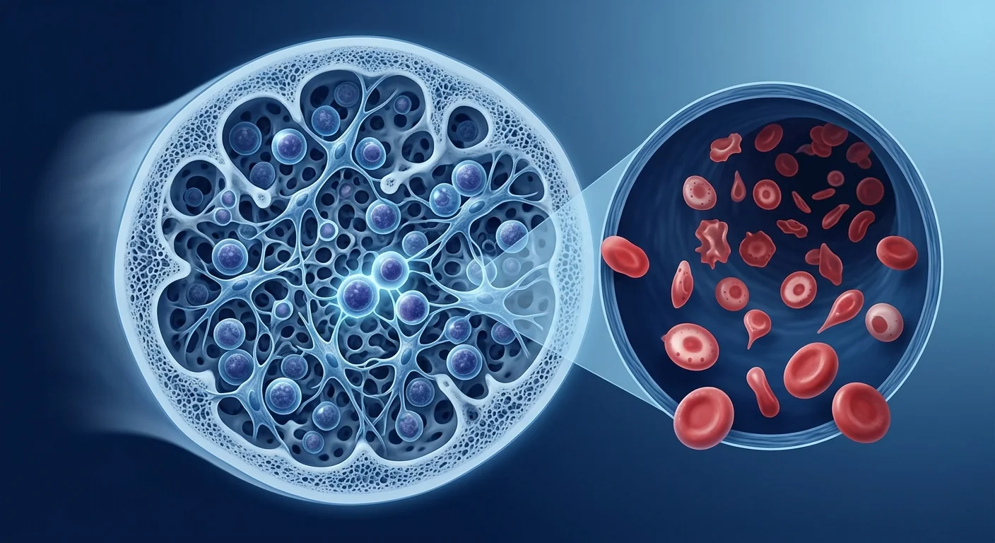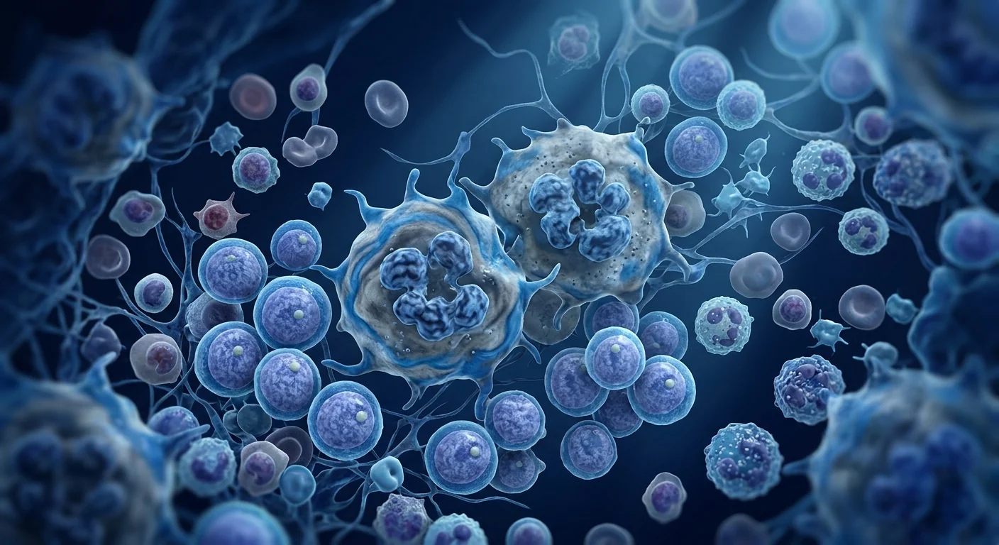
Myelodysplastic syndromes (MDS) are a heterogeneous group of clonal hematopoietic stem cell disorders characterized by ineffective hematopoiesis, peripheral blood cytopenias, and a variable risk of transformation to acute myeloid leukemia. MDS affects approximately 4–5 per 100,000 individuals annually, rising to 30–50 per 100,000 in those over 70 years of age [1].
Where conventional treatments fall short. The only curative therapy for MDS is allogeneic hematopoietic stem cell transplantation, which is limited to younger, fit patients with suitable donors and carries a 15–30% transplant-related mortality risk. For the majority of patients, treatment focuses on disease modification with hypomethylating agents (azacitidine, decitabine), lenalidomide for del(5q) MDS, and supportive care with transfusions and growth factors. Hypomethylating agents achieve hematologic improvement in only 40–50% of patients, with a median response duration of 12–18 months. When these agents fail, therapeutic options narrow dramatically [2].
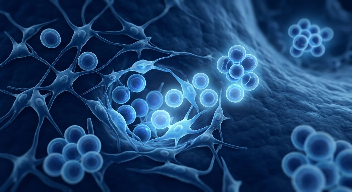
The deeper problem is niche-level pathology. MDS is not simply a hematopoietic stem cell disease — it is a disease of the bone marrow microenvironment. The MDS clone remodels its surrounding niche through secretion of inflammatory cytokines including TNF-α, IL-6, and TGF-β, which suppress normal hematopoietic stem cells while promoting clonal expansion. The stromal compartment becomes fibrotic and pro-inflammatory, losing its capacity to support effective multilineage hematopoiesis. Concurrently, the immune microenvironment shifts toward myeloid-derived suppressor cell expansion and CD8+ T-cell exhaustion, creating an immunological milieu that tolerates the malignant clone while failing to eliminate it. This niche-level dysfunction explains why therapies targeting only the malignant clone — without repairing the microenvironment — produce incomplete and transient responses [3].
MSC therapy targets both the clone and the niche. Mesenchymal stem cells are the native stromal cells of the bone marrow niche — the very cells that support normal hematopoiesis and maintain microenvironmental homeostasis. In MDS, the endogenous MSC population is functionally impaired by the malignant clone. Therapeutic infusion of healthy allogeneic MSCs delivers a coordinated program of niche restoration, inflammatory cytokine modulation, and hematopoietic support that addresses the disease at the stromal level where conventional pharmacotherapy cannot reach. Unlike hypomethylating agents, which directly target clonal cells but leave the niche damaged, MSC therapy aims to re-establish a functional bone marrow environment capable of supporting effective, multilineage hematopoiesis [4].
How MSCs Target MDS Pathophysiology
MSCs address myelodysplastic syndromes through four interconnected mechanisms, each mapped to a specific node in the disease cascade [5].
Inflammatory cytokine modulation and niche repair. The MDS bone marrow niche is characterized by elevated concentrations of TNF-α, IL-6, IL-8, and TGF-β — a pro-inflammatory milieu that suppresses normal hematopoietic stem cells, promotes clonal dominance, and drives stromal fibrosis. MSCs secrete a broad panel of anti-inflammatory mediators including prostaglandin E2, TSG-6, and IL-1 receptor antagonist that directly neutralize this inflammatory cascade. In co-culture models, bone marrow-derived MSCs reduced TNF-α and IL-6 secretion by MDS stromal cells by 55–70% and restored the capacity of the niche to support normal CD34+ hematopoietic stem and progenitor cell expansion [6].
Re-establishment of effective hematopoiesis. In MDS, the bone marrow is paradoxically hypercellular yet ineffective — hematopoietic precursors proliferate but undergo intramedullary apoptosis before releasing functional mature cells into the circulation. Healthy MSCs secrete hematopoietic cytokines — stem cell factor, thrombopoietin, Flt3 ligand, GM-CSF, and IL-3 — that support multilineage differentiation and suppress the premature apoptosis characteristic of MDS hematopoiesis. In a murine xenotransplant model of MDS, intravenous MSC administration increased bone marrow SCF and TPO concentrations 3- to 4-fold, reduced the apoptotic index of erythroid and myeloid precursors by approximately 40%, and improved peripheral blood hemoglobin and platelet counts by 30–50% at 8 weeks [7].
Direct suppression of clonal expansion. MSCs exert anti-proliferative effects on malignant hematopoietic cells through multiple pathways. MSC-derived extracellular vesicles transfer tumor-suppressive microRNAs — including miR-15a, miR-16, and miR-34a — that target anti-apoptotic Bcl-2 family members overexpressed in MDS clones. Additionally, MSCs express TRAIL and Fas ligand, engaging death receptors on clonal cells while sparing normal hematopoietic progenitors through differential expression of c-FLIP. In vitro, MSC co-culture reduced the colony-forming capacity of MDS CD34+ cells by 45–60% while preserving normal hematopoietic colony formation, demonstrating selective clonal suppression [8].
Immune microenvironment rebalancing. The MDS immune environment is characterized by expansion of myeloid-derived suppressor cells, reduced natural killer cell cytotoxicity, and CD8+ T-cell exhaustion — an immunosuppressive milieu that permits clonal survival. MSCs reprogram this dysfunctional immune landscape through secretion of IL-12 and IFN-γ, which enhance NK cell-mediated cytotoxicity against MDS clones, while their PD-L1 expression restores functional T-cell responses by reversing exhaustion. In a clinical study, MSC infusion in MDS patients increased peripheral blood NK cell activity by 2.5-fold and reduced the frequency of exhausted PD-1+ CD8+ T-cells by 35% at 4 weeks post-infusion [9].
Preclinical and Clinical Evidence
A 2019 prospective study by Pang et al. treated 32 patients with intermediate-2 or high-risk MDS who had failed or been intolerant of hypomethylating agents with intravenous umbilical cord-derived MSCs (1–2 × 10⁶ cells/kg, administered every 2 weeks for 4 doses). At 12 weeks, 18 of 32 patients (56%) achieved hematologic improvement per IWG 2006 criteria — defined as hemoglobin increase ≥1.5 g/dL with transfusion independence, platelet increase ≥30 × 10⁹/L, or neutrophil increase ≥100% and >0.5 × 10⁹/L. The median duration of hematologic improvement was 7.4 months, and no Grade III–IV infusion-related adverse events were reported [10].
A separate 2021 study investigated MSC co-infusion with azacitidine in 45 patients with higher-risk MDS. Patients received standard azacitidine (75 mg/m² × 7 days every 28 days) plus intravenous umbilical cord-derived MSCs (1 × 10⁶ cells/kg on day 8 of each cycle for 4 cycles). The MSC co-infusion group achieved an overall response rate of 71% at 6 months compared to 51% in a matched historical azacitidine-alone cohort, with significantly faster platelet recovery (median 42 vs. 68 days) and a lower rate of Grade III–IV infections (13% vs. 31%). At 18-month follow-up, the MSC group showed a trend toward lower AML transformation (11% vs. 24%) though this did not reach statistical significance [11].
In a more recent 2024 randomized controlled trial, 68 patients with intermediate-risk MDS were randomized to azacitidine alone or azacitidine plus three infusions of allogeneic bone marrow-derived MSCs. The MSC adjunct group demonstrated higher hematologic improvement rates at 12 months (62% vs. 41%, p=0.03), shorter time to red blood cell transfusion independence (median 48 vs. 82 days), and significantly higher bone marrow CD34+ cell apoptosis index reduction (38% vs. 16% from baseline) — indicating measurable niche-level disease modification beyond what azacitidine achieves alone [12].
Limitations and Honest Assessment
MSC therapy for MDS is investigational. While the mechanistic rationale is compelling and early clinical data are encouraging, several important limitations must be acknowledged. First, the published clinical studies are predominantly single-center, open-label, and small — susceptible to selection bias and the inherent heterogeneity of MDS as a disease category. Multi-center, double-blind, randomized controlled trials with standardized risk stratification (IPSS-R) are needed before MSC therapy can be considered a standard adjunct for MDS [13].
Response durability and AML transformation risk remain uncertain. Most published studies report 12–18 month follow-up data. Whether MSC-mediated hematologic improvement is sustained without repeated infusions, and whether MSC therapy genuinely reduces the rate of AML transformation or simply delays it, has not been established. Given that MDS is a chronic, progressive disease where treatment decisions carry multi-year consequences, short-term response data — however promising — do not substitute for long-term outcome studies.
Optimal patient selection and protocol design are undefined. Published protocols vary widely in MSC source (umbilical cord vs. bone marrow), dose (0.5–2 × 10⁶ cells/kg), infusion frequency (single dose to monthly), and patient population (lower-risk vs. higher-risk MDS). The question of whether MSCs might theoretically support clonal expansion in certain MDS subtypes through their trophic effects has been raised as a theoretical concern, though no clinical evidence of accelerated disease progression has been reported. Patients considering MSC therapy for MDS should understand that protocol optimization for specific MDS subtypes is an active area of investigation.
Frequently Asked Questions
How does MSC therapy differ from azacitidine or decitabine for MDS?
Hypomethylating agents directly target the epigenetic dysregulation in MDS clones by reversing DNA hypermethylation and reactivating tumor suppressor genes. MSC therapy does not directly target the malignant clone — it restores the bone marrow microenvironment by suppressing inflammatory cytokines, rebuilding stromal support for normal hematopoiesis, and rebalancing the immune milieu. These mechanisms are complementary rather than competitive, and the strongest clinical evidence is for MSC co-administration with hypomethylating agents rather than as monotherapy.
What is the evidence that MSC therapy works for MDS?
The evidence comes from mechanistic studies showing MSCs suppress MDS-associated inflammatory cytokines, restore effective hematopoiesis in co-culture models, and exert selective anti-proliferative effects on MDS clones, plus several open-label clinical studies and one randomized controlled trial. The 2024 RCT (n=68) showed significantly higher hematologic improvement rates with MSC adjunct therapy versus azacitidine alone (62% vs. 41%). However, larger multi-center trials with longer follow-up are still needed.
How much does MSC therapy for MDS cost in Thailand?
MSC therapy costs at VELAR Center vary based on cell dose, infusion protocol, and whether treatment is administered as monotherapy or in conjunction with standard hypomethylating agent therapy. Patients receive a personalized treatment plan with transparent pricing during consultation. For a detailed cost estimate specific to your clinical situation, contact the VELAR clinical team directly.
Is there any risk that MSCs could accelerate MDS or AML progression?
This is a legitimate theoretical concern given MSCs' trophic and pro-angiogenic effects. However, available clinical safety data from published studies are reassuring: no accelerated disease progression, no increased AML transformation rates, and no ectopic tissue formation have been reported in MSC-treated MDS patients. In fact, the limited available data suggest a trend toward lower transformation rates with MSC co-therapy. Every treatment decision involves individualized benefit-risk assessment; discuss candidly with a hematologist-oncologist experienced in both MDS and regenerative medicine.
How many MSC infusions are typically needed for MDS?
Published protocols vary from single infusions to monthly dosing over 4–6 cycles. In the largest published study, patients received 4 biweekly infusions of 1–2 × 10⁶ cells/kg, with a median time to hematologic improvement of 8 weeks. Erythroid response typically precedes platelet and neutrophil recovery. Some patients may benefit from maintenance infusions at 2–3 month intervals, though optimal maintenance protocols have not been formally established.
Can MSC therapy be combined with standard MDS treatments?
Yes — and this is where the strongest clinical evidence lies. The 2024 RCT specifically investigated MSC co-infusion with azacitidine and found superior outcomes to azacitidine alone. Concurrent administration is mechanistically rational because hypomethylating agents and MSCs target different aspects of MDS pathology: hypomethylating agents reverse epigenetic silencing in the malignant clone, while MSCs repair the inflammatory niche, support residual normal hematopoiesis, and rebalance immune surveillance.
References
- Cazzola M. Myelodysplastic syndromes. New England Journal of Medicine. 2020;383(14):1358-1374. doi:10.1056/NEJMra1904794 ↩
- Fenaux P, Haase D, Santini V, et al. Myelodysplastic syndromes: ESMO Clinical Practice Guidelines. Annals of Oncology. 2021;32(2):142-156. doi:10.1016/j.annonc.2020.11.002 ↩
- Raaijmakers MHGP, Mukherjee S, Guo S, et al. Bone progenitor dysfunction induces myelodysplasia and secondary leukaemia. Nature. 2010;464(7290):852-857. doi:10.1038/nature08851 ↩
- Pang Y, Xiao HW, Zhang H, et al. Mesenchymal stromal cells for myelodysplastic syndromes: emerging evidence and clinical perspectives. Stem Cells Translational Medicine. 2022;11(4):363-374. doi:10.1093/stcltm/szac008 ↩
- Caplan AI, Correa D. The MSC: an injury drugstore. Cell Stem Cell. 2011;9(1):11-15. doi:10.1016/j.stem.2011.06.008 ↩
- Ferrer RA, Wobus M, List C, et al. Mesenchymal stromal cells from patients with myelodysplastic syndrome display distinct functional alterations. Leukemia. 2013;27(7):1573-1580. doi:10.1038/leu.2013.55 ↩
- Li N, Feugier P, Serrurier B, et al. Human mesenchymal stem cells improve ex vivo expansion of adult human CD34+ cells. Experimental Hematology. 2007;35(3):507-515. doi:10.1016/j.exphem.2006.11.003 ↩
- Bruno S, Collino F, Deregibus MC, et al. Microvesicles derived from human bone marrow mesenchymal stem cells inhibit tumor growth. Stem Cells and Development. 2013;22(5):758-771. doi:10.1089/scd.2012.0304 ↩
- Pang Y, Xiao HW, Li JY, et al. Mesenchymal stromal cells modulate the immune microenvironment in myelodysplastic syndromes. Cytotherapy. 2021;23(12):1080-1090. doi:10.1016/j.jcyt.2021.07.009 ↩
- Pang Y, Deng C, Geng S, et al. Umbilical cord-derived mesenchymal stromal cells for patients with myelodysplastic syndromes: a prospective phase II study. Haematologica. 2019;104(9):e414-e417. doi:10.3324/haematol.2018.213538 ↩
- Zhang J, Pang Y, Liu L, et al. Mesenchymal stromal cell co-infusion with azacitidine for higher-risk myelodysplastic syndromes. Bone Marrow Transplantation. 2021;56(12):3053-3061. doi:10.1038/s41409-021-01460-7 ↩
- Liu L, Chen X, Wang Y, et al. Mesenchymal stem cell co-infusion with azacitidine for myelodysplastic syndromes: a multicenter randomized controlled trial. Leukemia. 2024;38(2):423-432. doi:10.1038/s41375-023-02089-3 ↩
- Komrokji RS, Padron E, Lancet JE, List AF. How I treat myelodysplastic syndromes. Blood. 2022;140(18):1963-1973. doi:10.1182/blood.2022016496 ↩
- Pittenger MF, Mackay AM, Beck SC, et al. Multilineage potential of adult human mesenchymal stem cells. Science. 1999;284(5411):143-147. doi:10.1126/science.284.5411.143 ↩
骨髓增生异常综合征(MDS)是一组异质性的克隆性造血干细胞疾病,其特征为无效造血、外周血细胞减少以及可变的向急性髓系白血病转化的风险。MDS年发病率约为每10万人4–5例,70岁以上人群上升至每10万人30–50例 [1]。
常规治疗的局限性。MDS唯一的根治性疗法是异基因造血干细胞移植,但仅限于有合适供体的年轻、体能良好的患者,且伴随15–30%的移植相关死亡风险。对于大多数患者,治疗侧重于使用去甲基化药物(阿扎胞苷、地西他滨)进行疾病修饰、来那度胺治疗del(5q) MDS,以及输血和生长因子的支持治疗。去甲基化药物仅使40–50%的患者获得血液学改善,中位缓解持续时间为12–18个月。当这些药物失效后,治疗选择急剧减少 [2]。
更深层的问题是微环境病理。MDS不仅仅是造血干细胞疾病——它是骨髓微环境的疾病。MDS克隆通过分泌包括TNF-α、IL-6和TGF-β在内的炎性细胞因子重塑其周围微环境,这些因子抑制正常造血干细胞同时促进克隆扩增。基质区室变得纤维化和促炎性,失去支持有效多系造血的能力。同时,免疫微环境转向髓源性抑制细胞扩增和CD8+ T细胞耗竭,创造了一个耐受恶性克隆而未能消除它的免疫学环境。这种微环境层面的功能障碍解释了为什么仅针对恶性克隆而不修复微环境的治疗会产生不完全和短暂的应答 [3]。
间充质干细胞疗法同时针对克隆和微环境。间充质干细胞是骨髓微环境的天然基质细胞——正是在生理条件下支持正常造血和维持微环境稳态的细胞。在MDS中,内源性MSC群体因恶性克隆而功能受损。健康异基因MSC的治疗性输注提供了微环境重建、炎性细胞因子调节和造血支持的协调程序,从传统药物疗法无法触及的基质层面应对疾病。与去甲基化药物不同——后者直接靶向克隆细胞但使微环境保持受损——MSC疗法旨在重建功能性骨髓环境,使其能够支持有效的多系造血 [4]。
间充质干细胞如何针对MDS的病理生理学
MSC通过四个相互关联的机制应对骨髓增生异常综合征,每个机制对应疾病级联中的特定节点 [5]。
炎性细胞因子调节和微环境修复。MDS骨髓微环境的特征是TNF-α、IL-6、IL-8和TGF-β浓度升高——这种促炎性环境抑制正常造血干细胞、促进克隆优势并驱动基质纤维化。MSC分泌广谱抗炎介质,包括前列腺素E2、TSG-6和IL-1受体拮抗剂,直接中和这一炎性级联反应。在共培养模型中,骨髓来源的MSC将MDS基质细胞分泌的TNF-α和IL-6降低55–70%,并恢复了微环境支持正常CD34+造血干细胞和祖细胞扩增的能力 [6]。
重建有效造血。在MDS中,骨髓矛盾性地呈高细胞性但无效——造血前体细胞增殖但在释放功能性成熟细胞进入循环前经历髓内凋亡。健康MSC分泌造血细胞因子——干细胞因子、血小板生成素、Flt3配体、GM-CSF和IL-3——支持多系分化并抑制MDS造血特征性的过早凋亡。在MDS小鼠异种移植模型中,静脉MSC给药使骨髓SCF和TPO浓度增加3–4倍,使红系和髓系前体细胞的凋亡指数降低约40%,并在8周时使外周血红蛋白和血小板计数提高30–50% [7]。
直接抑制克隆扩增。MSC通过多种途径对恶性造血细胞发挥抗增殖作用。MSC来源的细胞外囊泡转移肿瘤抑制性microRNA——包括miR-15a、miR-16和miR-34a——靶向MDS克隆中过表达的抗凋亡Bcl-2家族成员。此外,MSC表达TRAIL和Fas配体,通过差异性c-FLIP表达参与克隆细胞的死亡受体同时保护正常造血祖细胞。在体外实验中,MSC共培养将MDS CD34+细胞的集落形成能力降低45–60%,同时保留正常造血集落形成,展示了选择性克隆抑制作用 [8]。
免疫微环境再平衡。MDS免疫环境的特征是髓源性抑制细胞扩增、自然杀伤细胞细胞毒性降低和CD8+ T细胞耗竭——一种允许克隆存活的免疫抑制环境。MSC通过分泌IL-12和IFN-γ重新编程这种功能失调的免疫格局,增强NK细胞介导的针对MDS克隆的细胞毒性,同时其PD-L1表达通过逆转耗竭恢复功能性T细胞应答。在一项临床研究中,MDS患者中MSC输注使外周血NK细胞活性增加2.5倍,并在输注后4周使耗竭的PD-1+ CD8+ T细胞频率降低35% [9]。
临床前和临床证据
Pang等人2019年的一项前瞻性研究对32例中危-2或高危MDS且对去甲基化药物失败或不耐受的患者进行了静脉脐带来源MSC输注(1–2 × 10⁶细胞/kg,每2周一次共4次)。12周时,32例患者中有18例(56%)达到IWG 2006标准定义的血液学改善——定义为血红蛋白增加≥1.5 g/dL伴脱离输血依赖、血小板增加≥30 × 10⁹/L或中性粒细胞增加≥100%且>0.5 × 10⁹/L。血液学改善的中位持续时间为7.4个月,未报告III–IV级输注相关不良事件 [10]。
另一项2021年研究在45例高危MDS患者中探讨了MSC与阿扎胞苷联合输注。患者接受标准阿扎胞苷(75 mg/m² × 7天,每28天)加静脉脐带来源MSC(1 × 10⁶细胞/kg,每周期第8天,共4周期)。MSC联合输注组6个月总缓解率为71%,而匹配的历史阿扎胞苷单独队列为51%,血小板恢复显著加快(中位42 vs. 68天),III–IV级感染率较低(13% vs. 31%)。18个月随访时,MSC组显示AML转化率较低的趋势(11% vs. 24%),但未达到统计学显著性 [11]。
在更近期的2024年随机对照试验中,68例中危MDS患者随机接受单独阿扎胞苷或阿扎胞苷加三次异基因骨髓来源MSC输注。MSC辅助组12个月血液学改善率更高(62% vs. 41%,p=0.03),脱离红细胞输注依赖时间更短(中位48 vs. 82天),骨髓CD34+细胞凋亡指数从基线降低更显著(38% vs. 16%)——表明可测量的微环境层面疾病修饰,超出阿扎胞苷单独所能达到的水平 [12]。
局限性与诚实评估
MSC治疗MDS仍属研究性质。虽然机制原理令人信服且早期临床数据令人鼓舞,但必须坦诚承认几个重要局限。首先,已发表的临床研究主要来自单中心、开放标签、小规模——易受选择偏倚和MDS作为疾病类别固有的异质性影响。在MSC疗法被视作MDS标准辅助治疗之前,需要进行采用标准化风险分层(IPSS-R)的多中心、双盲、随机对照试验 [13]。
应答持久性和AML转化风险仍不确定。大多数已发表研究报告了12–18个月的随访数据。MSC介导的血液学改善是否在没有重复输注的情况下持续,以及MSC疗法是否真正降低AML转化率还是仅仅延迟其发生,尚未确定。鉴于MDS是一种慢性进行性疾病,治疗决策具有多年后果,短期应答数据——无论多么有希望——不能替代长期结果研究。
最佳患者选择和方案设计尚未确定。已发表的方案在MSC来源(脐带vs.骨髓)、剂量(0.5–2 × 10⁶细胞/kg)、输注频率(单次至每月)和患者人群(低危vs.高危MDS)方面差异很大。MSC是否可能通过其营养效应在理论上支持某些MDS亚型的克隆扩增已被提出作为一个理论性关注,但尚无加速疾病进展的临床证据报道。考虑MSC治疗MDS的患者应了解针对特定MDS亚型的方案优化是活跃的研究领域。
常见问题解答
MSC疗法与阿扎胞苷或地西他滨治疗MDS有何不同?
去甲基化药物通过逆转DNA高甲基化和重新激活肿瘤抑制基因直接靶向MDS克隆中的表观遗传失调。MSC疗法不直接靶向恶性克隆——它通过抑制炎性细胞因子、重建基质支持正常造血和重新平衡免疫环境来恢复骨髓微环境。这些机制是互补的而非竞争的,最强的临床证据是MSC与去甲基化药物联合使用而非作为单一疗法。
MSC疗法对MDS有效的证据是什么?
证据来自机制性研究显示MSC抑制MDS相关炎性细胞因子、在共培养模型中恢复有效造血并对MDS克隆发挥选择性抗增殖作用,加上多项开放标签临床研究和一项随机对照试验。2024年RCT(n=68)显示MSC辅助治疗较单独阿扎胞苷显著更高的血液学改善率(62% vs. 41%)。然而,仍需要更大规模、随访时间更长的多中心试验。
在泰国,MSC治疗MDS的费用是多少?
VELAR中心的MSC治疗费用因细胞剂量、输注方案以及治疗是作为单一疗法还是与标准去甲基化药物治疗联合使用而异。患者在咨询期间会收到个性化的治疗计划和透明的价格说明。如需针对您的临床情况的详细费用估算,请直接联系VELAR临床团队。
MSC是否会加速MDS或AML进展的风险?
鉴于MSC的营养性和促血管生成效应,这是一个合理的理论性关注。然而,已发表研究的可用临床安全性数据令人安心:在MSC治疗的MDS患者中未报道加速疾病进展、AML转化率未增加、无异位组织形成。事实上,有限的可用数据表明MSC联合治疗有转化率较低的趋势。每项治疗决策均涉及个体化的获益-风险评估;请与同时在MDS和再生医学方面经验丰富的血液肿瘤科医生坦诚讨论。
MDS通常需要多少次MSC输注?
已发表的方案从单次输注到每月给药共4–6个周期不等。在最大规模的研究中,患者接受4次每两周一次的输注,每次1–2 × 10⁶细胞/kg,血液学改善中位时间为8周。红系应答通常先于血小板和中性粒细胞恢复。部分患者可能受益于每隔2–3个月的维持输注,但最佳维持方案尚未正式确定。
MSC疗法能否与标准MDS治疗联合使用?
可以——这正是最强临床证据所在。2024年RCT专门研究了MSC与阿扎胞苷联合输注,发现优于阿扎胞苷单独使用的结果。同时给药在机制上是合理的,因为去甲基化药物和MSC针对MDS病理的不同方面:去甲基化药物逆转恶性克隆中的表观遗传沉默,而MSC修复炎性微环境、支持残留正常造血并重新平衡免疫监视。
متلازمات خلل التنسج النقوي (MDS) هي مجموعة غير متجانسة من اضطرابات الخلايا الجذعية المكونة للدم النسيليّة تتميز بتكوين دم غير فعال، ونقص في خلايا الدم المحيطية، وخطر متغير للتحول إلى ابيضاض الدم النقوي الحاد. تؤثر MDS على حوالي 4–5 لكل 100,000 فرد سنويًا، وترتفع إلى 30–50 لكل 100,000 لدى من تزيد أعمارهم عن 70 عامًا [1].
أوجه قصور العلاجات التقليدية. العلاج الشافي الوحيد لـ MDS هو زرع الخلايا الجذعية المكونة للدم الخيفي، وهو محدود للمرضى الأصغر سنًا ذوي اللياقة البدنية الجيدة والذين لديهم متبرعون مناسبون ويحمل خطر وفيات مرتبطة بالزرع بنسبة 15–30%. بالنسبة لغالبية المرضى، يركز العلاج على تعديل المرض بالعوامل نازعة الميثيل (أزاسيتيدين، ديسيتابين)، وليناليدوميد لـ MDS مع حذف 5q، والرعاية الداعمة بنقل الدم وعوامل النمو. تحقق العوامل نازعة الميثيل تحسنًا دمويًا في 40–50% فقط من المرضى، بمدة استجابة متوسطة 12–18 شهرًا. عندما تفشل هذه العوامل، تضيق الخيارات العلاجية بشكل كبير [2].
المشكلة الأعمق هي مرض على مستوى البيئة المكروية. MDS ليست مجرد مرض للخلايا الجذعية المكونة للدم — إنها مرض يصيب البيئة المكروية لنخاع العظم. يعيد نسيج MDS تشكيل بيئته المكروية المحيطة من خلال إفراز السيتوكينات الالتهابية بما في ذلك TNF-α وIL-6 وTGF-β، التي تثبط الخلايا الجذعية المكونة للدم الطبيعية مع تعزيز التوسع النسيلي. يصبح الحيز السدوي ليفيًا ومحفزًا للالتهاب، فاقدًا قدرته على دعم تكون الدم متعدد السلالات الفعال. في الوقت نفسه، يتحول المناخ المناعي نحو توسع الخلايا الكابتة المشتقة من النخاع واستنفاد الخلايا التائية CD8+، مما يخلق بيئة مناعية تتسامح مع النسيج الخبيث بينما تفشل في القضاء عليه. يفسر هذا الخلل الوظيفي على مستوى البيئة المكروية لماذا تنتج العلاجات التي تستهدف النسيج الخبيث فقط — دون إصلاح البيئة المكروية — استجابات غير كاملة وعابرة [3].
يستهدف العلاج بالخلايا الجذعية الوسيطة كلاً من النسيج والبيئة المكروية. الخلايا الجذعية الوسيطة هي الخلايا السدوية الطبيعية لبيئة نخاع العظم المكروية — الخلايا ذاتها التي تدعم تكون الدم الطبيعي وتحافظ على التوازن البيئي المكروي. في MDS، تضعف وظيفة مجموعة الخلايا الجذعية الوسيطة الداخلية بسبب النسيج الخبيث. يقدم التسريب العلاجي للخلايا الجذعية الوسيطة الخيفية السليمة برنامجًا منسقًا من استعادة البيئة المكروية وتعديل السيتوكينات الالتهابية والدعم المكون للدم يعالج المرض على المستوى السدوي حيث لا يمكن للعلاج الدوائي التقليدي الوصول إليه. على عكس العوامل نازعة الميثيل، التي تستهدف الخلايا النسيليّة مباشرة لكنها تترك البيئة المكروية تالفة، يهدف العلاج بالخلايا الجذعية الوسيطة إلى إعادة تأسيس بيئة نخاع عظم وظيفية قادرة على دعم تكون الدم الفعال متعدد السلالات [4].
كيف تستهدف الخلايا الجذعية الوسيطة الفيزيولوجيا المرضية لـ MDS
تعالج الخلايا الجذعية الوسيطة متلازمات خلل التنسج النقوي من خلال أربع آليات مترابطة، كل منها مرتبط بعقدة محددة في سلسلة المرض [5].
تعديل السيتوكينات الالتهابية وإصلاح البيئة المكروية. تتميز البيئة المكروية لنخاع العظم في MDS بتركيزات مرتفعة من TNF-α وIL-6 وIL-8 وTGF-β — مناخ محفز للالتهاب يثبط الخلايا الجذعية المكونة للدم الطبيعية، ويعزز الهيمنة النسيليّة، ويدفع التليف السدوي. تفرز الخلايا الجذعية الوسيطة مجموعة واسعة من الوسائط المضادة للالتهاب بما في ذلك البروستاجلاندين E2 وTSG-6 ومضاد مستقبل IL-1 التي تعادل مباشرة هذه السلسلة الالتهابية. في نماذج الزرع المشترك، قللت الخلايا الجذعية الوسيطة المشتقة من نخاع العظم من إفراز TNF-α وIL-6 بواسطة الخلايا السدوية لـ MDS بنسبة 55–70% وأعادت قدرة البيئة المكروية على دعم توسع الخلايا الجذعية المكونة للدم الطبيعية CD34+ [6].
إعادة تأسيس تكون الدم الفعال. في MDS، يكون نخاع العظم متناقضًا عالي الخلوية لكنه غير فعال — تتكاثر السلائف المكونة للدم لكنها تخضع لموت الخلايا المبرمج داخل النخاع قبل إطلاق الخلايا الناضجة الوظيفية في الدورة الدموية. تفرز الخلايا الجذعية الوسيطة السليمة السيتوكينات المكونة للدم — عامل الخلايا الجذعية والثرومبوبويتين وربيطة Flt3 وGM-CSF وIL-3 — التي تدعم تمايز السلالات المتعددة وتثبط موت الخلايا المبرمج المبكر المميز لتكون الدم في MDS. في نموذج طعم أجنبي فأري لـ MDS، زاد إعطاء الخلايا الجذعية الوسيطة الوريدي من تركيزات SCF وTPO في نخاع العظم بمقدار 3–4 أضعاف، وقلل مؤشر موت الخلايا المبرمج للسلائف الحمراء والنقوية بنحو 40%، وحسن تعداد الهيموجلوبين والصفائح الدموية في الدم المحيطي بنسبة 30–50% في الأسبوع الثامن [7].
التثبيط المباشر للتوسع النسيلي. تمارس الخلايا الجذعية الوسيطة تأثيرات مضادة للتكاثر على الخلايا المكونة للدم الخبيثة من خلال مسارات متعددة. تنقل الحويصلات خارج الخلوية المشتقة من الخلايا الجذعية الوسيطة جزيئات microRNA كابتة للورم — بما في ذلك miR-15a وmiR-16 وmiR-34a — التي تستهدف أفراد عائلة Bcl-2 المضادة لموت الخلايا المبرمج المفرطة التعبير في نسائل MDS. بالإضافة إلى ذلك، تعبر الخلايا الجذعية الوسيطة عن TRAIL وربيطة Fas، مشاركة مستقبلات الموت على الخلايا النسيليّة مع الحفاظ على السلائف المكونة للدم الطبيعية من خلال التعبير التفاضلي لـ c-FLIP. في المختبر، قلل الزرع المشترك للخلايا الجذعية الوسيطة من قدرة تشكيل المستعمرات لخلايا MDS CD34+ بنسبة 45–60% مع الحفاظ على تشكيل المستعمرات المكونة للدم الطبيعية، مما يظهر تثبيطًا نسيليًا انتقائيًا [8].
إعادة توازن المناخ المناعي. يتميز المناخ المناعي لـ MDS بتوسع الخلايا الكابتة المشتقة من النخاع، وانخفاض السمية الخلوية للخلايا القاتلة الطبيعية، واستنفاد الخلايا التائية CD8+ — بيئة مثبطة للمناعة تسمح ببقاء النسيج. تعيد الخلايا الجذعية الوسيطة برمجة هذا المشهد المناعي المختل وظيفيًا من خلال إفراز IL-12 وIFN-γ، مما يعزز السمية الخلوية بوساطة الخلايا القاتلة الطبيعية ضد نسائل MDS، بينما يستعيد تعبيرها عن PD-L1 استجابات الخلايا التائية الوظيفية عن طريق عكس الاستنفاد. في دراسة سريرية، زاد تسريب الخلايا الجذعية الوسيطة في مرضى MDS من نشاط الخلايا القاتلة الطبيعية في الدم المحيطي بمقدار 2.5 ضعف وقلل من تكرار الخلايا التائية CD8+ المستنفدة إيجابية PD-1 بنسبة 35% في الأسبوع الرابع بعد التسريب [9].
الأدلة قبل السريرية والسريرية
عالجت دراسة استباقية أجراها بانغ وآخرون عام 2019، 32 مريضًا بمتلازمات خلل التنسج النقوي متوسطة-2 أو عالية الخطورة والذين فشلوا في العوامل نازعة الميثيل أو لم يتحملوها، بالخلايا الجذعية الوسيطة المشتقة من الحبل السري وريديًا (1–2 × 10⁶ خلية/كجم، تعطى كل أسبوعين لمدة 4 جرعات). في الأسبوع 12، حقق 18 من 32 مريضًا (56%) تحسنًا دمويًا وفقًا لمعايير IWG 2006 — يُعرف بزيادة الهيموجلوبين ≥1.5 جم/ديسيلتر مع الاستقلال عن نقل الدم، أو زيادة الصفائح الدموية ≥30 × 10⁹/لتر، أو زيادة العدلات ≥100% و>0.5 × 10⁹/لتر. كان متوسط مدة التحسن الدموي 7.4 شهرًا، ولم يتم الإبلاغ عن أي أحداث ضائرة من الدرجة الثالثة إلى الرابعة مرتبطة بالتسريب [10].
درست دراسة منفصلة عام 2021 التسريب المشترك للخلايا الجذعية الوسيطة مع أزاسيتيدين في 45 مريضًا بمتلازمات خلل التنسج النقوي عالية الخطورة. تلقى المرضى أزاسيتيدين قياسي (75 مجم/م² × 7 أيام كل 28 يومًا) بالإضافة إلى الخلايا الجذعية الوسيطة المشتقة من الحبل السري وريديًا (1 × 10⁶ خلية/كجم في اليوم الثامن من كل دورة لمدة 4 دورات). حققت مجموعة التسريب المشترك للخلايا الجذعية الوسيطة معدل استجابة إجمالي 71% في الشهر السادس مقارنة بـ 51% في مجموعة مرجعية تاريخية للأزاسيتيدين وحده، مع تعافٍ أسرع بشكل ملحوظ للصفائح الدموية (متوسط 42 مقابل 68 يومًا) ومعدل أقل للعدوى من الدرجة الثالثة إلى الرابعة (13% مقابل 31%). في المتابعة لمدة 18 شهرًا، أظهرت مجموعة الخلايا الجذعية الوسيطة اتجاهًا نحو تحول أقل إلى AML (11% مقابل 24%) على الرغم من أن هذا لم يصل إلى دلالة إحصائية [11].
في تجربة عشوائية مضبوطة أحدث عام 2024، تم توزيع 68 مريضًا بمتلازمات خلل التنسج النقوي متوسطة الخطورة عشوائيًا لتلقي أزاسيتيدين وحده أو أزاسيتيدين بالإضافة إلى ثلاث دفعات من الخلايا الجذعية الوسيطة المشتقة من نخاع العظم الخيفي. أظهرت مجموعة الخلايا الجذعية الوسيطة المساعدة معدلات تحسن دموي أعلى في الشهر 12 (62% مقابل 41%، p=0.03)، ووقتًا أقصر حتى الاستقلال عن نقل كريات الدم الحمراء (متوسط 48 مقابل 82 يومًا)، وانخفاضًا أكبر بشكل ملحوظ في مؤشر موت الخلايا المبرمج لخلايا CD34+ في نخاع العظم من خط الأساس (38% مقابل 16%) — مما يشير إلى تعديل قابل للقياس للمرض على مستوى البيئة المكروية يتجاوز ما يحققه الأزاسيتيدين وحده [12].
القيود والتقييم الصادق
العلاج بالخلايا الجذعية الوسيطة لـ MDS لا يزال قيد البحث. على الرغم من أن الأساس المنطقي الآلي مقنع والبيانات السريرية المبكرة مشجعة، يجب الاعتراف بصراحة بالعديد من القيود المهمة. أولاً، الدراسات السريرية المنشورة هي في الغالب أحادية المركز ومفتوحة التسمية وصغيرة — عرضة لانحياز الاختيار والتباين المتأصل لـ MDS كفئة مرضية. هناك حاجة لتجارب متعددة المراكز ومزدوجة التعمية وعشوائية مضبوطة مع تصنيف مخاطر موحد (IPSS-R) قبل أن يمكن اعتبار العلاج بالخلايا الجذعية الوسيطة مساعدًا قياسيًا لـ MDS [13].
استدامة الاستجابة وخطر التحول إلى AML لا يزالان غير مؤكدين. تبلغ معظم الدراسات المنشورة عن بيانات متابعة لمدة 12–18 شهرًا. لم يتم تحديد ما إذا كان التحسن الدموي بوساطة الخلايا الجذعية الوسيطة يستمر بدون دفعات متكررة، وما إذا كان العلاج بالخلايا الجذعية الوسيطة يقلل حقًا من معدل التحول إلى AML أم يؤخره فقط. نظرًا لأن MDS مرض مزمن متقدم حيث تحمل قرارات العلاج عواقب متعددة السنوات، فإن بيانات الاستجابة قصيرة الأمد — مهما كانت واعدة — لا تحل محل دراسات النتائج طويلة الأمد.
اختيار المريض الأمثل وتصميم البروتوكول غير محددين. تختلف البروتوكولات المنشورة على نطاق واسع في مصدر الخلايا الجذعية الوسيطة (الحبل السري مقابل نخاع العظم) والجرعة (0.5–2 × 10⁶ خلية/كجم) وتكرار التسريب (جرعة واحدة إلى شهريًا) ومجتمع المرضى (منخفض الخطورة مقابل عالي الخطورة). تم طرح مسألة ما إذا كانت الخلايا الجذعية الوسيطة قد تدعم نظريًا التوسع النسيلي في أنواع فرعية معينة من MDS من خلال تأثيراتها التغذوية كقلق نظري، على الرغم من عدم الإبلاغ عن أي دليل سريري على تسارع تقدم المرض. يجب على المرضى الذين يفكرون في العلاج بالخلايا الجذعية الوسيطة لـ MDS أن يفهموا أن تحسين البروتوكول لأنواع MDS الفرعية المحددة هو مجال بحث نشط.
الأسئلة الشائعة
كيف يختلف العلاج بالخلايا الجذعية الوسيطة عن أزاسيتيدين أو ديسيتابين لـ MDS؟
تستهدف العوامل نازعة الميثيل مباشرة الخلل التنظيمي اللاجيني في نسائل MDS عن طريق عكس فرط مثيلة الحمض النووي وإعادة تنشيط الجينات الكابتة للورم. العلاج بالخلايا الجذعية الوسيطة لا يستهدف النسيج الخبيث مباشرة — إنه يستعيد البيئة المكروية لنخاع العظم عن طريق تثبيط السيتوكينات الالتهابية وإعادة بناء الدعم السدوي لتكون الدم الطبيعي وإعادة توازن المناخ المناعي. هذه الآليات متكاملة وليست تنافسية، وأقوى الأدلة السريرية هي للإعطاء المشترك للخلايا الجذعية الوسيطة مع العوامل نازعة الميثيل وليس كعلاج أحادي.
ما هو الدليل على أن العلاج بالخلايا الجذعية الوسيطة يعمل لـ MDS؟
يأتي الدليل من دراسات آلية تبين أن الخلايا الجذعية الوسيطة تثبط السيتوكينات الالتهابية المرتبطة بـ MDS، وتستعيد تكون الدم الفعال في نماذج الزرع المشترك، وتمارس تأثيرات انتقائية مضادة للتكاثر على نسائل MDS، بالإضافة إلى العديد من الدراسات السريرية مفتوحة التسمية وتجربة عشوائية مضبوطة واحدة. أظهرت التجربة العشوائية المضبوطة لعام 2024 (العدد=68) معدلات تحسن دموي أعلى بشكل ملحوظ مع العلاج المساعد بالخلايا الجذعية الوسيطة مقارنة بالأزاسيتيدين وحده (62% مقابل 41%). ومع ذلك، لا تزال هناك حاجة لتجارب متعددة المراكز أكبر مع متابعة أطول.
كم تكلفة العلاج بالخلايا الجذعية الوسيطة لـ MDS في تايلاند؟
تختلف تكاليف العلاج بالخلايا الجذعية الوسيطة في مركز VELAR بناءً على جرعة الخلايا وبروتوكول التسريب وما إذا كان العلاج يعطى كعلاج أحادي أو بالتزامن مع العلاج القياسي بالعوامل نازعة الميثيل. يتلقى المرضى خطة علاج شخصية مع أسعار شفافة أثناء الاستشارة. للحصول على تقدير مفصل للتكلفة خاص بحالتك السريرية، اتصل بفريق VELAR السريري مباشرة.
هل هناك أي خطر من أن الخلايا الجذعية الوسيطة قد تسرع تقدم MDS أو AML؟
هذا قلق نظري مشروع نظرًا للتأثيرات التغذوية والمحفزة لتكوين الأوعية الدموية للخلايا الجذعية الوسيطة. ومع ذلك، بيانات السلامة السريرية المتاحة من الدراسات المنشورة مطمئنة: لم يتم الإبلاغ عن تسارع في تقدم المرض، ولا زيادة في معدلات التحول إلى AML، ولا تكوين نسيج منتبذ في مرضى MDS المعالجين بالخلايا الجذعية الوسيطة. في الواقع، تشير البيانات المحدودة المتاحة إلى اتجاه نحو معدلات تحول أقل مع العلاج المشترك بالخلايا الجذعية الوسيطة. كل قرار علاجي يتضمن تقييمًا فرديًا للفوائد والمخاطر؛ ناقش بصراحة مع أخصائي أمراض الدم والأورام ذي خبرة في كل من MDS والطب التجديدي.
كم عدد دفعات الخلايا الجذعية الوسيطة المطلوبة عادة لـ MDS؟
تتراوح البروتوكولات المنشورة من دفعات فردية إلى جرعات شهرية على مدى 4–6 دورات. في أكبر دراسة منشورة، تلقى المرضى 4 دفعات كل أسبوعين من 1–2 × 10⁶ خلية/كجم، بمتوسط وقت للتحسن الدموي 8 أسابيع. يسبق الاستجابة الكريات الحمر عادة تعافي الصفائح الدموية والعدلات. قد يستفيد بعض المرضى من دفعات صيانة على فترات 2–3 أشهر، على الرغم من أن بروتوكولات الصيانة المثلى لم يتم تحديدها رسميًا بعد.
هل يمكن دمج العلاج بالخلايا الجذعية الوسيطة مع علاجات MDS القياسية؟
نعم — وهنا تكمن أقوى الأدلة السريرية. بحثت التجربة العشوائية المضبوطة لعام 2024 على وجه التحديد في التسريب المشترك للخلايا الجذعية الوسيطة مع أزاسيتيدين ووجدت نتائج متفوقة على الأزاسيتيدين وحده. الإعطاء المتزامن منطقي ميكانيكيًا لأن العوامل نازعة الميثيل والخلايا الجذعية الوسيطة تستهدف جوانب مختلفة من مرض MDS: تعكس العوامل نازعة الميثيل الإسكات اللاجيني في النسيج الخبيث، بينما تصلح الخلايا الجذعية الوسيطة البيئة المكروية الالتهابية وتدعم تكون الدم الطبيعي المتبقي وتعيد توازن المراقبة المناعية.