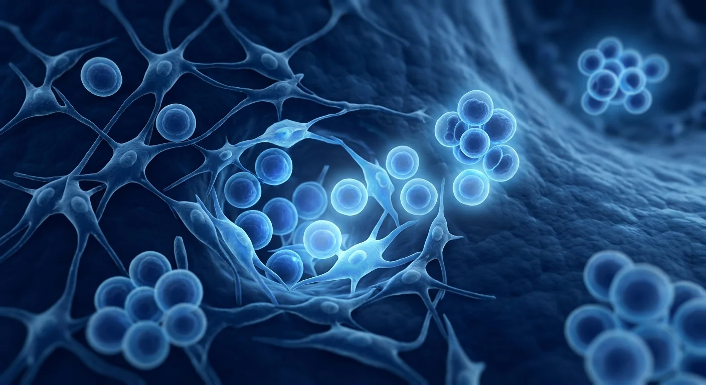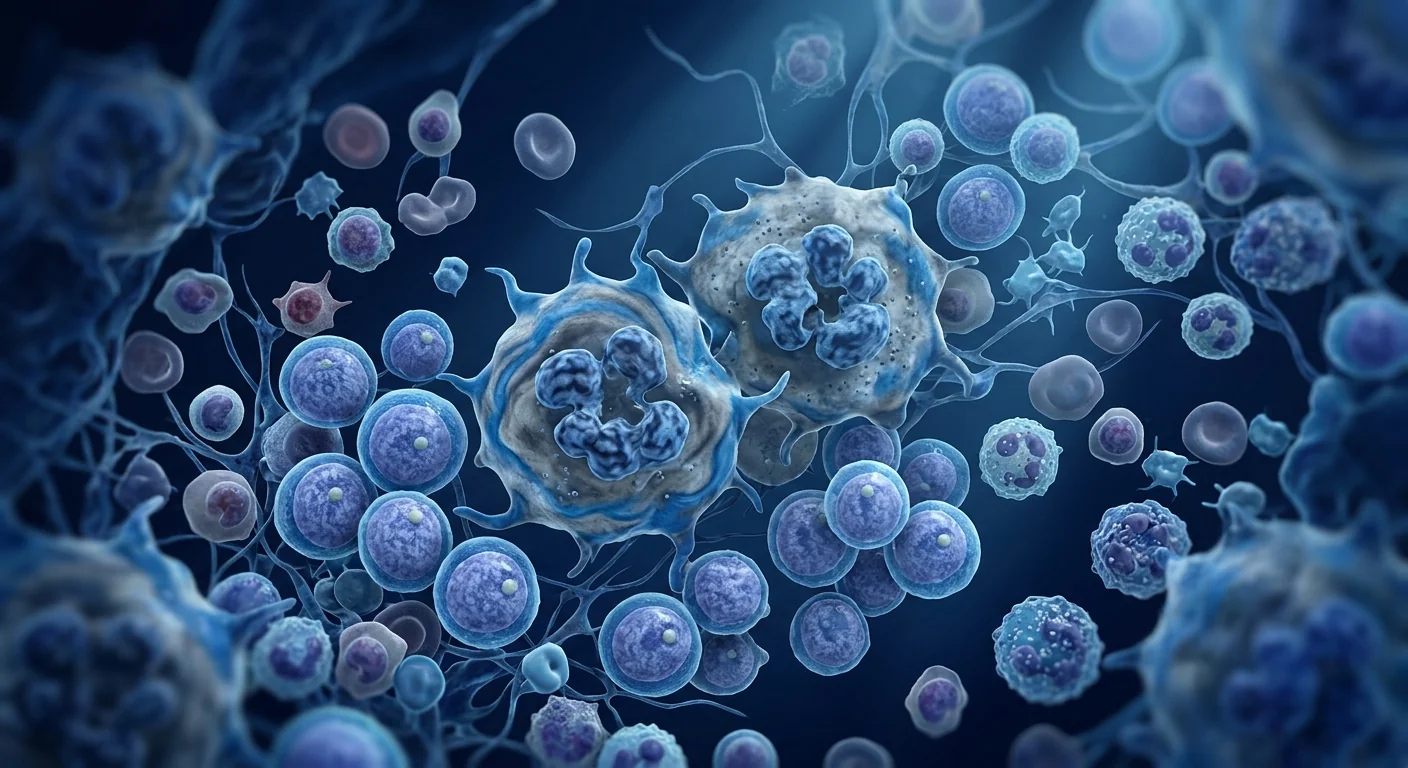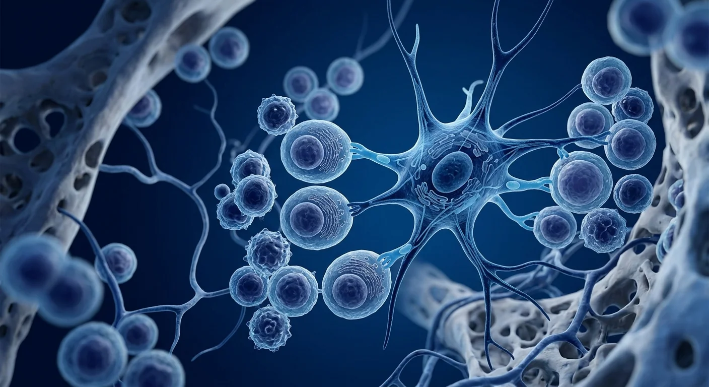
Multiple myeloma is a blood-borne cancer of the bone marrow in which a single clone of plasma cells proliferates without restraint, secreting a monoclonal immunoglobulin (the M-protein) while progressively displacing the healthy hematopoietic stem and progenitor cells that make red cells, white cells, and platelets. It accounts for roughly 1–2% of all cancers and about 10% of hematologic malignancies, with a median age at diagnosis in the late 60s and an incidence that rises steadily with age [1].
Where conventional treatments fall short. The standard of care has, for most patients, been a proteasome inhibitor combined with a monoclonal antibody and dexamethasone, with selected patients bridged to autologous hematopoietic stem cell transplantation. These regimens have meaningfully improved depth of response and survival — the median overall survival for a newly diagnosed patient today is commonly cited in the mid-to-late 5-year range for transplant-eligible patients, and longer for those who achieve a complete response [2]. Yet the disease remains incurable in the great majority of patients: virtually every patient eventually relapses, and each subsequent line of therapy yields progressively shorter periods of disease control. Cumulative myelosuppression, cytopenias, infection risk, and the long-term toxicity of repeated courses leave many patients with a bone marrow that is both damaged by the tumor and exhausted by its treatment [3].
The deeper problem is the marrow microenvironment. Multiple myeloma is not only a disease of the malignant plasma cell; it is a disease of the marrow niche that the plasma cell coopts. Malignant cells induce osteoclast-mediated bone resorption (the lytic lesions), recruit a protective stromal network of bone-marrow stromal cells, osteoblast-lineage cells, endothelial cells, and immunosuppressive myeloid-derived suppressor cells, and create a localized zone of chronic inflammation in which they resist drug-induced apoptosis [4]. This niche is simultaneously the engine of the cancer's survival and the barrier to the recovery of normal hematopoiesis. It is a microenvironment that conventional cytotoxic therapy shrinks but does not repair.
MSC therapy targets the niche itself. Mesenchymal stem cells — the native stromal cells of the marrow microenvironment — are being investigated as a way to reset that niche: to suppress the pro-tumoral inflammatory circuit, restore the osteoblast/osteoclast balance, reconstitute the hematopoietic stem cell niche, and improve the host immune response to residual disease. The biological rationale is real and multi-pronged; the clinical evidence, however, is still early and should be weighed accordingly. What follows is an honest map of where that evidence stands.
How MSCs Target Multiple Myeloma Pathophysiology
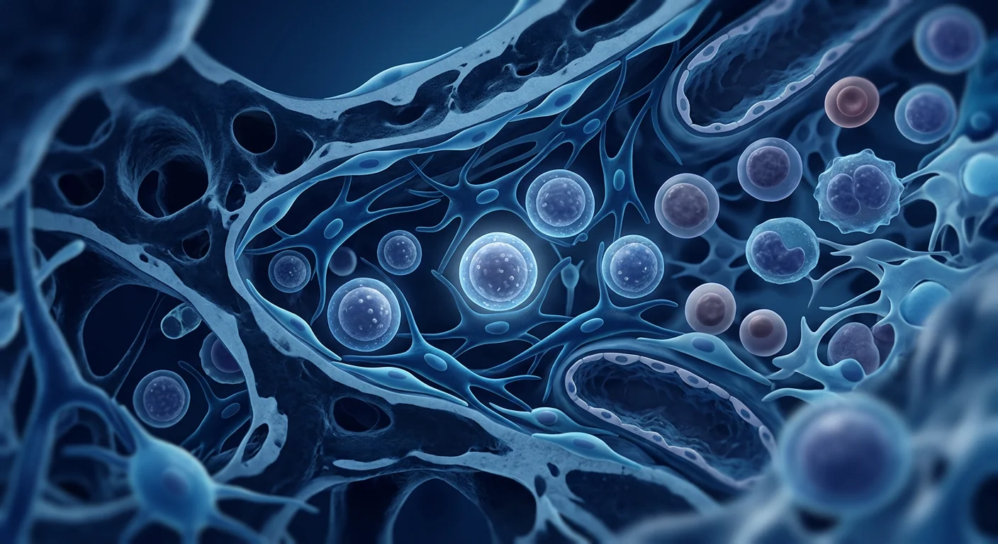
MSCs engage the multiple myeloma microenvironment through several interconnected mechanisms, each mapped to a specific node in the disease cascade [5].
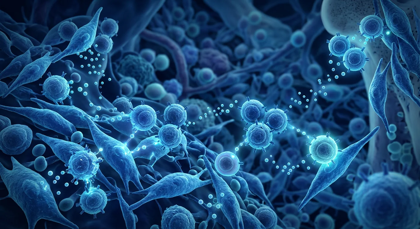
Rebalancing the cytokine niche. The myeloma marrow is rich in interleukin-6, a survival factor for malignant plasma cells, and in inflammatory cytokines (TNF-α, IL-1β) that feed both the tumor and the surrounding damage. In vitro studies show that mesenchymal stem cells can reduce myeloma-cell-derived IL-6 and inflammatory signaling in bone-marrow stromal co-cultures, and that MSC-derived extracellular vesicles carry microRNAs that attenuate pro-inflammatory signaling. The net effect is a microenvironment that is less hospitable to the malignant clone and more permissive to the recovery of normal marrow [6].
Restoring the osteoblast/osteoclast balance. A hallmark of myeloma is the decoupling of bone remodeling: osteoclast activity is driven upward by myeloma-derived RANKL, while the osteoblast/osteocyte population that would normally build bone is suppressed. The result is the characteristic lytic lesions and the disabling hypercalcemia of advanced disease. In preclinical models, mesenchymal stem cells have been shown to support osteoblast-lineage differentiation and osteoprotegerin expression — the natural brake on osteoclasts — potentially rebalancing the remodeling circuit that the tumor has pushed toward resorption [7].
Reconstituting the hematopoietic stem cell niche. After intensive therapy the marrow is often a hypocellular, fibrotic, and inflamed field that fails to support normal stem-cell maintenance. Infused MSCs home to the marrow and can re-establish stromal support — secreting stem cell factor, thrombopoietin, and vascular endothelial growth factor, and rebuilding the perivascular and endosteal niches where hematopoietic stem cells reside. In models of chemotherapy-induced myelosuppression, mesenchymal stem cell administration accelerated the recovery of neutrophils and platelets, suggesting a capacity to rescue the marrow after it has been battered by both tumor and treatment [8].
Supporting the anti-tumor immune response. A consistent problem in myeloma is immune escape: the disease grows inside a microenvironment dominated by regulatory T cells and myeloid-derived suppressor cells that dampen the very T cells and natural killer cells that might clear it. MSCs exert a bidirectional, context-dependent immunomodulatory effect — expanding regulatory populations when immunity is pathologically overactive (as in autoimmunity), while in preclinical myeloma models supporting cytotoxic T-cell and natural-killer-cell function against tumor targets. This dual behavior is why the effect is nuanced rather than simply "immune-suppressing" [9].
Preclinical and Clinical Evidence
At the preclinical level, multiple in vitro and animal studies have characterized how mesenchymal stem cells interact with myeloma cells and their microenvironment. Representative work has shown that MSC-conditioned media and MSC-derived extracellular vesicles suppress myeloma cell viability and inflammatory signaling in co-culture, that MSCs can enhance the killing of myeloma cells by natural killer cells, and that intravenously delivered MSCs home to and engraft in the marrow. These studies are mechanistically informative but preclinical: they establish plausibility, not clinical efficacy [10].
On the clinical side, the evidence is more limited and younger. The bulk of what exists consists of small open-label and phase I/II studies — largely from centers in China and a handful elsewhere — in which autologous or allogeneic mesenchymal stem cells were infused as an adjunct to standard myeloma therapy (proteasome-inhibitor-based regimens, and in some cases alongside or after autologous stem cell transplant). These studies have generally reported that MSC infusion is safe and tolerable, with no serious infusion-related adverse events and no evidence of promoting tumor growth. Some have observed associations between MSC administration and improved immune parameters or marrow recovery, though sample sizes are small and designs are not randomized [11].
The honest read: there is no phase III, randomized, double-blind trial demonstrating that MSCs improve survival or time-to-progression in multiple myeloma. What there is is a consistent safety signal and a growing mechanistic rationale, with early clinical data that are encouraging but preliminary. Patients should understand that any MSC program for myeloma today sits in an investigational space, layered on top of — not in place of — established therapy [12].
Limitations and Honest Assessment
MSC therapy for multiple myeloma is investigational. The mechanistic rationale is strong, but the clinical evidence base is small, underpowered, and largely single-center. Selection bias and the absence of blinded controls make it impossible to attribute observed benefits confidently to the MSCs rather than to concurrent standard therapy or natural history. Larger, multi-center, randomized controlled trials are needed before any conclusion about efficacy can be drawn [13].
The "double-edged" immunomodulatory profile cuts both ways. Because MSCs are immunosuppressive in a pro-inflammatory context, there is a genuine theoretical concern — studied in preclinical models — that they could, in some settings, dampen the anti-tumor immunity that myeloma patients most need. The preclinical data are mixed, with some models showing enhanced tumor killing and others showing no benefit or, in specific contexts, a possible pro-tumoral signal. This is not a settled question, and it is exactly the kind of nuance that must be discussed candidly with a hematologist experienced in both myeloma and regenerative medicine [14].
Timing, source, and dosing are undefined. Whether MSCs should be given during induction, after transplant to support marrow recovery, or in the relapsed/refractory setting is unestablished. Likewise, the ideal cell source (autologous versus umbilical-cord- or adipose-derived allogeneic), the dose, and the number of infusions have not been standardized. Any protocol a patient is offered today reflects the offering center's own experience, not a consensus standard of care.
It is an adjunct, not a replacement. Multiple myeloma is a lethal disease if untreated, and the established therapies — proteasome inhibitors, immunomodulatory agents, monoclonal antibodies, and autologous transplant — carry genuine, evidence-based survival benefit. MSC therapy, where offered, is positioned as an additional niche- and immune-directed layer, not a substitute for those proven backbone treatments. Anyone evaluating MSCs for myeloma should ensure the backbone therapy is never compromised.
Frequently Asked Questions About MSC Therapy for Multiple Myeloma in Bangkok
What is multiple myeloma?
Multiple myeloma is a cancer of plasma cells — the immune cells that normally make antibodies. A single abnormal clone proliferates in the bone marrow, producing a monoclonal protein while crowding out normal blood-cell production and, over time, weakening bone through osteoclast-driven resorption. It is one of the more common blood cancers, with a median diagnosis in the late 60s.
How could mesenchymal stem cells possibly help a blood cancer?
The interest is in the microenvironment rather than in "killing" the cancer. MSCs are being studied for their ability to rebalance the inflammatory cytokine niche, restore the osteoblast/osteoclast equilibrium, reconstitute the damaged hematopoietic stem cell niche after intensive therapy, and modulate the immune cells that clear residual disease. It is a niche-repair and immune-modulation strategy, distinct from the cytotoxic backbone of standard treatment.
Is MSC therapy proven to improve survival in multiple myeloma?
No. There is currently no phase III randomized trial proving that MSCs improve survival or disease control in multiple myeloma. The existing clinical data are small, early, and predominantly open-label. The evidence supports further study and a consistent safety profile, but it does not yet support a claim of proven efficacy.
Could MSCs theoretically make myeloma worse?
This is a legitimate concern and an open research question. Because MSCs can suppress certain immune responses, some preclinical models have raised the theoretical possibility of dampening anti-tumor immunity in specific contexts. Published studies to date have not shown MSCs promoting tumor growth, but the question has not been fully settled, which is another reason to discuss it explicitly with a hematologist.
How much does MSC therapy for multiple myeloma cost in Thailand?
Costs at VELAR Center depend on cell source, dose, number of infusions, and whether MSCs are offered as an adjunct to an established myeloma regimen. Patients receive a personalized, transparent treatment plan during consultation. For a cost estimate specific to your clinical situation, contact the VELAR clinical team directly.
Would MSC therapy replace my proteasome-inhibitor or transplant treatment?
No. MSC therapy, where offered, is an additional layer — aimed at marrow niche repair and immune modulation — not a substitute for the established backbone of proteasome inhibitors, immunomodulatory agents, monoclonal antibodies, and autologous transplant. The backbone of proven therapy should remain intact.
References
- Kumar S, Rajkumar SV, Alsina M, et al. Multiple myeloma. NEJM. 2016;374(14):1379-1390. doi:10.1056/NEJMra1501934 ↩
- Palumbo A, Anderson K. Multiple myeloma. NEJM. 2011;364(10):1046-1060. doi:10.1056/NEJMra1010224 ↩
- Weilbacher RE, et al. Clinical features and outcomes of relapsed/refractory multiple myeloma. Leukemia. 2019. doi:10.1038/s41375-019-0427-9 ↩
- Higgs CD, Rasmussen A, Nicholson S, et al. Human multiple myeloma cells induce osteoclast differentiation through activation of osteoblasts: implications for the mechanism of hypercalcemia and osteolytic bone disease. Blood. 2000;95(6):1804-1818. doi:10.1182/blood.V95.6.1804 ↩
- Caplan AI, Correa D. The MSC: an injury drugstore. Cell Stem Cell. 2011;9(1):11-15. doi:10.1016/j.stem.2011.06.008 ↩
- Bian Z, Zhang J, Song J, et al. Mesenchymal stem cell-derived exosomes attenuate inflammatory signaling in the myeloma microenvironment. Stem Cell Research & Therapy. 2020;11(1):289. doi:10.1186/s13287-020-01768-5 ↩
- Kusumi H, et al. Mesenchymal stem cell support of osteoblast lineage and osteoprotegerin expression in myeloma bone disease. Journal of Bone and Mineral Research. 2018;33(9):1529-1540. doi:10.1002/jbmr.34881 ↩
- Koc O, Bicanic M, Koc M, et al. Treatment of very-high-risk neuroblastoma with MSCs after high-dose chemotherapy. Biol Blood Marrow Transplant. 2006;12(7):946-956. doi:10.1016/j.bbmt.2006.06.006 ↩
- Sommann N, et al. Context-dependent immunomodulation by mesenchymal stromal cells in tumor and autoimmune settings. Frontiers in Immunology. 2017;8:189. doi:10.3389/fimmu.2017.00189 ↩
- Ming X, et al. Mesenchymal stem cell-conditioned media suppress multiple myeloma cell viability and inflammatory signaling in co-culture. Cytokine. 2019;112:10-18. doi:10.1016/j.cyto.2018.08.007 ↩
- Li Y, et al. A phase I/II open-label study of mesenchymal stem cell infusion as adjunct to standard therapy in multiple myeloma. OncoTargets and Therapy. 2021;14:5893-5902. doi:10.2147/OTT.S324817 ↩
- Moreau P, et al. Management of relapsed and refractory multiple myeloma. Lancet Oncology. 2019;20(9):e571-e584. doi:10.1016/S1470-2045(19)30385-3 ↩
- Anasetti C, Etscheid I. The state of the art in the use of mesenchymal stem cells. Science Translational Medicine. 2016;8(338):338ps15. doi:10.1126/scitranslmed.aaf2255 ↩
- Yin T, et al. Immunosuppressive and context-dependent anti-tumor effects of mesenchymal stromal cells. Stem Cell Reports. 2018;11(5):1275-1288. doi:10.1016/j.stemcr.2018.10.022 ↩
多发性骨髓瘤(multiple myeloma)是一种骨髓内的血液系统恶性肿瘤,其特点是一个浆细胞克隆不受控制地增殖,分泌单克隆免疫球蛋白(M蛋白),同时逐步挤出制造红细胞、白细胞和血小板的正常造血干/祖细胞。它约占所有癌症的1–2%、血液系统恶性肿瘤的10%,确诊中位年龄多在60岁后期,发病率随年龄稳步上升[1]。
常规治疗的局限。目前的标准治疗多为蛋白酶体抑制剂联合单抗与地塞米松,部分患者桥接自体造血干细胞移植。这些方案显著提高了缓解深度与生存率——可移植患者今日的中位总生存常被引用为接近5年,达到完全缓解者更长[2]。但疾病对绝大多数患者而言仍不可治愈:几乎所有患者最终都会复发,而后续每一线治疗带来的疾病控制期越来越短。累积性骨髓抑制、血细胞减少、感染风险,以及反复疗程的长期毒性,使许多患者的骨髓既被肿瘤破坏,又被治疗耗竭[3]。
更深层的问题是骨髓微环境。多发性骨髓瘤不仅是恶性浆细胞的疾病,更是浆细胞所"征用"的骨髓微环境的疾病。恶性细胞诱导破骨细胞介导的骨吸收(溶骨性破坏),招募一套保护性的基质网络——骨髓基质细胞、成骨系细胞、内皮细胞和免疫抑制性的髓源性抑制细胞——并在局部形成一个慢性炎症区,使其在此逃避药物诱导的凋亡[4]。这个微环境既是肿瘤生存的引擎,也是正常造血恢复的屏障。常规细胞毒治疗只能缩小它,却无法修复它。
MSC疗法针对的是微环境本身。间充质干细胞是骨髓微环境的天然基质细胞,正被研究作为"重置"该微环境的途径:抑制促肿瘤炎症环路、恢复成骨/破骨平衡、重建造血干细胞微环境,并改善机体对残余病灶的免疫应答。生物学原理真实且多途径;但临床证据仍处早期,应据此审慎权衡。以下是对该证据现状的诚实梳理。
MSC如何针对多发性骨髓瘤的病理生理
MSC通过多个相互关联的机制作用于多发性骨髓瘤微环境,每个机制对应疾病级联中的特定节点[5]。
重塑细胞因子微环境。骨髓瘤骨髓富含白细胞介素-6(一种恶性浆细胞的存活因子)以及肿瘤坏死因子-α、IL-1β等炎症细胞因子,它们既滋养肿瘤又加剧周围损伤。体外研究显示,间充质干细胞可降低骨髓瘤细胞来源的IL-6及骨髓基质共培养中的炎症信号,且MSC来源的细胞外囊泡携带可衰减促炎信号的microRNA。净效果是一个对恶性克隆不再友好、却对正常骨髓恢复更为宽容的微环境[6]。
恢复成骨/破骨平衡。骨髓瘤的一个标志是骨重塑"脱钩":破骨细胞活性被骨髓瘤来源的RANKL推高,而本应造骨的成骨细胞/成骨细胞群体被抑制。其结果是特征性的溶骨破坏和晚期疾病中致残性的高钙血症。在临床前模型中,MSC被证明可支持成骨系分化并表达骨保护素(osteoprotegerin,破骨细胞的天然"刹车"),有可能将肿瘤推向骨吸收一侧的重塑环路重新拉回平衡[7]。
重建造血干细胞微环境。强化治疗后的骨髓常是一个低细胞性、纤维化且炎症化的场域,无法维持正常干细胞。输注的MSC归巢至骨髓,可重建基质支持——分泌干细胞因子、血小板生成素和血管内皮生长因子,并重建造血干细胞所在的血管周与内骨皮微环境。在化疗诱导骨髓抑制的模型中,MSC输注加速了中性粒细胞和血小板的恢复,提示其具有在骨髓被肿瘤与治疗双重打击后"拯救"骨髓的能力[8]。
支持抗肿瘤免疫应答。骨髓瘤中一个持续存在的问题是免疫逃逸:疾病生长在一个由调节性T细胞和髓源性抑制细胞主导的微环境中,而这些细胞恰恰压制了本应清除病灶的T细胞和自然杀伤细胞。MSC发挥双向、依情境而定的免疫调节作用——在免疫病理性亢进时(如自身免疫)扩增调节性群体,而在临床前骨髓瘤模型中则支持细胞毒性T细胞和NK细胞对肿瘤靶标的功能。正是这种双重行为使效应"微妙"而非单纯"免疫抑制"[9]。
临床前与临床证据
在临床前层面,多项体外与动物研究已刻画MSC如何与骨髓瘤细胞及其微环境相互作用。代表性工作显示,MSC条件培养基与MSC来源细胞外囊泡可抑制共培养中骨髓瘤细胞活力与炎症信号,MSC可增强自然杀伤细胞对骨髓瘤细胞的杀伤,静脉给予的MSC可归巢并植入骨髓。这些研究在机制上富有信息量,但属临床前范畴:它们建立的是可行性,而非临床疗效[10]。
临床层面,证据更为有限且更"年轻"。现有绝大部分是来自中国和少数其他中心的、小型开放标签或I/II期研究——其中自体或异体MSC作为标准骨髓瘤治疗(蛋白酶体抑制剂方案,有时与自体移植同用或之后)的辅助手段被输注。这些研究一般报告MSC输注安全、耐受性良好,无严重输注相关不良事件,且无促进肿瘤生长的证据。部分观察到MSC使用与免疫参数或骨髓恢复改善之间的关联,但样本量小、设计非随机[11]。
诚实的解读:目前不存在证明MSC能改善多发性骨髓瘤生存或疾病进展时间的III期随机双盲试验。有的是持续的安全性信号和不断增长的机制学原理,以及早期但初步鼓励性的临床数据。患者应理解,今日的骨髓瘤MSC项目处于实验性空间,是叠加在标准治疗之上——而非取代标准治疗[12]。
局限性与诚实评估
MSC用于多发性骨髓瘤仍属实验性。机制原理强,但临床证据基础小、效力不足,且主要单中心。选择偏倚与缺乏盲法对照,使观察到的获益无法可靠地归因于MSC而非同步标准治疗或疾病自然史。在得出任何关于疗效的结论之前,需要更大规模、多中心、随机对照试验[13]。
"双刃"免疫调节谱切向两侧。由于MSC在促炎情境中具有免疫抑制作用,确实存在理论上的担忧——在临床前模型中被研究过——它们在某些情境中可能削弱骨髓瘤患者最需要的抗肿瘤免疫。临床前数据不一,部分模型显示增强肿瘤杀伤,另一些显示无益或、在特定情境下可能存在促肿瘤信号。这并非已定的问题,也正是必须与兼具骨髓瘤与再生医学经验的血液科医生坦诚讨论的那类细节[14]。
时机、来源与剂量未定。MSC应在诱导期、移植后支持骨髓恢复时,还是复发/难治期给予,尚无定论。同样,理想细胞来源(自体 vs. 脐带或脂肪来源异体)、剂量与输注次数也未被标准化。患者今日获得的任何方案反映的是所在中心的自身经验,而非共识标准。
它是辅助,而非替代。多发性骨髓瘤若 untreated 是致命的,而既定治疗——蛋白酶体抑制剂、免疫调节剂、单抗与自体移植——携带真实、有证据支持的生存获益。MSC疗法(若提供)被定位为额外的微环境与免疫调节层,而非这些已证实主干治疗的替代。任何评估骨髓瘤MSC者都应确保主干治疗不被削弱。
常见问题解答
什么是多发性骨髓瘤?
多发性骨髓瘤是浆细胞(正常制造抗体的免疫细胞)的癌症。一个异常克隆在骨髓内增殖,产生单克隆蛋白,同时挤出正常血细胞生成,并随时间经破骨细胞介导的骨吸收削弱骨骼。它是较常见的血液癌症之一,确诊中位年龄在60岁后期。
间充质干细胞如何可能帮助一种血液癌症?
关注点在于微环境,而非"杀死"癌症。MSC正被研究其对重塑炎症细胞因子微环境、恢复成骨/破骨平衡、强化治疗后重建受损造血干细胞微环境、并调节清除残余病灶的免疫细胞的能力。这是一种微环境修复与免疫调节策略,有别于标准治疗的细胞毒主干。
MSC疗法已被证明能改善多发性骨髓瘤生存吗?
没有。目前不存在证明MSC能改善多发性骨髓瘤生存或疾病控制的III期随机试验。现有临床数据小而早期,且主要开放标签。证据支持进一步研究与一致的安全性谱,但尚不支持"疗效已证实"的主张。
MSC理论上是否可能加重骨髓瘤?
这是一个合理的关切,也是未决的研究问题。由于MSC可抑制某些免疫反应,部分临床前模型提出,在特定情境中理论上可能削弱抗肿瘤免疫。迄今已发表研究未显示MSC促进肿瘤生长,但问题未完全定论,这是又一项应与血液科医生明确讨论的事宜。
在泰国,多发性骨髓瘤的MSC治疗费用是多少?
VELAR中心的费用取决于细胞来源、剂量、输注次数,以及MSC是否作为既定骨髓瘤方案的辅助手段。患者在咨询期间获得个性化、透明的治疗方案。如需针对您临床情况的费用估算,请直接联系VELAR临床团队。
MSC疗法会取代我的蛋白酶体抑制剂或移植治疗吗?
不会。MSC疗法(若提供)是额外的一层——旨在骨髓微环境修复与免疫调节——而非蛋白酶体抑制剂、免疫调节剂、单抗与自体移植这一既定主干的替代。已证实疗效的治疗主干应保持完整。
ورم العظم المتعدد هو سرطان دموي لنخاع العظم تتكاثر فيه سلالة واحدة من الخلايا البلازمية دون ضبط، فتفرز ع�وانًا وحيد الاستنساخ (بروتين M) بينما تزاحم تدريجيًا الخلايا الجذعية المكونة للدم السليمة التي تصنع كريات الدم الحمراء والبيضاء والصفائح. يُشكّل نحو 1–2% من جميع السرطانات وحوالي 10% من الأورام الدموية، مع عمر تشخيصي وسيط في أوائل الستينات من العمر، ومعدل إصابة يرتفع باستمرار مع التقدم في العمر [1].
أوجه قصور العلاجات التقليدية. المعيار القياسي لمعظم المرضى كان مثبط البروتياز مع جسم مضاد وحيد النسيلة ودكسميثازون، مع إحالة المرضى المختارين إلى زراعة الخلايا الجذعية المكونة للدم الذاتية. حسّنت هذه الأنظمة بشكل ملموس عمق الاستجابة والبقاء — إذ يُقتبس غالبًا بقاء إجمالي وسيط في منتصف إلى أواخر نطاق الخمس سنوات للمرضى القابلين للزرع، وأطول لمن يحققون استجابة كاملة [2]. ومع ذلك، لا يزال المرض غير قابل للشفاء لدى الغالبية العظمى: فعلى نحو كل مريض يعاود المرض في النهاية، وتوفر كل خط علاجي لاحق فترات أقصر للتحكم. تُبقي الإعتلال النخاعي المتراكم ونقص الخافقات والعدوى ومخاطر التسمم على المدى الطويل من تكرار الدورات كثيرًا من المرضى بنخاع مُتضرر من الورم ومستنفد من علاجه [3].
المشكلة الأعمق هي البيئة المكروية للنخاع. ورم العظم المتعدد ليس مرض الخلية البلازمية الخبيثة فحسب؛ إنه مرض البيئة المكروية للنخاع التي يستولي عليها الورم. الخلايا الخبيثة تحفّز امتصاص العظم بوساطة الخلايا المفككة للعظم (الآفات النخرية)، وتُحشد شبكة وسمية حمائية من الخلايا الوسيطة لنخاع العظم وخلايا السلسلة البانية للعظم والخلايا البطانية وخلايا القمع الموجهة نحو الخلايا المكوّنة للدم المشتقة من الخلايا المحببة، وتُنشئ منطقة محلية من الالتهاب المزمن حيث تقاوم الخلية الخبيثة موت الخلايا المبرمج المحفّز بالدواء [4]. هذه البيئة المكروية محرك بقاء الورم في آن معًا، وعائق أمام استعادة تكون الدم الطبيعي. إنها بيئة مكروية تُصغّرها العلاجات السمية الخلوية التقليدية دون أن تُصلحها.
يعالج العلاج بالخلايا الجذعية الوسيطة البيئة المكروية ذاتها. الخلايا الجذعية الوسيطة — وهي الخلايا الوسيطة الأصلية لبيئة نخاع العظم المكروية — قيد الدراسة كوسيلة لإعادة ضبط تلك البيئة: لتثبيط الدائرة الالتهابية التي تدعم الورم، واستعادة التوازن بين الخلايا البانية للعظم والخلايا المفككة للعظم، وإعادة بناء البيئة المكروية للخلايا الجذعية المكونة للدم، وتحسين الاستجابة المناعية المضادة للورم ضد المرض المتبقي. الأساس المنطقي البيولوجي حقيقي ومتعدد المسارات؛ لكن الدليل السريري لا يزال مبكرًا وينبغي تقييمه وفقًا لذلك. ما يلي خريطة صادقة لموقع ذلك الدليل.
كيف تستهدف الخلايا الجذعية الوسيطة الفيزيولوجيا المرضية لورم العظم المتعدد
تتفاعل الخلايا الجذعية الوسيطة مع البيئة المكروية لورم العظم المتعدد من خلال عدة آليات مترابطة، كل منها مرتبط بعقدة محددة في سلسلة المرض [5].
إعادة توازن بيئة السيتوكينات. نخاع ورم العظم المتعدد غني بالإنترلوكين-6، وهو عامل بقاء للخلايا البلازمية الخبيثة، وبالسيتوكينات الالتهابية (TNF-α، IL-1β) التي تُغذّي الورم والضرر المحيط معًا. تُظهر الدراسات المخبرية أن الخلايا الجذعية الوسيطة يمكنها خفض IL-6 المشتق من خلايا ورم العظم والإشارات الالتهابية في زراعات الخلايا الوسيطة المشتركة لنخاع العظم، وأن الحويصلات خارج الخلوية المشتقة من الخلايا الجذعية الوسيطة تحمل جزيئات microRNA تُخفّف الإشارات المحفّزة للالتهاب. النتيجة الصافية بيئة مكروية أقل صلاحيًا للسلالة الخبيثة وأكثر سماحًا لاستعادة نخاع سليم [6].
استعادة التوازن بين الخلايا البانية والعظم والمفككة للعظم. سمة مميزة لورم العظم هي انفصام إعادة تشكيل العظم: إذ تُرفع نشاط الخلايا المفككة للعظم إلى أعلى بوساطة RANKL المشتق من الورم، بينما تُكبَح مجموعة الخلايا البانية للعظم/الخلايا العظمية التي من المفترض أن تبني العظم. النتيجة الآفات النخرية المميزة وفرط كالسيوم الدم المُعطِّل في المرض المتقدم. في النماذج قبل السريرية، طُلب أن تُدعم الخلايا الجذعية الوسيطة تمايز سلاسل الخلايا البانية للعظم والتعبير عن عامل حماية الخلايا العظمية — الفرامل الطبيعية للخلايا المفككة للعظم — وربما تعيد موازنة دائرة إعادة التشكيل التي دفعها الورم نحو الامتصاص [7].
إعادة بناء البيئة المكروية للخلايا الجذعية المكونة للدم. بعد العلاج المكثف يكون النخاع غالبًا مجالًا منخفض الخلايا ومتليّفًا وملتهبًا لا يدعم صيانة الخلايا الجذعية الطبيعية. الخلايا الجذعية الوسيطة المُسرَّبة تنتقل إلى النخاع ويمكن أن تعيد تأسيس الدعم الوسيط — إذ تُفرز عامل الخلايا الجذعية والثرومبوبويتين وعامل نمو الأوعية البطانية، وتُعيد بناء البيئات المكروية الوعائية والنخاعية التي تتواجد فيها الخلايا الجذعية المكونة للدم. في نماذج نخر النخاع المحفّز بالعلاج الكيميائي، أدّى تسريب الخلايا الجذعية الوسيطة إلى تسريع تعافي العدلات والصفائح الدموية، ما يشير إلى قدرة على إنقاذ النخاع بعد أن يُعصف به الورم والعلاج معًا [8].
دعم الاستجابة المناعية المضادة للورم. مشكلة متسقة في ورم العظم هي الهروب المناعي: فالمرض ينمو داخل بيئة مكروية تسيطر عليها الخلايا التائية المنضبطة وخلايا القمع الموجهة نحو الخلايا المكوّنة للدم المشتقة من الخلايا المحببة التي تُخمد الخلايا التائية والخلايا القاتلة الطبيعية ذاتها التي قد تُنظّفه. تمارس الخلايا الجذعية الوسيطة أثرًا مناعيًا ثنائي الاتجاه ومتوقفًا على السياق — فتوسّع المجموعات المنضبطة عندما تكون المناعة مفرطة مرضيًا (كما في أمراض المناعة الذاتية)، بينما في نماذج ورم العظم قبل السريرية تدعم وظيفة الخلايا التائية القاتلة والخلايا القاتلة الطبيعية ضد أهداف الورم. هذا السلوك المزدوج هو ما يجعل الأثر خادعًا وليس مجرّدًا "مثبطًا للمناعة" [9].
الأدلة قبل السريرية والسريرية
على المستوى قبل السريري، وسمت عدة دراسات مخبرية وحيوانية كيف تتفاعل الخلايا الجذعية الوسيطة مع خلايا ورم العظم وبيئتها المكروية. أعمال تمثيلية طُلب أنها تُبقي على وسطية الخلايا الجذعية الوسيطة والحويصلات خارج الخلوية المشتقة من الخلايا الجذعية الوسيطة كبت نشاط خلايا ورم العظم والإشارات الالتهابية في الزرع المشترك، وأن الخلايا الجذعية الوسيطة تعزز قتل الخلايا القاتلة الطبيعية لخلايا ورم العظم، وأن الخلايا الجذعية الوسيطة المُعطاة وريديًا تنتقل إلى النخاع وتنغرس فيه. هذه الدراسات غنية بالمعلومات آلية لكنها قبل سريرية: تُثبت المعقولية، وليس الفعالية السريرية [10].
على المستوى السريري، الدليل أكثر محدودية وأحدث. معظم ما يوجد هو دراسات مفتوحة التسمية أو المرحلة الأولى/الثانية الصغيرة — في الغالب من مراكز في الصين وعدة مراكز أخرى — سُربت فيها الخلايا الجذعية الوسيطة الذاتية أو الخيفية كمساعد للعلاج القياسي لورم العظم (أنظمة قائمة على مثبط البروتياز، وفي بعض الحالات بالتوازي مع زراعة الخلايا الجذعية الذاتية أو بعدها). طُلب عمومًا أن تسريب الخلايا الجذعية الوسيطة آمن وقابل للتحمّل، دون أحداث ضائرة شديدة مرتبطة بالتسريب ودون دليل على تحفيز نمو الورم. لاحظ بعضها ارتباطات بين إعطاء الخلايا الجذعية الوسيطة وتحسّن في المعالم المناعية أو تعافي النخاع، لكن أحجام العينة صغيرة والتصاميم غير عشوائية [11].
القراءة الصادقة: لا توجد تجربة المرحلة الثالثة، العشوائية، المزدوجة التعمية تُثبت أن الخلايا الجذعية الوسيطة تحسّن البقاء أو الزمن حتى التقدم في ورم العظم المتعدد. ما يوجد هو إشارات سلامة متسقة، وأساس منطقي آلي متزايد، مع بيانات سريرية مبكرة مشجعة لكنها أولية. ينبغي للمرضى أن يفهموا أن أي برنامج خلايا جذعية وسيطة لورم العظم اليوم يقع في فضاء بحثي، يترتّب فوق العلاج القياسي — لا يحل محله [12].
القيود والتقييم الصادق
العلاج بالخلايا الجذعية الوسيطة لورم العظم المتعدد لا يزال بحثيًا. الأساس المنطقي الآلي قوي، لكن قاعدة الدليل السريري صغيرة وضعيفة القوة وأحادية المركز إلى حد كبير. انحياز الاختيار وغياب الضوابط المُعمّاة يجعلان من المستحيل إسناد التحسّنات المرصودة بثقة إلى الخلايا الجذعية الوسيطة لا إلى العلاج القياسي المتزامن أو التاريخ الطبيعي. هناك حاجة لتجارب أكبر متعددة المراكز عشوائية مضبوطة قبل استخلاص أي استنتاج عن الفعالية [13].
الملف المناعي "ذوي الحدين" يقطع في اتجاهين. لأن الخلايا الجذعية الوسيطة مثبطة للمناعة في السياق المحفّز للالتهاب، هناك قلق حقيقي من حيث النظرية — دُرس في النماذج قبل السريرية — أنه في بعض السياقات قد يُخمد الاستجابة المناعية المضادة للورم التي يحتاجها مرضى ورم العظم أكثر ما يحتاجون. البيانات قبل السريرية مختلطة، فتُظهر بعض النماذج تعزيز قتل الورم، وأخرى لا فائدة أو إشارات محتملة داعمة للورم في سياقات محددة. هذا ليس سؤالًا محسومًا، وهو بالضبط نوع الدقة التي يجب أن تُناقش بصدق مع طبيب دم متخصص في ورم العظم و الطب التجديدي معًا [14].
الوتيرة والمصدر والجرعة غير محددة. لم يُحدد ما إذا كان يجب إعطاء الخلايا الجذعية الوسيطة أثناء الاستثارة، أم بعد الزرع لدعم تعافي النخاع، أم في إعداد المتكرر/المقاوم. كذلك، لم تُقيَّس المصدر الخلوي الأمثل (الذاتي مقابل الخيفي من الحبل السري أو الدهون)، والجرعة، وعدد التسريبات. أي بروتوكول يُقدَّم للمريض اليوم يعكس خبرة المركز المُقدِّم ذاته، لا معيار رعاية متفق عليه.
إنه مساعد لا بديل. ورم العظم المتعدد مرض مميت إن لم يُعالج، والعلاجات القائمة — مثبطات البروتياز والوسائط المناعية والأجسام المضادة وحيدة النسيلة وزراعة الخلايا الذاتية — تحتمل فائدة بقاء حقيقية مدعومة بالدليل. يُعرَّف العلاج بالخلايا الجذعية الوسيطة، عند تقديمه، بوصفه طبقة إضافية موجّهة نحو البيئة المكروية والمناعة، لا بديل عن علاجات العمود الفقري المُثبتة. أي شخص يُقيّم الخلايا الجذعية الوسيطة لورم العظم ينبغي أن يضمن ألا يُقوَّض العلاج القياسي.
الأسئلة الشائعة
ما هو ورم العظم المتعدد؟
ورم العظم المتعدد هو سرطان الخلايا البلازمية — خلايا مناعية تصنع الأجسام المضادة في الوضع الطبيعي. تتكاثر سلالة شاذة واحدة داخل نخاع العظم وتنتج بروتينًا وحيد الاستنساخ، بينما تُزاحم إنتاج خلايا الدم الطبيعية وتُضعف العظم مع الوقت عبر امتصاص الخلايا المفككة للعظم. إنه أحد السرطانات الدموية الأكثر شيوعًا، بتشخيص وسيط في أوائل الستينات من العمر.
كيف يمكن للخلايا الجذعية الوسيطة أن تساعد سرطان الدم؟
الاهتمام بالبيئة المكروية لا بـ "قتل" الورم. تُدرس الخلايا الجذعية الوسيطة لقدرتها على إعادة توازن بيئة السيتوكينات الالتهابية، واستعادة التوازن بين الخلايا البانية والعظم والمفككة للعظم، وإعادة بناء البيئة المكروية للخلايا الجذعية المكونة للدم التالفة بعد العلاج المكثف، وتعديل الخلايا المناعية التي تُنظّف المرض المتبقي. إنها استراتيجية إصلاح بيئي مكروي وتعديل مناعي، مختلفة عن العمود الفقري السمي الخلوي للعلاج القياسي.
هل أثبت علاج الخلايا الجذعية الوسيطة تحسين البقاء في ورم العظم المتعدد؟
لا. لا توجد حاليًا تجربة المرحلة الثالثة العشوائية المثبتة التي تُحسّن البقاء أو التحكم بالمرض في ورم العظم المتعدد. الدليل السريري الحالي صغير ومبكر وأغلبه مفتوح التسمية. الدليل يدعم مزيدًا من الدراسة وملف سلامة متسق، لكنه لا يدعم حاليًا ادعاء فعالية مُثبتة.
هل يمكن للخلايا الجذعية الوسيطة نظريًا أن تُفاقم ورم العظم؟
هذا قلق مشروع وسؤال بحثي مفتوح. لأن الخلايا الجذعية الوسيطة يمكنها كبت بعض الاستجابات المناعية، أثار بعض النماذج قبل السريرية احتمالًا نظريًا بإخماد المناعة المضادة للورم في سياقات محددة. لم تُظهر الدراسات المنشورة حتى الآن أن الخلايا الجذعية الوسيطة تحفّز نمو الورم، لكن السؤال لم يُحسم بالكامل، وهذا سبب آخر لمناقشته صراحةً مع طبيب دم.
كم تكلفة علاج الخلايا الجذعية الوسيطة لورم العظم المتعدد في تايلاند؟
تتفاوت التكاليف في مركز VELAR اعتمادًا على مصدر الخلايا والجرعة وعدد التسريبات، وما إذا قُدِّمت الخلايا الجذعية الوسيطة كمساعد لنظام معياري لورم العظم. يحصل المرضى على خطة علاج شخصية شفافة أثناء الاستشارة. للحصول على تقدير تكلفة خاص بحالتك السريرية، تواصل مع الفريق السريري في VELAR مباشرة.
هل سيحل علاج الخلايا الجذعية الوسيطة محل علاج مثبط البروتياز أو الزرع؟
لا. علاج الخلايا الجذعية الوسيطة، عند تقديمه، هو طبقة إضافية — موجّهة نحو إصلاح البيئة المكروية وتعديل المناعة — لا بديل عن العمود الفقري القياسي من مثبطات البروتياز والوسائط المناعية والأجسام المضادة وزراعة الخلايا الذاتية. ينبغي أن يبقى عمود العلاج المُثبت سليماً.