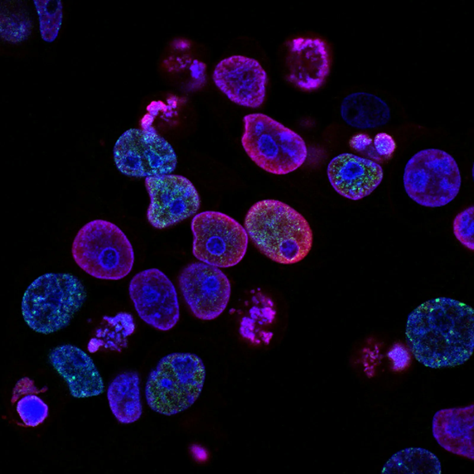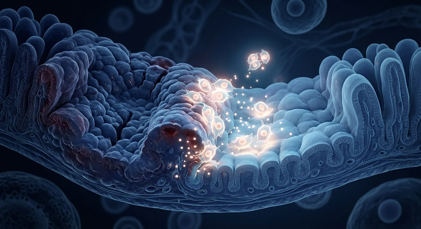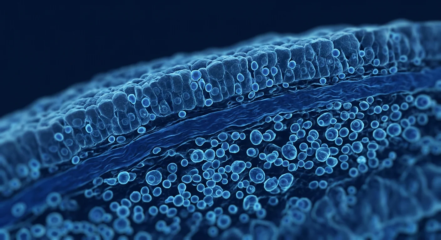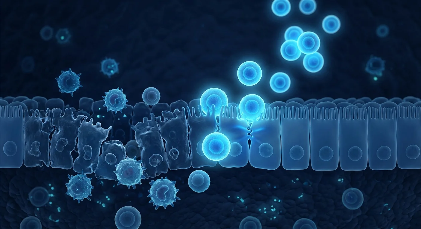
Microscopic colitis is a chronic inflammatory bowel disease characterized by persistent watery diarrhea, normal-appearing colonic mucosa on endoscopy, and characteristic histopathological findings on biopsy. It affects approximately 10–20 per 100,000 person-years, predominantly in women over 50, and is one of the most common causes of chronic non-bloody diarrhea in older adults. [1] Despite its prevalence, microscopic colitis remains underdiagnosed — many patients endure years of symptoms before a colonoscopy with biopsy provides the correct diagnosis.
Two subtypes, one clinical picture. Microscopic colitis encompasses two histological variants: collagenous colitis, defined by a thickened subepithelial collagen band (>10 μm), and lymphocytic colitis, characterized by increased intraepithelial lymphocytes without collagen thickening. [2] Clinically, both present similarly — chronic watery diarrhea, often nocturnal, with urgency, abdominal cramping, and weight loss. The distinction is histological, not clinical, and treatment approaches are largely the same.
Where conventional treatment falls short. First-line therapy is budesonide — a locally acting corticosteroid — which achieves clinical remission in approximately 80% of patients. [3] However, relapse after discontinuation is common, affecting 60–80% of responders within months. [4] Second-line options — including loperamide, bismuth subsalicylate, cholestyramine, and immunosuppressants such as azathioprine or methotrexate — offer variable efficacy and carry their own side-effect burdens. For patients with steroid-dependent or steroid-refractory disease, the therapeutic options narrow considerably. Anti-TNF biologics have shown some efficacy in small series, but evidence is limited and access is variable.
The tissue-level problem. The fundamental pathology in microscopic colitis is a dysregulated mucosal immune response to luminal antigens, resulting in chronic T-cell-mediated inflammation of the colonic epithelium. [5] Increased CD8+ intraepithelial lymphocytes, mucosal Th1/Th17 skewing, and epithelial barrier dysfunction are core features. [6] Nonsteroidal anti-inflammatory drugs (NSAIDs), proton pump inhibitors (PPIs), and selective serotonin reuptake inhibitors (SSRIs) have been implicated as triggers or exacerbating factors in susceptible individuals. [7] MSC therapy is being investigated as a potential disease-modifying approach that targets the underlying immune dysregulation rather than simply suppressing symptoms.
Why mesenchymal stem cells are studied for microscopic colitis
MSCs exert a broad immunomodulatory program that is particularly well-suited to the immunopathology of microscopic colitis. When exposed to an inflamed colonic environment, MSCs secrete anti-inflammatory mediators — including TGF-β, IL-10, PGE2, TSG-6, and indoleamine 2,3-dioxygenase (IDO) — that shift macrophages from a pro-inflammatory M1 phenotype to a reparative M2 phenotype, suppress effector T-cell proliferation, and expand regulatory T-cell populations. [8][9]
Reversing the Th1/Th17 skew. Microscopic colitis is driven by a Th1/Th17-predominant immune response, with elevated levels of interferon-gamma (IFN-γ), tumor necrosis factor-alpha (TNF-α), and interleukin-17 (IL-17) in the colonic mucosa. [10] MSCs suppress Th1 and Th17 differentiation while promoting Treg expansion through secretion of TGF-β and PGE2 — effectively rebalancing the mucosal immune environment away from chronic inflammation. [11] This mechanism directly addresses the CD8+ lymphocyte-driven epithelial injury that defines the condition.
Restoring epithelial barrier integrity. The colonic epithelium in microscopic colitis exhibits increased permeability, contributing to the translocation of luminal antigens that perpetuate the inflammatory cycle. [12] MSCs upregulate tight junction proteins — including occludin, claudin-1, and ZO-1 — and promote epithelial proliferation through secretion of keratinocyte growth factor (KGF), epidermal growth factor (EGF), and hepatocyte growth factor (HGF). [13] Restoration of barrier function may reduce the antigenic drive that sustains colonic mucosal inflammation.
Targeting the collagen band in collagenous colitis. The thickened subepithelial collagen band in collagenous colitis reflects excessive extracellular matrix deposition by activated myofibroblasts. [14] MSCs possess anti-fibrotic properties through modulation of the TGF-β/Smad signaling pathway, matrix metalloproteinase (MMP)/tissue inhibitor of metalloproteinase (TIMP) balance, and suppression of myofibroblast activation. [15] While direct evidence in collagenous colitis is lacking, the anti-fibrotic potential of MSCs is well-documented in other gastrointestinal fibrotic conditions.
Key MSC mechanisms relevant to microscopic colitis
- Th1/Th17 suppression + Treg expansion — rebalances mucosal adaptive immunity
- M1→M2 macrophage polarization — shifts colonic immune environment from inflammatory to reparative
- Epithelial barrier restoration — tight junction protein upregulation reduces gut permeability
- Anti-fibrotic activity — TGF-β/Smad modulation may reduce collagen band thickness in collagenous colitis
- CD8+ T-cell suppression — directly targets the intraepithelial lymphocytosis that defines the disease
What the clinical evidence says
The direct evidence is early. No large randomized controlled trial has specifically tested MSC therapy for microscopic colitis. However, the evidence base from related gastrointestinal inflammatory conditions — and from the broader MSC immunomodulation literature — provides a biologically plausible foundation.
Evidence from IBD trials. The most relevant clinical data comes from trials of MSC therapy in Crohn's disease and ulcerative colitis. Allogeneic MSCs (darvadstrocel / Cx601) received EMA approval for complex perianal fistulas in Crohn's disease based on the ADMIRE-CD trial, which demonstrated a significant improvement in combined remission at 24 weeks versus placebo. [16] Systemic MSC infusion for luminal Crohn's and ulcerative colitis has shown safety and modest efficacy signals in early-phase trials, with reductions in endoscopic disease activity and inflammatory biomarkers in some patients. [17]
Gut-specific immunomodulation. Intravenously infused MSCs exhibit tropism for sites of inflammation, including the gastrointestinal tract, through expression of chemokine receptors (CCR2, CCR4, CXCR4) that respond to inflammatory chemokine gradients. [18] Once localized to the inflamed colon, MSCs exert paracrine effects — secreting anti-inflammatory mediators and growth factors — without requiring long-term engraftment. This "hit-and-run" mechanism is well-suited to a condition like microscopic colitis, where chronic, low-grade inflammation is the primary pathology rather than structural tissue loss.
Case reports and clinical experience. A small number of case reports have described the use of MSC therapy in patients with refractory microscopic colitis, with outcomes including reductions in stool frequency, improvements in quality-of-life scores, and reductions in corticosteroid dependence. These are anecdotal and subject to publication bias — they do not constitute a formal evidence base. However, they are consistent with the mechanism of action and the safety profile observed in larger IBD cohorts.
How MSC therapy is delivered for gastrointestinal conditions
Intravenous (IV) infusion is the standard route. For systemic colonic conditions like microscopic colitis, MSCs are typically delivered via a peripheral intravenous infusion over 30–60 minutes. The cells travel through the pulmonary circulation and distribute to sites of active inflammation via chemokine-directed homing. This approach is non-invasive, well-tolerated, and does not require bowel preparation or endoscopic access.
What to expect during treatment. The infusion itself is straightforward. A standard IV line is placed, and the MSC suspension is administered in a clinical setting with vital-sign monitoring. Most patients experience no immediate symptoms. A small proportion may experience a mild, transient infusion reaction — typically a low-grade temperature elevation or mild flushing — that resolves within an hour. Patients return to normal activities the following day. There is no recovery period, no sedation, and no dietary restriction beyond the usual pre-IV hydration.
Treatment frequency and dosing. There is no established protocol specifically for microscopic colitis. In clinical experience with related gastrointestinal inflammatory conditions, a single intravenous infusion is the most common starting point, with repeat infusions at 3–6 month intervals guided by clinical response, stool frequency, and inflammatory biomarker trends. The total number of infusions is individualized — some patients achieve sustained improvement after a single treatment, while others benefit from periodic booster infusions.
What MSC therapy does NOT do for microscopic colitis
- Does not provide immediate symptom relief — immunomodulatory effects develop over weeks, not hours
- Does not eliminate the need for diagnostic colonoscopy — histological confirmation remains essential
- Does not replace budesonide for acute flares — MSC therapy targets the chronic inflammatory substrate, not acute exacerbations
- Does not cure the condition — the goal is disease modification and reduced pharmacotherapy dependence, not permanent remission in all cases
Recovery and expected timeline
MSC therapy is an outpatient procedure. Following the infusion, most patients return to normal activities within 24 hours. The timeline for symptomatic improvement in gastrointestinal conditions is gradual:
Week 1–2
Most patients notice no immediate change. Occasional mild fatigue or transient low-grade temperature. The immunomodulatory program is initiating — MSCs are engaging macrophages, T-cells, and the epithelial repair machinery at the cellular level. No acute symptom improvement should be expected during this phase.
Week 4–8
This is when initial clinical signals typically emerge. Some patients report gradual reductions in stool frequency and urgency, with improvements in abdominal comfort. The anti-inflammatory effects — macrophage polarization, Treg expansion, Th1/Th17 suppression — are becoming clinically apparent.
Month 3–6
The period of maximal immunomodulatory effect. Most responders see substantial reductions in diarrhea frequency and improvements in quality of life. Corticosteroid tapering may be possible under medical supervision. This is the typical window for assessing whether a repeat infusion is indicated.
Month 6–12
Sustained responders maintain reduced stool frequency and improved quality of life. The goal during this period is durable disease modification — reduced pharmacotherapy dependence, fewer corticosteroid courses, and stable or improved colonic mucosal histology on follow-up biopsy. Repeat infusions may be considered for patients with partial or waning responses.
How to evaluate a clinic for microscopic colitis treatment
Choosing where to receive MSC therapy — particularly for a condition where the evidence base is still emerging — requires careful due diligence. Several markers distinguish legitimate clinical providers from those that prioritize marketing over medicine:
- Cell source transparency. The clinic should clearly state the tissue source (umbilical cord Wharton's jelly, bone marrow, or adipose), whether cells are allogeneic or autologous, and the passage number at which cells are administered. For gastrointestinal inflammatory conditions, allogeneic Wharton's jelly-derived MSCs are the most commonly used due to their potent immunomodulatory profile and off-the-shelf availability. [19]
- Quality control documentation. Legitimate providers should share certificates of analysis — including cell viability (>90%), MSC marker expression (CD73+, CD90+, CD105+), sterility testing, and endotoxin levels — for each batch. These are standard in cGMP-compliant manufacturing.
- Candid discussion of uncertainty. Any clinic that guarantees a cure or makes specific claims about complete remission rates for microscopic colitis should be viewed with extreme skepticism. Honest providers will acknowledge the investigational nature of MSC therapy for this indication, present the evidence transparently, and set realistic expectations.
- Gastroenterology expertise. The ideal clinic integrates MSC therapy with conventional gastroenterological care — not as a replacement for budesonide, dietary modification, or specialist follow-up, but as a complement for patients with steroid-dependent or refractory disease.
- Follow-up protocol. A structured follow-up program — including stool frequency diaries, quality-of-life assessments, and repeat colonoscopy with biopsy where clinically indicated — separates serious clinical providers from transactional operators.
Frequently Asked Questions
How much does stem cell therapy for microscopic colitis cost in Thailand?
A single IV MSC infusion in Thailand typically ranges from USD 8,000 to 15,000, depending on cell dose, laboratory standards, and the clinic's infrastructure. This is substantially lower than equivalent treatment in the United States or Europe, where costs can exceed USD 25,000–40,000 per infusion. Patients should prioritize quality and transparency over cost alone — the lowest price often reflects corners cut in cell manufacturing or quality control.
Is stem cell therapy for microscopic colitis safe?
MSC therapy has an established safety record across thousands of patients treated in clinical trials for gastrointestinal, autoimmune, and inflammatory conditions. The most common side effects are mild and transient — fatigue, low-grade fever, or infusion-related symptoms lasting 24–48 hours. [20] Serious adverse events related to the MSC product itself are rare. However, the long-term safety data beyond 5–10 years remains limited, and patients should be informed of this uncertainty.
Can MSC therapy replace budesonide for microscopic colitis?
MSC therapy is not a replacement for acute pharmacotherapy. Budesonide remains the first-line treatment for active microscopic colitis and achieves high rates of clinical remission. MSC therapy is being investigated as a disease-modifying strategy for patients with steroid-dependent or frequently relapsing disease — potentially reducing the need for long-term corticosteroid exposure and interrupting the cycle of relapse and remission.
How long does it take to see results from MSC therapy?
Symptomatic improvement in gastrointestinal inflammatory conditions typically becomes apparent between 4 and 12 weeks after infusion, with the maximal effect developing over 3–6 months. This gradual timeline reflects the immunomodulatory mechanism — MSCs do not provide immediate symptom relief but rather reprogram the mucosal immune environment toward a less inflammatory state. Some patients may see earlier improvements; others may require a second infusion to achieve a clinically meaningful response.
Which microscopic colitis subtype responds better to MSC therapy?
There is no clinical data comparing MSC therapy outcomes between collagenous colitis and lymphocytic colitis specifically. Both subtypes share a common immunopathological substrate — T-cell-mediated inflammation of the colonic epithelium — suggesting that MSCs' broad immunomodulatory effects should be applicable to both. The anti-fibrotic properties of MSCs may offer additional theoretical benefit in collagenous colitis, where the thickened subepithelial collagen band is a defining histological feature, but this remains speculative.
Limitations and honest caveats
Several important limitations must be stated plainly. First, MSC therapy for microscopic colitis is investigational — it has not been validated in a randomized controlled trial for this specific indication. The evidence supporting its use is drawn from mechanistic plausibility, preclinical models of colonic inflammation, clinical experience in related gastrointestinal inflammatory conditions (Crohn's disease, ulcerative colitis), and a small number of anecdotal case reports — not from microscopic colitis-specific clinical trials. Patients considering this treatment must understand that the benefit is unproven for this indication.
Second, not every patient will respond. Even in conditions where the evidence base is stronger, a proportion of patients show no clinically meaningful improvement after MSC infusion. Predictors of response in gastrointestinal conditions are not well-characterized, and it is not currently possible to identify in advance who is most likely to benefit.
Third, MSC therapy does not cure microscopic colitis. The goal is disease modification — reducing the frequency and severity of symptoms, decreasing dependence on corticosteroids and other pharmacotherapy, and improving quality of life — not permanent remission in all cases. Budesonide, dietary modification, and specialist gastroenterological follow-up remain essential components of management.
Fourth, the cost is substantial and not covered by insurance. MSC therapy for microscopic colitis is a self-funded treatment. Patients should carefully consider the financial implications alongside the uncertain benefit before proceeding.
Fifth, long-term safety data beyond 5–10 years of follow-up are limited for MSC therapy in general, and essentially nonexistent for microscopic colitis specifically. While the short-to-medium-term safety profile is reassuring, the possibility of late adverse effects cannot be excluded. Patients must weigh these uncertainties against the potential for disease modification — particularly if they have exhausted conventional therapeutic options and face a future of chronic corticosteroid exposure or biologic therapy.
Sixth, the trigger avoidance component remains critical. NSAIDs, PPIs, SSRIs, and smoking have all been implicated in the development and exacerbation of microscopic colitis. MSC therapy cannot compensate for ongoing exposure to recognized triggers. A comprehensive management plan must include identification and elimination of modifiable risk factors alongside any cellular intervention.
References
- Tong J, Zheng Q, Zhang C, Lo R, Shen J, Ran Z. Incidence, prevalence, and temporal trends of microscopic colitis: a systematic review and meta-analysis. American Journal of Gastroenterology. 2015;110(2):265-276. doi:10.1038/ajg.2014.431 ↩
- Münch A, Aust D, Bohr J, et al. Microscopic colitis: current status, present and future challenges. Journal of Crohn's and Colitis. 2012;6(9):932-945. doi:10.1016/j.crohns.2012.05.014 ↩
- Miehlke S, Madisch A, Kupcinskas L, et al. Budesonide is more effective than mesalamine or placebo in short-term treatment of collagenous colitis. Gastroenterology. 2014;146(5):1222-1230. doi:10.1053/j.gastro.2014.01.019 ↩
- Miehlke S, Madisch A, Vinter-Jensen L, et al. Budesonide for maintenance of clinical remission in collagenous colitis: a randomized, placebo-controlled, 12-month trial. Gut. 2016;65(1):47-56. doi:10.1136/gutjnl-2014-308363 ↩
- Pisani LF, Tontini GE, Vecchi M, Pastorelli L. Microscopic colitis: what do we know about pathogenesis? Inflammatory Bowel Diseases. 2016;22(2):450-458. doi:10.1097/MIB.0000000000000628 ↩
- Kumawat AK, Strid H, Tysk C, Bohr J, Hörnquist EH. Microscopic colitis patients demonstrate a mixed Th17/Tc17 and Th1/Tc1 mucosal cytokine profile. Molecular Immunology. 2013;55(3-4):355-364. doi:10.1016/j.molimm.2013.03.007 ↩
- Verhaegh BP, de Vries F, Masclee AA, et al. High risk of drug-induced microscopic colitis with concomitant use of NSAIDs and proton pump inhibitors. Alimentary Pharmacology & Therapeutics. 2016;43(9):1004-1013. doi:10.1111/apt.13583 ↩
- Bernardo ME, Fibbe WE. Mesenchymal stromal cells: sensors and switchers of inflammation. Cell Stem Cell. 2013;13(4):392-402. doi:10.1016/j.stem.2013.09.006 ↩
- Shi Y, Wang Y, Li Q, et al. Immunoregulatory mechanisms of mesenchymal stem and stromal cells in inflammatory diseases. Nature Reviews Nephrology. 2018;14(8):493-507. doi:10.1038/s41581-018-0023-5 ↩
- Carrasco A, Fernández-Bañares F, Pedrosa E, et al. Immunological differences between lymphocytic and collagenous colitis. Journal of Crohn's and Colitis. 2016;10(9):1055-1066. doi:10.1093/ecco-jcc/jjw058 ↩
- Duffy MM, Ritter T, Ceredig R, Griffin MD. Mesenchymal stem cell effects on T-cell effector pathways. Stem Cell Research & Therapy. 2011;2(4):34. doi:10.1186/scrt75 ↩
- Barmeyer C, Erko I, Fromm A, et al. Ion transport and barrier function are disturbed in microscopic colitis. Annals of the New York Academy of Sciences. 2012;1258:143-148. doi:10.1111/j.1749-6632.2012.06531.x ↩
- Watanabe S, Arimura Y, Nagaishi K, et al. Conditioned mesenchymal stem cells produce pleiotropic gut trophic factors. Journal of Gastroenterology. 2014;49(2):270-282. doi:10.1007/s00535-013-0807-8 ↩
- Günther C, Neumann H, Neurath MF, Becker C. Apoptosis, necrosis and necroptosis: cell death regulation in the intestinal epithelium. Gut. 2013;62(7):1062-1071. doi:10.1136/gutjnl-2011-301364 ↩
- Usunier B, Benderitter M, Tamarat R, Chapel A. Management of fibrosis: the mesenchymal stromal cells breakthrough. Stem Cells International. 2014;2014:340257. doi:10.1155/2014/340257 ↩
- Panés J, García-Olmo D, Van Assche G, et al. Expanded allogeneic adipose-derived mesenchymal stem cells (Cx601) for complex perianal fistulas in Crohn's disease: a phase 3 randomised, double-blind controlled trial. The Lancet. 2016;388(10051):1281-1290. doi:10.1016/S0140-6736(16)31203-X ↩
- Duijvestein M, Vos AC, Roelofs H, et al. Autologous bone marrow-derived mesenchymal stromal cell treatment for refractory luminal Crohn's disease: results of a phase I study. Gut. 2010;59(12):1662-1669. doi:10.1136/gut.2010.215152 ↩
- Karp JM, Leng Teo GS. Mesenchymal stem cell homing: the devil is in the details. Cell Stem Cell. 2009;4(3):206-216. doi:10.1016/j.stem.2009.02.001 ↩
- Dominici M, Le Blanc K, Mueller I, et al. Minimal criteria for defining multipotent mesenchymal stromal cells. The International Society for Cellular Therapy position statement. Cytotherapy. 2006;8(4):315-317. doi:10.1080/14653240600855905 ↩
- Lalu MM, McIntyre L, Pugliese C, et al. Safety of cell therapy with mesenchymal stromal cells (SafeCell): a systematic review and meta-analysis of clinical trials. PLoS ONE. 2012;7(10):e47559. doi:10.1371/journal.pone.0047559 ↩
显微镜下结肠炎是一种慢性炎症性肠病,以持续性水样腹泻为特征,内镜下结肠黏膜外观正常,活检具有特征性组织病理学表现。该病的发病率约为每年每10万人中10–20例,主要影响50岁以上的女性,是老年人慢性非血性腹泻最常见的原因之一。[1]尽管患病率较高,显微镜下结肠炎仍常常被漏诊——许多患者在结肠镜活检确诊前已忍受多年症状。
两种亚型,同一临床表现。显微镜下结肠炎包含两种组织学变异:胶原性结肠炎,以增厚的上皮下胶原带(>10微米)为特征;淋巴细胞性结肠炎,以无胶原增厚的上皮内淋巴细胞增多为特征。[2]两者在临床上表现相似——慢性水样腹泻,常为夜间性,伴急迫感、腹部绞痛和体重下降。区别在于组织学,而非临床表现,治疗方法也大致相同。
常规治疗的局限性。一线治疗是布地奈德——一种局部作用的皮质类固醇——约80%的患者可获得临床缓解。[3]然而停药后复发率很高,60–80%的应答者在数月内复发。[4]二线选择——包括洛哌丁胺、次水杨酸铋、考来烯胺以及免疫抑制剂如硫唑嘌呤或甲氨蝶呤——疗效不一且有其自身的副作用负担。对于激素依赖或激素难治性患者,治疗选择显著收窄。抗TNF生物制剂在小型系列研究中显示一定疗效,但证据有限且可及性不一。
组织层面的问题。显微镜下结肠炎的基础病理是对管腔抗原的黏膜免疫反应失调,导致结肠上皮的慢性T细胞介导性炎症。[5]CD8+上皮内淋巴细胞增多、黏膜Th1/Th17偏移及上皮屏障功能障碍是核心特征。[6]非甾体抗炎药(NSAIDs)、质子泵抑制剂(PPIs)和选择性5-羟色胺再摄取抑制剂(SSRIs)被认为是易感个体的触发或加重因素。[7]间充质干细胞疗法正在被研究作为一种潜在的疾病修饰方法,靶向潜在的免疫失调而非简单抑制症状。
为何间充质干细胞被研究用于显微镜下结肠炎
间充质干细胞发挥广泛的免疫调节作用,特别适合显微镜下结肠炎的免疫病理学。当暴露于发炎的结肠环境时,间充质干细胞分泌抗炎介质——包括TGF-β、IL-10、PGE2、TSG-6和吲哚胺2,3-双加氧酶(IDO)——将巨噬细胞从促炎的M1表型转变为修复性的M2表型,抑制效应T细胞增殖并扩增调节性T细胞群体。[8][9]
逆转Th1/Th17偏移。显微镜下结肠炎由Th1/Th17主导的免疫反应驱动,结肠黏膜中干扰素-γ(IFN-γ)、肿瘤坏死因子-α(TNF-α)和白细胞介素-17(IL-17)水平升高。[10]间充质干细胞通过分泌TGF-β和PGE2抑制Th1和Th17分化,同时促进Treg扩增——有效地将黏膜免疫环境从慢性炎症中重新平衡。[11]这一机制直接针对定义了该疾病的CD8+淋巴细胞驱动的上皮损伤。
恢复上皮屏障完整性。显微镜下结肠炎的结肠上皮通透性增加,导致管腔抗原易位,使炎症循环持续。[12]间充质干细胞上调紧密连接蛋白——包括occludin、claudin-1和ZO-1——并通过分泌角质形成细胞生长因子(KGF)、表皮生长因子(EGF)和肝细胞生长因子(HGF)促进上皮增殖。[13]屏障功能的恢复可减少维持结肠黏膜炎症的抗原驱动。
靶向胶原性结肠炎中的胶原带。胶原性结肠炎中增厚的上皮下胶原带反映了活化肌成纤维细胞过度沉积细胞外基质。[14]间充质干细胞具有抗纤维化特性,通过调节TGF-β/Smad信号通路、基质金属蛋白酶(MMP)/组织金属蛋白酶抑制剂(TIMP)平衡以及抑制肌成纤维细胞活化。[15]虽然在胶原性结肠炎中缺乏直接证据,但间充质干细胞的抗纤维化潜力已在其他胃肠道纤维化疾病中得到充分记录。
间充质干细胞对显微镜下结肠炎的关键机制
- Th1/Th17抑制 + Treg扩增 — 重新平衡黏膜适应性免疫
- M1→M2巨噬细胞极化 — 将结肠免疫环境从炎性转变为修复性
- 上皮屏障修复 — 紧密连接蛋白上调降低肠道通透性
- 抗纤维化活性 — TGF-β/Smad调节可减少胶原性结肠炎中的胶原带厚度
- CD8+ T细胞抑制 — 直接针对定义该疾病的上皮内淋巴细胞增多
临床证据
直接证据尚处于早期。尚无大型随机对照试验专门测试间充质干细胞疗法对显微镜下结肠炎的效果。然而,来自相关胃肠道炎症疾病的证据基础——以及更广泛的间充质干细胞免疫调节文献——提供了一个具有生物学合理性的基础。
来自IBD试验的证据。最相关的临床数据来自间充质干细胞疗法在克罗恩病和溃疡性结肠炎中的试验。基于ADMIRE-CD试验,异体间充质干细胞(darvadstrocel / Cx601)获得EMA批准用于克罗恩病复杂性肛周瘘管,该试验显示与安慰剂相比,24周联合缓解率有显著改善。[16]系统性间充质干细胞输注用于管腔克罗恩病和溃疡性结肠炎在早期试验中显示了安全性和适度的疗效信号,部分患者的内镜疾病活动度和炎症生物标志物有所降低。[17]
肠道特异性免疫调节。静脉输注的间充质干细胞通过表达响应炎症趋化因子梯度的趋化因子受体(CCR2、CCR4、CXCR4)而向炎症部位(包括胃肠道)表现出趋向性。[18]一旦定位到发炎的结肠,间充质干细胞发挥旁分泌效应——分泌抗炎介质和生长因子——无需长期植入。这种"击中即走"机制非常适合显微镜下结肠炎这样的疾病,其主要病理是慢性低度炎症而非结构性组织损失。
病例报告和临床经验。少数病例报告描述了间充质干细胞疗法在难治性显微镜下结肠炎患者中的应用,结果包括排便次数减少、生活质量评分改善和皮质类固醇依赖减少。这些都是轶事性的且存在发表偏倚——它们不构成正式的证据基础。然而,这些结果与作用机制和在更大IBD队列中观察到的安全性特征一致。
间充质干细胞疗法在胃肠道疾病中的给药方式
静脉(IV)输注是标准途径。对于显微镜下结肠炎这样的系统性结肠疾病,间充质干细胞通常通过外周静脉输注给药,持续30–60分钟。细胞通过肺循环,经趋化因子定向归巢分布到活动性炎症部位。这种方法非侵入性,耐受性良好,不需要肠道准备或内镜通路。
治疗过程中的预期。输注本身很简单。放置标准IV管路,在临床环境中输注间充质干细胞悬液并进行生命体征监测。大多数患者不会立即出现症状。一小部分患者可能出现轻度、短暂的输注反应——通常是低度体温升高或轻度面部潮红——在一小时内消退。患者次日恢复正常活动。无恢复期,无镇静,除常规输液前补水外无饮食限制。
治疗频率和剂量。尚无专门针对显微镜下结肠炎的既定方案。在相关胃肠道炎症疾病的临床经验中,单次静脉输注是最常见的起点,根据临床反应、排便频率和炎症生物标志物趋势,间隔3–6个月重复输注。总输注次数个体化——部分患者在单次治疗后获得持续性改善,而其他患者则受益于定期加强输注。
间充质干细胞疗法对显微镜下结肠炎不能做什么
- 不提供即时症状缓解——免疫调节效应在数周内逐渐发展,而非数小时
- 不能替代诊断性结肠镜检查——组织学确认仍然必不可少
- 不能替代布地奈德用于急性发作——间充质干细胞疗法靶向慢性炎症基质,而非急性加重
- 不能治愈该疾病——目标是疾病修饰和减少药物治疗依赖,而非所有病例中的永久性缓解
恢复和预期时间线
间充质干细胞疗法是门诊手术。输注后,大多数患者在24小时内恢复正常活动。胃肠道疾病症状改善的时间线是渐进的:
第1–2周
大多数患者未注意到即时变化。偶有轻度疲劳或短暂低度发热。免疫调节程序正在启动——间充质干细胞在细胞水平上参与巨噬细胞、T细胞和上皮修复机制。在此阶段不应预期急性症状改善。
第4–8周
这是初始临床信号通常出现的时期。部分患者报告排便频率和急迫感逐渐减少,腹部舒适度改善。抗炎效应——巨噬细胞极化、Treg扩增、Th1/Th17抑制——正变得在临床上明显。
第3–6个月
最大免疫调节效应的时期。大多数应答者看到腹泻频率显著减少和生活质量改善。在医学监督下可能可以逐渐减少皮质类固醇剂量。这是评估是否需要重复输注的典型窗口期。
第6–12个月
持续应答者维持排便频率减少和生活质量改善。此期间的目标是持久的疾病修饰——减少药物治疗依赖,减少皮质类固醇疗程,以及随访活检中稳定或改善的结肠黏膜组织学。对部分或减弱应答的患者可考虑重复输注。
如何评估显微镜下结肠炎治疗的诊所
选择在哪里接受间充质干细胞疗法——尤其是对于证据基础仍在形成的疾病——需要仔细的尽职调查。几个标志可区分合法的临床提供者和那些将营销置于医学之上的提供者:
- 细胞来源透明度。诊所应清晰说明组织来源(脐带华通胶、骨髓或脂肪),细胞是异体还是自体,以及给药的传代次数。对于胃肠道炎症疾病,异体华通胶来源的间充质干细胞因其强大的免疫调节特性和现成可用性而最常用。[19]
- 质量控制文件。合法的提供者应分享每批次的分析证书——包括细胞活力(>90%)、间充质干细胞标志物表达(CD73+、CD90+、CD105+)、无菌检测和内毒素水平。这些都是cGMP合规生产中的标准要求。
- 诚实讨论不确定性。任何保证治愈或对显微镜下结肠炎做出特定完全缓解率声明的诊所都应受到极端怀疑。诚实的提供者将承认间充质干细胞疗法在该适应症中的研究性质,透明地呈现证据,并设定现实的期望。
- 胃肠病学专业知识。理想的诊所将间充质干细胞疗法与常规胃肠病护理整合——并非替代布地奈德、饮食调整或专科随访,而是作为激素依赖或难治性疾病患者的补充。
- 随访方案。结构化的随访计划——包括排便频率日记、生活质量评估,以及临床指征时的重复结肠镜活检——将严肃的临床提供者与交易性操作者区分开来。
常见问题
在泰国进行显微镜下结肠炎的干细胞疗法需要多少费用?
在泰国,单次IV间充质干细胞输注通常在8,000至15,000美元之间,取决于细胞剂量、实验室标准和诊所基础设施。这远低于美国或欧洲的等效治疗费用(每次输注可能超过25,000–40,000美元)。患者应将质量和透明度置于成本之上——最低价格往往反映了细胞制造或质量控制方面的缩水。
显微镜下结肠炎的干细胞疗法安全吗?
间充质干细胞疗法在数千名胃肠道、自身免疫和炎症疾病临床试验患者中具有已确立的安全性记录。最常见的副作用是轻度和短暂的——疲劳、低度发热或持续24–48小时的输注相关症状。[20]与间充质干细胞产品本身相关的严重不良事件罕见。然而,超过5–10年的长期安全性数据仍有限,患者应被告知这一不确定性。
间充质干细胞疗法能否替代布地奈德治疗显微镜下结肠炎?
间充质干细胞疗法不能替代急性药物治疗。布地奈德仍然是活动性显微镜下结肠炎的一线治疗,可获得高临床缓解率。间充质干细胞疗法正在被研究作为一种疾病修饰策略,用于激素依赖或频繁复发的患者——可能减少长期皮质类固醇暴露的需求并中断复发-缓解的循环。
间充质干细胞疗法需要多长时间才能看到效果?
胃肠道炎症疾病的症状改善通常在输注后4至12周内显现,最大效应在3–6个月内发展。这种渐进的时间线反映了免疫调节机制——间充质干细胞不提供即时症状缓解,而是将黏膜免疫环境重新编程为较低炎症状态。部分患者可能更早看到改善;其他患者可能需要第二次输注才能获得具有临床意义的应答。
哪种显微镜下结肠炎亚型对间充质干细胞疗法应答更好?
尚无临床数据专门比较胶原性结肠炎和淋巴细胞性结肠炎之间的间充质干细胞疗法结果。两种亚型共享共同的免疫病理基础——结肠上皮的T细胞介导性炎症——表明间充质干细胞的广泛免疫调节效应应适用于两者。间充质干细胞的抗纤维化特性可能在胶原性结肠炎中提供额外理论优势,该亚型以增厚的上皮下胶原带为决定性组织学特征,但这仍属推测。
局限性和诚实警示
必须坦率地陈述几项重要局限性。首先,间充质干细胞疗法对显微镜下结肠炎是研究性的——尚未在该特定适应症的随机对照试验中得到验证。支持其使用的证据来自机制合理性、结肠炎症的临床前模型、相关胃肠道炎症疾病(克罗恩病、溃疡性结肠炎)的临床经验以及少量轶事病例报告——而非显微镜下结肠炎特异性临床试验。考虑此治疗的患者必须理解该适应症的获益未经证实。
第二,并非每位患者都会应答。即使在证据基础更强的疾病中,一部分患者在间充质干细胞输注后也不会显示具有临床意义的改善。胃肠道疾病中的应答预测因素尚未明确,目前无法提前识别谁最可能受益。
第三,间充质干细胞疗法不能治愈显微镜下结肠炎。目标是疾病修饰——减少症状的频率和严重程度,减少对皮质类固醇和其他药物的依赖,并改善生活质量——而非所有病例中的永久性缓解。布地奈德、饮食调整和专科胃肠病随访仍然是管理的关键组成部分。
第四,费用可观且不受保险覆盖。显微镜下结肠炎的间充质干细胞疗法是自费治疗。患者在继续之前应仔细考虑财务影响以及不确定的获益。
第五,间充质干细胞疗法超过5–10年的长期安全性数据总体上有限,对显微镜下结肠炎则基本不存在。虽然短至中期安全性特征令人放心,但不能排除晚期不良效应的可能性。患者必须权衡这些不确定性与疾病修饰的潜力——特别是如果他们已耗尽常规治疗选择并面临长期皮质类固醇暴露或生物制剂治疗的未来。
第六,避免触发因素仍然至关重要。NSAIDs、PPIs、SSRIs和吸烟均与显微镜下结肠炎的发生和加重有关。间充质干细胞疗法无法补偿持续暴露于已知触发因素。全面的管理计划必须包括在任何细胞干预的同时识别和消除可改变的风险因素。
参考文献
- Tong J, Zheng Q, Zhang C, Lo R, Shen J, Ran Z. Incidence, prevalence, and temporal trends of microscopic colitis: a systematic review and meta-analysis. American Journal of Gastroenterology. 2015;110(2):265-276. doi:10.1038/ajg.2014.431 ↩
- Münch A, Aust D, Bohr J, et al. Microscopic colitis: current status, present and future challenges. Journal of Crohn's and Colitis. 2012;6(9):932-945. doi:10.1016/j.crohns.2012.05.014 ↩
- Miehlke S, Madisch A, Kupcinskas L, et al. Budesonide is more effective than mesalamine or placebo in short-term treatment of collagenous colitis. Gastroenterology. 2014;146(5):1222-1230. doi:10.1053/j.gastro.2014.01.019 ↩
- Miehlke S, Madisch A, Vinter-Jensen L, et al. Budesonide for maintenance of clinical remission in collagenous colitis: a randomized, placebo-controlled, 12-month trial. Gut. 2016;65(1):47-56. doi:10.1136/gutjnl-2014-308363 ↩
- Pisani LF, Tontini GE, Vecchi M, Pastorelli L. Microscopic colitis: what do we know about pathogenesis? Inflammatory Bowel Diseases. 2016;22(2):450-458. doi:10.1097/MIB.0000000000000628 ↩
- Kumawat AK, Strid H, Tysk C, Bohr J, Hörnquist EH. Microscopic colitis patients demonstrate a mixed Th17/Tc17 and Th1/Tc1 mucosal cytokine profile. Molecular Immunology. 2013;55(3-4):355-364. doi:10.1016/j.molimm.2013.03.007 ↩
- Verhaegh BP, de Vries F, Masclee AA, et al. High risk of drug-induced microscopic colitis with concomitant use of NSAIDs and proton pump inhibitors. Alimentary Pharmacology & Therapeutics. 2016;43(9):1004-1013. doi:10.1111/apt.13583 ↩
- Bernardo ME, Fibbe WE. Mesenchymal stromal cells: sensors and switchers of inflammation. Cell Stem Cell. 2013;13(4):392-402. doi:10.1016/j.stem.2013.09.006 ↩
- Shi Y, Wang Y, Li Q, et al. Immunoregulatory mechanisms of mesenchymal stem and stromal cells in inflammatory diseases. Nature Reviews Nephrology. 2018;14(8):493-507. doi:10.1038/s41581-018-0023-5 ↩
- Carrasco A, Fernández-Bañares F, Pedrosa E, et al. Immunological differences between lymphocytic and collagenous colitis. Journal of Crohn's and Colitis. 2016;10(9):1055-1066. doi:10.1093/ecco-jcc/jjw058 ↩
- Duffy MM, Ritter T, Ceredig R, Griffin MD. Mesenchymal stem cell effects on T-cell effector pathways. Stem Cell Research & Therapy. 2011;2(4):34. doi:10.1186/scrt75 ↩
- Barmeyer C, Erko I, Fromm A, et al. Ion transport and barrier function are disturbed in microscopic colitis. Annals of the New York Academy of Sciences. 2012;1258:143-148. doi:10.1111/j.1749-6632.2012.06531.x ↩
- Watanabe S, Arimura Y, Nagaishi K, et al. Conditioned mesenchymal stem cells produce pleiotropic gut trophic factors. Journal of Gastroenterology. 2014;49(2):270-282. doi:10.1007/s00535-013-0807-8 ↩
- Günther C, Neumann H, Neurath MF, Becker C. Apoptosis, necrosis and necroptosis: cell death regulation in the intestinal epithelium. Gut. 2013;62(7):1062-1071. doi:10.1136/gutjnl-2011-301364 ↩
- Usunier B, Benderitter M, Tamarat R, Chapel A. Management of fibrosis: the mesenchymal stromal cells breakthrough. Stem Cells International. 2014;2014:340257. doi:10.1155/2014/340257 ↩
- Panés J, García-Olmo D, Van Assche G, et al. Expanded allogeneic adipose-derived mesenchymal stem cells (Cx601) for complex perianal fistulas in Crohn's disease: a phase 3 randomised, double-blind controlled trial. The Lancet. 2016;388(10051):1281-1290. doi:10.1016/S0140-6736(16)31203-X ↩
- Duijvestein M, Vos AC, Roelofs H, et al. Autologous bone marrow-derived mesenchymal stromal cell treatment for refractory luminal Crohn's disease: results of a phase I study. Gut. 2010;59(12):1662-1669. doi:10.1136/gut.2010.215152 ↩
- Karp JM, Leng Teo GS. Mesenchymal stem cell homing: the devil is in the details. Cell Stem Cell. 2009;4(3):206-216. doi:10.1016/j.stem.2009.02.001 ↩
- Dominici M, Le Blanc K, Mueller I, et al. Minimal criteria for defining multipotent mesenchymal stromal cells. The International Society for Cellular Therapy position statement. Cytotherapy. 2006;8(4):315-317. doi:10.1080/14653240600855905 ↩
- Lalu MM, McIntyre L, Pugliese C, et al. Safety of cell therapy with mesenchymal stromal cells (SafeCell): a systematic review and meta-analysis of clinical trials. PLoS ONE. 2012;7(10):e47559. doi:10.1371/journal.pone.0047559 ↩
التهاب القولون المجهري هو مرض التهابي مزمن في الأمعاء يتميز بإسهال مائي مستمر، مع غشاء مخاطي قولوني طبيعي المظهر عند التنظير الداخلي، ونتائج نسيجية مرضية مميزة عند الخزعة. يصيب حوالي 10–20 لكل 100,000 شخص سنويًا، بشكل رئيسي لدى النساء فوق سن 50 عامًا، وهو أحد أكثر الأسباب شيوعًا للإسهال المزمن غير الدموي لدى كبار السن. [1] على الرغم من انتشاره، لا يزال التهاب القولون المجهري غير مشخص بشكل كافٍ — حيث يعاني العديد من المرضى من الأعراض لسنوات قبل أن يوفر تنظير القولون مع الخزعة التشخيص الصحيح.
نوعان فرعيان، صورة سريرية واحدة. يشمل التهاب القولون المجهري متغيرين نسيجيين: التهاب القولون الكولاجيني، المحدد بشريط كولاجين تحت ظهاري سميك (>10 ميكرومتر)، والتهاب القولون اللمفاوي، المميز بزيادة الخلايا اللمفاوية داخل الظهارية دون سماكة الكولاجين. [2] سريريًا، يتشابه كلاهما — إسهال مائي مزمن، غالبًا ليلي، مع إلحاح، وتقلصات بطنية، وفقدان الوزن. الفرق نسيجي وليس سريريًا، وطرق العلاج متشابهة إلى حد كبير.
أين تقصر العلاجات التقليدية. العلاج الأولي هو بوديزونيد — كورتيكوستيرويد موضعي المفعول — الذي يحقق هدوءًا سريريًا في حوالي 80% من المرضى. [3] ومع ذلك، فإن الانتكاس بعد التوقف شائع، حيث يصيب 60–80% من المستجيبين في غضون أشهر. [4] الخيارات الثانوية — بما في ذلك لوبيراميد، بسموث سبساليسيلات، كوليستيرامين، ومثبطات المناعة مثل أزاثيوبرين أو ميثوتريكسات — تقدم فعالية متباينة وتحمل أعباءها الجانبية الخاصة. بالنسبة للمرضى المعتمدين على الستيرويدات أو المقاومين لها، تضيق الخيارات العلاجية بشكل كبير. أظهرت الأدوية البيولوجية المضادة لـ TNF بعض الفعالية في سلاسل صغيرة، لكن الأدلة محدودة وإمكانية الوصول متباينة.
المشكلة على مستوى الأنسجة. المرض الأساسي في التهاب القولون المجهري هو استجابة مناعية مخاطية غير منتظمة للمستضدات اللمعية، مما يؤدي إلى التهاب مزمن بوساطة الخلايا التائية في ظهارة القولون. [5] زيادة الخلايا اللمفاوية CD8+ داخل الظهارية، والانحراف المخاطي Th1/Th17، وخلل الحاجز الظهاري هي سمات أساسية. [6] تم ربط مضادات الالتهاب غير الستيرويدية (NSAIDs)، ومثبطات مضخة البروتون (PPIs)، ومثبطات استرداد السيروتونين الانتقائية (SSRIs) كمحفزات أو عوامل مسببة للتفاقم لدى الأفراد المعرضين. [7] يتم دراسة العلاج بالخلايا الجذعية الوسيطة كنهج محتمل معدل للمرض يستهدف الاضطراب المناعي الأساسي بدلاً من مجرد تثبيط الأعراض.
لماذا تُدرس الخلايا الجذعية الوسيطة لالتهاب القولون المجهري
تمارس الخلايا الجذعية الوسيطة برنامجًا مناعيًا تنظيميًا واسعًا مناسبًا بشكل خاص للمناعة المرضية لالتهاب القولون المجهري. عند تعرضها لبيئة قولونية ملتهبة، تفرز الخلايا الجذعية الوسيطة وسطاء مضادة للالتهاب — بما في ذلك TGF-β، IL-10، PGE2، TSG-6، وإندوليامين 2,3-ديوكسيجيناز (IDO) — التي تحول البلاعم من النمط الظاهري الالتهابي M1 إلى النمط الإصلاحي M2، وتثبط تكاثر الخلايا التائية المستجيبة، وتوسع مجموعات الخلايا التائية التنظيمية. [8][9]
عكس انحراف Th1/Th17. يُدفع التهاب القولون المجهري باستجابة مناعية سائدة Th1/Th17، مع مستويات مرتفعة من إنترفيرون-غاما (IFN-γ)، وعامل نخر الورم-ألفا (TNF-α)، وإنترلوكين-17 (IL-17) في الغشاء المخاطي القولوني. [10] تثبط الخلايا الجذعية الوسيطة تمايز Th1 و Th17 مع تعزيز توسع Treg من خلال إفراز TGF-β و PGE2 — مما يعيد توازن البيئة المناعية المخاطية بعيدًا عن الالتهاب المزمن. [11] تستهدف هذه الآلية مباشرة إصابة الظهارة المدفوعة بالخلايا اللمفاوية CD8+ التي تحدد الحالة.
استعادة سلامة الحاجز الظهاري. تظهر ظهارة القولون في التهاب القولون المجهري نفاذية متزايدة، مما يساهم في انتقال المستضدات اللمعية التي تديم دورة الالتهاب. [12] ترفع الخلايا الجذعية الوسيطة بروتينات الوصل المحكم — بما في ذلك occludin و claudin-1 و ZO-1 — وتعزز تكاثر الظهارة من خلال إفراز عامل نمو الخلايا الكيراتينية (KGF)، وعامل نمو البشرة (EGF)، وعامل نمو الخلايا الكبدية (HGF). [13] قد يقلل استعادة وظيفة الحاجز من الدافع المستضدي الذي يديم التهاب الغشاء المخاطي القولوني.
استهداف شريط الكولاجين في التهاب القولون الكولاجيني. يعكس شريط الكولاجين تحت الظهاري السميك في التهاب القولون الكولاجيني ترسيبًا مفرطًا للمطرق خارج الخلوي بواسطة الخلايا الليفية العضلية المنشطة. [14] تمتلك الخلايا الجذعية الوسيطة خصائص مضادة للتليف من خلال تعديل مسار إشارات TGF-β/Smad، وتوازن ميتالوبروتيناز المطرق (MMP)/مثبط ميتالوبروتيناز الأنسجة (TIMP)، وتثبيط تنشيط الخلايا الليفية العضلية. [15] بينما تفتقر الأدلة المباشرة في التهاب القولون الكولاجيني، فإن الإمكانات المضادة للتليف للخلايا الجذعية الوسيطة موثقة جيدًا في حالات تليف الجهاز الهضمي الأخرى.
الآليات الرئيسية للخلايا الجذعية الوسيطة ذات الصلة بالتهاب القولون المجهري
- تثبيط Th1/Th17 + توسع Treg — إعادة توازن المناعة التكيفية المخاطية
- استقطاب البلاعم M1→M2 — تحويل البيئة المناعية القولونية من التهابية إلى إصلاحية
- استعادة الحاجز الظهاري — رفع بروتينات الوصل المحكم يقلل نفاذية الأمعاء
- النشاط المضاد للتليف — تعديل TGF-β/Smad قد يقلل سماكة شريط الكولاجين في التهاب القولون الكولاجيني
- تثبيط الخلايا التائية CD8+ — يستهدف مباشرة كثرة الخلايا اللمفاوية داخل الظهارية التي تحدد المرض
ما تقوله الأدلة السريرية
الأدلة المباشرة مبكرة. لم تختبر أي تجربة عشوائية محكومة كبيرة العلاج بالخلايا الجذعية الوسيطة تحديدًا لالتهاب القولون المجهري. ومع ذلك، توفر قاعدة الأدلة من حالات الالتهاب المعدي المعوي ذات الصلة — ومن أدبيات التنظيم المناعي الأوسع للخلايا الجذعية الوسيطة — أساسًا معقولاً بيولوجيًا.
أدلة من تجارب IBD. تأتي البيانات السريرية الأكثر صلة من تجارب العلاج بالخلايا الجذعية الوسيطة في داء كرون والتهاب القولون التقرحي. حصلت الخلايا الجذعية الوسيطة الخيفية (darvadstrocel / Cx601) على موافقة EMA للنواسير حول الشرجية المعقدة في داء كرون بناءً على تجربة ADMIRE-CD، التي أظهرت تحسنًا كبيرًا في الهدوء المشترك عند 24 أسبوعًا مقابل الدواء الوهمي. [16] أظهر التسريب الجهازي للخلايا الجذعية الوسيطة لداء كرون اللمعي والتهاب القولون التقرحي إشارات أمان وفعالية متواضعة في تجارب المرحلة المبكرة، مع انخفاضات في نشاط المرض التنظيري والمؤشرات الحيوية الالتهابية لدى بعض المرضى. [17]
التنظيم المناعي الخاص بالأمعاء. تظهر الخلايا الجذعية الوسيطة المحقونة وريديًا انتحاءً نحو مواقع الالتهاب، بما في ذلك الجهاز الهضمي، من خلال التعبير عن مستقبلات الكيموكين (CCR2، CCR4، CXCR4) التي تستجيب لتدرجات الكيموكين الالتهابية. [18] بمجرد توطينها في القولون الملتهب، تمارس الخلايا الجذعية الوسيطة تأثيرات باراكرينية — إفراز وسطاء مضادة للالتهاب وعوامل نمو — دون الحاجة إلى زرع طويل الأمد. هذه الآلية "اضرب واهرب" مناسبة تمامًا لحالة مثل التهاب القولون المجهري، حيث يكون الالتهاب المزمن منخفض الدرجة هو المرض الأساسي وليس فقدان الأنسجة الهيكلي.
تقارير الحالات والخبرة السريرية. وصفت عدد قليل من تقارير الحالات استخدام العلاج بالخلايا الجذعية الوسيطة في مرضى التهاب القولون المجهري المقاوم، مع نتائج شملت انخفاضات في تكرار التبرز، وتحسينات في درجات جودة الحياة، وانخفاضات في الاعتماد على الكورتيكوستيرويدات. هذه قصصية وتخضع لتحيز النشر — لا تشكل قاعدة أدلة رسمية. ومع ذلك، فهي متسقة مع آلية العمل وملف الأمان الملاحظ في مجموعات IBD الأكبر.
كيفية تقديم العلاج بالخلايا الجذعية الوسيطة للحالات المعدية المعوية
التسريب الوريدي (IV) هو الطريق القياسي. بالنسبة للحالات القولونية الجهازية مثل التهاب القولون المجهري، تُعطى الخلايا الجذعية الوسيطة عادةً عبر تسريب وريدي محيطي على مدى 30–60 دقيقة. تنتقل الخلايا عبر الدورة الدموية الرئوية وتتوزع إلى مواقع الالتهاب النشط عبر الانجذاب الموجه بالكيموكين. هذا النهج غير جراحي، جيد التحمل، ولا يتطلب تحضير الأمعاء أو الوصول التنظيري.
ما يمكن توقعه أثناء العلاج. التسريب نفسه مباشر. يتم وضع خط IV قياسي، ويتم إعطاء معلق الخلايا الجذعية الوسيطة في بيئة سريرية مع مراقبة العلامات الحيوية. لا يعاني معظم المرضى من أعراض فورية. قد تعاني نسبة صغيرة من تفاعل تسريب خفيف وعابر — عادةً ارتفاع طفيف في درجة الحرارة أو احمرار خفيف — يزول في غضون ساعة. يعود المرضى إلى الأنشطة الطبيعية في اليوم التالي. لا توجد فترة نقاهة، ولا تخدير، ولا قيود غذائية تتجاوز الترطيب المعتاد قبل IV.
تكرار العلاج والجرعات. لا يوجد بروتوكول محدد لالتهاب القولون المجهري تحديدًا. في الخبرة السريرية مع حالات الالتهاب المعدي المعوي ذات الصلة، فإن التسريب الوريدي الواحد هو نقطة البداية الأكثر شيوعًا، مع تكرار التسريب على فترات 3–6 أشهر بتوجيه من الاستجابة السريرية، وتكرار التبرز، واتجاهات المؤشرات الحيوية الالتهابية. العدد الإجمالي للتسريبات فردي — يحقق بعض المرضى تحسنًا مستدامًا بعد علاج واحد، بينما يستفيد آخرون من تسريبات معززة دورية.
ما لا يفعله العلاج بالخلايا الجذعية الوسيطة لالتهاب القولون المجهري
- لا يوفر راحة فورية من الأعراض — تتطور التأثيرات المناعية التنظيمية على مدى أسابيع، وليس ساعات
- لا يلغي الحاجة إلى تنظير القولون التشخيصي — يبقى التأكيد النسيجي أساسيًا
- لا يحل محل بوديزونيد للنوبات الحادة — يستهدف العلاج بالخلايا الجذعية الوسيطة الركيزة الالتهابية المزمنة، وليس التفاقمات الحادة
- لا يعالج الحالة — الهدف هو تعديل المرض وتقليل الاعتماد على العلاج الدوائي، وليس الهدوء الدائم في جميع الحالات
التعافي والجدول الزمني المتوقع
العلاج بالخلايا الجذعية الوسيطة هو إجراء خارجي. بعد التسريب، يعود معظم المرضى إلى الأنشطة الطبيعية في غضون 24 ساعة. الجدول الزمني لتحسن الأعراض في الحالات المعدية المعوية تدريجي:
الأسبوع 1–2
لا يلاحظ معظم المرضى تغييرًا فوريًا. تعب خفيف عرضي أو ارتفاع طفيف عابر في درجة الحرارة. يبدأ البرنامج المناعي التنظيمي — تتفاعل الخلايا الجذعية الوسيطة مع البلاعم، والخلايا التائية، وآلية إصلاح الظهارة على المستوى الخلوي. لا ينبغي توقع تحسن حاد في الأعراض خلال هذه المرحلة.
الأسبوع 4–8
هذا هو الوقت الذي تظهر فيه الإشارات السريرية الأولية عادةً. يبلغ بعض المرضى عن انخفاضات تدريجية في تكرار التبرز والإلحاح، مع تحسينات في الراحة البطنية. التأثيرات المضادة للالتهاب — استقطاب البلاعم، توسع Treg، تثبيط Th1/Th17 — تصبح واضحة سريريًا.
الشهر 3–6
فترة التأثير المناعي التنظيمي الأقصى. يرى معظم المستجيبين انخفاضات كبيرة في تكرار الإسهال وتحسينات في جودة الحياة. قد يكون التخفيض التدريجي للكورتيكوستيرويدات ممكنًا تحت إشراف طبي. هذه هي النافذة النموذجية لتقييم ما إذا كان التسريب المتكرر مطلوبًا.
الشهر 6–12
يحافظ المستجيبون المستدامون على انخفاض تكرار التبرز وتحسين جودة الحياة. الهدف خلال هذه الفترة هو تعديل دائم للمرض — تقليل الاعتماد على العلاج الدوائي، وتقليل دورات الكورتيكوستيرويد، ونسيج مخاطي قولوني مستقر أو محسن عند خزعة المتابعة. قد يُنظر في التسريبات المتكررة للمرضى ذوي الاستجابات الجزئية أو المتضائلة.
كيفية تقييم عيادة لعلاج التهاب القولون المجهري
اختيار مكان تلقي العلاج بالخلايا الجذعية الوسيطة — خاصةً لحالة لا تزال قاعدة أدلتها في طور التكوين — يتطلب عناية واجبة دقيقة. تميز عدة علامات مقدمي الخدمات السريرية الشرعيين عن أولئك الذين يعطون الأولوية للتسويق على الطب:
- شفافية مصدر الخلايا. يجب أن تذكر العيادة بوضوح مصدر الأنسجة (هلام وارتون من الحبل السري، أو نخاع العظم، أو الدهون)، وما إذا كانت الخلايا خيفية أو ذاتية، ورقم المرور الذي تُعطى عنده الخلايا. بالنسبة لحالات الالتهاب المعدي المعوي، فإن الخلايا الجذعية الوسيطة الخيفية المشتقة من هلام وارتون هي الأكثر استخدامًا نظرًا لملفها المناعي التنظيمي القوي وتوفرها الجاهز. [19]
- وثائق مراقبة الجودة. يجب على مقدمي الخدمات الشرعيين مشاركة شهادات التحليل — بما في ذلك حيوية الخلايا (>90%)، والتعبير عن علامات الخلايا الجذعية الوسيطة (CD73+، CD90+، CD105+)، واختبار العقم، ومستويات الإندوتوكسين — لكل دفعة. هذه معايير في التصنيع المتوافق مع cGMP.
- مناقشة صريحة لعدم اليقين. أي عيادة تضمن الشفاء أو تقدم ادعاءات محددة حول معدلات الهدوء الكامل لالتهاب القولون المجهري يجب النظر إليها بتشكك شديد. سيعترف مقدمو الخدمات الصادقون بالطبيعة البحثية للعلاج بالخلايا الجذعية الوسيطة لهذا المؤشر، ويقدمون الأدلة بشفافية، ويضعون توقعات واقعية.
- خبرة في أمراض الجهاز الهضمي. تدمج العيادة المثالية العلاج بالخلايا الجذعية الوسيطة مع الرعاية التقليدية لأمراض الجهاز الهضمي — ليس كبديل لبوديزونيد، أو التعديل الغذائي، أو متابعة الأخصائي، ولكن كمكمل للمرضى ذوي المرض المعتمد على الستيرويدات أو المقاوم.
- بروتوكول المتابعة. برنامج متابعة منظم — بما في ذلك يوميات تكرار التبرز، وتقييمات جودة الحياة، وتنظير القولون المتكرر مع الخزعة عند الحاجة السريرية — يفصل مقدمي الخدمات السريرية الجادين عن المشغلين التجاريين.
الأسئلة الشائعة
كم تكلفة العلاج بالخلايا الجذعية لالتهاب القولون المجهري في تايلاند؟
يتراوح تسريب IV واحد للخلايا الجذعية الوسيطة في تايلاند عادةً من 8,000 إلى 15,000 دولار أمريكي، اعتمادًا على جرعة الخلايا، ومعايير المختبر، والبنية التحتية للعيادة. هذا أقل بكثير من العلاج المكافئ في الولايات المتحدة أو أوروبا، حيث يمكن أن تتجاوز التكاليف 25,000–40,000 دولار لكل تسريب. يجب على المرضى إعطاء الأولوية للجودة والشفافية على التكلفة وحدها — غالبًا ما يعكس السعر الأدنى اختصارات في تصنيع الخلايا أو مراقبة الجودة.
هل العلاج بالخلايا الجذعية لالتهاب القولون المجهري آمن؟
العلاج بالخلايا الجذعية الوسيطة له سجل أمان راسخ عبر آلاف المرضى المعالجين في التجارب السريرية للحالات المعدية المعوية، والمناعية الذاتية، والالتهابية. الآثار الجانبية الأكثر شيوعًا خفيفة وعابرة — تعب، أو حمى منخفضة الدرجة، أو أعراض مرتبطة بالتسريب تستمر 24–48 ساعة. [20] الأحداث الضائرة الخطيرة المرتبطة بمنتج الخلايا الجذعية الوسيطة نفسه نادرة. ومع ذلك، تبقى بيانات الأمان طويلة الأمد بعد 5–10 سنوات محدودة، ويجب إبلاغ المرضى بهذا عدم اليقين.
هل يمكن للعلاج بالخلايا الجذعية الوسيطة أن يحل محل بوديزونيد لالتهاب القولون المجهري؟
العلاج بالخلايا الجذعية الوسيطة ليس بديلاً عن العلاج الدوائي الحاد. يبقى بوديزونيد العلاج الأولي لالتهاب القولون المجهري النشط ويحقق معدلات عالية من الهدوء السريري. يُبحث العلاج بالخلايا الجذعية الوسيطة كاستراتيجية معدلة للمرض للمرضى المعتمدين على الستيرويدات أو ذوي الانتكاس المتكرر — مما قد يقلل الحاجة إلى التعرض طويل الأمد للكورتيكوستيرويدات ويقطع دورة الانتكاس والهدوء.
كم من الوقت يستغرق رؤية نتائج العلاج بالخلايا الجذعية الوسيطة؟
يصبح التحسن العرضي في حالات الالتهاب المعدي المعوي واضحًا عادةً بين 4 و12 أسبوعًا بعد التسريب، مع تطور التأثير الأقصى على مدى 3–6 أشهر. يعكس هذا الجدول الزمني التدريجي آلية التنظيم المناعي — لا توفر الخلايا الجذعية الوسيطة راحة فورية من الأعراض بل تعيد برمجة البيئة المناعية المخاطية نحو حالة أقل التهابًا. قد يرى بعض المرضى تحسنات أبكر؛ قد يحتاج آخرون إلى تسريب ثانٍ لتحقيق استجابة ذات معنى سريري.
أي نوع فرعي من التهاب القولون المجهري يستجيب بشكل أفضل للعلاج بالخلايا الجذعية الوسيطة؟
لا توجد بيانات سريرية تقارن نتائج العلاج بالخلايا الجذعية الوسيطة بين التهاب القولون الكولاجيني والتهاب القولون اللمفاوي تحديدًا. يشترك كلا النوعين الفرعيين في ركيزة مناعية مرضية مشتركة — التهاب بوساطة الخلايا التائية في ظهارة القولون — مما يشير إلى أن التأثيرات المناعية التنظيمية الواسعة للخلايا الجذعية الوسيطة يجب أن تكون قابلة للتطبيق على كليهما. قد توفر الخصائص المضادة للتليف للخلايا الجذعية الوسيطة فائدة نظرية إضافية في التهاب القولون الكولاجيني، حيث يكون شريط الكولاجين تحت الظهاري السميك سمة نسيجية محددة، لكن هذا يبقى تخمينيًا.
القيود والتحذيرات الصادقة
يجب ذكر عدة قيود مهمة بصراحة. أولاً، العلاج بالخلايا الجذعية الوسيطة لالتهاب القولون المجهري بحثي — لم يتم التحقق من صحته في تجربة عشوائية محكومة لهذا المؤشر المحدد. الأدلة الداعمة لاستخدامه مستمدة من المعقولية الآلية، والنماذج قبل السريرية لالتهاب القولون، والخبرة السريرية في حالات الالتهاب المعدي المعوي ذات الصلة (داء كرون، التهاب القولون التقرحي)، وعدد قليل من تقارير الحالات القصصية — وليس من تجارب سريرية محددة لالتهاب القولون المجهري. يجب على المرضى الذين يفكرون في هذا العلاج أن يفهموا أن الفائدة غير مثبتة لهذا المؤشر.
ثانيًا، لن يستجيب كل مريض. حتى في الحالات التي تكون فيها قاعدة الأدلة أقوى، لا تظهر نسبة من المرضى تحسنًا ذا معنى سريري بعد تسريب الخلايا الجذعية الوسيطة. عوامل التنبؤ بالاستجابة في الحالات المعدية المعوية غير محددة جيدًا، وليس من الممكن حاليًا تحديد من هو الأكثر احتمالاً للاستفادة مقدمًا.
ثالثًا، العلاج بالخلايا الجذعية الوسيطة لا يعالج التهاب القولون المجهري. الهدف هو تعديل المرض — تقليل تكرار وشدة الأعراض، وتقليل الاعتماد على الكورتيكوستيرويدات والأدوية الأخرى، وتحسين جودة الحياة — وليس الهدوء الدائم في جميع الحالات. يبقى بوديزونيد، والتعديل الغذائي، والمتابعة التخصصية لأمراض الجهاز الهضمي مكونات أساسية للإدارة.
رابعًا، التكلفة كبيرة وغير مغطاة بالتأمين. العلاج بالخلايا الجذعية الوسيطة لالتهاب القولون المجهري هو علاج يموله المريض ذاتيًا. يجب على المرضى النظر بعناية في الآثار المالية إلى جانب الفائدة غير المؤكدة قبل المتابعة.
خامسًا، بيانات الأمان طويلة الأمد بعد 5–10 سنوات من المتابعة محدودة للعلاج بالخلايا الجذعية الوسيطة بشكل عام، وغير موجودة أساسًا لالتهاب القولون المجهري تحديدًا. بينما ملف الأمان على المدى القصير إلى المتوسط مطمئن، لا يمكن استبعاد إمكانية الآثار الضائرة المتأخرة. يجب على المرضى موازنة حالات عدم اليقين هذه مقابل إمكانية تعديل المرض — خاصةً إذا كانوا قد استنفدوا الخيارات العلاجية التقليدية ويواجهون مستقبلاً من التعرض المزمن للكورتيكوستيرويدات أو العلاج البيولوجي.
سادسًا، يبقى مكون تجنب المحفزات حاسمًا. تم ربط NSAIDs و PPIs و SSRIs والتدخين جميعها بتطور وتفاقم التهاب القولون المجهري. لا يمكن للعلاج بالخلايا الجذعية الوسيطة أن يعوض عن التعرض المستمر للمحفزات المعترف بها. يجب أن تتضمن خطة الإدارة الشاملة تحديد وإزالة عوامل الخطر القابلة للتعديل إلى جانب أي تدخل خلوي.
المراجع
- Tong J, Zheng Q, Zhang C, Lo R, Shen J, Ran Z. Incidence, prevalence, and temporal trends of microscopic colitis: a systematic review and meta-analysis. American Journal of Gastroenterology. 2015;110(2):265-276. doi:10.1038/ajg.2014.431 ↩
- Münch A, Aust D, Bohr J, et al. Microscopic colitis: current status, present and future challenges. Journal of Crohn's and Colitis. 2012;6(9):932-945. doi:10.1016/j.crohns.2012.05.014 ↩
- Miehlke S, Madisch A, Kupcinskas L, et al. Budesonide is more effective than mesalamine or placebo in short-term treatment of collagenous colitis. Gastroenterology. 2014;146(5):1222-1230. doi:10.1053/j.gastro.2014.01.019 ↩
- Miehlke S, Madisch A, Vinter-Jensen L, et al. Budesonide for maintenance of clinical remission in collagenous colitis: a randomized, placebo-controlled, 12-month trial. Gut. 2016;65(1):47-56. doi:10.1136/gutjnl-2014-308363 ↩
- Pisani LF, Tontini GE, Vecchi M, Pastorelli L. Microscopic colitis: what do we know about pathogenesis? Inflammatory Bowel Diseases. 2016;22(2):450-458. doi:10.1097/MIB.0000000000000628 ↩
- Kumawat AK, Strid H, Tysk C, Bohr J, Hörnquist EH. Microscopic colitis patients demonstrate a mixed Th17/Tc17 and Th1/Tc1 mucosal cytokine profile. Molecular Immunology. 2013;55(3-4):355-364. doi:10.1016/j.molimm.2013.03.007 ↩
- Verhaegh BP, de Vries F, Masclee AA, et al. High risk of drug-induced microscopic colitis with concomitant use of NSAIDs and proton pump inhibitors. Alimentary Pharmacology & Therapeutics. 2016;43(9):1004-1013. doi:10.1111/apt.13583 ↩
- Bernardo ME, Fibbe WE. Mesenchymal stromal cells: sensors and switchers of inflammation. Cell Stem Cell. 2013;13(4):392-402. doi:10.1016/j.stem.2013.09.006 ↩
- Shi Y, Wang Y, Li Q, et al. Immunoregulatory mechanisms of mesenchymal stem and stromal cells in inflammatory diseases. Nature Reviews Nephrology. 2018;14(8):493-507. doi:10.1038/s41581-018-0023-5 ↩
- Carrasco A, Fernández-Bañares F, Pedrosa E, et al. Immunological differences between lymphocytic and collagenous colitis. Journal of Crohn's and Colitis. 2016;10(9):1055-1066. doi:10.1093/ecco-jcc/jjw058 ↩
- Duffy MM, Ritter T, Ceredig R, Griffin MD. Mesenchymal stem cell effects on T-cell effector pathways. Stem Cell Research & Therapy. 2011;2(4):34. doi:10.1186/scrt75 ↩
- Barmeyer C, Erko I, Fromm A, et al. Ion transport and barrier function are disturbed in microscopic colitis. Annals of the New York Academy of Sciences. 2012;1258:143-148. doi:10.1111/j.1749-6632.2012.06531.x ↩
- Watanabe S, Arimura Y, Nagaishi K, et al. Conditioned mesenchymal stem cells produce pleiotropic gut trophic factors. Journal of Gastroenterology. 2014;49(2):270-282. doi:10.1007/s00535-013-0807-8 ↩
- Günther C, Neumann H, Neurath MF, Becker C. Apoptosis, necrosis and necroptosis: cell death regulation in the intestinal epithelium. Gut. 2013;62(7):1062-1071. doi:10.1136/gutjnl-2011-301364 ↩
- Usunier B, Benderitter M, Tamarat R, Chapel A. Management of fibrosis: the mesenchymal stromal cells breakthrough. Stem Cells International. 2014;2014:340257. doi:10.1155/2014/340257 ↩
- Panés J, García-Olmo D, Van Assche G, et al. Expanded allogeneic adipose-derived mesenchymal stem cells (Cx601) for complex perianal fistulas in Crohn's disease: a phase 3 randomised, double-blind controlled trial. The Lancet. 2016;388(10051):1281-1290. doi:10.1016/S0140-6736(16)31203-X ↩
- Duijvestein M, Vos AC, Roelofs H, et al. Autologous bone marrow-derived mesenchymal stromal cell treatment for refractory luminal Crohn's disease: results of a phase I study. Gut. 2010;59(12):1662-1669. doi:10.1136/gut.2010.215152 ↩
- Karp JM, Leng Teo GS. Mesenchymal stem cell homing: the devil is in the details. Cell Stem Cell. 2009;4(3):206-216. doi:10.1016/j.stem.2009.02.001 ↩
- Dominici M, Le Blanc K, Mueller I, et al. Minimal criteria for defining multipotent mesenchymal stromal cells. The International Society for Cellular Therapy position statement. Cytotherapy. 2006;8(4):315-317. doi:10.1080/14653240600855905 ↩
- Lalu MM, McIntyre L, Pugliese C, et al. Safety of cell therapy with mesenchymal stromal cells (SafeCell): a systematic review and meta-analysis of clinical trials. PLoS ONE. 2012;7(10):e47559. doi:10.1371/journal.pone.0047559 ↩