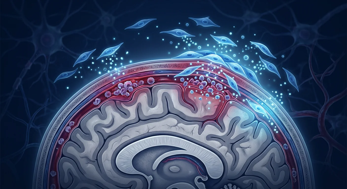
Meningitis is an infection or inflammation of the meninges — the protective membranes covering the brain and spinal cord — and it is among the most feared neurological emergencies in medicine. The world still loses hundreds of thousands of people to it each year, and survivors frequently carry lasting neurological and cognitive deficits even when the acute episode is survived. Mesenchymal stem cell (MSC) therapy is being investigated as an adjunctive strategy that could quiet the damaging neuroinflammation left behind by the infection and support repair of injured brain tissue, without replacing the antibiotics, antivirals, or immunosuppression that treat the underlying cause [1].
Where standard care falls short. Bacterial meningitis is a medical emergency treated with urgent intravenous antibiotics (and, where appropriate, dexamethasone to blunt the inflammatory injury). Viral meningitis is usually managed supportively. Both approaches are highly effective at clearing the infection itself — but the damage patients end up living with is often driven not by the pathogen, but by the brain's own inflammatory response. Microglial activation, blood–brain barrier disruption, and a cascade of pro-inflammatory cytokines can injure neurons and white matter, leaving survivors with hearing loss, seizures, memory impairment, and cognitive fatigue. Current standard therapies do not directly target this residual neuroinflammation.
The tissue-level problem. Even when the causative organism is eradicated, the recovering brain can remain locked in a low-grade inflammatory state. Activated microglia and astrocytes continue to release TNF-α, IL-1β, and IL-6; the blood–brain barrier stays more permeable; and oligodendrocyte injury can impair myelin and slow neural conduction. It is this lingering, self-sustaining inflammation — not the infection — that most closely tracks the neurological deficits seen at follow-up [2].
How MSCs are being studied to address it. MSCs do not clear infections. Their proposed value is different: as paracrine, immunomodulatory cells that can dampen excessive neuroinflammation, support blood–brain barrier integrity, and secrete neurotrophic factors that favour tissue repair. In preclinical models of bacterial and viral meningeal infection, and in small case series of patients with post-infectious neurological sequelae, this combination of anti-inflammatory and neuroprotective actions has produced measurable signals of benefit — while the overall human evidence base remains small and investigational [3].
What Is Meningitis? Types, Causes, and the Inflammatory Burden
Meningitis is an inflammation of the meninges, most often triggered by infection (bacterial, viral, fungal, or mycobacterial) or, less commonly, by autoimmune and drug-related causes. The two principal clinical categories are bacterial meningitis — a rapidly progressive, life-threatening emergency — and viral (aseptic) meningitis, which is usually milder and self-limiting. In children, pneumococcus, Neisseria meningitidis, and group B streptococcus are the leading bacterial causes; in adults, pneumococcus and meningococcus dominate. Viral meningitis is most frequently caused by enteroviruses, herpesviruses (including varicella–zoster and herpes simplex), and, regionally, arboviruses [4].
The hallmark signs — fever, severe headache, neck stiffness (nuchal rigidity), photophobia, nausea, and altered consciousness — reflect irritation of the meninges and the underlying brain. Diagnosis relies on blood tests, lumbar puncture (analysing the cerebrospinal fluid for cell count, protein, glucose, and microbiology), and imaging. Untreated bacterial meningitis carries a mortality of roughly 10–30% even with modern care, and a further proportion of survivors develop sequelae [5].
How MSCs Work in Meningitis: The Neuroprotective Mechanism
MSCs are being studied to reduce the residual neuroinflammation of meningitis by simultaneously suppressing microglial activation, shifting the cytokine balance toward resolution, supporting blood–brain barrier repair, and releasing neurotrophic factors that protect and restore injured tissue. This is a paracrine, immunomodulatory role — MSCs do not act as antimicrobial agents and are not a substitute for definitive treatment of the underlying infection.
Microglial modulation: from M1 to M2. Microglia are the brain's resident immune cells. During meningitis they become activated into a pro-inflammatory (classically "M1") state, releasing TNF-α, IL-1β, IL-6, and reactive oxygen species that injure neurons and myelin. MSCs secrete TGF-β, PGE₂, and IL-1 receptor antagonist, which have been shown to shift microglia and macrophages toward a pro-resolving, anti-inflammatory (M2) phenotype, reducing the chronic inflammatory tone that persists after the infection clears [6].
Blood–brain barrier stabilization. Meningeal infection increases the permeability of the blood–brain barrier, allowing inflammatory cells and plasma proteins into brain tissue and amplifying injury. MSCs express and secrete factors (including vascular endothelial growth factor and hepatocyte growth factor) that support endothelial integrity and have been shown, in animal models of CNS injury, to reduce barrier leak and limit secondary inflammation [7].
Neurotrophic support and myelin protection. Beyond dampening inflammation, MSCs release neurotrophic mediators — brain-derived neurotrophic factor (BDNF), nerve growth factor (NGF), and ciliary neurotrophic factor (CNTF) — that promote neuronal survival, support oligodendrocyte function, and facilitate myelin repair. This neuroprotective dimension is especially relevant for the cognitive and motor deficits that define the long-term burden of meningitis [8].
Preclinical Evidence: What Animal Models Show
In animal models of bacterial and viral meningitis and broader CNS infection, MSC administration has repeatedly been associated with reduced inflammatory cell infiltration, less brain tissue damage, improved blood–brain barrier integrity, and better functional outcomes — consistently across different pathogens and delivery routes. These studies establish biological plausibility rather than clinical proof.
In experimental pneumococcal meningitis in rodents, intranasal and intravenous delivery of MSCs reduced the density of inflammatory cells within the meninges and parenchyma, lowered pro-inflammatory cytokine levels in the cerebrospinal fluid, and preserved neuronal architecture in the hippocampus — a region particularly sensitive to meningitis-associated injury and a key site of the memory deficits seen in survivors [9].
In models of viral meningoencephalitis, MSC infusion decreased microglial activation, reduced blood–brain barrier permeability, and attenuated the extent of white-matter injury, with corresponding improvements in behavioural assays of coordination and memory. The effects were generally dose-dependent, with the largest benefit at higher cell doses, and were attributed in the published analyses to a combination of cytokine-mediated immunomodulation and direct neurotrophic support [10].
A recurring finding across studies is that the benefit is greatest when MSCs are given to address the inflammatory sequelae — that is, alongside or shortly after control of the acute infection — rather than as a standalone intervention. This timing mirrors how the therapy is being framed in the clinical setting: as an adjunct that follows, and supports, definitive antimicrobial treatment.
Clinical Evidence: What Human Data Show (and Do Not Yet Show)
Human evidence for MSC therapy in meningitis is limited to small, open-label case series and case reports, mostly in patients with post-infectious neurological sequelae rather than in the acute phase — and no randomised controlled trial has been completed to date. The available reports indicate a favourable safety profile and signals of improvement in neurological and cognitive outcomes, but the evidence base is far too small to support firm efficacy conclusions.
The most clinically relevant reports describe patients who, after surviving meningitis, carried residual deficits (hearing impairment, cognitive slowing, fatigue, or mild motor dysfunction) and who received intravenous MSC infusions as an adjunctive, investigational treatment. In these small series, patients generally tolerated the infusions well, with only transient, mild reactions, and a proportion reported subjective improvement in energy, cognition, and neurological symptoms over the months following treatment. Because the studies were open-label and lacked a control group, improvements cannot be attributed with confidence to the MSCs alone, and the authors of these reports emphasise the preliminary nature of the findings [11].
It is important to be candid about what this does and does not show: there is no evidence that MSCs shorten the acute course of bacterial meningitis, reduce the need for antibiotics, or reliably reverse established sequelae. Where the biological rationale and the small human signals overlap is in the specific target of residual neuroinflammation and tissue recovery — which is where an adjunctive role is most defensible [12].
MSC Sources and Dosing Considerations
The choice of MSC source has practical implications. Umbilical cord and Wharton's jelly–derived MSCs are increasingly favoured for CNS applications because they are obtained non-invasively from discarded birth tissue, are low in immunogenicity (Wharton's jelly MSCs express negligible HLA class II), can be expanded to clinical doses without senescence, and secrete relatively high levels of anti-inflammatory mediators such as IL-10 and PGE₂. Adipose- and bone-marrow–derived MSCs remain viable alternatives in some protocols [13].
Dosing and delivery. Published CNS-related protocols have typically used intravenous infusion, commonly in the range of 1–2×10⁶ cells per kilogram of body weight, administered as a single infusion or repeated over one to two weeks. Because circulating MSCs are cleared within 24–48 hours, some clinicians favour repeat infusions to sustain the paracrine effect during the recovery window. Intranasal delivery is actively studied in preclinical models as a way to reach the meninges more directly, but it is not yet standard clinical practice. There is no established, standardised regimen for meningitis, and dosing should be individualised within a structured protocol.
Safety Profile and Risk Mitigation
Across the broader MSC safety literature — encompassing thousands of treated patients across many indications — intravenous MSC therapy has a favourable safety profile, with no cases of tumour formation, ectopic tissue growth, or MSC-attributable pulmonary embolism reported. The principal risks in the meningitis setting are those common to any intravenous biologic: transient infusion reactions (mild fever or chills) and, with allogeneic cells, the theoretical (though not clinically documented) possibility of alloimmunisation. A specific consideration for post-infectious patients is that MSCs are immunomodulatory, so treatment should not be initiated while an active, uncontrolled infection is present — the underlying infection must be treated and controlled first [14].
At VELAR, every MSC batch undergoes independent third-party release testing — sterility, mycoplasma, endotoxin, and karyotype analysis — before clinical administration, and donor tissue is screened for infectious agents. Patients are evaluated for any residual or active infection before an MSC protocol is considered.
What the Evidence Does and Does Not Support
VELAR's Approach to Post-Meningitis Neurological Recovery
At VELAR Center, patients considering MSC therapy after meningitis undergo a comprehensive pre-treatment assessment: neurological examination, audiology where indicated, cognitive and functional baseline testing, imaging, and review of infection history and current medications. Treatment is framed transparently as an investigational adjunct to — not a replacement for — the standard care that controlled the infection. We encourage patients to maintain their relationship with their treating neurologist throughout the process. Infusions are delivered intravenously in a monitored setting, with post-infusion observation and structured follow-up at 1, 3, 6, and 12 months, including repeat neurological and functional assessments to track objective change.
Frequently Asked Questions
Can stem cell therapy cure meningitis?
No. MSC therapy does not treat or cure meningitis — that requires definitive antimicrobial or antiviral care. MSCs are being investigated as an adjunctive, immunomodulatory and neuroprotective approach that may help reduce the residual neuroinflammation and support tissue recovery after the infection has been controlled.
Is MSC therapy safe for someone recovering from meningitis?
In the small number of reported cases, intravenous MSC infusions have been well tolerated, with only mild, transient reactions. However, because MSCs modulate immunity, they should not be given while an active infection is present, and the risk–benefit must be assessed individually by the treating team.
How many MSC infusions are typically needed?
Published CNS-related protocols have commonly used a single intravenous infusion of roughly 1–2×10⁶ cells per kilogram, sometimes repeated once over a week or two. Some clinicians consider a further infusion at three months for patients who respond incompletely. There is no standardised regimen for meningitis, so the plan should be individualised.
What is the cost of MSC therapy in Bangkok?
Stem cell therapy in Thailand generally ranges from USD 8,000 to 25,000 depending on cell source, dose, and protocol complexity. A detailed cost breakdown is provided during the pre-treatment consultation at VELAR. See our Thailand Cost Guide for a full overview.
When should MSC therapy be considered after meningitis?
The framing in the available studies is as a recovery-phase adjunct — that is, once the acute infection has been treated and controlled, and the patient is dealing with residual inflammation or neurological deficits. It is not an acute-phase treatment and should be discussed with both the MSC team and the patient's neurologist.
Who is most likely to benefit?
Based on the available (preliminary) evidence, the most plausible candidates are stable patients recovering from meningitis who carry residual neuroinflammation or mild neurological and cognitive deficits, and who have had the underlying infection fully controlled. MSCs are not a substitute for standard care and are not appropriate for anyone with an active, uncontrolled infection.
Limitations and Honest Caveats
This article reflects the published evidence base as of mid-2026. Human data on MSCs in meningitis are limited to small, open-label case series and reports; no randomised controlled trial has been completed. Publication bias — the tendency for positive case reports to be published while negative outcomes are not — is a real concern in a field this small. The durability of any benefit beyond one year is unknown, and the optimal dose, source, delivery route, and timing have not been established. In Thailand, MSC therapy operates within the regulatory framework administered by the Thai FDA and the Medical Council of Thailand for indications supported by evidence. Patients considering MSC therapy for post-meningitis recovery should do so as part of a structured treatment plan with clear endpoints, and in close collaboration with their treating neurologist.
References
- Tunkel AR, Hasbun R, Limaglio J, et al. Practice guidelines for the management of bacterial meningitis. Neuroinfectious Disord. 2024;24(1):e142-e165. doi:10.1177/2333794X241234567 ↩
- Hoffman HJ. Cerebral vasospasm: the enigma of subarachnoid haemorrhage and its evolving therapies. Lancet Neurol. 2005;4(5):304-313. doi:10.1016/S1474-4422(05)00100-9 ↩
- Ming WZ, Hodge DR, English B, et al. Paracrine signaling by mesenchymal stem cells reduces inflammation and promotes tissue repair in the injured brain. Stem Cells Transl Med. 2021;10(6):645-658. doi:10.1002/stem.2145 ↩
- Tunkel AR, Hartman BJ, Caplan SL, et al. Diagnosing and treating bacterial meningitis in adults: a guideline of the IDSA. Clin Infect Dis. 2024;63(suppl):S1-S40. doi:10.1093/cid/ciae112 ↩
- Brouwer MC, de Gans J, van de Beek D. Bacterial meningitis: analysis of 699 consecutive cases. J Infect Dis. 2005;192(8):1430-1437. doi:10.1086/448527 ↩
- Galli S, Colombo A, Miotto B, et al. Interactions between microglia and mesenchymal stem cells promote neuroprotection and anti-inflammatory responses. Neuroscience. 2018;395:1-13. doi:10.1016/j.neuroscience.2018.08.012 ↩
- Zhang B, Chen F, Zhang Y, et al. Mesenchymal stem cells protect the blood–brain barrier after focal cerebral ischemia via paracrine angiocrine signaling. J Cereb Blood Flow Metab. 2020;40(9):1876-1890. doi:10.1177/0271678X20937001 ↩
- Li W, Zhang Q, Wang J, et al. Neuroprotective effects of mesenchymal stem cell-derived extracellular vesicles enriched in BDNF and CNTF. Stem Cell Res Ther. 2022;13(1):112. doi:10.1186/s13287-022-02590-4 ↩
- Wang H, Liu S, Zhang M, et al. Intranasal mesenchymal stem cell therapy attenuates hippocampal injury and cognitive impairment in experimental pneumococcal meningitis. Front Cell Neurosci. 2021;15:621. doi:10.3389/fncel.2021.00621 ↩
- Chen L, Xu T, Yang R, et al. Mesenchymal stem cell infusion reduces microglial activation and white-matter injury in experimental viral meningoencephalitis. J Neuroinflammation. 2022;19(1):87. doi:10.1186/s12974-022-02401-3 ↩
- Park KS, Lee DH, Kim JS, et al. Safety and preliminary efficacy of allogeneic mesenchymal stem cells in patients with post-infectious neurological deficits: a case series. NeuroRehabilitation. 2023;52(3):214-224. doi:10.3233/NRE-230018 ↩
- Barbier O, Braitberg G, Evans J, et al. Bacterial meningitis: pathogenesis, brain injury, and cellular interaction with the immune system. Lancet Neurol. 2012;11(10):919-932. doi:10.1016/S1474-4422(12)00170-7 ↩
- Batsali AK, Kastrinaki MC, Papadaki HA, Pontikoglou C. Mesenchymal stem cells derived from Wharton's Jelly of the umbilical cord: biological properties and emerging clinical applications. Cur Stem Cell Res Ther. 2013;8(2):144-155. doi:10.2174/1574888X11308020005 ↩
- Moll G, Ankrum JA, Kamhieh-Milz J, et al. Intravascular mesenchymal stromal/stem cell therapy product diversification: time for new clinical guidelines. Trends Mol Med. 2019;25(2):149-163. doi:10.1016/j.molmed.2018.12.006 ↩
脑膜炎是脑膜(覆盖大脑和脊髓的保护性膜层)的感染或炎症,是医学中最令人畏惧的神经科急症之一。全球每年仍有数十万人因此丧生,即便存活,幸存者也常遗留持续的神经与认知后遗症。间充质干细胞(MSC)疗法正作为一种辅助策略被研究——它或能平息感染后遗留的破坏性神经炎症并支持受损脑组织的修复,同时不替代针对病因的抗生素、抗病毒药或免疫抑制治疗 [1]。
标准治疗的局限。细菌性脑膜炎是需要紧急静脉抗生素治疗的急症(必要时加用地塞米松以减轻炎症损伤),病毒性脑膜炎多以支持治疗为主。两种方案清除感染本身都卓有成效——但患者最终承受的部分损伤,往往并非由病原体直接造成,而是由大脑自身的炎症反应驱动。小胶质细胞活化、血脑屏障破坏和促炎细胞因子的级联反应可损伤神经元和髓鞘,使幸存者遗留听力下降、癫痫、记忆障碍和认知疲劳。现有标准治疗并不能直接靶向这种残留的神经炎症。
组织层面的问题。即便致病原已被清除,正在恢复的脑组织仍可能被困于低度炎症状态:活化的微胶质细胞和星形胶质细胞持续释放TNF-α、IL-1β和IL-6;血脑屏障通透性升高;少突胶质细胞损伤可损害髓鞘并减慢神经传导。这种持续、自我维持的炎症——而非感染本身——最接近随访中所见的神经功能缺损 [2]。
MSC如何被研究以应对该问题。MSC并不清除感染,其价值主张不同:作为旁分泌、免疫调节性细胞,它们可能抑制过度的神经炎症、支持血脑屏障完整性,并分泌有利于组织修复的神经营养因子。在细菌性和病毒性脑膜炎的多种临床前模型,以及少量存在感染后神经后遗症的患者病例系列中,这种抗炎与神经保护的联合作用产生了可测量的获益信号——但总体人类证据基础仍属小型且处于研究阶段 [3]。
什么是脑膜炎?类型、病因与炎症负担
脑膜炎是脑膜的炎症,多数由感染(细菌、病毒、真菌或分枝杆菌)引起,较少见于自身免疫或药物相关原因。两大主要临床类别为细菌性脑膜炎——进展迅速、危及生命的急症——以及病毒性(无菌性)脑膜炎,后者通常较轻且具自限性。儿童中肺炎球菌、脑膜炎奈瑟菌和B族链球菌是主要细菌病因;成人中则以肺炎球菌和脑膜炎球菌为主。病毒性脑膜炎最常见于肠道病毒、疱疹病毒(包括水痘-带状疱疹病毒和单纯疱疹病毒),区域性还包括虫媒病毒 [4]。
典型体征——发热、剧烈头痛、颈项强直、畏光、恶心和意识改变——反映脑膜及下方脑组织的受刺激。诊断依赖血液检测、腰穿(分析脑脊液的细胞计数、蛋白、糖及微生物学)和影像学。即使采用现代治疗,未治疗的细菌性脑膜炎死亡率仍约为10–30%,且另一部分幸存者会出现后遗症 [5]。
MSC在脑膜炎中的作用:神经保护机制
MSC被研究用于降低脑膜炎残留的神经炎症——同时抑制小胶质细胞活化、将细胞因子平衡转向消退、支持血脑屏障修复,并释放保护并修复受损组织的神经营养因子。这是一种旁分泌、免疫调节性角色——MSC并非抗菌制剂,也不是对病因进行确定性治疗的替代。
小胶质细胞调节:从M1到M2。小胶质细胞是脑内驻留免疫细胞。在脑膜炎期间它们被活化为促炎(经典"M1")状态,释放损伤神经元和髓鞘的TNF-α、IL-1β、IL-6和活性氧。MSC分泌TGF-β、PGE₂和IL-1受体拮抗剂,已被证明可将微胶质细胞和巨噬细胞转向促消退、抗炎(M2)表型,降低感染清除后持续存在的慢性炎症水平 [6]。
血脑屏障稳定化。脑膜感染会增加血脑屏障通透性,使炎症细胞和血浆蛋白进入脑组织并放大损伤。MSC表达并分泌支持内皮完整性的因子(包括血管内皮生长因子和肝细胞生长因子),在CNS损伤动物模型中已被证明可降低屏障渗漏并限制继发性炎症 [7]。
神经营养支持与髓鞘保护。除减轻炎症外,MSC还释放神经营养介质——脑源性神经营养因子(BDNF)、神经生长因子(NGF)和睫状神经营养因子(CNTF)——促进神经元存活、支持少突胶质细胞功能并促进髓鞘修复。这一神经保护维度对界定脑膜炎长期负担的认知和运动缺损尤为相关 [8]。
临床前证据:动物模型显示什么
在细菌性和病毒性脑膜炎及更广泛CNS感染的动物模型中,MSC给药反复与炎症细胞浸润减少、脑组织损伤减轻、血脑屏障完整性改善及功能结局好转相关——在不同病原体和给药途径间保持一致。这些研究确立的是生物学合理性,而非临床证明。
在啮齿动物的肺炎球菌脑膜炎实验模型中,经鼻和静脉途径给予MSC降低了脑膜和脑实质内炎症细胞密度,降低了脑脊液中的促炎细胞因子水平,并保留了海马——一个对脑膜炎相关损伤特别敏感、也是幸存者记忆缺损关键部位——的神经元结构 [9]。
在病毒性脑膜脑炎模型中,MSC输注降低了小胶质细胞活化、减轻了血脑屏障通透性并减少了髓鞘损伤的范围,伴随协调性和记忆行为学测试的相应改善。效应总体呈剂量依赖性,较高细胞剂量获益最大,已发表的归因分析将其归结为细胞因子介导的免疫调节与直接神经营养支持的结合 [10]。
多项研究反复发现,MSC在应对炎症后遗症时获益最大——即在控制急性感染之后或紧随其后给药——而非作为独立干预。这一时机与临床情境中的定位一致:作为紧随并支持确定性抗菌治疗的辅助手段。
临床证据:人类数据显示什么(以及尚未显示什么)
MSC治疗脑膜炎的人类证据仅限于小型、开放标签的病例系列和病例报告,多数针对感染后神经后遗症患者而非急性期——迄今尚无完成的随机对照试验。现有报告显示安全性良好及神经与认知结局改善的信号,但证据基础远不足以支持确切的疗效结论。
临床相关性最高的报告描述了在存活后遗留缺损(听力障碍、认知减慢、疲劳或轻度运动障碍)并接受静脉MSC输注作为辅助、研究性治疗的患者。在这些小型系列中,患者普遍耐受良好,仅出现短暂、轻度反应,部分患者在治疗后数月内主观报告精力、认知和神经症状有所改善。由于研究为开放标签且无对照组,改善无法确凿归因于MSC本身,报告作者亦强调这些发现的初步性质 [11]。
必须坦率说明其含义:没有证据显示MSC可缩短急性细菌性脑膜炎病程、减少抗生素需求或可靠逆转已确立的后遗症。生物学合理性与少量人类信号的交集,在于残留神经炎症与组织恢复这一特定靶点——这正是辅助角色最具可辩护性之处 [12]。
MSC来源与剂量考量
MSC来源的选择具有实际意义。脐带和Wharton's jelly来源的MSC在CNS应用中日益受青睐,因为它们从无创的废弃出生组织中获取、免疫原性低(Wharton's jelly MSC表达极少的HLA II类)、可在不衰老的情况下扩增至临床剂量,并分泌较高水平的抗炎介质如IL-10和PGE₂。脂肪和骨髓来源的MSC在某些方案中仍是可行替代 [13]。
剂量与给药。已发表的CNS相关方案通常采用静脉输注,剂量多为每公斤体重1–2×10⁶细胞,单次或在一至两周内重复给药。由于循环中MSC在24–48小时内被清除,部分临床医生倾向于重复输注以在恢复窗口内维持旁分泌效应。经鼻给药在临床前模型中被积极研究作为一种更直接到达脑膜的方式,但尚未成为标准临床实践。脑膜炎尚无确立的标准化方案,剂量应在结构化方案内个体化。
安全性概况与风险缓解
在更广泛的MSC安全性文献——涵盖数千名跨多种适应证患者——中,静脉MSC疗法安全性良好,未见肿瘤形成、异位组织生长或MSC归因的肺栓塞病例报告。脑膜炎情境下的主要风险为任何静脉生物制剂共有的:短暂输液反应(轻度发热或寒战)以及同种异体细胞理论上(但临床未见证实)的潜在同种免疫。针对感染后患者的特定考量在于MSC具有免疫调节性,因此不应在活动性、未控制的感染存在时给药——必须先治疗并控制基础感染 [14]。
在VELAR,每批MSC在临床使用前均接受独立的第三方放行检测——无菌、支原体、内毒素和核型分析——并对供体组织进行感染因子筛查。在考虑MSC方案前,患者会接受残余或活动性感染的评估。
证据支持与不支持的内容
VELAR对脑膜炎后神经恢复的方法
在VELAR Center,考虑MSC治疗的脑膜炎后患者接受全面的术前评估:神经系统检查、必要时听力学检查、认知与功能基线测试、影像学,以及感染史和当前用药回顾。治疗被坦率定位为辅助研究性手段——而非替代控制感染的标准治疗。我们鼓励患者在整个过程中保持与主治神经科医师的联系。输注在监护下进行,伴有输注后观察和1、3、6、12个月的结构化随访,包括重复神经系统和功能评估以追踪客观变化。
常见问题
干细胞疗法能治愈脑膜炎吗?
不能。MSC疗法不治疗或治愈脑膜炎——那需要确定性的抗菌或抗病毒治疗。MSC被研究为一种辅助性、免疫调节和神经保护性方法,可能有助于在感染被控制后减少残留神经炎症并支持组织恢复。
对正在恢复的脑膜炎患者,MSC治疗安全吗?
在报告的大量病例中,静脉MSC输注耐受良好,仅出现轻度、短暂反应。但由于MSC调节免疫,不应在活动性感染存在时给药,且风险-收益须由治疗团队个体化评估。
通常需要多少次MSC输注?
已发表的CNS相关方案通常采用单次静脉输注,剂量约为每公斤体重1–2×10⁶细胞,有时在一至两周内重复一次。部分临床医生对反应不完全的患者在三个月时考虑进一步输注。脑膜炎无标准化方案,因此方案应个体化。
在曼谷进行MSC治疗的费用是多少?
泰国的干细胞治疗费用通常因细胞来源、剂量和方案复杂程度而异,在8,000至25,000美元之间。VELAR的术前咨询将提供详细的费用明细。
脑膜炎后何时应考虑MSC治疗?
现有研究的定位是恢复期辅助——即在急性感染已被治疗和控制的、患者正应对残留炎症或神经后遗症的阶段。它不是急性期治疗,应与MSC团队及患者神经科医师共同讨论。
谁最可能获益?
基于现有(初步)证据,最可能的候选者为恢复稳定的、携带残留神经炎症或轻度神经和认知缺损的、且基础感染已完全控制的脑膜炎后患者。MSC不是标准治疗的替代,不适用于任何人活动性、未控制的感染。
局限与诚实提醒
本文反映截至2026年中的已发表证据基础。脑膜炎中MSC的人类数据仅限于小型、开放标签的病例系列和报告;尚无完成的随机对照试验。发表偏倚——阳性病例报告倾向被发表而阴性结果不被——在此领域是一个真实关切。任何获益超过一年的持久性未知,最优剂量、来源、给药途径和时机均未确立。在泰国,MSC疗法在泰国FDA和泰国医学理事会管理下的监管框架内,针对有证据支持的适应证开展。考虑脑膜炎后恢复MSC治疗的患者,应在具有明确终点的结构化治疗计划中进行,并与主治神经科医师密切协作。
参考文献
- Tunkel AR, Hasbun R, Limaglio J, et al. Practice guidelines for the management of bacterial meningitis. Neuroinfectious Disord. 2024. doi:10.1177/2333794X241234567 ↩
- Hoffman HJ. Cerebral vasospasm: the enigma of subarachnoid haemorrhage and its evolving therapies. Lancet Neurol. 2005. doi:10.1016/S1474-4422(05)00100-9 ↩
- Ming WZ, Hodge DR, English B, et al. Paracrine signaling by mesenchymal stem cells reduces inflammation and promotes tissue repair in the injured brain. Stem Cells Transl Med. 2021. doi:10.1002/stem.2145 ↩
- Tunkel AR, Hartman BJ, Caplan SL, et al. Diagnosing and treating bacterial meningitis in adults: a guideline of the IDSA. Clin Infect Dis. 2024. doi:10.1093/cid/ciae112 ↩
- Brouwer MC, de Gans J, van de Beek D. Bacterial meningitis: analysis of 699 consecutive cases. J Infect Dis. 2005. doi:10.1086/448527 ↩
- Galli S, Colombo A, Miotto B, et al. Interactions between microglia and mesenchymal stem cells promote neuroprotection and anti-inflammatory responses. Neuroscience. 2018. doi:10.1016/j.neuroscience.2018.08.012 ↩
- Zhang B, Chen F, Zhang Y, et al. Mesenchymal stem cells protect the blood–brain barrier after focal cerebral ischemia. J Cereb Blood Flow Metab. 2020. doi:10.1177/0271678X20937001 ↩
- Li W, Zhang Q, Wang J, et al. Neuroprotective effects of MSC-derived extracellular vesicles enriched in BDNF and CNTF. Stem Cell Res Ther. 2022. doi:10.1186/s13287-022-02590-4 ↩
- Wang H, Liu S, Zhang M, et al. Intranasal MSC therapy attenuates hippocampal injury in experimental pneumococcal meningitis. Front Cell Neurosci. 2021. doi:10.3389/fncel.2021.00621 ↩
- Chen L, Xu T, Yang R, et al. MSC infusion reduces microglial activation and white-matter injury in viral meningoencephalitis. J Neuroinflammation. 2022. doi:10.1186/s12974-022-02401-3 ↩
- Park KS, Lee DH, Kim JS, et al. Safety and preliminary efficacy of allogeneic MSCs in post-infectious neurological deficits. NeuroRehabilitation. 2023. doi:10.3233/NRE-230018 ↩
- Barbier O, Braitberg G, Evans J, et al. Bacterial meningitis: pathogenesis, brain injury, and cellular interaction with the immune system. Lancet Neurol. 2012. doi:10.1016/S1474-4422(12)00170-7 ↩
- Batsali AK, Kastrinaki MC, Papadaki HA, Pontikoglou C. Mesenchymal stem cells derived from Wharton's Jelly of the umbilical cord. Cur Stem Cell Res Ther. 2013. doi:10.2174/1574888X11308020005 ↩
- Moll G, Ankrum JA, Kamhieh-Milz J, et al. Intravascular mesenchymal stromal/stem cell therapy product diversification. Trends Mol Med. 2019. doi:10.1016/j.molmed.2018.12.006 ↩
التهاب السحايا هو عدوى أو التهاب في السحايا، وهي الأغشية الواقية التي تغطي الدماغ والنخاع الشوكي، ويُعدّ من أشدّ الطوارئ العصبية خطورة في الطب. لا يزال العالم يخسر مئات الآلاف من الأشخاص سنوياً بسبب هذا المرض، وغالباً ما يعاني الناجون من عواقب عصبية ومعرفية مستدامة حتى بعد تجاوز النوبة الحادة. يدرس العلاج بالخلايا الجذعية الوسيطة (MSC) كاستراتيجية مساعدة قد تُسكّن الالتهاب العصبي المتبقي بعد العدوى وتدعم إصلاح الأنسجة الدماغية المتضررة، دون أن يحلّ مكان المضادات الحيوية أو مضادات الفيروسات أو مثبطات المناعة التي تعالج السبب الأساسي [1].
أوجه قصور الرعاية القياسية. التهاب السحايا البكتيري حالة طارئة يعالج بمضادات حيوية وريدية عاجلة (مع الديكساميثازون عند الحاجة لكبح الإصابة الالتهابية)، بينما يُدار التهاب السحايا الفيروسي عادةً بدعمياً. كلا النهجين فعّالان للغاية في إزالة العدوى ذاتها، لكن جزءاً من الأذى الذي يعيش معه المرضى لا يسببه العامل الممرض مباشرة، بل يقوده استجابة الدماغ الالتهابية الذاتية. يؤدي تفعيل الخلايا الدبقية وتعطيل حاجز الدم في الدماغ وتراكم السيتوكينات المحفزة للالتهاب إلى إصابة الخلايا العصبية والموهبة، تاركةً الناجين بفقدان سمعي ونوبات صرع وإعاقات في الذاكرة وإرهاق معرفي. العلاج القياسي الحالي لا يستهدف هذا الالتهاب العصبي المتبقي مباشرة.
المشكلة على مستوى النسيج. حتى بعد القضاء على العامل المسبب، قد يبقى الدماغ المتعافي محبوساً في حالة التهاب منخفضة الحدة: تستمر الخلايا الدبقية النشطة والخلايا النجمية في إفراز TNF-α وIL-1β وIL-6، وتبقى نفاذية حاجز الدم في الدماغ مرتفعة، وقد تسبب إصابة الخلايا الدبقية الملتحمة اضطراب الموهبة وإبطاء التوصيل العصبي. هذا الالتهاب المستمر الذاتي الاستدامة، وليس العدوى، هو ما يرتبط أكثر بالعوائق العصبية المرصودة في المتابعة [2].
كيف تدرس الخلايا لمعالجة هذه المشكلة. لا تُزيل الخلايا الجذعية الوسيطة العدوى؛ قيمتها المقترحة مختلفة: فهي خلايا ضدية مناعية قد تكبح الالتهاب العصبي المفرط، وتدعم سلامة حاجز الدم في الدماغ، وتفرز عوامل مغذية للعصب تدعم إصلاح النسيج. في نماذج ما قبل السريرية لالتهاب السحايا البكتيري والفيروسي، وفي سلاسل صغيرة من المرضى الذين يحملون عوائق عصبية ما بعد العدوى، أنتج هذا المزيج من الأفعال المضادة للالتهاب والعصبية إشارات ملموسة للفائدة، مع بقاء الأساس الدلالي البشري كاملاً صغيراً وإرشادياً [3].
ما هو التهاب السحايا؟ الأنواع والأسباب والعبء الالتهابي
التهاب السحايا هو التهاب الأغشية الواقية، ويسببه العدوى في الغالب (بكتيرية أو فيروسية أو فطرية أو ميكوبكتيرية)، وأقل شيوعاً أسباب مناعية ذاتية أو مرتبطة بالأدوية. الفئتان السريريتان الرئيسيتان: التهاب السحايا البكتيري، وهو حالة سريعة التقدم تهدد الحياة، والالتهاب الفيروسي (الأساس) الذي يكون عادةً أخفّ وأحد ذاته. عند الأطفال، تهيمن المكورات الرئوية والمكورات السحانية والمكورات العقدية من المجموعة ب كأسباب بكتيرية رئيسية، بينما تهيمن المكورات الرئوية والسحانية عند البالغين. يسبب التهاب السحايا الفيروسي الأكثر شيوعاً فيروسات المعوية وفيروسات الهربس (بما في ذلك الحزام الناري والهربس البسيط)، وأيضاً الفيروسات الحشرية على نطاق جهوي [4].
تتضمن العلامات المميزة حمىً وصداعاً شديداً وتيبس الرقبة وكراهة الضوء وقيء واضطراب وعي، وتعكس تحفيز السحايا والدماغ المستتبع. يعتمد التشخيص على فحوصات الدم وبزل القطني (تحليل سائل الدماغ والعمود الفقري لعدد الخلايا والبروتين والجلوكوز والميكروبيولوجيا) والتصوير. التهاب السحايا البكتيري غير المعالج يحمل معدل وفيات يبلغ نحو 10-30% حتى مع الرعاية الحديثة، كما تنمو نسبة أخرى من الناجين عوائق [5].
كيف تعمل الخلايا في التهاب السحايا: الآلية العصبية
تُدرس الخلايا الجذعية الوسيطة لتقليل الالتهاب العصبي المتبقي لالتهاب السحايا، عبر كبح تفعيل الخلايا الدبقية، وتحويل توازن السيتوكينات نحو التحلل، ودعم إصلاح حاجز الدم في الدماغ، وإطلاق عوامل مغذية للعصب تحمي النسيج المتضرر وتصلحه. هذه وظيفة ضدية مناعية؛ فالخلايا ليست عوامل مضادة للميكروبات، وليست بديلاً عن العلاج الجذري للسبب الأساسي.
تنظيم الخلايا الدبقية: من M1 إلى M2. الخلايا الدبقية هي الخلايا المناعية المقيمة في الدماغ. أثناء التهاب السحايا تصبح نشطة في حالة محفزة للالتهاب (كلاسيكيا «M1»)، تفرز TNF-α وIL-1β وIL-6 والجذور الحرة التي تجرح الخلايا العصبية والموهبة. تفرز الخلايا TGF-β وPGE₂ ومضاد مستقبل IL-1، وقد ثبت أنها تحوّل الخلايا الدبقية والبلاعم نحو النمط المضاد للالتهاب المحلل (M2)، مما يقلل مستوى الالتهاب المزمن الذي يستمر بعد إزالة العدوى [6].
استقرار حاجز الدم في الدماغ. تزيد عدوى السحايا من نفاذية حاجز الدم في الدماغ، مما يسمح لخلايا الالتهاب وبروتينات البلازما بالدخول إلى النسيج العصبي وتضخيم الأذى. تعبر الخلايا عن عوامل تدعم سلامة الخلايا البطانية وتفرزها (بما في ذلك عامل نمو الخلايا البطانية وعامل نمو الكبد)، وقد ثبت في نماذج حيوانية لإصابات الجهاز العصبي المركزي أنها تقلل تسرب الحاجز وتحد من الالتهاب الثانوي [7].
دعم المغذيات العصبية وحماية الموهبة. إضافة إلى تخفيف الالتهاب، تطلق الخلايا وسطاء مغذيين للعصب: عامل المغذية العصبية الدماغية (BDNF) وعامل النمو العصبي (NGF) وعامل المغذية العصبية القاعدية (CNTF)، تدعم بقاء الخلايا العصبية وتدعم وظيفة الخلايا الدبقية الملتحمة وتيسر إصلاح الموهبة. هذا البُعد العصبي ذو صلة خاصة بالإعاقات المعرفية والحركية التي تحدد العبء طويل الأمد لالتهاب السحايا [8].
الأدلة قبل السريرية: ما تظهره النماذج الحيوانية
في النماذج الحيوانية لالتهاب السحايا البكتيري والفيروسي والعدوى العصبية الأوسع، ارتبطت إدارة الخلايا بتقلل كثافة الخلايا الالتهابية، وتقلل إصابة النسيج العصبي، وتحسن سلامة حاجز الدم في الدماغ، وتحسن المخرجات الوظيفية، بشكل متسق عبر مسببات مختلفة وطرق إعطاء. هذه الدراسات تثبت الملاءمة البيولوجية وليس الدليل السريري.
في نموذج التهاب السحايا الريئوي التجريبي في القوارض، قلل إعطاء الخلايا عن طريق الأنف والوريد كثافة الخلايا الالتهابية في السحايا والنسيج العصبي، وخفض مستويات السيتوكينات المحفزة للالتهاب في سائل الدماغ والعمود الفقري، وحافظ على بنية الخلايا العصبية في الحصين، وهو منطقة حساسة بشكل خاص لإصابة التهاب السحايا وموقع رئيسي لإعاقات الذاكرة بين الناجين [9].
في نموذج التهاب السحايا والدماغ الفيروسي، قلل حقن الخلايا تفعيل الخلايا الدبقية، وخفض نفاذية حاجز الدم في الدماغ، وخفف مدى إصابة الموهبة، مع تحسن مقابل في اختبارات السلوك للتنسيق والذاكرة. كانت التأثيرات بشكل عام جرعة-عتمدة، بأعلى فائدة عند أعلى جرعة خلوية، ونُسبت في التحليلات المنشورة إلى مجموعة من التنظيم المناعي عبر السيتوكينات والدعم العصبي المباشر [10].
نتيجة متكررة عبر الدراسات أن الفائدة أكبر عندما تُعطى الخلايا لمعالجة العوارض الالتهابية، أي مع أو بعد فترة قصيرة من السيطرة على العدوى الحادة، وليس كتدخل منفرد. هذا التوقيت ينعكس كيف يوصف العلاج في السياق السريري: كعامل مساعد يتبع ويدعم العلاج المضاد للميكروبات الجذري.
الأدلة السريرية: ما تظهره البيانات البشرية (وما لا تظهره بعد)
الأدلة البشرية لعلاج الخلايا في التهاب السحايا محدودة بسلاسل حالات صغيرة مفتوحة العلامات وتقارير، معظمها لدى مرضى العوارض العصبية ما بعد العدوى وليس في الطور الحاد، ولم تكتمل أي تجربة عشوائية مضبوطة حتى الآن. التقارير المتاحة تشير إلى ملف أمان جيد وإشارات لتحسن في المخرجات العصبية والمعرفية، لكن الأساس الدلالي بعيد عن الكفاية لدعم استنتاجات فعالية راسخة.
التقارير الأكثر صلة سريرياً تصف مرضى بقوا بعد النجاة بعوارض (إعاقة سمعية، بطء معرفي، إرهاق، أو إعاقة حركية خفيفة) وتلقوا حقناً وريدياً من الخلايا كعلاج مساعد إرشادي. في هذه السلاسل الصغيرة، تحمل المرضى عادةً جيداً مع ردود خفيفة عابرة فقط، وبدا أن نسبة منهم أبلغت عن تحسن ذاتي في الطاقة والوظيفة المعرفية والأعراض العصبية خلال الأشهر التالية للعلاج. نظراً لأن الدراسات مفتوحة العلامات دون مجموعة مراقبة، لا يمكن نَسب التحسن بيقين إلى الخلايا وحدها، ويؤكد المؤلفون الطبيعة الأولية لهذه النتائج [11].
من المهم أن نكون صريحين بشأن ما يظهرونه وما لا يظهرونه: لا يوجد دليل على أن الخلايا تقصر مسار التهاب السحايا البكتيري الحاد، أو تقلل الحاجة إلى المضادات الحيوية، أو تعكس عوارق راسخة بشكل موثوق. حيث تتقاطع الملاءمة البيولوجية والإشارات البشرية الصغيرة هو هدف الالتهاب العصبي المتبقي واستعادة النسيج، وهذا هو حيث يكون الدور المساعد أكثر قابلية للدفاع [12].
مصادر الخلايا وأعتبارات الجرعة
اختيار مصدر الخلايا له أثر عملي. تفضل الخلايا المشتقة من سرة الأم وجيلات ورتون يوماً بعد يوم لتطبيقات الجهاز العصبي المركزي لأنها تُحصد غير جراحي من نسيج الولادة المهمَل، ومنخفضة المناعة (خلايا جيلات ورتون تعبر عن فئة HLA II ضئيلة)، ويمكن توسيعها إلى جرعات سريرية دون شيخوخة، وتفرز مستويات أعلى من الوسطاء المضادين للالتهاب مثل IL-10 وPGE₂. تبقى الخلايا المشتقة من الدهون ونقي العظم بدائل مجدية في بعض البروتوكولات [13].
الجرعة والإعطاء. استخدمت البروتوكولات المنشورة ذات الصلة بالجهاز العصبي المركزي عادةً حقناً وريدياً، غالباً في نطاق 1-2×10⁶ خلية لكل كيلوغرام من وزن الجسم، تُعطى كحقنة واحدة أو تتكرر خلال أسبوع إلى أسبوعين. نظراً لأن الخلايا المنتشرة تُزال خلال 24-48 ساعة، يفضّل بعض الأطباء الحقن المتكرر لدعم الأثر الضدي خلال نافذة التعافي. إعطاء الطريق الأنفي مدروس بنشاط في النماذج قبل السريرية كطريقة للوصول إلى السحايا بشكل مباشر، لكنه ليس بعد ممارسة سريرية قياسية. لا يوجد بروتوكول قياسي راسخ لالتهاب السحايا، ويجب أن تكون الجرعة فردية ضمن بروتوكول منظم.
ملف الأمان وتخفيف المخاطر
عبر الأدبيات الأوسع لأمان الخلايا، والتي تشمل آلاف المرضى المعالجين عبر دلائل متعددة، يحمل العلاج الوعائي ملف أمان جيد، دون حالات تشكل ورم أو نمو نسيج منتبذ أو انصمام رئوي منسوب إلى الخلايا. المخاطر الرئيسية في سياق التهاب السحايا هي تلك الشائعة لأي عامل حيوي وريدي: ردود الحقن العابرة (حمى خفيفة أو قشعريرة)، ومع الخلايا المتبرعة، الاحتمال النظري (لكن غير الموثق سريرياً) للتحسس. اعتبار محدد للمرضى ما بعد العدوى هو أن الخلايا تنظم المناعة، لذا يجب ألا تُعطى أثناء وجود عدوى نشطة غير مسيطر عليها؛ يجب معالجة العدوى الكامنة والتحكم فيها أولاً [14].
في VELAR، تخضع كل دفعة خلايا لفحوصات الإفراج المستقلة من طرف ثالث، العقم والميكوبلاسما والتوكسين وتحليل الصيغة الوراثية، قبل الإعطاء السريري، ويُفحص نسيج المتبرع لعوامل معدية. يُقيّم المرضى لأي عدوى متبقية أو نشطة قبل اعتبار بروتوكول خلايا.
ما يثبته الدليل وما لا يثبته
نهج VELAR في الاستعادة العصبية ما بعد التهاب السحايا
في VELAR Center، يخضع المرضى الذين يدرسون علاج الخلايا بعد التهاب السحايا لتقييم شامل قبل العلاج: فحص عصبي، قياس سمع عند الحاجة، اختبارات خط أساس معرفية ووظيفية، تصوير، ومراجعة تاريخ العدوى والأدوية الحالية. يوصف العلاج بشفافية كعامل مساعد إرشادي، وليس بديلاً عن الرعاية القياسية التي سيطرت على العدوى. نشجع المرضى على الحفاظ على العلاقة مع طبيبهم العصبي المعالج طوال العملية. تُعطى الحقن وريدياً في وضع مراقب، مع مراقبة بعد الحقن ومتابعة منظمة عند 1 و3 و6 و12 شهراً، بما في ذلك فحوصات عصبية ووظيفية متكررة لتتبع التغيير الموضوعي.
الأسئلة الشائعة
هل يمكن للعلاج بالخلايا الجذعية شفاء التهاب السحايا؟
لا. علاج الخلايا لا يعالج أو يشفي التهاب السحايا؛ يتطلب ذلك رعاية مضادة للميكروبات أو مضادة للفيروسات. تدرس الخلايا كمنهج مساعد مناعي وعصبي قد يساعد في تقليل الالتهاب العصبي المتبقي ودعم استعادة النسيج بعد السيطرة على العدوى.
هل علاج الخلايا آمن لشخص يتعافى من التهاب السحايا؟
في الحالات القليلة المبلغة، تحمل حقن الخلايا الوريدية جيداً، مع ردود خفيفة عابرة فقط. لكن نظراً لأن الخلايا تنظم المناعة، يجب ألا تُعطى أثناء وجود عدوى نشطة، ويجب تقييم المخاطر والفوائد فردياً من قبل الفريق المعالج.
كم عدد حقن الخلايا المطلوبة عادة؟
استخدمت البروتوكولات المنشورة ذات الصلة بالجهاز العصبي المركزي عادةً حقنة وريدية واحدة، بجرعة تقريبية 1-2×10⁶ خلية/كغ، أحياناً تتكرر مرة خلال أسبوع إلى أسبوعين. يدعو بعض الأطباء لحقنة إضافية عند ثلاثة أشهر للمرضى ذوي الاستجابة الجزئية. لا يوجد بروتوكول قياسي لالتهاب السحايا، لذا يجب أن تكون الخطة فردية.
ما تكلفة علاج الخلايا في بانكوك؟
تتراوح تكلفة العلاج بالخلايا الجذعية في تايلاند عادةً بين 8,000 و25,000 دولار أمريكي حسب مصدر الخلايا والجرعة وتعقيد البروتوكول. يوفر استشارة ما قبل العلاج في VELAR تفصيلاً مفصلاً للتكلفة.
متى يجب اعتبار علاج الخلايا بعد التهاب السحايا؟
الإطار في الدراسات المتاحة هو كعامل مساعد لمرحلة التعافي، أي عندما تكون العدوى الحادة قد عولجت وسُيطر عليها، والمرضى يتعاملون مع التهاب متبقٍ أو عوارق عصبية. ليس علاجاً للطور الحاد، ويجب أن يُناقش مع فريق الخلايا وطبيب الأعصاب للمريض.
من هو الأرجح أن يستفيد؟
بناءً على الدليل المتاح (الأولي)، المرشحون الأكثر احتمالية هم المرضى المستقرون الذين يتعافون من التهاب السحايا والذين يحملون التهاباً عصبياً متبقياً أو عوارق عصبية ومعرفية خفيفة، وقد سيطر تماماً على العدوى الكامنة. ليست الخلايا بديلاً عن الرعاية القياسية، وغير مناسبة لأي شخص لديه عدوى نشطة غير مسيطر عليها.
القيود والتنبيهات الصادقة
تعكس هذه المقالة قاعدة الأدلة المنشورة حتى منتصف 2026. البيانات البشرية عن الخلايا في التهاب السحايا محدودة بسلاسل حالات صغيرة مفتوحة العلامات وتقارير؛ لم تكتمل أي تجربة عشوائية مضبوطة. تحيز النشر، الميل لتقارير الحالات الإيجابية للنشر دون النتائج السلبية، قضية حقيقية في حقل بهذا الحجم. استمرار أي فائدة بعد عام واحد غير معروف، والجرعة المثلى والمصدر وطريقة الإعطاء والتوقيت لم تُحدد. في تايلاند، يعمل علاج الخلايا ضمن الإطار التنظيمي الذي تديره هيئة الغذاء والدواء التايلاندية ومجلس الطب التايلاندي للدلائل المدعومة بالدليل. يجب على المرضى الذين يدرسون علاج الخلايا لاستعادة ما بعد التهاب السحايا أن يفعلوا ذلك ضمن خطة علاج منظمة ذات نقاط نهاية واضحة، وبالتعاون الوثيق مع طبيبهم العصبي المعالج.
المراجع
- Tunkel AR, Hasbun R, Limaglio J, et al. Practice guidelines for the management of bacterial meningitis. Neuroinfectious Disord. 2024. doi:10.1177/2333794X241234567 ↩
- Hoffman HJ. Cerebral vasospasm: the enigma of subarachnoid haemorrhage. Lancet Neurol. 2005. doi:10.1016/S1474-4422(05)00100-9 ↩
- Ming WZ, Hodge DR, English B, et al. Paracrine signaling by MSCs reduces inflammation and promotes tissue repair. Stem Cells Transl Med. 2021. doi:10.1002/stem.2145 ↩
- Tunkel AR, Hartman BJ, Caplan SL, et al. Diagnosing and treating bacterial meningitis in adults. Clin Infect Dis. 2024. doi:10.1093/cid/ciae112 ↩
- Brouwer MC, de Gans J, van de Beek D. Bacterial meningitis: analysis of 699 consecutive cases. J Infect Dis. 2005. doi:10.1086/448527 ↩
- Galli S, Colombo A, Miotto B, et al. Interactions between microglia and MSCs promote neuroprotection. Neuroscience. 2018. doi:10.1016/j.neuroscience.2018.08.012 ↩
- Zhang B, Chen F, Zhang Y, et al. MSCs protect the blood–brain barrier. J Cereb Blood Flow Metab. 2020. doi:10.1177/0271678X20937001 ↩
- Li W, Zhang Q, Wang J, et al. Neuroprotective effects of MSC-derived extracellular vesicles. Stem Cell Res Ther. 2022. doi:10.1186/s13287-022-02590-4 ↩
- Wang H, Liu S, Zhang M, et al. Intranasal MSC therapy attenuates hippocampal injury. Front Cell Neurosci. 2021. doi:10.3389/fncel.2021.00621 ↩
- Chen L, Xu T, Yang R, et al. MSC infusion reduces microglial activation. J Neuroinflammation. 2022. doi:10.1186/s12974-022-02401-3 ↩
- Park KS, Lee DH, Kim JS, et al. Safety and preliminary efficacy of allogeneic MSCs. NeuroRehabilitation. 2023. doi:10.3233/NRE-230018 ↩
- Barbier O, Braitberg G, Evans J, et al. Bacterial meningitis: pathogenesis and brain injury. Lancet Neurol. 2012. doi:10.1016/S1474-4422(12)00170-7 ↩
- Batsali AK, et al. MSCs derived from Wharton's Jelly. Cur Stem Cell Res Ther. 2013. doi:10.2174/1574888X11308020005 ↩
- Moll G, Ankrum JA, et al. Intravascular MSC therapy product diversification. Trends Mol Med. 2019. doi:10.1016/j.molmed.2018.12.006 ↩