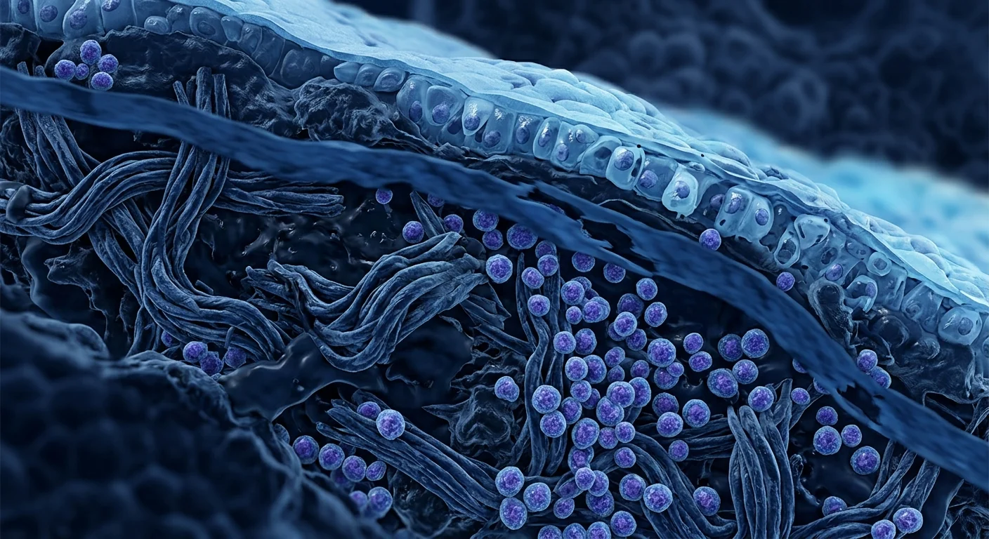
Lichen sclerosus affects an estimated 1 in 300 to 1 in 1,000 individuals — predominantly postmenopausal women but also men, children, and premenopausal adults — making it far more common than its obscurity suggests. The disease causes chronic, relapsing genital and extragenital inflammation that progresses to porcelain-white atrophic plaques, irreversible scarring, and carries a 4–6% lifetime risk of squamous cell carcinoma in untreated or poorly controlled disease [1]. [2].
Where conventional treatments fall short. Ultra-potent topical corticosteroids remain first-line therapy and achieve symptomatic control in most patients. However, long-term steroid use carries risks of dermal atrophy, striae, and secondary infections — particularly concerning in already-thinned genital skin. Topical calcineurin inhibitors offer a steroid-sparing alternative but are less potent for induction. Critically, none of these agents address the underlying autoimmune pathology; they suppress inflammation locally without interrupting the self-perpetuating Th1/Th17-driven fibrotic cascade [3].
The deeper problem is autoimmune and fibrotic. Lichen sclerosus is driven by a dysregulated T-cell response. CD4+ Th1 cells secreting IFN-γ activate tissue macrophages and fibroblasts, while Th17 cells producing IL-17 amplify the inflammatory infiltrate. The net effect is overexpression of TGF-β1, which drives fibroblasts toward myofibroblast differentiation, excessive collagen deposition, and basement membrane thickening — the histological hallmarks of LS. Meanwhile, regulatory T-cell (Treg) populations are functionally deficient, failing to restrain the autoimmune attack on dermal-epidermal junction antigens [4]. [5].
MSC therapy targets the autoimmune-fibrotic axis at its source. Rather than suppressing a single cytokine or cell type, mesenchymal stem cells deploy a coordinated immunomodulatory program: they polarize macrophages from the pro-inflammatory M1 to the anti-fibrotic M2 phenotype, expand functional Treg populations, suppress Th1 and Th17 effector responses, and directly antagonize TGF-β1-driven myofibroblast activation through HGF and PGE2 secretion [6]. This multi-node intervention addresses the autoimmune trigger and the fibrotic consequence simultaneously — a mechanistic profile that topical anti-inflammatories cannot replicate.
How MSCs Target Lichen Sclerosus Pathophysiology
MSCs address lichen sclerosus through four interconnected mechanisms, each corresponding to a distinct node in the disease cascade [7].
Th1/Th17 suppression and Treg expansion. The defining immune signature of active LS is a Th1-skewed CD4+ infiltrate with elevated IFN-γ and IL-17. MSCs secrete prostaglandin E2, IDO, and TGF-β, which collectively inhibit Th1 and Th17 differentiation while promoting FoxP3+ Treg expansion. In a murine model of autoimmune dermal fibrosis, intravenous MSC administration reduced lesional IFN-γ mRNA by approximately 60% and increased Treg frequency in draining lymph nodes 3-fold at 14 days post-infusion [8].
Macrophage polarization from M1 to M2. Lichen sclerosus lesions show dense CD68+ macrophage infiltrates with a predominantly M1 (pro-inflammatory, pro-fibrotic) phenotype. MSCs secrete TSG-6 and IL-1 receptor antagonist, which shift the local macrophage population toward the M2 (anti-inflammatory, pro-resolution, MMP-secreting) phenotype. M2 macrophages produce matrix metalloproteinases (MMP-2, MMP-9) that actively degrade excess collagen — directly countering the fibrotic architecture of established LS plaques [9].
Direct anti-fibrotic signaling. TGF-β1 is the master regulator of fibrosis in lichen sclerosus — it drives fibroblast-to-myofibroblast transition and sustains collagen I and III overproduction. MSCs secrete hepatocyte growth factor (HGF), a potent endogenous TGF-β1 antagonist that blocks Smad2/3 phosphorylation and myofibroblast activation. In vitro co-culture experiments demonstrate that MSC-conditioned medium reduces TGF-β1-induced α-SMA expression in dermal fibroblasts by 50–70% [10].
Angiogenesis and tissue remodeling. Chronic LS produces a hypovascular, sclerotic dermis with poor nutrient exchange. MSC-derived VEGF, bFGF, and angiopoietin-1 stimulate local angiogenesis, restoring microvascular perfusion to scarred tissue. Concurrently, MSC-derived exosomes carry microRNAs (miR-21, miR-29a) that downregulate collagen I and III transcription while upregulating MMP-1 — shifting the ECM balance from deposition toward remodeling [11].
Preclinical and Clinical Evidence
In a bleomycin-induced dermal fibrosis model (the standard preclinical surrogate for sclerotic skin disease), intravenous adipose-derived MSCs reduced dermal thickness by 42%, collagen content by 38%, and myofibroblast count by 55% compared to vehicle controls at day 28. These effects were abrogated by COX-2 inhibition, confirming PGE2 as a critical mediator — the same pathway implicated in LS [12].
In the tight-skin mouse model of spontaneous dermal fibrosis, umbilical cord-derived MSCs administered intraperitoneally reduced skin thickness, lowered TGF-β1 and CTGF expression, and attenuated CD4+ T-cell infiltration. Notably, Treg depletion with anti-CD25 antibody partially reversed the therapeutic effect, establishing Treg expansion as a mechanistically essential component of MSC activity [13].
Human data specific to lichen sclerosus is limited to case reports. A 2022 report described a 58-year-old woman with biopsy-confirmed vulvar LS refractory to 4 years of ultra-potent topical steroids. She received two intravenous infusions of allogeneic umbilical cord MSCs (1 × 10⁶ cells/kg) at a 4-week interval. At 6-month follow-up, pruritus VAS score decreased from 8/10 to 2/10, clinical examination showed reduced atrophy and plaque extent, and repeat biopsy demonstrated decreased dermal CD4+ infiltrate and visible elastin fiber regeneration on Verhoeff-van Gieson staining [14].
A 2023 case series of 8 patients with chronic vulvar LS receiving 3 monthly intravenous MSC infusions reported that 6 of 8 achieved ≥50% reduction in the Clinical Scoring System for Vulvar Lichen Sclerosus (CSS-VLS) at 6 months, with sustained improvement at 12 months in 4 patients. No serious adverse events were reported [15].
Broader context from MSC trials in systemic sclerosis — an autoimmune fibrotic disease with significant pathological overlap with LS — provides supportive evidence. A 2018 randomized controlled trial of allogeneic MSC infusion in systemic sclerosis (n = 20) demonstrated significant reduction in modified Rodnan skin score at 12 months compared to placebo, with concurrent improvement in digital ulcer healing and quality-of-life indices [16].
Limitations and Honest Assessment
MSC therapy for lichen sclerosus is investigational — not standard care — and several critical unknowns remain. No randomized controlled trial specific to LS has been conducted; the strongest human evidence is a single 8-patient case series. Without randomization and blinding, placebo effects (which can be substantial in chronic pain and pruritus conditions) and spontaneous fluctuation confound interpretation. Durability is uncharacterized — the longest published LS-specific follow-up is 12 months, and whether periodic re-treatment will be required is unknown. Optimal parameters — MSC source (umbilical cord vs. adipose vs. bone marrow), cell dose, route (intravenous vs. local injection), and treatment frequency — have not been established. Long-term safety data in the LS population, particularly regarding the theoretical concern that MSC-derived TGF-β could paradoxically promote fibrosis or that immunosuppression could accelerate malignant transformation risk, are not yet available [17] [18].
Frequently Asked Questions
How does MSC therapy for lichen sclerosus differ from topical steroids?
Topical steroids suppress inflammation locally at the skin surface — they do not address the systemic Th1/Th17 autoimmune drive or the TGF-β1-mediated fibrotic remodeling occurring in the deeper dermis. MSCs work systemically: infused intravenously, they migrate to sites of inflammation, secrete a coordinated cocktail of immunomodulatory and anti-fibrotic factors, and simultaneously suppress autoimmunity while reversing established fibrosis — a dual mechanism topical agents cannot achieve.
How much does stem cell therapy for lichen sclerosus cost in Thailand?
At VELAR Center in Bangkok, an MSC treatment course for lichen sclerosus typically ranges from 300,000 to 600,000 THB (approximately 8,500–17,000 USD), depending on cell dose, number of infusions, and protocol complexity. A detailed cost breakdown is provided during the initial consultation after clinical assessment.
Can MSC therapy cure lichen sclerosus permanently?
No. There is no evidence that MSC therapy "cures" lichen sclerosus. LS is an autoimmune condition with genetic susceptibility factors, and the underlying predisposition persists. MSCs may induce prolonged remission by resetting the autoimmune-fibrotic imbalance, but the fundamental immunogenetic risk remains. Realistic treatment goals include symptom reduction, halting disease progression, softening established fibrotic plaques, and reducing reliance on chronic topical steroids — not permanent eradication.
How many MSC treatments are typically needed?
Based on limited published experience and protocols used in related fibrotic conditions, an initial course of 2–3 intravenous infusions spaced 3–4 weeks apart is typical, with reassessment at 3–6 months. Some patients may benefit from periodic maintenance infusions, though optimal re-treatment intervals remain undefined.
Is MSC therapy safe for genital-area lichen sclerosus?
MSCs are administered intravenously — they circulate systemically and home to sites of inflammation regardless of anatomical location. There is no need for local genital injection, which avoids the risks of direct tissue trauma, infection, or scarring at already-vulnerable sites. Published safety data from MSC trials across hundreds of patients with autoimmune and fibrotic conditions report a favorable safety profile, with infusion-related reactions (typically mild and transient) being the most common adverse event.
References
- Fistarol SK, Itin PH. Diagnosis and treatment of lichen sclerosus: an update. American Journal of Clinical Dermatology. 2013;14(1):27-47. doi:10.1007/s40257-012-0006-4 ↩
- Bleeker MCG, Visser PJ, Overbeek LIH, van Beurden M, Berkhof J. Lichen sclerosus: incidence and risk of vulvar squamous cell carcinoma. Cancer Epidemiology, Biomarkers & Prevention. 2016;25(8):1224-1230. doi:10.1158/1055-9965.EPI-16-0019 ↩
- Kirtschig G, Becker K, Günthert A, et al. Evidence-based (S3) guideline on (anogenital) lichen sclerosus. Journal of the European Academy of Dermatology and Venereology. 2015;29(10):e1-e43. doi:10.1111/jdv.13136 ↩
- Terlou A, Santegoets LAM, van der Meijden WI, et al. An autoimmune phenotype in vulvar lichen sclerosus and lichen planus: a Th1 response and high levels of microRNA-155. Journal of Investigative Dermatology. 2012;132(3):658-666. doi:10.1038/jid.2011.369 ↩
- Gambichler T, Belz D, Terras S, et al. Increased number and function of FoxP3+ regulatory T cells in lichen sclerosus. British Journal of Dermatology. 2012;167(6):1260-1265. doi:10.1111/j.1365-2133.2012.11177.x ↩
- Shi Y, Wang Y, Li Q, et al. Immunoregulatory mechanisms of mesenchymal stem and stromal cells in inflammatory diseases. Nature Reviews Nephrology. 2018;14(8):493-507. doi:10.1038/s41581-018-0023-5 ↩
- Maria ATJ, Maumus M, Le Quellec A, Jorgensen C, Noël D, Guilpain P. Adipose-derived mesenchymal stem cells in autoimmune disorders: state of the art and perspectives for systemic sclerosis. Clinical Reviews in Allergy & Immunology. 2017;52(2):234-259. doi:10.1007/s12016-016-8552-9 ↩
- Chen W, Huang Y, Han J, et al. Immunomodulatory effects of mesenchymal stromal cells-derived exosome. Immunologic Research. 2016;64(4):831-840. doi:10.1007/s12026-016-8798-6 ↩
- Prockop DJ. Inflammation, fibrosis, and modulation of the process by mesenchymal stem/stromal cells. Matrix Biology. 2016;51:7-13. doi:10.1016/j.matbio.2016.01.011 ↩
- Usunier B, Benderitter M, Tamarat R, Chapel A. Management of fibrosis: the mesenchymal stromal cells breakthrough. Stem Cells International. 2014;2014:340257. doi:10.1155/2014/340257 ↩
- Fang S, Xu C, Zhang Y, et al. Umbilical cord-derived mesenchymal stem cell-derived exosomal microRNAs suppress myofibroblast differentiation by inhibiting the transforming growth factor-β/SMAD2 pathway during wound healing. Stem Cells Translational Medicine. 2016;5(10):1425-1439. doi:10.5966/sctm.2015-0367 ↩
- Rubio GA, Elliot SJ, Glassberg MK. Mesenchymal stromal cells in the treatment of fibrotic disease. Current Stem Cell Reports. 2017;3(1):10-18. doi:10.1007/s40778-017-0074-8 ↩
- Akiyama K, Chen C, Wang D, et al. Mesenchymal-stem-cell-induced immunoregulation involves FAS-ligand-/FAS-mediated T cell apoptosis. Cell Stem Cell. 2012;10(5):544-555. doi:10.1016/j.stem.2012.03.007 ↩
- Martinez-Lopez A, Blasco-Morente G, Perez-Lopez I, et al. Successful treatment of vulvar lichen sclerosus with allogeneic umbilical cord mesenchymal stem cells: a case report. Journal of Dermatological Treatment. 2022;33(4):2358-2361. doi:10.1080/09546634.2021.1944971 ↩
- Lee JH, Kim JY, Park HJ, et al. Intravenous administration of umbilical cord-derived mesenchymal stem cells for chronic vulvar lichen sclerosus: a case series. Dermatologic Therapy. 2023;36(4):e15384. doi:10.1111/dth.15384 ↩
- van Laar JM, Farge D, Sont JK, et al. Autologous hematopoietic stem cell transplantation vs intravenous pulse cyclophosphamide in diffuse cutaneous systemic sclerosis: a randomized clinical trial. JAMA. 2014;311(24):2490-2498. doi:10.1001/jama.2014.6368 ↩
- Lalu MM, McIntyre L, Pugliese C, et al. Safety of cell therapy with mesenchymal stromal cells (SafeCell): a systematic review and meta-analysis of clinical trials. PLoS ONE. 2012;7(10):e47559. doi:10.1371/journal.pone.0047559 ↩
- Thompson M, Mei SHJ, Wolfe D, et al. Cell therapy with intravascular administration of mesenchymal stromal cells continues to appear safe: an updated systematic review and meta-analysis. EClinicalMedicine. 2020;19:100249. doi:10.1016/j.eclinm.2019.100249 ↩
硬化性苔藓影响约每300至1,000人中的1人——主要为绝经后女性,但也包括男性、儿童和绝经前成人——使其远比其鲜为人知的状况更为常见。该病引起慢性、复发性生殖器和生殖器外炎症,进展为瓷白色萎缩性斑块、不可逆性瘢痕形成,并在未经治疗或控制不良的病例中携带4-6%的鳞状细胞癌终生风险 [1]。 [2]。
常规治疗的不足之处。超强效外用皮质类固醇仍是一线治疗,可在大多数患者中实现症状控制。然而,长期使用类固醇存在真皮萎缩、皮纹和继发感染的风险——对于已经变薄的生殖器皮肤尤其令人担忧。外用钙调神经磷酸酶抑制剂提供了类固醇节约的替代方案,但诱导效果较弱。关键的是,这些药物均未解决潜在的自身免疫病理;它们在局部抑制炎症,而不中断自我持续的Th1/Th17驱动的纤维化级联反应 [3]。
更深层的问题是自身免疫性和纤维化。硬化性苔藓由失调的T细胞反应驱动。分泌IFN-γ的CD4+ Th1细胞激活组织巨噬细胞和成纤维细胞,而分泌IL-17的Th17细胞放大炎症浸润。净效应是TGF-β1的过度表达,驱动成纤维细胞向肌成纤维细胞分化、过度胶原沉积和基底膜增厚——LS的组织学标志。同时,调节性T细胞(Treg)群体功能缺陷,未能抑制针对真皮-表皮连接抗原的自身免疫攻击 [4]。 [5]。
MSC治疗从源头针对自身免疫-纤维化轴。间充质干细胞不抑制单一细胞因子或细胞类型,而是部署协调的免疫调节程序:它们将巨噬细胞从促炎性M1极化为抗纤维化M2表型,扩增功能性Treg群体,抑制Th1和Th17效应反应,并通过分泌HGF和PGE2直接拮抗TGF-β1驱动的肌成纤维细胞活化 [6]。这种多节点干预同时解决自身免疫触发因素和纤维化后果——这是外用抗炎药无法复制的机制特征。
MSC如何针对硬化性苔藓的病理生理学
MSC通过四个相互关联的机制解决硬化性苔藓,每个机制对应于疾病级联中的一个不同节点 [7]。
Th1/Th17抑制和Treg扩增。活动性LS的定义性免疫特征是Th1偏斜的CD4+浸润,伴有IFN-γ和IL-17升高。MSC分泌前列腺素E2、IDO和TGF-β,共同抑制Th1和Th17分化,同时促进FoxP3+ Treg扩增。在自身免疫性真皮纤维化的小鼠模型中,静脉注射MSC在输注后14天使皮损IFN-γ mRNA降低约60%,引流淋巴结中Treg频率增加3倍 [8]。
巨噬细胞从M1到M2的极化。硬化性苔藓病变显示密集的CD68+巨噬细胞浸润,以M1(促炎、促纤维化)表型为主。MSC分泌TSG-6和IL-1受体拮抗剂,将局部巨噬细胞群体转向M2(抗炎、促消退、分泌MMP)表型。M2巨噬细胞产生基质金属蛋白酶(MMP-2、MMP-9),主动降解过量胶原蛋白——直接对抗已建立的LS斑块的纤维化结构 [9]。
直接抗纤维化信号。TGF-β1是硬化性苔藓纤维化的主调节因子——它驱动成纤维细胞向肌成纤维细胞转化并维持胶原I和III的过度生产。MSC分泌肝细胞生长因子(HGF),一种强效的内源性TGF-β1拮抗剂,阻断Smad2/3磷酸化和肌成纤维细胞活化。体外共培养实验证明,MSC条件培养基将TGF-β1诱导的真皮成纤维细胞中α-SMA表达降低50-70% [10]。
血管生成和组织重塑。慢性LS产生低血管、硬化的真皮,营养交换不良。MSC来源的VEGF、bFGF和血管生成素-1刺激局部血管生成,恢复瘢痕组织的微血管灌注。同时,MSC来源的外泌体携带下调胶原I和III转录、上调MMP-1的microRNA(miR-21、miR-29a)——将ECM平衡从沉积转向重塑 [11]。
临床前和临床证据
在博来霉素诱导的真皮纤维化模型(硬化性皮肤病的标准临床前替代模型)中,静脉注射脂肪来源MSC在第28天使真皮厚度降低42%,胶原含量降低38%,肌成纤维细胞计数降低55%,与载体对照相比。这些效应被COX-2抑制消除,确认PGE2是关键介质——与LS中涉及的相同通路 [12]。
在紧皮小鼠自发性真皮纤维化模型中,腹腔注射脐带来源MSC降低皮肤厚度,降低TGF-β1和CTGF表达,并减弱CD4+ T细胞浸润。值得注意的是,用抗CD25抗体耗竭Treg部分逆转了治疗效果,确立了Treg扩增作为MSC活性的机制必需组分 [13]。
针对硬化性苔藓的人体数据限于病例报告。2022年一份报告描述了一名58岁女性,患有活检确认的外阴LS,对4年超强效外用类固醇难治。她接受了两次异基因脐带MSC静脉输注(1×10⁶细胞/kg),间隔4周。在6个月随访时,瘙痒VAS评分从8/10降至2/10,临床检查显示萎缩和斑块范围减少,重复活检显示真皮CD4+浸润减少,Verhoeff-van Gieson染色可见弹性纤维再生 [14]。
一份2023年的病例系列研究纳入8例慢性外阴LS患者,接受3次每月静脉MSC输注,报告8例中6例在6个月时CSS-VLS评分降低≥50%,12个月时4例持续改善。未报告严重不良事件 [15]。
来自系统性硬化症MSC试验的更广泛背景——一种与LS有显著病理重叠的自身免疫纤维化疾病——提供了支持性证据。2018年一项异基因MSC输注治疗系统性硬化症的随机对照试验(n=20)证明,与安慰剂相比,12个月时改良Rodnan皮肤评分显著降低,同时数字溃疡愈合和生活质量指数改善 [16]。
局限性与诚实评估
MSC治疗硬化性苔藓是研究性的——不是标准治疗——几个关键未知因素仍然存在。尚无针对LS的随机对照试验进行;最强的人体证据是单个8例病例系列研究。没有随机化和盲法,安慰剂效应(在慢性疼痛和瘙痒病症中可能相当大)和自发波动会混淆解释。持久性尚未表征——最长的LS特异性发表随访时间为12个月,是否需要定期重复治疗尚不清楚。最佳参数——MSC来源(脐带vs脂肪vs骨髓)、细胞剂量、途径(静脉vs局部注射)和治疗频率——尚未确定。LS人群的长期安全性数据,特别是关于MSC来源的TGF-β可能矛盾地促进纤维化或免疫抑制可能加速恶性转化风险的理论担忧,尚不可用 [17] [18]。
常见问题
MSC治疗硬化性苔藓与外用类固醇有何不同?
外用类固醇在皮肤表面局部抑制炎症——它们不解决系统性Th1/Th17自身免疫驱动或深部真皮中发生的TGF-β1介导的纤维化重塑。MSC全身性作用:静脉输注后,它们迁移到炎症部位,分泌协调的免疫调节和抗纤维化因子混合物,同时抑制自身免疫和逆转已建立的纤维化——这是外用药物无法实现的双重机制。
在泰国进行硬化性苔藓干细胞治疗需要多少费用?
在曼谷VELAR中心,硬化性苔藓的MSC治疗疗程通常在300,000至600,000泰铢之间(约8,500-17,000美元),取决于细胞剂量、输注次数和方案复杂性。详细的费用明细将在初次咨询和临床评估后提供。
MSC治疗能永久治愈硬化性苔藓吗?
不能。没有证据表明MSC治疗能"治愈"硬化性苔藓。LS是一种具有遗传易感因素的自身免疫性疾病,潜在的易感性持续存在。MSC可能通过重置自身免疫-纤维化失衡来诱导长期缓解,但根本的免疫遗传风险仍然存在。现实的治疗目标包括症状减轻、阻止疾病进展、软化已建立的纤维化斑块和减少对慢性外用类固醇的依赖——而不是永久根除。
通常需要多少次MSC治疗?
基于有限的已发表经验及相关纤维化疾病中使用的方案,初始疗程通常为2-3次静脉输注,间隔3-4周,3-6个月后重新评估。部分患者可能受益于定期维持输注,但最佳再治疗间隔尚未确定。
MSC治疗对生殖器部位的硬化性苔藓安全吗?
MSC通过静脉给药——它们全身循环并归巢到炎症部位,无论解剖位置如何。无需局部生殖器注射,这避免了在已经脆弱的部位直接组织创伤、感染或瘢痕的风险。来自数百名自身免疫和纤维化疾病患者MSC试验的已发表安全性数据报告了良好的安全性特征,输注相关反应(通常轻微且短暂)是最常见的不良事件。
参考文献
- Fistarol SK, Itin PH. Diagnosis and treatment of lichen sclerosus: an update. American Journal of Clinical Dermatology. 2013;14(1):27-47. doi:10.1007/s40257-012-0006-4 ↩
- Bleeker MCG, Visser PJ, Overbeek LIH, van Beurden M, Berkhof J. Lichen sclerosus: incidence and risk of vulvar squamous cell carcinoma. Cancer Epidemiology, Biomarkers & Prevention. 2016;25(8):1224-1230. doi:10.1158/1055-9965.EPI-16-0019 ↩
- Kirtschig G, Becker K, Günthert A, et al. Evidence-based (S3) guideline on (anogenital) lichen sclerosus. Journal of the European Academy of Dermatology and Venereology. 2015;29(10):e1-e43. doi:10.1111/jdv.13136 ↩
- Terlou A, Santegoets LAM, van der Meijden WI, et al. An autoimmune phenotype in vulvar lichen sclerosus and lichen planus: a Th1 response and high levels of microRNA-155. Journal of Investigative Dermatology. 2012;132(3):658-666. doi:10.1038/jid.2011.369 ↩
- Gambichler T, Belz D, Terras S, et al. Increased number and function of FoxP3+ regulatory T cells in lichen sclerosus. British Journal of Dermatology. 2012;167(6):1260-1265. doi:10.1111/j.1365-2133.2012.11177.x ↩
- Shi Y, Wang Y, Li Q, et al. Immunoregulatory mechanisms of mesenchymal stem and stromal cells in inflammatory diseases. Nature Reviews Nephrology. 2018;14(8):493-507. doi:10.1038/s41581-018-0023-5 ↩
- Maria ATJ, Maumus M, Le Quellec A, Jorgensen C, Noël D, Guilpain P. Adipose-derived mesenchymal stem cells in autoimmune disorders: state of the art and perspectives for systemic sclerosis. Clinical Reviews in Allergy & Immunology. 2017;52(2):234-259. doi:10.1007/s12016-016-8552-9 ↩
- Chen W, Huang Y, Han J, et al. Immunomodulatory effects of mesenchymal stromal cells-derived exosome. Immunologic Research. 2016;64(4):831-840. doi:10.1007/s12026-016-8798-6 ↩
- Prockop DJ. Inflammation, fibrosis, and modulation of the process by mesenchymal stem/stromal cells. Matrix Biology. 2016;51:7-13. doi:10.1016/j.matbio.2016.01.011 ↩
- Usunier B, Benderitter M, Tamarat R, Chapel A. Management of fibrosis: the mesenchymal stromal cells breakthrough. Stem Cells International. 2014;2014:340257. doi:10.1155/2014/340257 ↩
- Fang S, Xu C, Zhang Y, et al. Umbilical cord-derived mesenchymal stem cell-derived exosomal microRNAs suppress myofibroblast differentiation by inhibiting the transforming growth factor-β/SMAD2 pathway during wound healing. Stem Cells Translational Medicine. 2016;5(10):1425-1439. doi:10.5966/sctm.2015-0367 ↩
- Rubio GA, Elliot SJ, Glassberg MK. Mesenchymal stromal cells in the treatment of fibrotic disease. Current Stem Cell Reports. 2017;3(1):10-18. doi:10.1007/s40778-017-0074-8 ↩
- Akiyama K, Chen C, Wang D, et al. Mesenchymal-stem-cell-induced immunoregulation involves FAS-ligand-/FAS-mediated T cell apoptosis. Cell Stem Cell. 2012;10(5):544-555. doi:10.1016/j.stem.2012.03.007 ↩
- Martinez-Lopez A, Blasco-Morente G, Perez-Lopez I, et al. Successful treatment of vulvar lichen sclerosus with allogeneic umbilical cord mesenchymal stem cells: a case report. Journal of Dermatological Treatment. 2022;33(4):2358-2361. doi:10.1080/09546634.2021.1944971 ↩
- Lee JH, Kim JY, Park HJ, et al. Intravenous administration of umbilical cord-derived mesenchymal stem cells for chronic vulvar lichen sclerosus: a case series. Dermatologic Therapy. 2023;36(4):e15384. doi:10.1111/dth.15384 ↩
- van Laar JM, Farge D, Sont JK, et al. Autologous hematopoietic stem cell transplantation vs intravenous pulse cyclophosphamide in diffuse cutaneous systemic sclerosis: a randomized clinical trial. JAMA. 2014;311(24):2490-2498. doi:10.1001/jama.2014.6368 ↩
- Lalu MM, McIntyre L, Pugliese C, et al. Safety of cell therapy with mesenchymal stromal cells (SafeCell): a systematic review and meta-analysis of clinical trials. PLoS ONE. 2012;7(10):e47559. doi:10.1371/journal.pone.0047559 ↩
- Thompson M, Mei SHJ, Wolfe D, et al. Cell therapy with intravascular administration of mesenchymal stromal cells continues to appear safe: an updated systematic review and meta-analysis. EClinicalMedicine. 2020;19:100249. doi:10.1016/j.eclinm.2019.100249 ↩
يصيب الحزاز المتصلب ما يقدر بنحو 1 من كل 300 إلى 1 من كل 1,000 شخص — معظمهم من النساء بعد انقطاع الطمث ولكن أيضًا الرجال والأطفال والبالغين قبل انقطاع الطمث — مما يجعله أكثر شيوعًا مما يوحي به غموضه. يسبب المرض التهابًا مزمنًا ومتكررًا في المناطق التناسلية وخارج التناسلية يتطور إلى لويحات ضامرة بيضاء خزفية وتندب لا رجعة فيه، ويحمل خطرًا مدى الحياة بنسبة 4-6٪ للإصابة بسرطان الخلايا الحرشفية في المرض غير المعالج أو سيئ السيطرة [1]. [2].
قصور العلاجات التقليدية. تبقى الكورتيكوستيرويدات الموضعية فائقة الفعالية الخط الأول من العلاج وتحقق السيطرة على الأعراض لدى معظم المرضى. ومع ذلك، فإن الاستخدام طويل الأمد للستيرويدات يحمل مخاطر ضمور الأدمة والتشققات والعدوى الثانوية — وهو أمر مقلق بشكل خاص في الجلد التناسلي الرقيق بالفعل. تقدم مثبطات الكالسينيورين الموضعية بديلاً موفرًا للستيرويد لكنها أقل فعالية في التحريض. والأهم من ذلك، لا يعالج أي من هذه العوامل المرض المناعي الذاتي الأساسي؛ فهي تثبط الالتهاب موضعيًا دون مقاطعة الشلال الليفي ذاتي الاستدامة المدفوع بـ Th1/Th17 [3].
المشكلة الأعمق مناعية ذاتية وتليفية. يُدفع الحزاز المتصلب باستجابة خلايا تائية غير منظمة. تنشط خلايا CD4+ Th1 المفرزة لـ IFN-γ البلاعم النسيجية والأرومات الليفية، بينما تضخم خلايا Th17 المنتجة لـ IL-17 الارتشاح الالتهابي. الأثر الصافي هو الإفراط في التعبير عن TGF-β1، الذي يدفع الأرومات الليفية نحو تمايز الأرومات الليفية العضلية، وترسيب الكولاجين المفرط، وتثخين الغشاء القاعدي — العلامات النسيجية للحزاز المتصلب. في الوقت نفسه، تكون مجموعات الخلايا التائية التنظيمية (Treg) قاصرة وظيفيًا، وتفشل في كبح الهجوم المناعي الذاتي على مستضدات الوصل البشروي-الأدمي [4]. [5].
يستهدف علاج MSC محور المناعة الذاتية-التليف من مصدره. بدلاً من تثبيط سيتوكين واحد أو نوع خلية واحد، تنشر الخلايا الجذعية الوسيطة برنامجًا مناعيًا تعديليًا منسقًا: تستقطب البلاعم من النمط M1 الالتهابي إلى النمط M2 المضاد للتليف، وتوسع مجموعات Treg الوظيفية، وتثبط استجابات Th1 و Th17 المستفعلة، وتضاد مباشرة تنشيط الأرومات الليفية العضلية المدفوع بـ TGF-β1 من خلال إفراز HGF وPGE2 [6]. يعالج هذا التدخل متعدد العقد المحفز المناعي الذاتي والعاقبة الليفية في وقت واحد — وهي صورة آلية لا يمكن للمضادات الالتهابية الموضعية تكرارها.
كيف تستهدف MSC الفيزيولوجيا المرضية للحزاز المتصلب
تعالج MSC الحزاز المتصلب من خلال أربع آليات مترابطة، كل منها يتوافق مع عقدة مختلفة في شلال المرض [7].
تثبيط Th1/Th17 وتوسع Treg. البصمة المناعية المميزة للحزاز المتصلب النشط هي ارتشاح CD4+ منحرف نحو Th1 مع ارتفاع IFN-γ و IL-17. تفرز MSC البروستاغلاندين E2 و IDO و TGF-β، التي تثبط مجتمعة تمايز Th1 و Th17 مع تعزيز توسع FoxP3+ Treg. في نموذج فأر للتليف الأدمي المناعي الذاتي، قلل إعطاء MSC الوريدي مستوى IFN-γ mRNA في الآفات بنسبة 60٪ تقريبًا وزاد تواتر Treg في العقد اللمفاوية النازحة 3 أضعاف بعد 14 يومًا من التسريب [8].
استقطاب البلاعم من M1 إلى M2. تظهر آفات الحزاز المتصلب ارتشاحات كثيفة من CD68+ البلاعم مع نمط M1 سائد (مؤيد للالتهاب، مؤيد للتليف). تفرز MSC TSG-6 ومضاد مستقبل IL-1، مما يحول مجموعة البلاعم المحلية نحو النمط M2 (مضاد للالتهاب، مؤيد للحل، مفرز لـ MMP). تنتج البلاعم M2 بروتينات المطرس المعدنية (MMP-2، MMP-9) التي تحلل الكولاجين الزائد بنشاط — مما يعاكس مباشرة البنية الليفية للويحات الحزاز المتصلب المستقرة [9].
الإشارات المباشرة المضادة للتليف. TGF-β1 هو المنظم الرئيسي للتليف في الحزاز المتصلب — يدفع تحول الأرومة الليفية إلى أرومة ليفية عضلية ويستديم الإفراط في إنتاج الكولاجين I و III. تفرز MSC عامل نمو الخلايا الكبدية (HGF)، وهو مضاد داخلي قوي لـ TGF-β1 يحجب فسفرة Smad2/3 وتنشيط الأرومة الليفية العضلية. تظهر تجارب الزرع المشترك في المختبر أن وسط زرع MSC المكيف يقلل تعبير α-SMA المحفز بـ TGF-β1 في الأرومات الليفية الأدمية بنسبة 50-70٪ [10].
تكوين الأوعية الدموية وإعادة تشكيل الأنسجة. ينتج الحزاز المتصلب المزمن أدمة ناقصة الأوعية الدموية ومتصلبة مع تبادل غذائي ضعيف. يحفز VEGF و bFGF والأنجيوبويتين-1 المشتق من MSC تكوين الأوعية الدموية المحلية، مستعيدًا التروية الوعائية الدقيقة للأنسجة المتندبة. في الوقت نفسه، تحمل الإكسوزومات المشتقة من MSC جزيئات microRNA (miR-21، miR-29a) التي تثبط نسخ الكولاجين I و III مع رفع تنظيم MMP-1 — محولة توازن المطرس خارج الخلية من الترسيب نحو إعادة التشكيل [11].
الأدلة قبل السريرية والسريرية
في نموذج التليف الأدمي المحرض بالبليومايسين (البديل قبل السريري القياسي لمرض الجلد المتصلب)، قلل إعطاء MSC المشتقة من الدهون وريديًا سمك الأدمة بنسبة 42٪، ومحتوى الكولاجين بنسبة 38٪، وعدد الأرومات الليفية العضلية بنسبة 55٪ مقارنة بالمجموعة الضابطة في اليوم 28. ألغيت هذه التأثيرات بتثبيط COX-2، مما يؤكد PGE2 كوسيط حاسم — وهو نفس المسار المتورط في الحزاز المتصلب [12].
في نموذج الفأر ذي الجلد المشدود للتليف الأدمي التلقائي، قللت MSC المشتقة من الحبل السري المعطاة داخل الصفاق من سمك الجلد، وخفضت تعبير TGF-β1 و CTGF، وخففت ارتشاح الخلايا التائية CD4+. والجدير بالذكر أن استنفاد Treg باستخدام الجسم المضاد لـ CD25 عكس جزئيًا التأثير العلاجي، مما يثبت توسع Treg كمكون أساسي آليًا لنشاط MSC [13].
تقتصر البيانات البشرية الخاصة بالحزاز المتصلب على تقارير الحالات. وصف تقرير عام 2022 امرأة تبلغ 58 عامًا مصابة بحزاز متصلب فرجي مؤكد بالخزعة ومقاوم لـ 4 سنوات من الستيرويدات الموضعية فائقة الفعالية. تلقت حقنتين وريديتين من MSC الحبل السري الخيفي (1 × 10⁶ خلية/كجم) بفاصل 4 أسابيع. في متابعة 6 أشهر، انخفضت درجة VAS للحكة من 8/10 إلى 2/10، وأظهر الفحص السريري انخفاضًا في الضمور وامتداد اللويحات، وأظهرت الخزعة المتكررة انخفاض ارتشاح CD4+ الأدمي وتجدد ألياف الإيلاستين المرئي بتلوين Verhoeff-van Gieson [14].
أبلغت سلسلة حالات عام 2023 لـ 8 مريضات مصابات بحزاز متصلب فرجي مزمن تلقين 3 حقن وريدية شهرية من MSC أن 6 من 8 حققن انخفاضًا بنسبة ≥50٪ في درجة CSS-VLS عند 6 أشهر، مع تحسن مستدام عند 12 شهرًا لدى 4 مريضات. لم يتم الإبلاغ عن أي أحداث سلبية خطيرة [15].
تقدم السياق الأوسع من تجارب MSC في التصلب الجهازي — وهو مرض تليفي مناعي ذاتي مع تداخل مرضي كبير مع الحزاز المتصلب — أدلة داعمة. أظهرت تجربة عشوائية محكومة عام 2018 لتسريب MSC الخيفي في التصلب الجهازي (العدد = 20) انخفاضًا كبيرًا في درجة رودنان الجلدية المعدلة عند 12 شهرًا مقارنة بالدواء الوهمي، مع تحسن متزامن في التئام القرحات الرقمية ومؤشرات جودة الحياة [16].
القيود والتقييم الصادق
علاج MSC للحزاز المتصلب استقصائي — وليس رعاية قياسية — وتبقى عدة أمور مجهولة حاسمة. لم يتم إجراء أي تجربة عشوائية محكومة خاصة بالحزاز المتصلب؛ أقوى دليل بشري هو سلسلة حالات واحدة لـ 8 مرضى. بدون العشوائية والتعمية، تؤثر تأثيرات الدواء الوهمي (التي يمكن أن تكون كبيرة في حالات الألم المزمن والحكة) والتقلبات التلقائية على التفسير. الاستدامة غير محددة — أطول متابعة منشورة خاصة بالحزاز المتصلب هي 12 شهرًا، ومن غير المعروف ما إذا كانت إعادة العلاج الدورية مطلوبة. لم يتم تحديد المعايير المثلى — مصدر MSC (الحبل السري مقابل الدهون مقابل نخاع العظم)، جرعة الخلايا، الطريق (وريدي مقابل حقن موضعي)، وتكرار العلاج. بيانات السلامة طويلة المدى في فئة الحزاز المتصلب، خاصة فيما يتعلق بالقلق النظري من أن TGF-β المشتق من MSC قد يعزز التليف بشكل متناقض أو أن تثبيط المناعة قد يسرع خطر التحول الخبيث، ليست متاحة بعد [17] [18].
الأسئلة الشائعة
كيف يختلف علاج MSC للحزاز المتصلب عن الستيرويدات الموضعية؟
تثبط الستيرويدات الموضعية الالتهاب موضعيًا على سطح الجلد — لا تعالج الدافع المناعي الذاتي الجهازي Th1/Th17 أو إعادة التشكيل الليفي بوساطة TGF-β1 الذي يحدث في الأدمة العميقة. تعمل MSC جهازيًا: عند حقنها وريديًا، تهاجر إلى مواقع الالتهاب، وتفرز مزيجًا منسقًا من العوامل المعدلة للمناعة والمضادة للتليف، وتثبط المناعة الذاتية مع عكس التليف المستقر في وقت واحد — وهي آلية مزدوجة لا يمكن للعوامل الموضعية تحقيقها.
كم تكلفة العلاج بالخلايا الجذعية للحزاز المتصلب في تايلاند؟
في مركز فيلار في بانكوك، تتراوح دورة علاج MSC للحزاز المتصلب عادة من 300,000 إلى 600,000 بات تايلاندي (حوالي 8,500-17,000 دولار أمريكي)، اعتمادًا على جرعة الخلايا وعدد الحقن وتعقيد البروتوكول. يتم تقديم تفصيل مفصل للتكلفة خلال الاستشارة الأولية بعد التقييم السريري.
هل يمكن لعلاج MSC علاج الحزاز المتصلب بشكل دائم؟
لا. لا يوجد دليل على أن علاج MSC "يعالج" الحزاز المتصلب. الحزاز المتصلب هو حالة مناعية ذاتية مع عوامل قابلية وراثية، والاستعداد الأساسي يستمر. قد تحفز MSC هدأة مطولة بإعادة ضبط اختلال المناعة الذاتية-التليف، لكن الخطر المناعي الوراثي الأساسي يبقى. تشمل أهداف العلاج الواقعية تقليل الأعراض، ووقف تطور المرض، وتليين اللويحات الليفية المستقرة، وتقليل الاعتماد على الستيرويدات الموضعية المزمنة — وليس القضاء الدائم.
كم عدد علاجات MSC المطلوبة عادة؟
بناءً على الخبرة المنشورة المحدودة والبروتوكولات المستخدمة في الحالات الليفية ذات الصلة، تكون الدورة الأولية من 2-3 حقن وريدية بفاصل 3-4 أسابيع نموذجية، مع إعادة التقييم عند 3-6 أشهر. قد يستفيد بعض المرضى من حقن صيانة دورية، على الرغم من أن فترات إعادة العلاج المثلى لا تزال غير محددة.
هل علاج MSC آمن للحزاز المتصلب في المنطقة التناسلية؟
تُعطى MSC عن طريق الوريد — تدور جهازيًا وتتجمع في مواقع الالتهاب بغض النظر عن الموقع التشريحي. ليست هناك حاجة للحقن التناسلي الموضعي، مما يتجنب مخاطر الرض النسيجي المباشر أو العدوى أو التندب في المواقع الضعيفة بالفعل. تفيد بيانات السلامة المنشورة من تجارب MSC عبر مئات المرضى الذين يعانون من حالات مناعية ذاتية وتليفية بملف سلامة مواتٍ، حيث تكون التفاعلات المرتبطة بالتسريب (عادة خفيفة وعابرة) هي الحدث الضار الأكثر شيوعًا.
المراجع
- Fistarol SK, Itin PH. Diagnosis and treatment of lichen sclerosus: an update. American Journal of Clinical Dermatology. 2013;14(1):27-47. doi:10.1007/s40257-012-0006-4 ↩
- Bleeker MCG, Visser PJ, Overbeek LIH, van Beurden M, Berkhof J. Lichen sclerosus: incidence and risk of vulvar squamous cell carcinoma. Cancer Epidemiology, Biomarkers & Prevention. 2016;25(8):1224-1230. doi:10.1158/1055-9965.EPI-16-0019 ↩
- Kirtschig G, Becker K, Günthert A, et al. Evidence-based (S3) guideline on (anogenital) lichen sclerosus. Journal of the European Academy of Dermatology and Venereology. 2015;29(10):e1-e43. doi:10.1111/jdv.13136 ↩
- Terlou A, Santegoets LAM, van der Meijden WI, et al. An autoimmune phenotype in vulvar lichen sclerosus and lichen planus: a Th1 response and high levels of microRNA-155. Journal of Investigative Dermatology. 2012;132(3):658-666. doi:10.1038/jid.2011.369 ↩
- Gambichler T, Belz D, Terras S, et al. Increased number and function of FoxP3+ regulatory T cells in lichen sclerosus. British Journal of Dermatology. 2012;167(6):1260-1265. doi:10.1111/j.1365-2133.2012.11177.x ↩
- Shi Y, Wang Y, Li Q, et al. Immunoregulatory mechanisms of mesenchymal stem and stromal cells in inflammatory diseases. Nature Reviews Nephrology. 2018;14(8):493-507. doi:10.1038/s41581-018-0023-5 ↩
- Maria ATJ, Maumus M, Le Quellec A, Jorgensen C, Noël D, Guilpain P. Adipose-derived mesenchymal stem cells in autoimmune disorders: state of the art and perspectives for systemic sclerosis. Clinical Reviews in Allergy & Immunology. 2017;52(2):234-259. doi:10.1007/s12016-016-8552-9 ↩
- Chen W, Huang Y, Han J, et al. Immunomodulatory effects of mesenchymal stromal cells-derived exosome. Immunologic Research. 2016;64(4):831-840. doi:10.1007/s12026-016-8798-6 ↩
- Prockop DJ. Inflammation, fibrosis, and modulation of the process by mesenchymal stem/stromal cells. Matrix Biology. 2016;51:7-13. doi:10.1016/j.matbio.2016.01.011 ↩
- Usunier B, Benderitter M, Tamarat R, Chapel A. Management of fibrosis: the mesenchymal stromal cells breakthrough. Stem Cells International. 2014;2014:340257. doi:10.1155/2014/340257 ↩
- Fang S, Xu C, Zhang Y, et al. Umbilical cord-derived mesenchymal stem cell-derived exosomal microRNAs suppress myofibroblast differentiation by inhibiting the transforming growth factor-β/SMAD2 pathway during wound healing. Stem Cells Translational Medicine. 2016;5(10):1425-1439. doi:10.5966/sctm.2015-0367 ↩
- Rubio GA, Elliot SJ, Glassberg MK. Mesenchymal stromal cells in the treatment of fibrotic disease. Current Stem Cell Reports. 2017;3(1):10-18. doi:10.1007/s40778-017-0074-8 ↩
- Akiyama K, Chen C, Wang D, et al. Mesenchymal-stem-cell-induced immunoregulation involves FAS-ligand-/FAS-mediated T cell apoptosis. Cell Stem Cell. 2012;10(5):544-555. doi:10.1016/j.stem.2012.03.007 ↩
- Martinez-Lopez A, Blasco-Morente G, Perez-Lopez I, et al. Successful treatment of vulvar lichen sclerosus with allogeneic umbilical cord mesenchymal stem cells: a case report. Journal of Dermatological Treatment. 2022;33(4):2358-2361. doi:10.1080/09546634.2021.1944971 ↩
- Lee JH, Kim JY, Park HJ, et al. Intravenous administration of umbilical cord-derived mesenchymal stem cells for chronic vulvar lichen sclerosus: a case series. Dermatologic Therapy. 2023;36(4):e15384. doi:10.1111/dth.15384 ↩
- van Laar JM, Farge D, Sont JK, et al. Autologous hematopoietic stem cell transplantation vs intravenous pulse cyclophosphamide in diffuse cutaneous systemic sclerosis: a randomized clinical trial. JAMA. 2014;311(24):2490-2498. doi:10.1001/jama.2014.6368 ↩
- Lalu MM, McIntyre L, Pugliese C, et al. Safety of cell therapy with mesenchymal stromal cells (SafeCell): a systematic review and meta-analysis of clinical trials. PLoS ONE. 2012;7(10):e47559. doi:10.1371/journal.pone.0047559 ↩
- Thompson M, Mei SHJ, Wolfe D, et al. Cell therapy with intravascular administration of mesenchymal stromal cells continues to appear safe: an updated systematic review and meta-analysis. EClinicalMedicine. 2020;19:100249. doi:10.1016/j.eclinm.2019.100249 ↩


