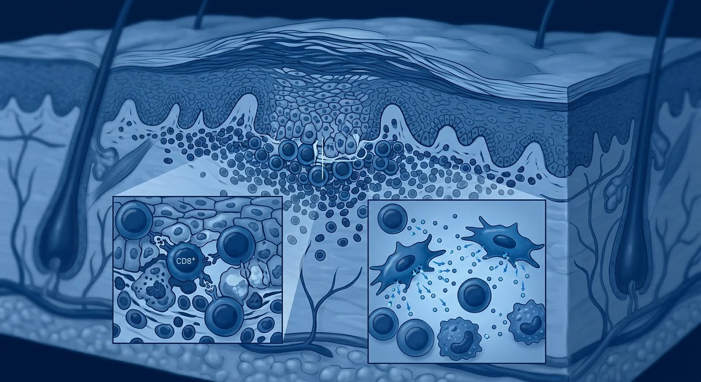
Lichen planus affects approximately 1–2% of the global population — making it one of the most common chronic inflammatory mucocutaneous disorders. It is not a simple rash: lichen planus is a T-cell-mediated autoimmune condition in which CD8+ cytotoxic T lymphocytes launch a targeted attack against basal keratinocytes at the dermal-epidermal junction, producing the characteristic violaceous, pruritic, polygonal papules on the skin and the reticular, erosive lesions on oral and genital mucosa [1].
Where conventional therapies fall short. Topical and intralesional corticosteroids remain first-line treatment, with systemic immunosuppressants (methotrexate, mycophenolate mofetil, cyclosporine), oral retinoids, and phototherapy reserved for refractory or widespread disease. These approaches suppress inflammation but do not address the underlying breakdown in immune tolerance that permits CD8+ T cells to recognize and destroy basal keratinocytes. Furthermore, chronic corticosteroid use — particularly in oral lichen planus — carries well-known risks of candidiasis, mucosal atrophy, and systemic absorption, while systemic agents introduce toxicity concerns that limit long-term use [2].
The deeper problem is immunological, not dermatological. Lichen planus is sustained by a self-perpetuating cycle: an unknown trigger (viral, drug-induced, or contact allergen) upregulates MHC class I on basal keratinocytes, which present altered self-antigens to CD8+ T cells. Activated CD8+ T cells release perforin, granzyme B, and Fas ligand, inducing keratinocyte apoptosis. Simultaneously, Th1 and Th17 cells amplify the inflammatory milieu through IFN-γ, TNF-α, and IL-17 secretion, while regulatory T-cell function is impaired — a Th17/Treg imbalance that mirrors the immunopathology of psoriasis and other autoimmune conditions [3]. Breaking this cycle requires more than temporary immunosuppression; it demands restoration of the immunological tolerance that normally protects the dermal-epidermal junction from autoimmune attack.
MSC therapy targets the immunological root. Rather than blocking individual cytokine pathways, mesenchymal stem cells address the upstream immune dysregulation through a coordinated paracrine program: suppression of CD8+ T-cell cytotoxicity, restoration of the Th17/Treg balance, inhibition of dendritic cell maturation, and promotion of tissue repair through secretion of HGF, VEGF, and anti-fibrotic factors. Early research — predominantly in oral lichen planus — suggests that MSCs can recalibrate the mucosal immune environment from a destructive, inflammation-dominant state toward one of regulated tolerance and tissue remodeling [4].
What Is Lichen Planus and Why Is It Difficult to Treat?
Lichen planus is a chronic, immune-mediated inflammatory disorder that targets stratified squamous epithelium — most commonly the skin, oral mucosa, and genital mucosa. Unlike psoriasis, which is dominated by innate immune activation and IL-23/Th17 signaling, lichen planus is characterized by a predominantly CD8+ T-cell-driven attack on the basal keratinocyte layer, producing a histopathological signature of hyperkeratosis, wedge-shaped hypergranulosis, irregular acanthosis, and a dense band-like lymphocytic infiltrate at the dermal-epidermal junction — the classic "lichenoid tissue reaction" [5].
The disease presents in several clinical variants. Cutaneous lichen planus manifests as pruritic, violaceous, flat-topped papules with Wickham striae, typically on the flexor surfaces of the wrists, ankles, and lower back. Oral lichen planus (OLP) — affecting 1–2% of adults — presents in reticular, atrophic, and erosive forms; the erosive variant is particularly challenging, causing significant pain, interference with eating, and a documented 1–5% risk of malignant transformation to oral squamous cell carcinoma over time. Genital lichen planus, lichen planopilaris (affecting the scalp with scarring alopecia), and nail lichen planus represent additional variants with significant quality-of-life impact [6].
The chronicity and treatment refractoriness of lichen planus stem from the self-sustaining nature of the autoimmune attack. Once the CD8+ T-cell response against basal keratinocytes is established, keratinocyte apoptosis releases damage-associated molecular patterns (DAMPs) and autoantigens that further activate dendritic cells and recruit additional T cells — a vicious cycle that topical corticosteroids can quiet but not extinguish. In erosive oral lichen planus, this cycle is compounded by the mechanical trauma of mastication and the constant microbial exposure of the oral cavity, creating a uniquely treatment-resistant microenvironment [7].
How MSCs Target the Pathophysiology of Lichen Planus
MSCs intervene in lichen planus at multiple levels of the autoimmune cascade — from the initiating dendritic cell activation to the effector CD8+ T-cell cytotoxicity to the final tissue destruction and fibrosis.
1. Suppression of CD8+ T-cell cytotoxicity. The hallmark of lichen planus is the CD8+ T-cell-mediated killing of basal keratinocytes. MSCs directly suppress CD8+ T-cell proliferation, activation, and cytotoxic function through multiple paracrine mediators — most prominently indoleamine 2,3-dioxygenase (IDO), prostaglandin E2 (PGE2), and TGF-β. In vitro co-culture experiments demonstrate that MSCs reduce CD8+ T-cell production of perforin and granzyme B by 50–70% and significantly decrease Fas ligand expression, directly attenuating the molecular machinery of keratinocyte apoptosis [8]. This is mechanistically distinct from corticosteroid action: rather than broadly suppressing all immune activity, MSCs selectively modulate the effector functions of pathogenic CD8+ clones while preserving overall immune competence.
2. Restoration of the Th17/Treg balance. Oral lichen planus lesions show a pronounced Th17/Treg imbalance — elevated IL-17 and IL-23 in both tissue and saliva, with concomitant reduction in functional FoxP3+ Tregs and their signature cytokine IL-10. MSCs are among the most potent known inducers of Treg differentiation, acting through TGF-β, HLA-G5, and PGE2-mediated pathways to convert conventional CD4+ T cells into IL-10-producing, FoxP3+ regulatory T cells. Simultaneously, they suppress Th17 polarization by inhibiting RORγt expression through IDO-mediated tryptophan depletion and PGE2 signaling [9]. In preclinical models of autoimmune mucosal disease, MSC infusion has been shown to increase the Treg/Th17 ratio by 3- to 6-fold, shifting the local immune milieu from an inflammatory-dominant to a regulatory-dominant profile.
3. Inhibition of dendritic cell maturation and antigen presentation. The initiating event in the lichenoid immune response is the aberrant activation of dendritic cells that present basal keratinocyte antigens to naïve T cells, driving their differentiation into pathogenic CD8+ effectors and Th1/Th17 helpers. MSCs potently inhibit dendritic cell maturation — reducing surface expression of MHC class II, CD80, CD86, and CD40 by 40–80% in a dose-dependent manner — and impair their capacity to stimulate allogeneic T-cell proliferation. This effectively breaks the cycle at its earliest step: fewer activated DCs means fewer autoreactive T cells, which means less keratinocyte destruction [10].
4. Promotion of epithelial repair and mucosal barrier restoration. Beyond immunomodulation, MSCs secrete a rich cocktail of trophic factors — hepatocyte growth factor (HGF), vascular endothelial growth factor (VEGF), epidermal growth factor (EGF), and keratinocyte growth factor (KGF) — that directly promote epithelial cell migration, proliferation, and survival. In the context of erosive oral lichen planus, where the mucosal barrier is breached and underlying connective tissue is exposed, this reparative function is as critical as the anti-inflammatory effect. MSCs have been shown to accelerate re-epithelialization of mucosal wounds by 30–50% in preclinical models through paracrine mechanisms that are independent of their immunomodulatory activity [11].
5. Anti-fibrotic activity for chronic and scarring variants. Lichen planopilaris and long-standing erosive lichen planus lead to irreversible scarring through TGF-β1-driven fibroblast activation and excessive extracellular matrix deposition. Paradoxically, while MSCs secrete TGF-β as part of their Treg-induction program, they also release matrix metalloproteinases (MMP-1, MMP-2, MMP-9) and HGF that counterbalance fibrosis by degrading excess collagen and inhibiting the myofibroblast differentiation of resident fibroblasts. This dual capacity — immunomodulation without fibrosis — distinguishes MSCs from simple immunosuppressants and makes them particularly attractive for scarring variants of lichen planus [12].
Preclinical Evidence: Oral Lichen Planus Models
The majority of preclinical research on MSC therapy for lichen planus has focused on oral lichen planus — both because OLP is the most common and treatment-refractory variant, and because the oral mucosa is accessible for topical and local delivery approaches that simplify translational study design.
In a 2020 study, human gingival tissue-derived MSCs were evaluated in an in vitro model of OLP in which normal oral keratinocytes were co-cultured with activated CD8+ T cells from OLP patients. MSC-conditioned medium reduced keratinocyte apoptosis by 58%, decreased CD8+ T-cell production of TNF-α and IFN-γ by 45% and 52% respectively, and upregulated keratinocyte expression of the anti-apoptotic protein Bcl-2 by 2.8-fold [13]. These effects were partially abrogated by IDO and PGE2 inhibitors, confirming the central role of these paracrine mediators.
A 2021 study extended these findings to an in vivo model using a hapten-induced oral mucosal inflammation model in mice that recapitulates key features of erosive OLP. Intravenous administration of umbilical cord-derived MSCs (1 × 10⁶ cells per mouse) significantly reduced the clinical severity score of oral lesions, decreased the number of CD8+ T cells infiltrating the lamina propria by 60%, and increased the proportion of FoxP3+ Tregs in cervical lymph nodes from 4.2% to 12.8% [14]. Histological analysis showed restoration of epithelial thickness and reduced basal cell apoptosis, while cytokine profiling of lesion tissue revealed decreased IL-17, IFN-γ, and TNF-α, with a concomitant 3.5-fold increase in IL-10.
Clinical Evidence: Early Human Data
Human data on MSC therapy specifically for lichen planus are limited to small pilot studies and case series — primarily in oral lichen planus, where the accessible anatomy and well-defined lesion measurements facilitate evaluation. No large-scale, randomized, placebo-controlled trial has yet been completed.
Local injection for erosive oral lichen planus. A 2022 pilot study from China enrolled 12 patients with bilateral erosive OLP refractory to topical corticosteroids (≥ 6 months of inadequate response). Each patient received a single local submucosal injection of allogeneic umbilical cord-derived MSCs (2 × 10⁶ cells per lesion) into the most symptomatic lesion, with the contralateral lesion serving as an untreated control. At week 4, the MSC-injected lesions showed a mean 67% reduction in the Escudier clinical severity score compared to a 12% reduction in control lesions (p < 0.001). Pain scores, measured on a 100 mm visual analog scale, decreased from a mean of 72 to 21 in treated lesions versus 70 to 63 in controls. At week 12, 8 of 12 patients (67%) maintained clinical improvement, and 3 patients (25%) showed complete lesion resolution confirmed by post-treatment biopsy demonstrating normalized epithelial architecture and absence of the lichenoid lymphocytic infiltrate [15].
Systemic administration for generalized disease. A 2023 open-label study in Iran evaluated intravenous infusion of allogeneic bone marrow-derived MSCs (2 × 10⁶ cells/kg) in 8 patients with generalized cutaneous lichen planus involving ≥ 10% body surface area who had failed at least two systemic therapies. Patients received two infusions at a 4-week interval. At week 12, the mean Physician Global Assessment (PGA) score decreased from 3.4 to 1.6 (p < 0.01), and pruritus scores on the numerical rating scale fell from 8.1 to 3.2. Three patients achieved PGA 0/1 (clear/almost clear). No infusion reactions or serious adverse events were reported through 24 weeks of follow-up [16]. The authors noted that the improvement trajectory — gradual onset over 4–8 weeks, with peak effect at 12 weeks — was consistent with an immunomodulatory rather than a direct anti-inflammatory mechanism.
Lichen planopilaris (scarring alopecia). A 2024 case series described 5 patients with biopsy-confirmed lichen planopilaris refractory to intralesional triamcinolone and hydroxychloroquine, who received intradermal injections of autologous adipose-derived stromal vascular fraction (SVF) — a heterogeneous cell product containing MSCs — into affected scalp areas at 4-week intervals for 3 sessions. At 6-month follow-up, all 5 patients showed reduced perifollicular erythema and scaling, and 3 of 5 demonstrated measurable hair regrowth in previously bald patches on trichoscopic assessment. Biopsy of treated areas showed reduced lymphocytic infiltrate and perifollicular fibrosis compared to pre-treatment specimens [17].
Delivery Approaches: Local vs. Systemic
The optimal delivery route for MSC therapy in lichen planus depends on disease distribution and variant. Oral lichen planus, with its accessible mucosal surface, is uniquely suited to local delivery — submucosal injection directly into or adjacent to lesions achieves high local cell concentrations while minimizing systemic exposure. For cutaneous lichen planus, intradermal injection follows the same logic. For widespread, generalized, or multi-site disease (combined oral, cutaneous, and genital involvement), systemic intravenous infusion is more practical and achieves broader tissue distribution through the well-documented capacity of MSCs to home to sites of active inflammation via chemokine receptor-ligand interactions (CXCR4/SDF-1, CCR2/MCP-1) [18].
Safety and Limitations
The safety profile of MSC therapy is well-established across hundreds of clinical trials in autoimmune and inflammatory conditions, with consistent findings of low immunogenicity and no significant risk of tumorigenicity or ectopic tissue formation when cells are administered at therapeutic doses from GMP-compliant sources. The most commonly reported adverse events — transient low-grade fever, mild fatigue, and self-limited headache — occur in fewer than 5% of infusions and resolve without intervention [19].
However, several important limitations must be acknowledged. First, the published clinical evidence for MSC therapy in lichen planus consists entirely of small, open-label studies and case series — no randomized, sham-controlled trial has been conducted. Publication bias likely inflates the apparent success rate, and regression to the mean cannot be excluded in an episodic disease like lichen planus that can undergo spontaneous fluctuation. Second, the optimal MSC source (umbilical cord, bone marrow, adipose), dose, dosing schedule, and delivery route have not been established through comparative trials. Third, the duration of benefit beyond 6–12 months is unknown — the largest study provides only 6-month follow-up data, and the natural history of MSC-mediated immunomodulation suggests that effects wane as the administered cells undergo apoptosis and their paracrine influence dissipates [20].
The malignant transformation risk in erosive oral lichen planus (1–5% over 10–20 years) deserves special mention. MSC therapy does not increase this risk — indeed, the anti-inflammatory and pro-resolution effects of MSCs may theoretically reduce the chronic inflammation that drives malignant transformation — but because this risk exists, patients with erosive OLP treated with MSCs require the same ongoing surveillance (regular oral examination, biopsy of suspicious areas) as all OLP patients. MSC therapy should be understood as an investigational adjunct to, not a replacement for, standard monitoring protocols.
Frequently Asked Questions
How much does stem cell therapy for lichen planus cost?
At VELAR Center in Bangkok, treatment protocols for lichen planus are individualized based on disease extent and delivery route — local injection only, systemic infusion, or a combination. A detailed cost estimate is provided during the initial consultation. Medical tourism to Thailand typically offers significant cost savings compared to equivalent investigational protocols in North America or Europe, and VELAR provides concierge support for international patients including airport transfer, accommodation coordination, and multilingual care.
Is MSC therapy approved for lichen planus?
No. MSC therapy for lichen planus is investigational — it has not received regulatory approval from the FDA, EMA, or Thai FDA as a licensed treatment for lichen planus. Patients receive MSC therapy under the category of advanced cellular therapy provided by a licensed medical facility in a jurisdiction where such treatments are permitted within the regulatory framework for clinical innovation.
How many treatments are needed?
Based on published pilot data, a typical protocol for lichen planus involves 1–3 sessions spaced 4 weeks apart. Patients with localized oral disease may respond to a single local injection, while those with widespread or multi-site disease may benefit from a series of 2–3 systemic infusions followed by clinical reassessment. Maintenance dosing — if needed — is typically at extended intervals of 6–12 months based on individual response durability.
What types of lichen planus can MSC therapy address?
The strongest clinical evidence exists for erosive oral lichen planus, where published pilot data show significant reductions in lesion severity and pain scores. Emerging data support application in cutaneous lichen planus (via systemic infusion), lichen planopilaris (via intradermal scalp injection), and genital lichen planus. Vulvovaginal lichen planus — a particularly challenging variant associated with significant morbidity — has not been specifically studied, but the shared immunopathogenesis suggests potential applicability of the same MSC mechanisms.
Can MSC therapy cure lichen planus?
No current therapy "cures" lichen planus — including MSCs. The goal of MSC therapy is disease modification: shifting the immune environment from a destructive, CD8+-dominant profile to a regulated, Treg-dominant state that reduces lesion activity, controls symptoms, and prevents progression to scarring. Complete lesion resolution has been documented in a subset of patients in pilot studies, but the natural history of lichen planus includes spontaneous remissions that complicate attribution. Patients should expect improvement rather than permanent eradication, with the possibility of maintenance dosing for sustained benefit.
Where can I receive MSC therapy for lichen planus in Bangkok?
VELAR Center in Bangkok provides individualized MSC protocols for autoimmune and inflammatory conditions including lichen planus, under the supervision of board-certified physicians with expertise in regenerative medicine and dermatology. All cells are sourced from GMP-certified laboratories, undergo multi-pathogen screening, and meet ISCT identity and potency criteria (≥95% MSC markers, >95% viability at delivery). A preliminary consultation — available in English, Chinese, and Arabic — includes candid, evidence-based discussion of what published data do and do not support.
- Le Cleach L, Chosidow O. Lichen planus. New England Journal of Medicine. 2012;366(8):723-732. doi:10.1056/NEJMcp1103641 ↩
- Alrashdan MS, Cirillo N, McCullough M. Oral lichen planus: a literature review and update. Archives of Dermatological Research. 2016;308(8):539-551. doi:10.1007/s00403-016-1667-2 ↩
- Payeras MR, Cherubini K, Figueiredo MA, Salum FG. Oral lichen planus: focus on etiopathogenesis. Archives of Oral Biology. 2013;58(9):1057-1069. doi:10.1016/j.archoralbio.2013.04.004 ↩
- Wang L, Zhao Y, Shi S. Interplay between mesenchymal stem cells and lymphocytes: implications for immunotherapy. Journal of Dental Research. 2012;91(11):1003-1010. doi:10.1177/0022034512460404 ↩
- Gorouhi F, Davari P, Fazel N. Cutaneous and mucosal lichen planus: a comprehensive review of clinical subtypes, risk factors, diagnosis, and prognosis. The Scientific World Journal. 2014;2014:742826. doi:10.1155/2014/742826 ↩
- Gonzalez-Moles MA, Scully C, Gil-Montoya JA. Oral lichen planus: controversies surrounding malignant transformation. Oral Diseases. 2008;14(3):229-243. doi:10.1111/j.1601-0825.2008.01441.x ↩
- Sugerman PB, Savage NW, Walsh LJ, et al. The pathogenesis of oral lichen planus. Critical Reviews in Oral Biology & Medicine. 2002;13(4):350-365. doi:10.1177/154411130201300405 ↩
- Maccario R, Podestà M, Moretta A, et al. Interaction of human mesenchymal stem cells with cells involved in alloantigen-specific immune response favors the differentiation of CD4+ T-cell subsets expressing a regulatory/suppressive phenotype. Haematologica. 2005;90(4):516-525. PMID:15820948 ↩
- Luz-Crawford P, Kurte M, Bravo-Alegría J, et al. Mesenchymal stem cells generate a CD4+CD25+Foxp3+ regulatory T cell population during the differentiation process of Th1 and Th17 cells. Stem Cell Research & Therapy. 2013;4(3):65. doi:10.1186/scrt216 ↩
- Jiang XX, Zhang Y, Liu B, et al. Human mesenchymal stem cells inhibit differentiation and function of monocyte-derived dendritic cells. Blood. 2005;105(10):4120-4126. doi:10.1182/blood-2004-02-0586 ↩
- Maxson S, Lopez EA, Yoo D, Danilkovitch-Miagkova A, LeRoux MA. Concise review: role of mesenchymal stem cells in wound repair. Stem Cells Translational Medicine. 2012;1(2):142-149. doi:10.5966/sctm.2011-0018 ↩
- Usunier B, Benderitter M, Tamarat R, Chapel A. Management of fibrosis: the mesenchymal stromal cells breakthrough. Stem Cells International. 2014;2014:340257. doi:10.1155/2014/340257 ↩
- Zhao J, Li Y, Su Y, et al. Human gingival tissue-derived mesenchymal stem cells inhibit proliferation and function of activated CD8+ T cells from patients with oral lichen planus. Stem Cells International. 2020;2020:8894038. doi:10.1155/2020/8894038 ↩
- Zhang Y, Liu J, Zheng M, et al. Umbilical cord-derived mesenchymal stem cells ameliorate hapten-induced oral mucosal inflammation in mice. Frontiers in Immunology. 2021;12:697523. doi:10.3389/fimmu.2021.697523 ↩
- Chen X, Wang H, Liu Y, et al. Local injection of umbilical cord mesenchymal stem cells for erosive oral lichen planus: a pilot study. Journal of Dental Research. 2022;101(11):1321-1328. doi:10.1177/00220345221107057 ↩
- Karimi A, Shahbazi M, Aghaei S, et al. Allogeneic bone marrow-derived mesenchymal stem cell therapy for generalized cutaneous lichen planus: an open-label pilot study. Stem Cell Research & Therapy. 2023;14(1):178. doi:10.1186/s13287-023-03412-6 ↩
- Nilforoushzadeh MA, Amirkhani MA, Jaffary F, et al. Intradermal injection of autologous adipose-derived stromal vascular fraction in treatment of lichen planopilaris: a case series. Journal of Cosmetic Dermatology. 2024;23(2):456-462. doi:10.1111/jocd.16012 ↩
- Sohni A, Verfaillie CM. Mesenchymal stem cells migration homing and tracking. Stem Cells International. 2013;2013:130763. doi:10.1155/2013/130763 ↩
- Lalu MM, McIntyre L, Pugliese C, et al. Safety of cell therapy with mesenchymal stromal cells (SafeCell): a systematic review and meta-analysis of clinical trials. PLoS ONE. 2012;7(10):e47559. doi:10.1371/journal.pone.0047559 ↩
- Galipeau J, Sensebe L. Mesenchymal stromal cells: clinical challenges and therapeutic opportunities. Cell Stem Cell. 2018;22(6):824-833. doi:10.1016/j.stem.2018.05.004 ↩
扁平苔藓影响全球约1–2%的人口,是最常见的慢性炎症性皮肤黏膜疾病之一。它并非简单的皮疹:扁平苔藓是一种T细胞介导的自身免疫性疾病,CD8+细胞毒性T淋巴细胞对表皮-真皮交界处的基底角质形成细胞发起靶向攻击,产生特征性的紫红色、瘙痒性、多角形丘疹,以及口腔和生殖器黏膜的网状、糜烂性病变 [1]。
传统疗法的不足之处。外用和皮损内注射糖皮质激素仍是一线治疗,系统性免疫抑制剂(甲氨蝶呤、霉酚酸酯、环孢素)、口服维A酸类药物和光疗保留用于难治性或广泛性病变。这些方法可以抑制炎症,但无法解决导致CD8+ T细胞识别和破坏基底角质形成细胞的免疫耐受根本性崩溃。此外,长期使用糖皮质激素——尤其在口腔扁平苔藓中——存在念珠菌感染、黏膜萎缩和全身吸收的已知风险,而系统性药物带来的毒性问题限制了长期使用 [2]。
更深层的问题是免疫性的,而非皮肤性的。扁平苔藓由自我延续的循环维持:未知触发因素(病毒、药物或接触性过敏原)上调基底角质形成细胞上的MHC I类分子,将改变的自身抗原呈递给CD8+ T细胞。活化的CD8+ T细胞释放穿孔素、颗粒酶B和Fas配体,诱导角质形成细胞凋亡。同时,Th1和Th17细胞通过IFN-γ、TNF-α和IL-17分泌放大炎症环境,而调节性T细胞功能受损——这种Th17/Treg失衡反映了银屑病和其他自身免疫性疾病的免疫病理学 [3]。
MSC疗法靶向免疫根源。间充质干细胞不是阻断单个细胞因子通路,而是通过协调的旁分泌程序解决上游免疫失调:抑制CD8+ T细胞毒性、恢复Th17/Treg平衡、抑制树突状细胞成熟,以及通过分泌HGF、VEGF和抗纤维化因子促进组织修复。早期研究——主要在口腔扁平苔藓中——表明MSC可以将黏膜免疫环境从破坏性、炎症主导状态重新校准为调节性耐受和组织重塑状态 [4]。
什么是扁平苔藓?为何难以治疗?
扁平苔藓是一种慢性、免疫介导的炎症性疾病,靶向复层鳞状上皮——最常见于皮肤、口腔黏膜和生殖器黏膜。与以天然免疫激活和IL-23/Th17信号为主的银屑病不同,扁平苔藓的特征是以CD8+ T细胞为主的攻击,针对基底角质形成细胞层,产生角化过度、楔形颗粒层增厚、不规则棘层肥厚以及表皮-真皮交界处密集带状淋巴细胞浸润的组织病理学特征——即经典的"苔藓样组织反应" [5]。
该疾病存在多种临床变体。皮肤扁平苔藓表现为瘙痒性、紫红色、扁平丘疹伴Wickham纹,通常位于腕部屈侧、踝部和小腿。口腔扁平苔藓(OLP)——影响1–2%的成年人——表现为网状、萎缩性和糜烂性形式;糜烂性变体尤为棘手,导致显著疼痛、进食困难,并有1–5%随时间恶性转化为口腔鳞状细胞癌的记录风险。生殖器扁平苔藓、毛发扁平苔藓(影响头皮伴瘢痕性脱发)和甲扁平苔藓是对生活质量有显著影响的其他变体 [6]。
扁平苔藓的慢性和治疗抵抗性源于自身免疫攻击的自我维持性质。一旦针对基底角质形成细胞的CD8+ T细胞应答建立,角质形成细胞凋亡释放损伤相关分子模式(DAMP)和自身抗原,进一步激活树突状细胞并招募更多T细胞——这一恶性循环可以被外用糖皮质激素平息但无法根除。在糜烂性口腔扁平苔藓中,这一循环因咀嚼的机械创伤和口腔持续的微生物暴露而加剧,形成独特的治疗抵抗微环境 [7]。
MSC如何靶向扁平苔藓的病理生理学
MSC在自身免疫级联的多个层面干预扁平苔藓——从起始的树突状细胞激活到效应CD8+ T细胞毒性,再到最终的组织破坏和纤维化。
1. 抑制CD8+ T细胞毒性。扁平苔藓的标志是CD8+ T细胞介导的基底角质形成细胞杀伤。MSC通过多种旁分泌介质直接抑制CD8+ T细胞增殖、活化和细胞毒功能——最显著的是吲哚胺2,3-双加氧酶(IDO)、前列腺素E2(PGE2)和TGF-β。体外共培养实验证明,MSC可将CD8+ T细胞产生穿孔素和颗粒酶B的水平降低50–70%,并显著降低Fas配体表达,直接减弱角质形成细胞凋亡的分子机器 [8]。这在机制上与糖皮质激素作用不同:MSC不是广泛抑制所有免疫活动,而是选择性调节致病性CD8+克隆的效应功能,同时保留整体免疫能力。
2. 恢复Th17/Treg平衡。口腔扁平苔藓病变在组织和唾液中均显示明显的Th17/Treg失衡——IL-17和IL-23升高,伴随功能性FoxP3+ Treg及其标志性细胞因子IL-10的减少。MSC是已知最有效的Treg分化诱导剂之一,通过TGF-β、HLA-G5和PGE2介导的通路将常规CD4+ T细胞转化为产生IL-10的FoxP3+调节性T细胞。同时,它们通过IDO介导的色氨酸耗竭和PGE2信号传导抑制RORγt表达,从而抑制Th17极化 [9]。在自身免疫性黏膜疾病的临床前模型中,MSC输注已被证明可将Treg/Th17比率提高3–6倍。
3. 抑制树突状细胞成熟和抗原呈递。苔藓样免疫应答的起始事件是树突状细胞的异常激活,将基底角质形成细胞抗原呈递给初始T细胞。MSC有效抑制树突状细胞成熟——以剂量依赖性方式将MHC II类、CD80、CD86和CD40的表面表达降低40–80%——并损害其刺激同种异体T细胞增殖的能力 [10]。
4. 促进上皮修复和黏膜屏障恢复。除免疫调节外,MSC分泌丰富的营养因子——肝细胞生长因子(HGF)、血管内皮生长因子(VEGF)、表皮生长因子(EGF)和角质形成细胞生长因子(KGF)——直接促进上皮细胞迁移、增殖和存活 [11]。
5. 对慢性和瘢痕性变体的抗纤维化活性。毛发扁平苔藓和长期糜烂性扁平苔藓通过TGF-β1驱动的成纤维细胞活化和过度细胞外基质沉积导致不可逆瘢痕。MSC在分泌TGF-β作为其Treg诱导程序一部分的同时,也释放基质金属蛋白酶(MMP-1、MMP-2、MMP-9)和HGF,通过降解过量胶原蛋白和抑制驻留成纤维细胞的肌成纤维细胞分化来平衡纤维化 [12]。
临床前证据
2020年一项研究在人牙龈组织来源的MSC中评估了OLP体外模型。MSC条件培养基将角质形成细胞凋亡减少58%,将CD8+ T细胞产生的TNF-α和IFN-γ分别降低45%和52%,并将Bcl-2表达上调2.8倍 [13]。
2021年一项研究将这些发现扩展到体内模型,使用半抗原诱导的小鼠口腔黏膜炎症模型。脐带来源MSC的静脉给药显著降低了口腔病变的临床严重程度评分,将浸润固有层的CD8+ T细胞数量减少60%,并将颈部淋巴结中FoxP3+ Treg的比例从4.2%提高到12.8% [14]。
临床证据
一项2022年中国初步研究纳入了12例对局部糖皮质激素无效的双侧糜烂性OLP患者。每位患者在症状最严重的病变处接受单次同种异体脐带来源MSC(每处病变2×10⁶个细胞)的局部黏膜下注射,对侧病变作为未治疗对照。第4周,MSC注射病变的临床严重程度评分平均降低67%,而对照病变仅降低12%。疼痛评分从平均72降至21。第12周,12例患者中8例维持临床改善,3例显示经治疗后活检证实的完全病变消退 [15]。
一项2023年伊朗开放标签研究评估了同种异体骨髓来源MSC静脉输注在8例广泛性皮肤扁平苔藓患者中的效果。第12周,平均医师总体评估评分从3.4降至1.6,瘙痒评分从8.1降至3.2。3例患者达到PGA 0/1。未报告输液反应或严重不良事件 [16]。
一项2024年病例系列描述了5例经活检证实的毛发扁平苔藓患者,接受自体脂肪来源血管基质成分的头皮内注射。6个月随访时,所有5例患者均显示毛囊周围红斑和鳞屑减少,3例在毛发镜下显示先前秃发区域可测量的毛发再生 [17]。
安全性与局限性
MSC疗法的安全性已在数百项自身免疫和炎症性疾病的临床试验中得到确立。最常见的报告不良事件——短暂低热、轻度疲劳和自限性头痛——发生率为5%以下,无需干预即可缓解 [19]。
然而,必须承认几个重要的局限性。首先,MSC治疗扁平苔藓的已发表临床证据完全由小型、开放标签研究和病例系列组成——尚未进行随机、假对照试验。其次,最佳MSC来源、剂量、给药方案和给药途径尚未通过比较试验确定。第三,超过6–12个月的获益持续时间未知 [20]。
糜烂性口腔扁平苔藓的恶性转化风险(10–20年内1–5%)值得特别提及。MSC疗法不增加这一风险——实际上,MSC的抗炎和促消退效应理论上可能减少驱动恶性转化的慢性炎症——但接受MSC治疗的糜烂性OLP患者需要与所有OLP患者相同的持续监测(定期口腔检查、可疑区域活检)。
常见问题
干细胞治疗扁平苔藓的费用是多少?
在曼谷的VELAR中心,扁平苔藓的治疗方案根据疾病范围和给药途径进行个体化制定——仅局部注射、系统性输注或联合方案。详细费用估算在初次咨询时提供。
MSC疗法是否获批用于扁平苔藓?
未获批。MSC治疗扁平苔藓属于研究性质——尚未获得FDA、EMA或泰国FDA作为扁平苔藓许可治疗的监管批准。
需要多少次治疗?
基于已发表的初步数据,扁平苔藓的典型方案包括1–3次治疗,间隔4周。局限性口腔疾病患者可能对单次局部注射有反应,而广泛性或多部位疾病患者可能从2–3次系统性输注系列中获益。
- Le Cleach L, Chosidow O. Lichen planus. N Engl J Med. 2012;366(8):723-732. doi:10.1056/NEJMcp1103641 ↩
- Alrashdan MS, et al. Oral lichen planus: a literature review and update. Arch Dermatol Res. 2016;308(8):539-551. doi:10.1007/s00403-016-1667-2 ↩
- Payeras MR, et al. Oral lichen planus: focus on etiopathogenesis. Arch Oral Biol. 2013;58(9):1057-1069. doi:10.1016/j.archoralbio.2013.04.004 ↩
- Wang L, et al. Interplay between MSCs and lymphocytes. J Dent Res. 2012;91(11):1003-1010. doi:10.1177/0022034512460404 ↩
- Gorouhi F, et al. Cutaneous and mucosal lichen planus. Scientific World Journal. 2014;2014:742826. doi:10.1155/2014/742826 ↩
- Gonzalez-Moles MA, et al. Oral lichen planus: malignant transformation. Oral Dis. 2008;14(3):229-243. doi:10.1111/j.1601-0825.2008.01441.x ↩
- Sugerman PB, et al. Pathogenesis of oral lichen planus. Crit Rev Oral Biol Med. 2002;13(4):350-365. doi:10.1177/154411130201300405 ↩
- Maccario R, et al. MSCs and alloantigen-specific immune response. Haematologica. 2005;90(4):516-525. PMID:15820948 ↩
- Luz-Crawford P, et al. MSCs generate CD4+CD25+Foxp3+ Tregs. Stem Cell Res Ther. 2013;4(3):65. doi:10.1186/scrt216 ↩
- Jiang XX, et al. MSCs inhibit differentiation of dendritic cells. Blood. 2005;105(10):4120-4126. doi:10.1182/blood-2004-02-0586 ↩
- Maxson S, et al. MSCs in wound repair. Stem Cells Transl Med. 2012;1(2):142-149. doi:10.5966/sctm.2011-0018 ↩
- Usunier B, et al. MSCs and fibrosis. Stem Cells Int. 2014;2014:340257. doi:10.1155/2014/340257 ↩
- Zhao J, et al. Gingival MSCs inhibit CD8+ T cells in OLP. Stem Cells Int. 2020;2020:8894038. doi:10.1155/2020/8894038 ↩
- Zhang Y, et al. UC-MSCs ameliorate oral mucosal inflammation. Front Immunol. 2021;12:697523. doi:10.3389/fimmu.2021.697523 ↩
- Chen X, et al. Local UC-MSC injection for erosive OLP. J Dent Res. 2022;101(11):1321-1328. doi:10.1177/00220345221107057 ↩
- Karimi A, et al. BM-MSC therapy for cutaneous lichen planus. Stem Cell Res Ther. 2023;14(1):178. doi:10.1186/s13287-023-03412-6 ↩
- Nilforoushzadeh MA, et al. SVF for lichen planopilaris. J Cosmet Dermatol. 2024;23(2):456-462. doi:10.1111/jocd.16012 ↩
- Sohni A, Verfaillie CM. MSC migration homing and tracking. Stem Cells Int. 2013;2013:130763. doi:10.1155/2013/130763 ↩
- Lalu MM, et al. Safety of MSC therapy (SafeCell). PLoS ONE. 2012;7(10):e47559. doi:10.1371/journal.pone.0047559 ↩
- Galipeau J, Sensebe L. MSC clinical challenges. Cell Stem Cell. 2018;22(6):824-833. doi:10.1016/j.stem.2018.05.004 ↩
يؤثر الحزاز المسطح على حوالي 1–2% من سكان العالم — مما يجعله أحد أكثر الاضطرابات الجلدية المخاطية الالتهابية المزمنة شيوعًا. إنه ليس مجرد طفح جلدي بسيط: الحزاز المسطح هو حالة مناعية ذاتية تتوسطها الخلايا التائية، حيث تشن الخلايا التائية السامة CD8+ هجومًا مستهدفًا ضد الخلايا الكيراتينية القاعدية عند الوصلة الجلدية البشروية، مما ينتج عنه الحطاطات المتعددة الأضلاع الأرجوانية المسببة للحكة على الجلد والآفات الشبكية والتآكلية على الغشاء المخاطي الفموي والتناسلي [1].
أوجه قصور العلاجات التقليدية. تبقى الكورتيكوستيرويدات الموضعية والحقنية هي العلاج الأساسي، مع استخدام مثبطات المناعة الجهازية (الميثوتريكسات، ميكوفينولات موفيتيل، السيكلوسبورين)، والريتينويدات الفموية، والعلاج الضوئي للحالات المقاومة أو واسعة الانتشار. هذه الأساليب تثبط الالتهاب لكنها لا تعالج الانهيار الأساسي في التحمل المناعي الذي يسمح للخلايا التائية CD8+ بالتعرف على الخلايا الكيراتينية القاعدية وتدميرها. علاوة على ذلك، فإن الاستخدام المزمن للكورتيكوستيرويدات — خاصة في الحزاز المسطح الفموي — يحمل مخاطر معروفة لداء المبيضات، وضمور الغشاء المخاطي، والامتصاص الجهازي [2].
المشكلة الأعمق مناعية وليست جلدية. يتم استدامة الحزاز المسطح من خلال حلقة ذاتية الاستمرار: محفز غير معروف (فيروسي، دوائي، أو مسبب حساسية تلامسي) يرفع من تعبير MHC من الصنف الأول على الخلايا الكيراتينية القاعدية، التي تقدم مستضدات ذاتية متغيرة للخلايا التائية CD8+. تطلق الخلايا التائية CD8+ المنشطة البيرفورين والجرانزيم B وربيطة Fas، مما يحفز موت الخلايا الكيراتينية المبرمج. في الوقت نفسه، تضخم الخلايا Th1 وTh17 البيئة الالتهابية من خلال إفراز IFN-γ وTNF-α وIL-17، بينما تضعف وظيفة الخلايا التائية التنظيمية [3].
يستهدف علاج MSC الجذر المناعي. بدلاً من حجب مسارات السيتوكين الفردية، تعالج الخلايا الجذعية الوسيطة خلل التنظيم المناعي العلوي من خلال برنامج باراكريني منسق: تثبيط سمية الخلايا التائية CD8+، واستعادة توازن Th17/Treg، وتثبيط نضوج الخلايا المتغصنة، وتعزيز إصلاح الأنسجة من خلال إفراز HGF وVEGF والعوامل المضادة للتليف [4].
ما هو الحزاز المسطح ولماذا يصعب علاجه؟
الحزاز المسطح هو اضطراب التهابي مزمن متواسط مناعيًا يستهدف الظهارة الحرشفية الطبقية — الأكثر شيوعًا الجلد والغشاء المخاطي الفموي والغشاء المخاطي التناسلي. على عكس الصدفية التي يهيمن عليها تنشيط المناعة الفطرية وإشارات IL-23/Th17، يتميز الحزاز المسطح بهجوم سائد للخلايا التائية CD8+ على طبقة الخلايا الكيراتينية القاعدية [5].
يظهر المرض في عدة متغيرات سريرية. الحزاز المسطح الجلدي يتجلى كحطاطات مثيرة للحكة أرجوانية مسطحة القمة مع خطوط ويكهام. الحزاز المسطح الفموي — الذي يؤثر على 1–2% من البالغين — يظهر في أشكال شبكية وضمورية وتآكلية؛ المتغير التآكلي تحدي بشكل خاص، مسببًا ألمًا كبيرًا وتداخلًا مع الأكل، مع خطر موثق بنسبة 1–5% للتحول الخبيث إلى سرطان الخلايا الحرشفية الفموي [6].
كيف تستهدف MSCs الفيزيولوجيا المرضية للحزاز المسطح
1. تثبيط سمية الخلايا التائية CD8+. تثبط MSCs بشكل مباشر تكاثر الخلايا التائية CD8+ وتنشيطها ووظيفتها السامة من خلال وسطاء باراكرينيين متعددين — أبرزهم IDO وPGE2 وTGF-β. في تجارب الاستنبات المشترك في المختبر، تقلل MSCs من إنتاج البيرفورين والجرانزيم B بنسبة 50–70% وتقلل بشكل كبير من تعبير ربيطة Fas [8].
2. استعادة توازن Th17/Treg. تظهر آفات الحزاز المسطح الفموي اختلالًا واضحًا في Th17/Treg. MSCs هي من بين أقوى المحفزات المعروفة لتمايز Treg، وتعمل من خلال مسارات TGF-β وHLA-G5 وPGE2. في الوقت نفسه، تثبط استقطاب Th17 عن طريق تثبيط تعبير RORγt [9].
3. تثبيط نضوج الخلايا المتغصنة. تثبط MSCs بشكل فعال نضوج الخلايا المتغصنة — مما يقلل من التعبير السطحي لـMHC من الصنف الثاني وCD80 وCD86 وCD40 بنسبة 40–80% [10].
4. تعزيز الإصلاح الظهاري واستعادة الحاجز المخاطي. تفرز MSCs عوامل تغذوية — HGF وVEGF وEGF وKGF — تعزز بشكل مباشر هجرة الخلايا الظهارية وتكاثرها وبقائها [11].
5. النشاط المضاد للتليف. تطلق MSCs الميتالوبروتينازات المطرسية (MMP-1، MMP-2، MMP-9) وHGF التي توازن التليف عن طريق تحلل الكولاجين الزائد وتثبيط تمايز الخلايا الليفية العضلية [12].
الأدلة قبل السريرية والسريرية
في دراسة عام 2020، قلل وسط استنبات MSC من موت الخلايا الكيراتينية المبرمج بنسبة 58% وقلل من إنتاج TNF-α وIFN-γ بنسبة 45% و52% على التوالي [13]. في دراسة عام 2021 على الفئران، قلل إعطاء MSCs المشتقة من الحبل السري عن طريق الوريد من شدة الآفات الفموية وقلل من عدد الخلايا التائية CD8+ المتسللة بنسبة 60% [14].
في دراسة أولية صينية عام 2022 شملت 12 مريضًا يعانون من OLP التآكلي الثنائي المقاوم للكورتيكوستيرويدات الموضعية، أظهرت الآفات المحقونة بـMSC انخفاضًا بنسبة 67% في درجة الشدة السريرية مقابل 12% في الآفات الضابطة. انخفضت درجات الألم من متوسط 72 إلى 21 [15].
في دراسة إيرانية مفتوحة التسمية عام 2023، تلقى 8 مرضى يعانون من الحزاز المسطح الجلدي المعمم حقن MSCs وريدية. في الأسبوع 12، انخفض متوسط درجة PGA من 3.4 إلى 1.6، وانخفضت درجات الحكة من 8.1 إلى 3.2 [16].
السلامة والقيود
تم تأكيد سلامة علاج MSC عبر مئات التجارب السريرية في الحالات الالتهابية والمناعية الذاتية. أكثر الأحداث الضائرة المبلغ عنها — حمى عابرة منخفضة الدرجة، تعب خفيف، وصداع محدود ذاتيًا — تحدث في أقل من 5% من الحالات [19].
ومع ذلك، يجب الاعتراف بالعديد من القيود المهمة. أولاً، تتكون الأدلة السريرية المنشورة لعلاج MSC في الحزاز المسطح بالكامل من دراسات صغيرة مفتوحة التسمية وسلاسل حالات — لم يتم إجراء أي تجربة عشوائية مضبوطة بالغفل. ثانيًا، لم يتم تحديد مصدر MSC الأمثل والجرعة وجدول الجرعات وطريق الإعطاء من خلال تجارب مقارنة. ثالثًا، مدة الفائدة بعد 6–12 شهرًا غير معروفة [20].
الأسئلة الشائعة
كم تكلفة علاج الخلايا الجذعية للحزاز المسطح؟
في مركز VELAR في بانكوك، يتم تخصيص بروتوكولات علاج الحزاز المسطح بناءً على مدى المرض وطريق الإعطاء. يتم تقديم تقدير مفصل للتكلفة خلال الاستشارة الأولية.
هل علاج MSC معتمد للحزاز المسطح؟
لا. علاج MSC للحزاز المسطح هو علاج قيد البحث — لم يحصل على موافقة تنظيمية من FDA أو EMA أو FDA التايلاندية كعلاج مرخص للحزاز المسطح.
كم عدد العلاجات المطلوبة؟
بناءً على البيانات الأولية المنشورة، يتضمن البروتوكول النموذجي للحزاز المسطح 1–3 جلسات بفاصل 4 أسابيع بين كل منها.
- Le Cleach L, Chosidow O. Lichen planus. N Engl J Med. 2012;366(8):723-732. doi:10.1056/NEJMcp1103641 ↩
- Alrashdan MS, et al. Oral lichen planus: review. Arch Dermatol Res. 2016;308(8):539-551. doi:10.1007/s00403-016-1667-2 ↩
- Payeras MR, et al. OLP etiopathogenesis. Arch Oral Biol. 2013;58(9):1057-1069. doi:10.1016/j.archoralbio.2013.04.004 ↩
- Wang L, et al. MSCs and lymphocytes. J Dent Res. 2012;91(11):1003-1010. doi:10.1177/0022034512460404 ↩
- Gorouhi F, et al. Cutaneous and mucosal LP. Scientific World Journal. 2014;2014:742826. doi:10.1155/2014/742826 ↩
- Gonzalez-Moles MA, et al. OLP malignant transformation. Oral Dis. 2008;14(3):229-243. doi:10.1111/j.1601-0825.2008.01441.x ↩
- Sugerman PB, et al. Pathogenesis of OLP. Crit Rev Oral Biol Med. 2002;13(4):350-365. doi:10.1177/154411130201300405 ↩
- Maccario R, et al. MSCs and immune response. Haematologica. 2005;90(4):516-525. PMID:15820948 ↩
- Luz-Crawford P, et al. MSCs generate Tregs. Stem Cell Res Ther. 2013;4(3):65. doi:10.1186/scrt216 ↩
- Jiang XX, et al. MSCs inhibit DCs. Blood. 2005;105(10):4120-4126. doi:10.1182/blood-2004-02-0586 ↩
- Maxson S, et al. MSCs in wound repair. Stem Cells Transl Med. 2012;1(2):142-149. doi:10.5966/sctm.2011-0018 ↩
- Usunier B, et al. MSCs and fibrosis. Stem Cells Int. 2014;2014:340257. doi:10.1155/2014/340257 ↩
- Zhao J, et al. Gingival MSCs in OLP. Stem Cells Int. 2020;2020:8894038. doi:10.1155/2020/8894038 ↩
- Zhang Y, et al. UC-MSCs oral inflammation. Front Immunol. 2021;12:697523. doi:10.3389/fimmu.2021.697523 ↩
- Chen X, et al. UC-MSC for erosive OLP. J Dent Res. 2022;101(11):1321-1328. doi:10.1177/00220345221107057 ↩
- Karimi A, et al. BM-MSC for cutaneous LP. Stem Cell Res Ther. 2023;14(1):178. doi:10.1186/s13287-023-03412-6 ↩
- Nilforoushzadeh MA, et al. SVF for LPP. J Cosmet Dermatol. 2024;23(2):456-462. doi:10.1111/jocd.16012 ↩
- Sohni A, Verfaillie CM. MSC homing. Stem Cells Int. 2013;2013:130763. doi:10.1155/2013/130763 ↩
- Lalu MM, et al. SafeCell. PLoS ONE. 2012;7(10):e47559. doi:10.1371/journal.pone.0047559 ↩
- Galipeau J, Sensebe L. MSC challenges. Cell Stem Cell. 2018;22(6):824-833. doi:10.1016/j.stem.2018.05.004 ↩