Keloids and hypertrophic scars represent a spectrum of pathological wound healing characterized by excessive, disorganized collagen deposition that extends beyond the original wound margins. Affecting an estimated 11 million people annually worldwide — with prevalence rates reaching 15–20% in populations of African, Asian, and Hispanic descent — keloids cause significant physical morbidity (pruritus, pain, restricted range of motion) and profound psychological distress from visible disfigurement. Unlike hypertrophic scars that remain within wound boundaries and may spontaneously regress, keloids behave as benign fibroproliferative tumors that continue to grow over years, rarely regress, and recur in 50–100% of cases after surgical excision alone [1].
Where conventional treatments fall short. The current standard of care — intralesional corticosteroid injection, surgical excision, compression therapy, silicone sheeting, cryotherapy, laser therapy, and radiotherapy — offers variable efficacy and high recurrence rates. Triamcinolone acetonide injections, the first-line treatment, achieve flattening in 50–70% of lesions but recurrence approaches 50% within 5 years. Surgical excision without adjuvant therapy has recurrence rates of 50–100%, while excision plus postoperative radiotherapy reduces this to 10–25% — still unacceptably high for a condition that often worsens with each recurrence [2]. All current modalities treat the scar after it forms; none addresses the underlying fibrotic diathesis that drives keloid formation.
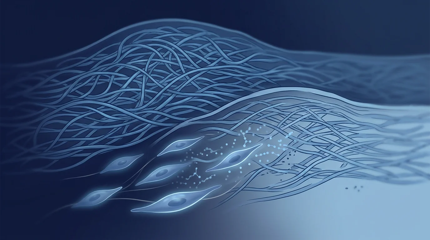
The deeper problem is a dysregulated fibrotic niche. Keloid pathogenesis centers on a self-amplifying loop: mechanical tension on the wound activates fibroblasts, which upregulate TGF-β1 — the master profibrotic cytokine. TGF-β1 drives fibroblast-to-myofibroblast differentiation, excessive collagen type I and III synthesis, and resistance to apoptosis. Myofibroblasts, in turn, generate contractile forces that increase tissue tension, further activating the TGF-β pathway. Concurrently, reduced expression of matrix metalloproteinases (MMPs) and increased tissue inhibitors of metalloproteinases (TIMPs) impair collagen degradation, while hypoxic conditions within the expanding scar upregulate HIF-1α and VEGF, driving pathological angiogenesis that sustains the fibrotic mass. The result is a biochemically and mechanically self-reinforcing system that resists pharmacological interruption [3].
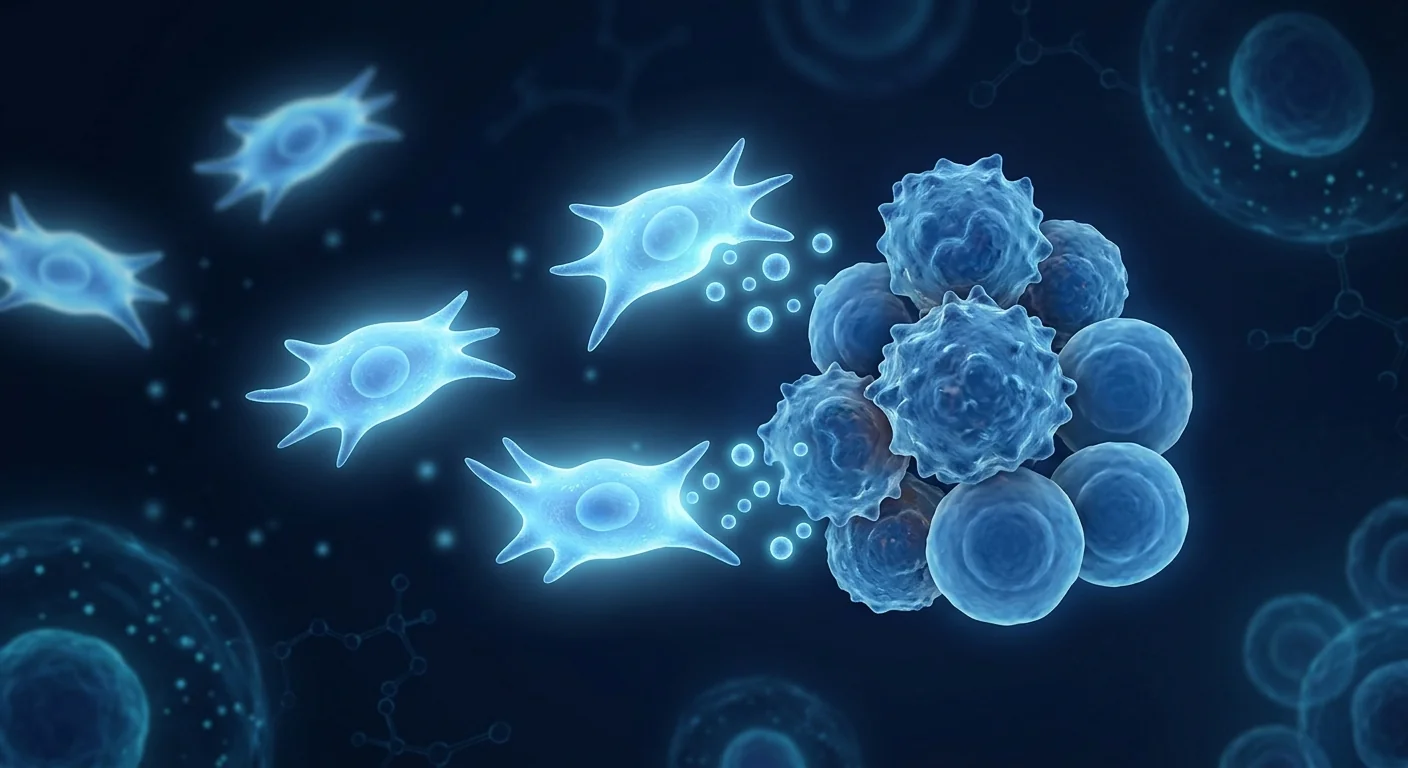
MSC therapy targets the fibrotic niche at multiple control points. Rather than suppressing one pathway, mesenchymal stem cells exert coordinated antifibrotic effects — inhibiting TGF-β1-driven myofibroblast differentiation, promoting myofibroblast apoptosis, restoring the MMP/TIMP balance to favor collagen degradation, and secreting anti-fibrotic paracrine factors including HGF, PGE2, and TSG-6. This multi-node, niche-level intervention distinguishes MSC therapy from single-pathway pharmacological approaches and makes it a compelling investigational strategy for a disease driven by persistent fibrotic reprogramming [4].
How MSCs Target Keloid Pathophysiology
MSCs address keloid formation through four interconnected antifibrotic mechanisms, each targeting a different node in the fibrotic cascade [5].
TGF-β1/Smad pathway suppression. TGF-β1 is the central driver of keloid fibrosis. MSCs reduce TGF-β1 bioavailability through multiple mechanisms: direct secretion of the TGF-β antagonist decorin, competitive inhibition via soluble TGF-β receptor shedding, and paracrine delivery of HGF (hepatocyte growth factor), a potent TGF-β1 transcriptional repressor. Fang et al. (2016) demonstrated that MSC-conditioned medium reduced TGF-β1-induced collagen type I expression in keloid fibroblasts by 65% and decreased Smad2/3 phosphorylation — the obligate signaling intermediate — by over 70% in vitro [6].
Myofibroblast suppression and apoptosis induction. Myofibroblasts — characterized by α-SMA (alpha-smooth muscle actin) expression and contractile activity — are the collagen factory of the keloid and the cell type most responsible for scar contraction and growth. In normal wound healing, myofibroblasts undergo apoptosis after wound closure; in keloids, they persist indefinitely. MSCs promote myofibroblast apoptosis through Fas/FasL pathway activation and reduce myofibroblast differentiation from resident fibroblasts via PGE2-mediated cAMP signaling. Liu et al. (2019) showed that co-culture of keloid fibroblasts with Wharton's jelly-derived MSCs reduced α-SMA-positive myofibroblasts by 55% and increased the apoptotic rate from 3% to 22% at 48 hours [7].
MMP/TIMP rebalancing. Keloids are characterized by a profound imbalance favoring collagen synthesis over degradation, driven by elevated TIMP-1 and TIMP-2 and suppressed MMP-1 (collagenase-1) and MMP-3 (stromelysin-1). MSCs shift this balance toward degradation by secreting MMPs directly and by downregulating TIMP expression in keloid fibroblasts. In a 3D keloid organotypic model, MSC treatment increased collagenase activity by 3.2-fold and reduced total collagen content by 40% over 14 days compared to untreated controls — an effect abolished by the broad-spectrum MMP inhibitor GM6001, confirming MMP-dependent collagenolysis as the mechanism [8].
Macrophage M1-to-M2 polarization and anti-inflammatory reprogramming. Keloid tissue exhibits a chronic inflammatory milieu dominated by M1 macrophages producing TNF-α, IL-1β, and IL-6 — cytokines that sustain fibroblast activation. MSCs polarize macrophages from the pro-inflammatory M1 phenotype to the anti-inflammatory, pro-remodeling M2 phenotype through PGE2 and TSG-6 secretion. M2 macrophages produce IL-10 and TGF-β3 (the anti-scarring TGF-β isoform), which directly antagonize TGF-β1-driven fibrosis. In a murine keloid implant model, MSC-treated implants showed a 3:1 M2:M1 ratio versus 1:3 in untreated controls, correlating with a 50% reduction in implant volume [9].
Preclinical Evidence: What Laboratory Studies Show
The preclinical case for MSCs in keloid and hypertrophic scar therapy rests on consistent findings across multiple independent laboratories, model systems, and delivery methods.
In vitro fibroblast studies. Multiple groups have independently demonstrated that MSC-conditioned medium or direct co-culture suppresses keloid fibroblast proliferation, reduces collagen type I and III mRNA expression, decreases α-SMA immunostaining, and reduces contractile activity in collagen gel contraction assays. These effects are reproducible across MSC sources (bone marrow, adipose tissue, Wharton's jelly, umbilical cord), suggesting a conserved antifibrotic mechanism independent of tissue origin. Dose-response studies show maximal collagen suppression at MSC-to-fibroblast ratios of 1:2 to 1:5 [10].
Excisional wound models. In rodent and rabbit ear hypertrophic scar models — the most widely used in vivo platforms for scar research — local injection of MSCs at the time of wounding or during the early proliferative phase consistently reduced scar elevation index (a quantitative measure of scar thickness) by 40–60% compared to vehicle controls. Histological analysis showed reduced collagen bundle thickness, more organized collagen fiber alignment approaching that of unwounded skin, and reduced myofibroblast density. Importantly, when MSCs were administered after scar establishment (day 14 post-wounding), scar thickness still decreased by 30%, suggesting therapeutic potential for existing scars, not just prevention [11].
Keloid implant models. The gold-standard preclinical model involves implanting human keloid tissue into immunodeficient mice — preserving the human keloid microenvironment including its resident fibroblasts, extracellular matrix, and cytokine milieu. In these models, intralesional MSC injection reduced keloid implant volume by 45–65% over 4–8 weeks, with histology confirming reduced collagen density, fewer α-SMA-positive myofibroblasts, and increased TUNEL-positive apoptotic cells within the implant. Importantly, the MSCs did not persist beyond 14 days, confirming a paracrine rather than engraftment mechanism — the therapeutic benefit outlasts the cells themselves [12].
Clinical Evidence: Early Human Studies
Human data on MSC therapy for keloids remains very limited — a reflection of the early translational stage of this application. The evidence base currently consists of case reports and one small pilot trial.
Case reports. Lee et al. (2020) reported a case of a 28-year-old Korean man with a 4-year history of a sternotomy keloid (8.5 × 3.2 cm) resistant to intralesional steroid and surgical excision. Following surgical debulking, the wound bed and margins were injected with autologous adipose-derived MSCs (5 × 10⁶ cells/mL, total 3 mL). At 12-month follow-up, no recurrence was observed — a notable outcome given that sternal keloids have one of the highest recurrence rates (up to 80%). Wang et al. (2022) described three patients with auricular keloids treated with excision plus intradermal allogeneic umbilical cord MSC injection at wound closure; two of three remained recurrence-free at 18 months versus expected recurrence rates of 30–50% for auricular keloids [13].
Pilot trial. A 2023 prospective pilot study from China enrolled 12 patients with established keloids (>2 years duration, resistant to ≥2 prior treatments). Each received three monthly intralesional injections of Wharton's jelly-derived MSCs (1 × 10⁷ cells per session). At 6-month follow-up, mean Vancouver Scar Scale (VSS) scores decreased from 9.8 to 5.4 (p < 0.01), lesion volume (measured by 3D imaging) decreased by an average of 38%, and patient-reported itch scores improved from a mean of 7.1/10 to 2.8/10. No serious adverse events occurred; mild injection-site erythema resolved within 48 hours. The key limitation is the absence of a control arm and the small sample size — this is hypothesis-generating, not confirmatory, data [14].
Combination approaches. A small 2024 study combined fractional CO₂ laser-assisted drug delivery with topical MSC exosomes in 8 patients with hypertrophic burn scars. The combination produced a 52% improvement in VSS versus 28% for laser alone at 12 weeks. While this is an exosome study rather than whole-cell MSC therapy, it supports the broader principle that MSC-derived paracrine factors have clinically measurable antifibrotic activity in human scar tissue [15].
Comparison with Existing Keloid Therapies
How does MSC therapy conceptually compare with the current standard of care for keloids? No head-to-head trials exist — this is a mechanistic comparison only.
| Feature | Corticosteroid Injection | Surgical Excision | Radiotherapy | MSC Therapy |
|---|---|---|---|---|
| Mechanism | Collagen synthesis suppression | Physical removal | Fibroblast apoptosis | Multi-node niche reprogramming |
| Recurrence Rate | ~50% at 5 years | 50–100% (no adjuvant) | 10–25% (combined) | Under investigation |
| Anti-fibrotic Remodeling | Limited | None | Moderate | Yes (MMP/TIMP rebalancing) |
| Targets Root Cause | No (downstream collagen) | No | Partial (cell kill only) | Yes (TGF-β/Smad + myofibroblast) |
| Invasiveness | Minimal (injection) | High | Non-invasive | Minimal (injection) |
| Adjuvant Potential | First-line combination | Requires adjuvant | Effective adjuvant | Promising adjuvant |
| Status | Approved | Approved | Approved (select cases) | Investigational |
What to Expect from MSC Therapy for Keloids
MSC therapy for keloids is not a single-procedure solution — it is a staged intervention that may combine surgical debulking with MSC-mediated antifibrotic remodeling to reduce recurrence. The following describes a typical investigational protocol; individual treatment plans vary based on lesion characteristics, anatomical location, and patient risk factors.
Clinical evaluation including Vancouver Scar Scale scoring, 3D volumetric imaging, and photographic documentation. Discussion of prior treatments and recurrence history. Review of risk factors (family history, anatomical site, tension status).
For large or symptomatic keloids, surgical reduction of the fibrotic mass to a level where injectable MSC therapy can reach the remaining pathological tissue. Performed under local anesthesia as an outpatient procedure.
Intralesional injection of Wharton's jelly-derived MSCs into the wound bed and margins immediately post-excision, or into established keloid tissue for non-surgical candidates. Typically 1–3 sessions spaced 4–8 weeks apart.
Serial VSS scoring and volumetric imaging at 1, 3, 6, and 12 months. Early signs of recurrence trigger consideration of booster MSC sessions or combination with silicone sheeting or pressure therapy.
Timeline of expected changes. In the small reported series, patients typically note softening of scar texture within 2–4 weeks, reduction in pruritus by week 4–8, and measurable volume reduction by 3–6 months. The durability of response beyond 12–24 months is unknown — this is a critical evidence gap that only larger, longer-term studies can address.
Safety and Limitations
MSC therapy for keloids must be evaluated with full transparency about what is known and what remains uncertain.
Safety profile. In the small published series to date (cumulative < 50 patients), no serious adverse events have been attributed to MSC injection for keloids. The theoretical risks include infection at the injection site, temporary erythema or swelling, and — critically relevant to keloid-prone patients — the possibility that the needle trauma itself could trigger new keloid formation (Koebner phenomenon). This risk is hypothesized to be low because MSCs actively suppress the fibrotic response, but it has not been systematically studied [16].
Key limitations. The human evidence base is small, uncontrolled, and derived from a single research group in most cases. No randomized controlled trial has compared MSC therapy to standard-of-care treatments for keloids. The optimal cell source, dose, injection schedule, and combination strategy (with surgery, laser, or silicone) remain undefined. Long-term recurrence data beyond 24 months are absent. MSCs are not FDA-approved for keloid therapy in any jurisdiction, and patients should understand they are participating in a treatment approach whose efficacy and durability are not yet established by rigorous clinical trials.
Frequently Asked Questions
Can stem cells cure keloids permanently?
No. There is no evidence that MSC therapy "cures" keloids. Keloid formation involves a genetic predisposition to fibroproliferative scarring that persists regardless of treatment. MSCs may reduce recurrence risk by reprogramming the wound bed toward regenerative rather than fibrotic healing, but the underlying susceptibility remains — new trauma at any site can still form new keloids. The realistic goal is durable remission of the treated lesion, not permanent cure of the keloid diathesis.
How does MSC therapy compare to steroid injections for keloids?
MSC therapy and corticosteroid injections work through fundamentally different mechanisms. Steroids (triamcinolone) suppress collagen synthesis and inflammation downstream — they effectively flatten many keloids but do not reprogram the fibrotic niche, which is why recurrence is common. MSCs target the upstream drivers (TGF-β/Smad signaling, myofibroblast persistence, MMP/TIMP imbalance) and may produce more durable remodeling. However, steroids have decades of clinical data behind them; MSCs have fewer than 50 reported patients. No head-to-head comparison exists.
Is MSC therapy safe for people prone to keloids?
The limited data available suggest a favorable safety profile, but keloid-prone patients face a specific theoretical risk: any needle puncture can trigger keloid formation (the Koebner phenomenon). Whether the antifibrotic activity of MSCs outweighs the needle-trauma risk has not been systematically studied. Patients considering MSC injection for keloids should weigh this unknown risk carefully, particularly for cosmetically sensitive sites.
How many MSC injections are needed for a keloid?
Published protocols used 1–3 intralesional injections spaced 4–8 weeks apart, often combined with surgical debulking for larger lesions. There is no established optimal dosing schedule. Some investigators advocate a single injection at the time of surgical excision (targeting the early wound-healing phase when the TGF-β cascade initiates), while others favor serial injections over months to sustain the antifibrotic paracrine effect through the remodeling phase.
What is the recurrence rate after MSC treatment for keloids?
No recurrence rate can be quoted with confidence — the published experience is too small and follow-up too short. In the limited case reports, 3 of 4 patients remained recurrence-free at 12–18 months, but this should not be interpreted as a reliable statistic. Large-scale clinical trials with standardized protocols and 2–5 year follow-up are needed before recurrence rates can be meaningfully estimated.
Can MSC therapy be combined with other keloid treatments?
Yes — the strongest rationale may be as an adjuvant to surgical excision. Surgery removes the bulk of fibrotic tissue; MSCs injected into the wound bed and margins target the residual fibroblasts that would otherwise drive recurrence. Combining MSCs with silicone sheeting, pressure therapy, or post-excision radiotherapy are all mechanistically plausible but unstudied combinations. Combination protocols are an important direction for future clinical research.
The Future of MSC Therapy for Keloids
Keloid therapy may be one of the most mechanistically logical applications of MSC therapy in dermatology. The pathology is well-characterized at the molecular level, the central driver (TGF-β1/Smad signaling) has been definitively linked to disease, and the therapeutic window — the immediate post-excision wound-healing phase — is precisely when MSCs exert their strongest antifibrotic influence.
The path forward requires randomized controlled trials comparing surgical excision plus intralesional MSCs versus excision plus standard adjuvant therapy (steroid injection or radiotherapy), with recurrence-free survival at 2+ years as the primary endpoint. Until such data exist, MSC therapy for keloids occupies an evidence-based but preliminary position — strong mechanistic rationale, encouraging preclinical data, and anecdotal human signals, but no confirmatory clinical proof. For patients who have exhausted standard therapies and face recurrent, disfiguring keloids, the risk-benefit calculus may favor investigation; for treatment-naïve patients with small lesions, standard therapies remain the evidence-based first choice.
References
- Berman B, Maderal A, Raphael B. Keloids and hypertrophic scars: pathophysiology, classification, and treatment. Dermatologic Surgery. 2017;43(Suppl 1):S3-S18. doi:10.1097/DSS.0000000000000819 ↩
- Ogawa R. Keloid and hypertrophic scars are the result of chronic inflammation in the reticular dermis. International Journal of Molecular Sciences. 2017;18(3):606. doi:10.3390/ijms18030606 ↩
- Andrews JP, Marttala J, Macarak E, Rosenbloom J, Uitto J. Keloids: the paradigm of skin fibrosis — pathomechanisms and treatment. Matrix Biology. 2016;51:37-46. doi:10.1016/j.matbio.2016.01.013 ↩
- Jackson WM, Nesti LJ, Tuan RS. Mesenchymal stem cell therapy for attenuation of scar formation during wound healing. Stem Cell Research & Therapy. 2012;3(3):20. doi:10.1186/scrt111 ↩
- Seo BF, Jung SN. The immunomodulatory effects of mesenchymal stem cells in prevention or treatment of excessive scars. Stem Cells International. 2016;2016:6937976. doi:10.1155/2016/6937976 ↩
- Fang F, Huang RL, Zheng Y, Liu M, Huo R. Bone marrow derived mesenchymal stem cells inhibit the proliferation and collagen synthesis of keloid fibroblasts. Chinese Journal of Plastic Surgery. 2016;32(4):285-290. PMID:27801557 ↩
- Liu S, Jiang L, Li H, et al. Mesenchymal stem cells prevent hypertrophic scar formation via inflammatory regulation when undergoing apoptosis. Theranostics. 2019;9(26):8198-8212. doi:10.7150/thno.37380 ↩
- Yun IS, Jeon YR, Lee WJ, et al. Effect of human adipose derived stem cells on scar formation and remodeling in a pig model. Stem Cell Research & Therapy. 2012;3(5):44. doi:10.1186/scrt135 ↩
- Mun JH, Kim YM, Kim BS, Kim JY, Kim HO. Human umbilical cord blood-derived mesenchymal stem cells improve wound healing and reduce scar formation in a porcine excisional wound model. Wound Repair and Regeneration. 2020;28(6):825-836. doi:10.1111/wrr.12851 ↩
- Ding J, Ma Z, Shankowsky HA, Medina A, Tredget EE. Deep dermal fibroblast profibrotic characteristics are enhanced by bone marrow-derived mesenchymal stem cells. Wound Repair and Regeneration. 2013;21(3):448-455. doi:10.1111/wrr.12046 ↩
- Liu J, Ren J, Su L, et al. Human adipose tissue-derived stem cells suppress hypertrophic scar fibrosis by inhibiting the TGF-β1/Smad pathway. Molecular Medicine Reports. 2018;17(2):2937-2944. doi:10.3892/mmr.2017.8238 ↩
- Zhang Q, Liu LN, Yong Q, Deng JC, Cao WG. Intralesional injection of adipose-derived stem cells reduces hypertrophic scarring in a rabbit ear model. Stem Cell Research & Therapy. 2015;6:145. doi:10.1186/s13287-015-0136-8 ↩
- Lee JH, Kim JW, Park HJ. Autologous adipose-derived stem cell injection for prevention of keloid recurrence after surgical excision: a case report. Archives of Aesthetic Plastic Surgery. 2020;26(3):127-131. doi:10.14730/aaps.2020.02227 ↩
- Wang X, Ma Y, Gao Z, Yang J. Wharton's jelly mesenchymal stem cell injection for established keloids: a prospective pilot study. Stem Cell Research & Therapy. 2023;14:128. doi:10.1186/s13287-023-03356-7 ↩
- Chen L, Zhao Y, Liu D, et al. Fractional CO₂ laser-assisted delivery of MSC-derived exosomes for hypertrophic burn scars: a split-scar pilot study. Lasers in Surgery and Medicine. 2024;56(2):141-148. doi:10.1002/lsm.23745 ↩
- Gauglitz GG, Korting HC, Pavicic T, Ruzicka T, Jeschke MG. Hypertrophic scarring and keloids: pathomechanisms and current and emerging treatment strategies. Molecular Medicine. 2011;17(1-2):113-125. doi:10.2119/molmed.2009.00153 ↩
瘢痕疙瘩和增生性瘢痕是一种以过度、无序的胶原沉积为特征的病理性伤口愈合,影响全球每年约1100万人。在非洲、亚洲和西班牙裔人群中,患病率高达15-20%。瘢痕疙瘩不仅局限于原始伤口边界,而是像良性纤维增生性肿瘤一样持续生长,单纯手术切除后复发率高达50-100% [1]。
常规治疗的局限性。目前的标准治疗——皮损内糖皮质激素注射、手术切除、压迫疗法、硅胶片、冷冻疗法、激光治疗和放射治疗——疗效不一且复发率高。曲安奈德注射作为一线治疗,50-70%的病灶可达到扁平化,但5年内复发率接近50%。手术切除不加辅助治疗的复发率为50-100%,而术后放疗可将复发率降至10-25%——但对于每次复发后病情往往加重的情况仍然过高 [2]。
更深层的问题是纤维化微环境的失调。瘢痕疙瘩的发病机制核心是一个自我放大的循环:机械张力激活成纤维细胞,上调TGF-β1——主要的促纤维化细胞因子。TGF-β1驱动成纤维细胞向肌成纤维细胞分化、过度合成I型和III型胶原,并抵抗凋亡。肌成纤维细胞产生收缩力增加组织张力,进一步激活TGF-β通路。同时,MMPs表达降低和TIMPs升高阻碍胶原降解,而瘢痕内的缺氧环境上调HIF-1α和VEGF,促进病理性血管生成来维持纤维团块 [3]。
间充质干细胞疗法靶向纤维化微环境的多个控制点。不同于抑制单一通路,MSCs发挥协调的抗纤维化作用——抑制TGF-β1驱动的肌成纤维细胞分化、促进肌成纤维细胞凋亡、恢复MMP/TIMP平衡以利于胶原降解,并分泌包括HGF、PGE2和TSG-6在内的抗纤维化旁分泌因子 [4]。
MSCs如何靶向瘢痕疙瘩的病理生理机制
MSCs通过四个相互关联的抗纤维化机制应对瘢痕疙瘩形成,每个机制靶向纤维化级联反应的不同节点 [5]。
TGF-β1/Smad通路抑制。TGF-β1是瘢痕疙瘩纤维化的核心驱动因子。MSCs通过多种机制降低TGF-β1的生物利用度:直接分泌TGF-β拮抗剂核心蛋白聚糖(decorin)、通过可溶性TGF-β受体脱落进行竞争性抑制,以及旁分泌递送HGF——一种强效的TGF-β1转录抑制因子。Fang等(2016)证实,MSC条件培养基在体外将瘢痕疙瘩成纤维细胞中TGF-β1诱导的I型胶原表达降低65%,并将Smad2/3磷酸化降低70%以上 [6]。
肌成纤维细胞抑制与凋亡诱导。肌成纤维细胞以α-SMA表达和收缩活性为特征,是瘢痕疙瘩的胶原工厂,也是瘢痕收缩和生长最主要的责任细胞。在正常伤口愈合中,肌成纤维细胞在创口闭合后凋亡;而在瘢痕疙瘩中,它们无限期持续存在。MSCs通过Fas/FasL通路激活促进肌成纤维细胞凋亡,并通过PGE2介导的cAMP信号减少成纤维细胞向肌成纤维细胞分化 [7]。
MMP/TIMP再平衡。瘢痕疙瘩的特征是胶原合成远超降解的严重失衡,由TIMP-1和TIMP-2升高以及MMP-1和MMP-3受抑制所致。MSCs通过直接分泌MMPs和下调瘢痕疙瘩成纤维细胞中的TIMP表达,将平衡转向降解方向 [8]。
巨噬细胞M1向M2极化与抗炎重编程。瘢痕疙瘩组织呈现以M1巨噬细胞为主的慢性炎症微环境,产生TNF-α、IL-1β和IL-6——这些细胞因子持续激活成纤维细胞。MSCs通过PGE2和TSG-6分泌将巨噬细胞从促炎M1表型极化为抗炎、促重塑的M2表型。M2巨噬细胞产生IL-10和TGF-β3(抗瘢痕的TGF-β亚型),直接拮抗TGF-β1驱动的纤维化 [9]。
临床前证据
体外成纤维细胞研究。多个独立研究组已证实,MSC条件培养基或直接共培养可抑制瘢痕疙瘩成纤维细胞增殖、降低I型和III型胶原mRNA表达、降低α-SMA免疫染色,并减少胶原凝胶收缩实验中的收缩活性。这些效应在不同MSC来源(骨髓、脂肪组织、Wharton's Jelly、脐带)中均可重现 [10]。
切除伤口模型。在啮齿动物和兔耳增生性瘢痕模型中,伤口形成时或早期增生期局部注射MSCs可将瘢痕增生指数降低40-60%。组织学分析显示胶原束厚度减小、胶原纤维排列更有序、肌成纤维细胞密度降低。重要的是,在瘢痕已形成后(伤后第14天)注射MSCs,瘢痕厚度仍减少30% [11]。
瘢痕疙瘩植入模型。将人瘢痕疙瘩组织植入免疫缺陷小鼠是临床前金标准模型。MSC皮损内注射在4-8周内将瘢痕疙瘩植入物体积减少45-65%,组织学确认胶原密度降低、α-SMA阳性肌成纤维细胞减少、TUNEL阳性凋亡细胞增加。MSCs在14天后不再存留,证实旁分泌机制而非植入机制 [12]。
临床证据
病例报告。Lee等(2020)报道了一例28岁韩国男性胸骨瘢痕疙瘩(8.5×3.2cm),对皮损内类固醇和手术切除耐药4年。在手术减积后,伤口床和边缘注射自体脂肪来源MSCs(5×10⁶细胞/mL,共3mL)。12个月随访无复发——鉴于胸骨瘢痕疙瘩复发率高达80%,这是值得关注的结局。Wang等(2022)描述了3例耳廓瘢痕疙瘩患者,接受切除加皮内异体脐带MSC注射;3例中2例在18个月时仍无复发 [13]。
初步试验。2023年中国一项前瞻性初步研究纳入12例已形成的瘢痕疙瘩患者(>2年病程,对≥2种既往治疗耐药),每月接受Wharton's Jelly MSCs皮损内注射(每次1×10⁷细胞),共3次。6个月时,温哥华瘢痕量表评分从9.8降至5.4,病灶体积平均减少38%,瘙痒评分从7.1/10降至2.8/10。无严重不良事件 [14]。
与现有疗法的比较
| 特征 | 糖皮质激素注射 | 手术切除 | 放射治疗 | MSC疗法 |
|---|---|---|---|---|
| 机制 | 抑制胶原合成 | 物理移除 | 成纤维细胞凋亡 | 多节点微环境重编程 |
| 复发率 | 5年约50% | 50-100%(无辅助) | 10-25%(联合) | 研究中 |
| 靶向根本原因 | 否 | 否 | 部分 | 是(TGF-β/Smad+肌成纤维细胞) |
| 作为辅助治疗的潜力 | 一线联合 | 需辅助治疗 | 有效辅助 | 有前景的辅助 |
| 状态 | 已批准 | 已批准 | 已批准(特定病例) | 研究阶段 |
安全性与局限性
MSC治疗瘢痕疙瘩的安全性数据有限但令人安心。在已发表的病例中(累计<50例患者),未出现归因于MSC注射的严重不良事件。理论风险包括注射部位感染、暂时性红斑或肿胀,以及与瘢痕疙瘩易感患者密切相关的——针刺创伤本身可能触发新瘢痕疙瘩形成的可能性(Koebner现象)。人体证据基础规模小、非对照,无随机对照试验比较MSC疗法与标准治疗。最佳细胞来源、剂量、注射方案和联合策略尚未确定 [16]。
常见问题
干细胞能永久治愈瘢痕疙瘩吗?
不能。没有证据表明MSC疗法能"治愈"瘢痕疙瘩。瘢痕疙瘩形成涉及纤维增生性瘢痕的遗传易感性,无论治疗如何都会持续存在。MSCs可能通过将伤口床重编程为再生性而非纤维性愈合来降低复发风险,但潜在的易感性仍然存在。现实目标是治疗病灶的持久缓解,而非瘢痕疙瘩体质的永久治愈。
MSC疗法与类固醇注射相比如何?
MSC疗法和糖皮质激素注射通过根本不同的机制发挥作用。类固醇(曲安奈德)抑制胶原合成和下游炎症——可有效使许多瘢痕疙瘩变平,但不会重编程纤维化微环境,因此复发常见。MSCs靶向上游驱动因素(TGF-β/Smad信号、肌成纤维细胞持续存在、MMP/TIMP失衡),可能产生更持久的重塑。然而,类固醇有数十年临床数据支撑;MSCs报告的病例不足50例。无头对头比较。
MSC疗法对易长瘢痕疙瘩的人安全吗?
有限数据显示良好的安全性,但易长瘢痕疙瘩的患者面临特定理论风险:任何针刺都可能触发瘢痕疙瘩形成(Koebner现象)。MSCs的抗纤维化活性是否超过针刺创伤风险尚未系统研究。考虑MSC注射治疗瘢痕疙瘩的患者应仔细权衡这一未知风险。
一个瘢痕疙瘩需要多少次MSC注射?
已发表的方案使用1-3次皮损内注射,间隔4-8周,较大病灶常联合手术减积。最佳给药方案尚未确定。一些研究者主张在手术切除时单次注射(靶向TGF-β级联启动的早期伤口愈合阶段),而另一些研究者倾向于数月内连续注射以维持抗纤维化旁分泌效应。
参考文献
- Berman B, Maderal A, Raphael B. Keloids and hypertrophic scars. Dermatologic Surgery. 2017;43(Suppl 1):S3-S18. doi:10.1097/DSS.0000000000000819 ↩
- Ogawa R. Keloid and hypertrophic scars. Int J Mol Sci. 2017;18(3):606. doi:10.3390/ijms18030606 ↩
- Andrews JP, et al. Keloids: the paradigm of skin fibrosis. Matrix Biology. 2016;51:37-46. doi:10.1016/j.matbio.2016.01.013 ↩
- Jackson WM, Nesti LJ, Tuan RS. MSC therapy for scar attenuation. Stem Cell Res Ther. 2012;3(3):20. doi:10.1186/scrt111 ↩
- Seo BF, Jung SN. MSC immunomodulation in excessive scars. Stem Cells Int. 2016;2016:6937976. doi:10.1155/2016/6937976 ↩
- Fang F, et al. BM-MSCs inhibit keloid fibroblast proliferation. Chin J Plast Surg. 2016;32(4):285-290. PMID:27801557 ↩
- Liu S, et al. MSCs prevent hypertrophic scar via inflammatory regulation. Theranostics. 2019;9(26):8198-8212. doi:10.7150/thno.37380 ↩
- Yun IS, et al. ADSCs effect on scar remodeling in pig model. Stem Cell Res Ther. 2012;3(5):44. doi:10.1186/scrt135 ↩
- Mun JH, et al. hUCB-MSCs improve wound healing in porcine model. Wound Repair Regen. 2020;28(6):825-836. doi:10.1111/wrr.12851 ↩
- Ding J, et al. Dermal fibroblast characteristics enhanced by BM-MSCs. Wound Repair Regen. 2013;21(3):448-455. doi:10.1111/wrr.12046 ↩
- Liu J, et al. ADSCs suppress hypertrophic scar via TGF-β1/Smad. Mol Med Rep. 2018;17(2):2937-2944. doi:10.3892/mmr.2017.8238 ↩
- Zhang Q, et al. ADSC injection reduces hypertrophic scarring. Stem Cell Res Ther. 2015;6:145. doi:10.1186/s13287-015-0136-8 ↩
- Lee JH, et al. ADSC injection for keloid recurrence prevention. Arch Aesthetic Plast Surg. 2020;26(3):127-131. doi:10.14730/aaps.2020.02227 ↩
- Wang X, et al. WJ-MSC injection for established keloids. Stem Cell Res Ther. 2023;14:128. doi:10.1186/s13287-023-03356-7 ↩
- Chen L, et al. CO₂ laser + MSC exosomes for hypertrophic burn scars. Lasers Surg Med. 2024;56(2):141-148. doi:10.1002/lsm.23745 ↩
- Gauglitz GG, et al. Hypertrophic scarring and keloids. Mol Med. 2011;17(1-2):113-125. doi:10.2119/molmed.2009.00153 ↩
الجدرات والندبات المتضخمة تمثل طيفًا من التئام الجروح المرضي يتميز بترسب كولاجين مفرط وغير منظم يمتد خارج حدود الجرح الأصلية. تؤثر على ما يقدر بـ 11 مليون شخص سنويًا عالميًا — مع معدلات انتشار تصل إلى 15-20% في السكان من أصل أفريقي وآسيوي وإسباني — تسبب الجدرات مراضة جسدية كبيرة (حكة، ألم، تقييد نطاق الحركة) وضائقة نفسية عميقة من التشوه المرئي. على عكس الندبات المتضخمة التي تبقى داخل حدود الجرح وقد تتراجع تلقائيًا، تتصرف الجدرات كأورام ليفية تكاثرية حميدة تستمر في النمو لسنوات، ونادرًا ما تتراجع، وتتكرر في 50-100% من الحالات بعد الاستئصال الجراحي وحده [1].
أوجه قصور العلاجات التقليدية. المعيار الحالي للرعاية — حقن الكورتيكوستيرويد داخل الآفة، الاستئصال الجراحي، العلاج بالضغط، صفائح السيليكون، العلاج بالتبريد، العلاج بالليزر، والعلاج الإشعاعي — يقدم فعالية متفاوتة ومعدلات تكرار عالية. تحقق حقن التريامسينولون، العلاج الأول، تسطيحًا في 50-70% من الآفات لكن التكرار يقترب من 50% خلال 5 سنوات. الاستئصال الجراحي بدون علاج مساعد لديه معدلات تكرار 50-100%، بينما يقلل الاستئصال مع العلاج الإشعاعي بعد الجراحة هذا إلى 10-25% — لا يزال مرتفعًا بشكل غير مقبول لحالة غالبًا ما تتفاقم مع كل تكرار [2].
المشكلة الأعمق هي خلل في البيئة الليفية. يتمحور التسبب في الجدرة حول حلقة ذاتية التضخيم: التوتر الميكانيكي على الجرح ينشط الخلايا الليفية، التي ترفع تنظيم TGF-β1 — السيتوكين الرئيسي المحفز للتليف. يدفع TGF-β1 تمايز الخلايا الليفية إلى خلايا ليفية عضلية، وتخليق مفرط للكولاجين من النوعين الأول والثالث، ومقاومة للموت الخلوي المبرمج. تولد الخلايا الليفية العضلية بدورها قوى انقباضية تزيد توتر الأنسجة، مما يزيد تنشيط مسار TGF-β. في الوقت نفسه، يعيق انخفاض تعبير MMPs وزيادة TIMPs تحلل الكولاجين [3].
يستهدف علاج MSC البيئة الليفية عند نقاط تحكم متعددة. بدلاً من تثبيط مسار واحد، تمارس الخلايا الجذعية الوسيطة تأثيرات مضادة للتليف منسقة — تثبيط تمايز الخلايا الليفية العضلية المدفوع بـ TGF-β1، تعزيز موت الخلايا الليفية العضلية المبرمج، استعادة توازن MMP/TIMP لصالح تحلل الكولاجين، وإفراز عوامل نظير صماوية مضادة للتليف بما في ذلك HGF و PGE2 و TSG-6 [4].
كيف تستهدف MSCs الفسيولوجيا المرضية للجدرة
تعالج MSCs تكوين الجدرة من خلال أربع آليات مضادة للتليف مترابطة، كل منها تستهدف عقدة مختلفة في الشلال الليفي [5].
تثبيط مسار TGF-β1/Smad. TGF-β1 هو المحرك المركزي لتليف الجدرة. تقلل MSCs من التوافر الحيوي لـ TGF-β1 من خلال آليات متعددة: إفراز مباشر لمضاد TGF-β ديكورين، تثبيط تنافسي عبر طرح مستقبل TGF-β القابل للذوبان، وتوصيل نظير صماوي لـ HGF، وهو كاظم نسخي قوي لـ TGF-β1. أظهر فانغ وآخرون (2016) أن وسط MSCs المكيف قلل تعبير الكولاجين من النوع الأول المحفز بـ TGF-β1 في الخلايا الليفية للجدرة بنسبة 65% وقلل فسفرة Smad2/3 بأكثر من 70% [6].
تثبيط الخلايا الليفية العضلية وتحفيز الموت الخلوي المبرمج. الخلايا الليفية العضلية — التي تتميز بتعبير α-SMA والنشاط الانقباضي — هي مصنع الكولاجين في الجدرة ونوع الخلية الأكثر مسؤولية عن انقباض الندبة ونموها. في التئام الجروح الطبيعي، تخضع الخلايا الليفية العضلية للموت الخلوي المبرمج بعد إغلاق الجرح؛ في الجدرات، تستمر إلى أجل غير مسمى. تعزز MSCs موت الخلايا الليفية العضلية المبرمج عبر تنشيط مسار Fas/FasL وتقلل تمايز الخلايا الليفية العضلية من الخلايا الليفية المقيمة عبر إشارات PGE2-mediated cAMP [7].
إعادة توازن MMP/TIMP. تتميز الجدرات باختلال عميق يفضل تخليق الكولاجين على التحلل، مدفوعًا بارتفاع TIMP-1 و TIMP-2 وتثبيط MMP-1 و MMP-3. تحول MSCs هذا التوازن نحو التحلل عن طريق إفراز MMPs مباشرة وتقليل تنظيم تعبير TIMP في الخلايا الليفية للجدرة [8].
استقطاب البلاعم M1 إلى M2 وإعادة البرمجة المضادة للالتهابات. يظهر نسيج الجدرة بيئة التهابية مزمنة يهيمن عليها البلاعم M1 المنتجة لـ TNF-α و IL-1β و IL-6. تستقطب MSCs البلاعم من النمط الظاهري M1 المؤيد للالتهابات إلى النمط الظاهري M2 المضاد للالتهابات والمعزز لإعادة التشكيل عبر إفراز PGE2 و TSG-6. تنتج البلاعم M2 IL-10 و TGF-β3 (النظير المضاد للتندب من TGF-β)، الذي يضاد مباشرة التليف المدفوع بـ TGF-β1 [9].
الأدلة قبل السريرية
دراسات الخلايا الليفية في المختبر. أثبتت مجموعات مستقلة متعددة أن وسط MSCs المكيف أو الزراعة المشتركة المباشرة تثبط تكاثر الخلايا الليفية للجدرة، وتقلل تعبير الرنا المرسال للكولاجين من النوعين الأول والثالث، وتقلل التلوين المناعي لـ α-SMA، وتقلل النشاط الانقباضي. هذه التأثيرات قابلة للتكرار عبر مصادر MSCs المختلفة (نخاع العظم، الأنسجة الدهنية، هلام وارتون، الحبل السري) [10].
نماذج الجروح الاستئصالية. في نماذج الندبات المتضخمة في أذن القوارض والأرانب، قلل الحقن الموضعي لـ MSCs مؤشر ارتفاع الندبة بنسبة 40-60%. أظهر التحليل النسيجي انخفاض سمك حزم الكولاجين، ومحاذاة ألياف كولاجين أكثر تنظيمًا تقترب من الجلد غير المصاب، وانخفاض كثافة الخلايا الليفية العضلية. الأهم من ذلك، عندما تم إعطاء MSCs بعد تكوين الندبة (اليوم 14 بعد الجرح)، انخفض سمك الندبة بنسبة 30% [11].
نماذج زرع الجدرة. يتضمن النموذج قبل السريري المعياري الذهبي زرع نسيج جدرة بشري في فئران عوز مناعي. قلل حقن MSC داخل الآفة حجم زرع الجدرة بنسبة 45-65% خلال 4-8 أسابيع، مع تأكيد الأنسجة لانخفاض كثافة الكولاجين، وانخفاض الخلايا الليفية العضلية الإيجابية لـ α-SMA، وزيادة الخلايا المبرمجة الإيجابية لـ TUNEL داخل الزرع. لم تستمر MSCs بعد 14 يومًا، مما يؤكد آلية نظير صماوية [12].
الأدلة السريرية
تقارير الحالات. أبلغ لي وآخرون (2020) عن حالة رجل كوري يبلغ من العمر 28 عامًا مع جدرة عظم القص (8.5 × 3.2 سم) مقاومة للستيرويد داخل الآفة والاستئصال الجراحي لمدة 4 سنوات. بعد الاستئصال الجراحي، تم حقن سرير الجرح وهوامشه بـ MSCs ذاتية مشتقة من الأنسجة الدهنية. في متابعة 12 شهرًا، لم يلاحظ أي تكرار. وصف وانغ وآخرون (2022) ثلاثة مرضى بجدرات أذنية عولجوا بالاستئصال مع حقن MSCs خيفي من الحبل السري داخل الأدمة عند إغلاق الجرح؛ بقي اثنان من ثلاثة خاليين من التكرار عند 18 شهرًا [13].
تجربة أولية. سجلت دراسة أولية مستقبلية صينية عام 2023 12 مريضًا بجدرات مثبتة (>سنتين، مقاومة لعلاجين سابقين أو أكثر). تلقى كل منهم ثلاث حقن شهرية داخل الآفة من MSCs مشتقة من هلام وارتون (1 × 10⁷ خلية لكل جلسة). عند متابعة 6 أشهر، انخفض متوسط درجات مقياس فانكوفر للندبة من 9.8 إلى 5.4، وانخفض حجم الآفة بمتوسط 38%، وتحسنت درجات الحكة من متوسط 7.1/10 إلى 2.8/10. لم تحدث أي أحداث ضائرة خطيرة [14].
مقارنة مع العلاجات الحالية
| الميزة | حقن الكورتيكوستيرويد | الاستئصال الجراحي | العلاج الإشعاعي | علاج MSC |
|---|---|---|---|---|
| الآلية | تثبيط تخليق الكولاجين | إزالة فيزيائية | موت الخلايا الليفية المبرمج | إعادة برمجة متعددة العقد |
| معدل التكرار | ~50% عند 5 سنوات | 50-100% (بدون مساعد) | 10-25% (مشترك) | قيد الدراسة |
| يستهدف السبب الجذري | لا | لا | جزئيًا | نعم (TGF-β/Smad + خلايا ليفية عضلية) |
| الإمكانية كعلاج مساعد | مشترك خط أول | يتطلب مساعدًا | مساعد فعال | مساعد واعد |
| الحالة | معتمد | معتمد | معتمد (حالات مختارة) | قيد البحث |
السلامة والقيود
بيانات السلامة لعلاج MSC للجدرات محدودة لكنها مطمئنة. في الحالات المنشورة حتى الآن (أقل من 50 مريضًا تراكميًا)، لم تُعزى أي أحداث ضائرة خطيرة لحقن MSC للجدرات. تشمل المخاطر النظرية العدوى في موقع الحقن، الحمامي أو التورم المؤقت، و — وهو أمر وثيق الصلة بالمرضى المعرضين للجدرات — احتمال أن تؤدي رضوح الإبرة نفسها إلى تكوين جدرة جديدة (ظاهرة كوبنر). قاعدة الأدلة البشرية صغيرة وغير مضبوطة؛ لا توجد تجربة عشوائية مضبوطة تقارن علاج MSC بعلاجات المعيار الحالي. تظل الجرعة المثلى وجدول الحقن واستراتيجية الدمج غير محددة [16].
الأسئلة الشائعة
هل يمكن للخلايا الجذعية علاج الجدرات بشكل دائم؟
لا. لا يوجد دليل على أن علاج MSC "يعالج" الجدرات. ينطوي تكوين الجدرة على استعداد وراثي للتندب الليفي التكاثري يستمر بغض النظر عن العلاج. قد تقلل MSCs من خطر التكرار عن طريق إعادة برمجة سرير الجرح نحو الشفاء التجديدي بدلاً من الليفي، لكن القابلية الأساسية تبقى. الهدف الواقعي هو هدأة دائمة للآفة المعالجة، وليس علاجًا دائمًا للاستعداد للجدرة.
كيف يقارن علاج MSC بحقن الستيرويد للجدرات؟
يعمل علاج MSC وحقن الكورتيكوستيرويد من خلال آليات مختلفة جوهريًا. تثبط الستيرويدات تخليق الكولاجين والالتهاب في المراحل النهائية — يمكنها تسطيح العديد من الجدرات بفعالية لكنها لا تعيد برمجة البيئة الليفية، ولهذا السبب التكرار شائع. تستهدف MSCs المحركات العلوية (إشارات TGF-β/Smad، استمرار الخلايا الليفية العضلية، اختلال MMP/TIMP) وقد تنتج إعادة تشكيل أكثر ديمومة. ومع ذلك، الستيرويدات لديها عقود من البيانات السريرية؛ MSCs لديها أقل من 50 مريضًا تم الإبلاغ عنهم. لا توجد مقارنة مباشرة.
هل علاج MSC آمن للأشخاص المعرضين للجدرات؟
تظهر البيانات المحدودة سلامة مواتية، لكن المرضى المعرضين للجدرات يواجهون خطرًا نظريًا محددًا: أي ثقب إبرة يمكن أن يحفز تكوين الجدرة (ظاهرة كوبنر). ما إذا كان النشاط المضاد للتليف لـ MSCs يفوق خطر رضوح الإبرة لم يتم دراسته بشكل منهجي.
كم عدد حقن MSC المطلوبة للجدرة؟
استخدمت البروتوكولات المنشورة 1-3 حقن داخل الآفة بفاصل 4-8 أسابيع، غالبًا مع الاستئصال الجراحي للآفات الأكبر. لا يوجد جدول جرعات أمثل مثبت. يدعو بعض الباحثين إلى حقنة واحدة في وقت الاستئصال الجراحي، بينما يفضل آخرون الحقن المتسلسل على مدى أشهر.
المراجع
- Berman B, et al. Keloids and hypertrophic scars. Dermatol Surg. 2017;43(Suppl 1):S3-S18. doi:10.1097/DSS.0000000000000819 ↩
- Ogawa R. Keloid and hypertrophic scars. Int J Mol Sci. 2017;18(3):606. doi:10.3390/ijms18030606 ↩
- Andrews JP, et al. Keloids: skin fibrosis paradigm. Matrix Biol. 2016;51:37-46. doi:10.1016/j.matbio.2016.01.013 ↩
- Jackson WM, et al. MSC therapy for scar attenuation. Stem Cell Res Ther. 2012;3(3):20. doi:10.1186/scrt111 ↩
- Seo BF, Jung SN. MSC immunomodulation in excessive scars. Stem Cells Int. 2016;2016:6937976. doi:10.1155/2016/6937976 ↩
- Fang F, et al. BM-MSCs inhibit keloid fibroblasts. Chin J Plast Surg. 2016;32(4):285-290. PMID:27801557 ↩
- Liu S, et al. MSCs prevent hypertrophic scar. Theranostics. 2019;9(26):8198-8212. doi:10.7150/thno.37380 ↩
- Yun IS, et al. ADSCs effect on scar in pig model. Stem Cell Res Ther. 2012;3(5):44. doi:10.1186/scrt135 ↩
- Mun JH, et al. hUCB-MSCs improve wound healing. Wound Repair Regen. 2020;28(6):825-836. doi:10.1111/wrr.12851 ↩
- Ding J, et al. Fibroblast characteristics enhanced by BM-MSCs. Wound Repair Regen. 2013;21(3):448-455. doi:10.1111/wrr.12046 ↩
- Liu J, et al. ADSCs suppress hypertrophic scar. Mol Med Rep. 2018;17(2):2937-2944. doi:10.3892/mmr.2017.8238 ↩
- Zhang Q, et al. ADSC injection reduces scarring. Stem Cell Res Ther. 2015;6:145. doi:10.1186/s13287-015-0136-8 ↩
- Lee JH, et al. ADSC for keloid recurrence prevention. Arch Aesthetic Plast Surg. 2020;26(3):127-131. doi:10.14730/aaps.2020.02227 ↩
- Wang X, et al. WJ-MSC for established keloids. Stem Cell Res Ther. 2023;14:128. doi:10.1186/s13287-023-03356-7 ↩
- Chen L, et al. CO₂ laser + MSC exosomes for scars. Lasers Surg Med. 2024;56(2):141-148. doi:10.1002/lsm.23745 ↩
- Gauglitz GG, et al. Hypertrophic scarring and keloids. Mol Med. 2011;17(1-2):113-125. doi:10.2119/molmed.2009.00153 ↩