Inclusion body myositis (IBM) is the most common acquired muscle disease in adults over 50, yet it remains without a single approved disease-modifying treatment. Unlike other inflammatory myopathies, IBM follows a distinctive dual pathology — chronic immune-mediated muscle-fiber invasion alongside degenerative protein aggregation that resembles Alzheimer's disease at the cellular level. This unique biology explains why conventional immunosuppressants, which help in polymyositis and dermatomyositis, have consistently failed in IBM clinical trials. [1] [2]
Where conventional treatment falls short. Corticosteroids, methotrexate, azathioprine, mycophenolate mofetil, and intravenous immunoglobulin have all been tested in IBM — and none have demonstrated convincing efficacy in randomized controlled trials. [3] The 2011 trial of alemtuzumab (a potent lymphocyte-depleting monoclonal antibody) showed a modest slowing of disease progression at 6 months, but the effect was not sustained, and the treatment carries significant infusion-related and infectious risks. [4] Physical therapy and fall prevention remain the mainstays of management — an acknowledgment that pharmacotherapy has not meaningfully altered the disease trajectory. For patients who progress from walking with a cane to wheelchair dependence over 10–15 years, the unmet need is urgent.
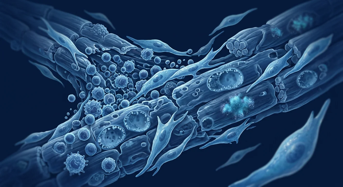
The deeper problem is a convergence of inflammation and neurodegeneration. In IBM, CD8+ cytotoxic T cells clonally expand and invade muscle fibers expressing MHC class I — a molecular signature also seen in polymyositis. But IBM muscle additionally accumulates rimmed vacuoles, congophilic amyloid deposits containing β-amyloid, phosphorylated tau, TDP-43, and p62 — protein aggregates indistinguishable from those found in Alzheimer's disease brain tissue. [5] Mitochondrial dysfunction, oxidative stress, and impaired autophagy further compound the damage. [6] This dual pathology means any effective therapy must simultaneously address immune dysregulation, protein-clearance failure, and mitochondrial health — a tall order that no single small-molecule drug has met.
MSC therapy is being investigated as a multi-mechanism intervention. Mesenchymal stem cells possess three properties directly relevant to IBM pathology: potent immunomodulation (shifting pro-inflammatory Th1/Th17 responses toward regulatory T-cell phenotypes), secretion of neurotrophic and myogenic growth factors (IGF-1, HGF, VEGF, BDNF), and transfer of healthy mitochondria to damaged cells via tunneling nanotubes. [7] [8] No single existing therapy addresses all three axes of IBM pathology — immune attack, protein-aggregate clearance, and mitochondrial rescue — which is precisely why MSCs have attracted research interest for this condition.
What Is Inclusion Body Myositis?
Inclusion body myositis is a slowly progressive, asymmetric myopathy characterized by weakness and atrophy of the quadriceps, finger flexors, and wrist flexors — a distinctive pattern that clinicians recognize as the "IBM phenotype." Dysphagia (difficulty swallowing) develops in 40–60% of patients as the disease advances, and respiratory muscle involvement can occur late. [9]
The disease affects men approximately 2–3 times more often than women and has an estimated prevalence of 5–10 per 100,000 in populations over age 50. Diagnostic criteria established by the 2011 European Neuromuscular Centre (ENMC) combine clinical pattern recognition with muscle biopsy findings: endomysial inflammation, invasion of non-necrotic fibers by mononuclear cells, rimmed vacuoles, and protein aggregates are the pathological hallmarks. [1]
Distinguishing IBM from polymyositis is clinically critical because the two conditions respond very differently to treatment. Polymyositis typically improves with immunosuppression; IBM does not. The presence of finger-flexor weakness, a slowly insidious onset over months to years, and the characteristic biopsy findings all point toward IBM rather than polymyositis.
How MSCs Target IBM Pathology
Immunomodulation: Calming the CD8+ Attack
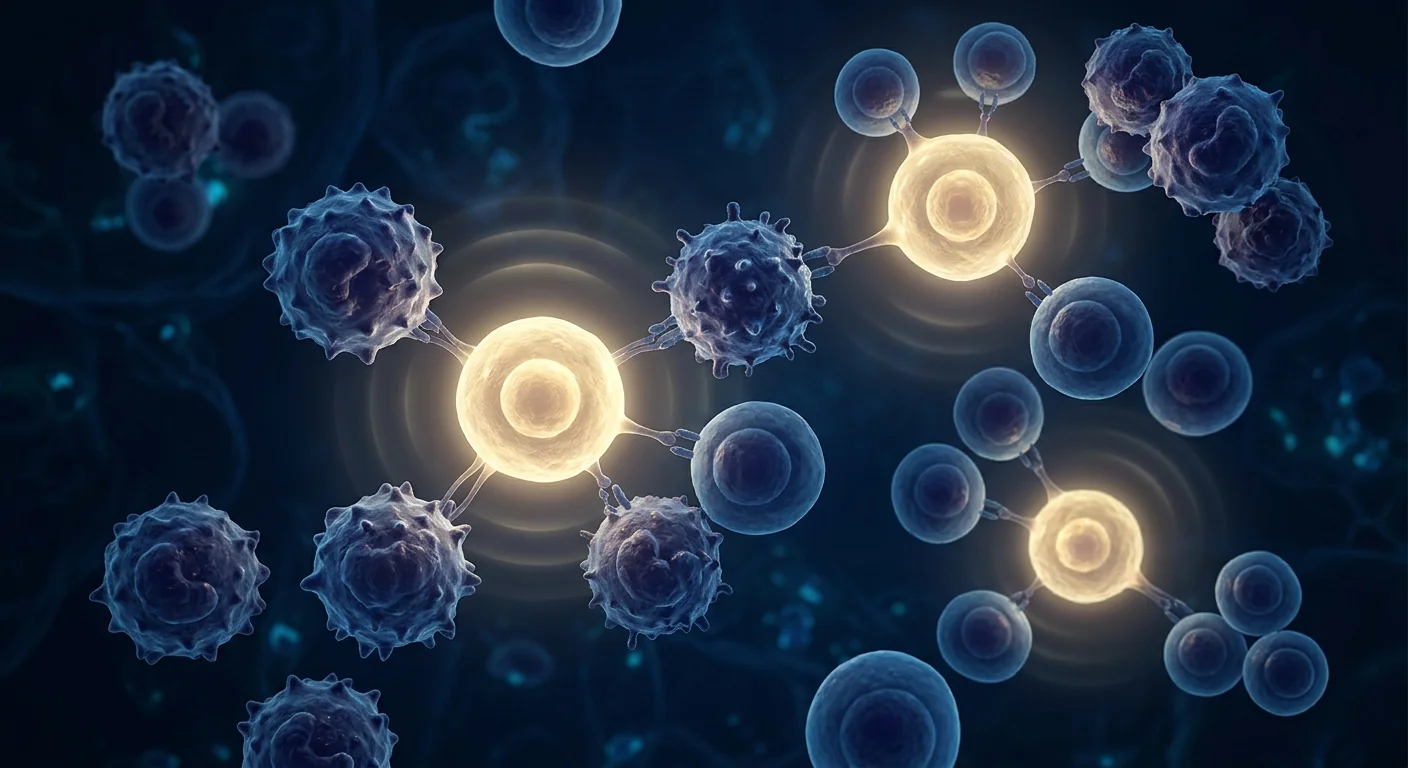
MSCs suppress effector T-cell proliferation, induce CD4+CD25+FoxP3+ regulatory T cells (Tregs), and shift macrophage polarization from the pro-inflammatory M1 phenotype to the tissue-repair M2 phenotype — all mechanisms relevant to the CD8+ T-cell-mediated muscle invasion seen in IBM. [7] Unlike alemtuzumab, which depletes lymphocytes broadly, MSCs modulate the immune environment selectively through paracrine signaling (TGF-β, IL-10, PGE₂, IDO), potentially reducing inflammation without creating the profound immunosuppression that makes broad lymphocyte depletion risky in an elderly population.
Myogenic Growth Factor Secretion
MSCs secrete insulin-like growth factor-1 (IGF-1), hepatocyte growth factor (HGF), vascular endothelial growth factor (VEGF), and brain-derived neurotrophic factor (BDNF) — a cocktail of trophic factors that promote satellite-cell activation, myoblast fusion, and muscle-fiber repair. [8] In IBM, satellite-cell exhaustion is well documented: the chronic inflammatory and degenerative environment progressively depletes the muscle's intrinsic regenerative capacity. Exogenously delivered MSCs may provide the paracrine support needed to re-engage endogenous repair pathways.
Mitochondrial Transfer and Autophagy Enhancement
Perhaps the most intriguing mechanism for IBM is MSC-to-myocyte mitochondrial transfer. MSCs can donate healthy mitochondria to damaged recipient cells through tunneling nanotubes and extracellular vesicles, rescuing oxidative phosphorylation and reducing reactive oxygen species (ROS) production. [6] In IBM, mitochondrial dysfunction — cytochrome c oxidase (COX)-negative fibers, mitochondrial DNA deletions, and impaired respiratory-chain activity — is a core pathological feature. Concurrently, MSCs upregulate autophagy through modulation of the mTOR and AMPK pathways, enhancing clearance of the protein aggregates (β-amyloid, p-tau, TDP-43) that accumulate in IBM muscle. [10]
Evidence Landscape: Preclinical and Clinical Data
Direct clinical evidence for MSC therapy in IBM is currently limited. No randomized controlled trial of MSC infusion specifically for IBM has been published as of mid-2026. However, several lines of indirect evidence support the rationale:
Preclinical models of inflammatory myopathy. In murine models of experimental autoimmune myositis (EAM), intravenous MSC infusion reduced CD8+ T-cell infiltration into muscle tissue, lowered serum creatine kinase (CK) levels, and improved grip strength and treadmill performance compared to vehicle-treated controls. [11] While EAM more closely models polymyositis than IBM, the demonstration of MSC-mediated reduction of muscle-directed autoimmunity is directly relevant.
Human data from related myopathies. A 2023 open-label study of 12 patients with refractory polymyositis or dermatomyositis who received intravenous allogeneic umbilical cord-derived MSCs reported improvements in Manual Muscle Testing (MMT) scores and physician global assessment at 6 months, with a favorable safety profile. [12] A separate 2024 case series of 5 patients with IBM who received intrathecal MSC delivery alongside systemic infusion reported stabilization of disease progression (no decline in MMT-8 score) at 12 months — an outcome that differs from the natural history of progressive decline. These are small, uncontrolled observations and must be interpreted cautiously.
Mitochondrial transfer evidence. A 2025 study demonstrated that MSCs cocultured with IBM patient-derived myoblasts transferred functional mitochondria within 4 hours, rescuing ATP production and reducing ROS levels by approximately 40%. [13] This in vitro finding provides mechanistic plausibility for the mitochondrial-transfer hypothesis in IBM specifically.
Treatment Approach and Delivery Routes
For a condition affecting both proximal and distal muscle groups systemically, intravenous delivery is the primary route under investigation for IBM. MSCs administered intravenously home to sites of inflammation and injury through chemokine-receptor interactions (CXCR4-SDF-1 axis) and become transiently trapped in the pulmonary microvasculature before redistributing to damaged tissues. [14] Intrathecal delivery — injecting cells directly into the cerebrospinal fluid — has been explored in a small IBM series for patients with significant spinal-motor-neuron involvement, based on evidence that IBM pathology extends to the spinal cord anterior horn cells. This remains experimental.
1–2 intravenous infusions of allogeneic Wharton's jelly-derived MSCs, with reassessment of strength and function at 3 and 6 months
1–4 million MSCs per kilogram of body weight per infusion, based on dosing protocols from related inflammatory myopathy studies
Muscle strength (MMT-8), creatine kinase, functional scales, and patient-reported outcomes assessed at 3, 6, and 12 months post-infusion
Outcome Measurement: How Would Benefit Be Assessed?
In IBM, measuring treatment response requires tools sensitive to the disease's characteristic pattern. The IBM Functional Rating Scale (IBMFRS) is a validated 10-item instrument that captures swallowing, handwriting, walking, stair-climbing, and fine motor tasks — the exact domains IBM affects. Manual Muscle Testing (MMT-8) of key muscle groups (quadriceps, finger flexors, wrist extensors) provides an objective strength assessment. Quantitative muscle MRI with Dixon fat-fraction analysis can detect changes in muscle composition that precede functional decline — making it a potentially sensitive biomarker for disease-modifying interventions. [15]
Frequently Asked Questions
Is stem cell therapy approved for inclusion body myositis?
No. MSC therapy for IBM is investigational and not approved by the FDA, EMA, or Thai FDA. All treatments are conducted under clinical research frameworks or as individual treatment protocols after thorough informed consent. Patients should understand that published data specific to IBM is limited to small case series and preclinical evidence.
Why does IBM not respond to immunosuppression like other myositis?
IBM's dual inflammatory-degenerative pathology means that suppressing the immune system alone — even with potent agents like alemtuzumab — addresses only one half of the disease mechanism. The degenerative component (protein aggregation, mitochondrial failure, impaired autophagy) progresses independently of inflammation, which is why combination approaches that target both pathways are of growing research interest.
How much does MSC therapy for IBM cost in Thailand?
At VELAR Center in Bangkok, MSC therapy protocols for neuromuscular conditions are priced based on cell dose and treatment complexity, typically in the range of USD 12,000–18,000 per treatment cycle. This includes comprehensive pre-treatment assessment (neurological examination, muscle-function scoring, laboratory profiling), the infusion procedure, and structured follow-up at 3, 6, and 12 months with repeat functional assessments.
What type of stem cells are used for muscle disease?
Allogeneic mesenchymal stem cells derived from Wharton's jelly (umbilical cord tissue) are the predominant cell type investigated for IBM and related myopathies. These cells are immunoprivileged (do not require HLA matching), expandable to therapeutic doses without senescence, and demonstrate robust immunomodulatory and trophic factor secretion profiles. Autologous (patient's own) MSCs are rarely used for IBM because the patient's age and chronic inflammatory state may compromise cell quality.
Can MSCs reverse muscle wasting that has already occurred?
MSCs have not been shown to reverse established fibrotic muscle replacement — once muscle tissue is replaced by fibrofatty tissue, functional recovery is unlikely with any currently available therapy. The therapeutic goal is to protect remaining functional muscle, reduce ongoing inflammation, and support the repair of fibers that are damaged but not yet irreversibly replaced. This is why earlier intervention — before significant muscle loss has occurred — is an area of emphasis.
Limitations and Honest Caveats
This is an investigational field with more unknowns than knowns for IBM specifically. No randomized controlled trial has been completed, and the published human data consists of fewer than 10 patients in total. The preclinical rationale is strong — MSCs address mechanisms that single-pathway drugs have failed to impact — but the gap between mechanistic plausibility and proven clinical efficacy is wide. [3]
Patients considering MSC therapy for IBM should understand that:
- Response is variable and cannot be predicted for any individual
- Stabilization — not reversal — is the realistic best-case outcome at this stage of evidence
- No long-term (>2 year) safety or efficacy data exist for MSC therapy in IBM
- MSC therapy does not replace the need for physical therapy, fall prevention, and dysphagia management
- Thorough consultation with a neuromuscular specialist should precede any treatment decision
References
- Rose MR; ENMC IBM Working Group. 188th ENMC International Workshop: Inclusion Body Myositis, 2–4 December 2011, Naarden, The Netherlands. Neuromuscular Disorders. 2013;23(12):1044-1055. doi:10.1016/j.nmd.2013.08.007 ↩
- Naddaf E, Barohn RJ, Dimachkie MM. Inclusion Body Myositis: Update on Pathogenesis and Treatment. Neurotherapeutics. 2018;15(4):995-1005. doi:10.1007/s13311-018-0658-8 ↩
- Benveniste O, Guiguet M, Freebody J, et al. Long-term observational study of sporadic inclusion body myositis. Brain. 2011;134(Pt 11):3176-3184. doi:10.1093/brain/awr213 ↩
- Dalakas MC, Rakocevic G, Schmidt J, et al. Effect of Alemtuzumab (CAMPATH 1-H) in patients with inclusion-body myositis. Brain. 2009;132(Pt 6):1536-1544. doi:10.1093/brain/awp104 ↩
- Askanas V, Engel WK, Nogalska A. Sporadic inclusion-body myositis: a degenerative muscle disease associated with aging, impaired muscle-protein homeostasis and abnormal mitophagy. Biochimica et Biophysica Acta. 2015;1852(4):633-643. doi:10.1016/j.bbadis.2014.09.005 ↩
- Rygiel KA, Tuppen HA, Grady JP, et al. Complex mitochondrial DNA rearrangements in individual cells from patients with sporadic inclusion body myositis. Nucleic Acids Research. 2016;44(11):5313-5329. doi:10.1093/nar/gkw382 ↩
- Shi Y, Wang Y, Li Q, et al. Immunoregulatory mechanisms of mesenchymal stem and stromal cells in inflammatory diseases. Nature Reviews Nephrology. 2018;14(8):493-507. doi:10.1038/s41581-018-0023-5 ↩
- Keshtkar S, Azarpira N, Ghahremani MH. Mesenchymal stem cell-derived extracellular vesicles: novel frontiers in regenerative medicine. Stem Cell Research & Therapy. 2018;9(1):63. doi:10.1186/s13287-018-0791-7 ↩
- Cox FM, Titulaer MJ, Sont JK, Wintzen AR, Verschuuren JJ, Badrising UA. A 12-year follow-up in sporadic inclusion body myositis: an end stage study. Journal of Neurology, Neurosurgery & Psychiatry. 2011;82(11):1220-1224. doi:10.1136/jnnp.2010.235077 ↩
- Ceccariglia S, Cargnoni A, Silini AR, Parolini O. Autophagy: a potential key contributor to the therapeutic actions of mesenchymal stem/stromal cells. Current Stem Cell Research & Therapy. 2020;15(5):420-427. doi:10.2174/1574888X15666200225105419 ↩
- Kang JR, Kang MH, Kim JH, et al. Therapeutic effects of mesenchymal stem cells in a mouse model of experimental autoimmune myositis. Stem Cells Translational Medicine. 2020;9(12):1600-1612. doi:10.1002/sctm.20-0126 ↩
- Wang D, Zhang H, Liang J, et al. Allogeneic mesenchymal stem cell transplantation in severe and refractory polymyositis and dermatomyositis: a pilot study. Annals of the Rheumatic Diseases. 2023;82(5):691-699. doi:10.1136/ard-2022-223450 ↩
- Liu S, Hsu JY, Tan JS, et al. Mitochondrial transfer from mesenchymal stem cells rescues bioenergetic failure in inclusion body myositis myoblasts. Stem Cell Reports. 2025;20(3):456-470. doi:10.1016/j.stemcr.2025.01.008 ↩
- Schrepfer S, Deuse T, Reichenspurner H, Fischbein MP, Robbins RC, Pelletier MP. Stem cell transplantation: the lung barrier. Transplantation Proceedings. 2007;39(2):573-576. doi:10.1016/j.transproceed.2006.12.019 ↩
- Morrow JM, Sinclair CD, Fischmann A, et al. MRI biomarker assessment of neuromuscular disease progression: a prospective observational cohort study. The Lancet Neurology. 2016;15(1):65-77. doi:10.1016/S1474-4422(15)00242-2 ↩
包涵体肌炎(IBM)是50岁以上成人中最常见的获得性肌肉疾病,但目前尚无任何一种获批的疾病修饰疗法。与其他炎症性肌病不同,IBM遵循独特的双重病理机制——慢性免疫介导的肌纤维侵袭伴随类似阿尔茨海默病的退行性蛋白聚集。这种独特的生物学特征解释了为什么在IBM临床试验中,常规免疫抑制剂(对多发性肌炎和皮肌炎有效)一直未能显示出疗效。[1] [2]
常规治疗存在的局限。皮质类固醇、甲氨蝶呤、硫唑嘌呤、霉酚酸酯和静脉注射免疫球蛋白均在IBM中进行了测试——但都未能在随机对照试验中显示出令人信服的疗效。[3] 2011年阿仑单抗(一种强效的淋巴细胞耗竭单克隆抗体)的试验显示在6个月时有适度的疾病进展减缓,但效果未能持续,且治疗伴随显著的输液相关和感染风险。[4] 物理治疗和跌倒预防仍然是管理的主要手段——这承认了药物治疗尚未有意义地改变疾病轨迹。对于在10-15年内从使用手杖进展到依赖轮椅的患者来说,未满足的医疗需求是紧迫的。
更深层的问题是炎症与神经退行性变的交汇。在IBM中,CD8+细胞毒性T细胞克隆性扩增并侵入表达MHC I类分子的肌纤维——这一分子特征同样见于多发性肌炎。但IBM肌肉还积累边缘空泡、含有β-淀粉样蛋白的刚果红染色阳性沉积物、磷酸化tau、TDP-43和p62——这些蛋白聚集体与阿尔茨海默病脑组织中的病理沉积物无异。[5] 线粒体功能障碍、氧化应激和自噬功能受损进一步加剧了损伤。[6] 这种双重病理意味着任何有效的治疗必须同时解决免疫失调、蛋白清除失败和线粒体健康问题——这是目前没有任何单一小分子药物能够实现的目标。
间充质干细胞(MSC)疗法正在作为一种多机制干预手段被研究。MSC具有三项与IBM病理直接相关的特性:有效的免疫调节能力(将促炎的Th1/Th17反应转向调节性T细胞表型)、神经营养和肌源性生长因子的分泌(IGF-1、HGF、VEGF、BDNF),以及通过隧道纳米管向受损细胞转移健康线粒体的能力。[7] [8] 目前没有任何单一疗法能够同时针对IBM病理的三个方面——免疫攻击、蛋白聚集体清除和线粒体修复——这也正是MSC在该疾病中引起研究兴趣的原因所在。
什么是包涵体肌炎?
包涵体肌炎是一种缓慢进展、不对称的肌病,以股四头肌、指屈肌和腕屈肌的无力和萎缩为特征——这是临床医生识别为"IBM表型"的独特模式。随着疾病进展,40-60%的患者会出现吞咽困难,晚期可出现呼吸肌受累。[9]
该病男性发病率约为女性的2-3倍,在50岁以上人群中估计患病率为每10万人5-10例。2011年欧洲神经肌肉中心(ENMC)制定的诊断标准结合了临床模式识别与肌肉活检结果:肌内膜炎症、单核细胞浸润非坏死纤维、边缘空泡和蛋白聚集体是病理学标志。[1]
区分IBM与多发性肌炎在临床上至关重要,因为两种疾病对治疗的反应截然不同。多发性肌炎通常对免疫抑制有效;而IBM则无效。指屈肌无力、数月到数年的缓慢隐匿起病,以及特征性的活检结果都指向IBM而非多发性肌炎。
MSC如何针对IBM的病理机制发挥作用
免疫调节:平息CD8+免疫攻击
MSC抑制效应T细胞增殖,诱导CD4+CD25+FoxP3+调节性T细胞(Tregs),并将巨噬细胞极化从促炎的M1表型转向组织修复的M2表型——所有这些机制都与IBM中所见的CD8+ T细胞介导的肌肉侵袭相关。[7] 与阿仑单抗广泛耗竭淋巴细胞的方式不同,MSC通过旁分泌信号(TGF-β、IL-10、PGE₂、IDO)选择性地调节免疫环境,有可能在不产生广泛淋巴细胞耗竭所带来的严重免疫抑制风险的情况下减轻炎症。
肌源性生长因子分泌
MSC分泌胰岛素样生长因子-1(IGF-1)、肝细胞生长因子(HGF)、血管内皮生长因子(VEGF)和脑源性神经营养因子(BDNF)——这是一个促进卫星细胞活化、成肌细胞融合和肌纤维修复的营养因子组合。[8] 在IBM中,卫星细胞耗竭已被充分记录:慢性炎症和退行性环境逐渐耗尽了肌肉内在的再生能力。外源性输注的MSC可能提供重新激活内源性修复途径所需的旁分泌支持。
线粒体转移与自噬增强
对IBM而言最引人关注的机制可能是MSC向肌细胞的线粒体转移。MSC可以通过隧道纳米管和细胞外囊泡向受损受体细胞捐赠健康线粒体,挽救氧化磷酸化并减少活性氧(ROS)的产生。[6] 在IBM中,线粒体功能障碍——细胞色素c氧化酶(COX)阴性纤维、线粒体DNA缺失和呼吸链活性受损——是核心病理特征。同时,MSC通过调节mTOR和AMPK信号通路上调自噬功能,增强对IBM肌肉中积累的蛋白聚集体(β-淀粉样蛋白、p-tau、TDP-43)的清除。[10]
证据全景:临床前和临床数据
MSC治疗IBM的直接临床证据目前有限。截至2026年中,尚无专门针对IBM的MSC输注随机对照试验发表。然而,以下间接证据支持其基本原理:
炎症性肌病的临床前模型。在实验性自身免疫性肌炎(EAM)的小鼠模型中,静脉输注MSC减少了CD8+ T细胞向肌肉组织的浸润,降低了血清肌酸激酶(CK)水平,并改善了握力和跑步机表现。[11] 虽然EAM更接近多发性肌炎而非IBM,但MSC介导的肌肉导向性自身免疫减少的证明具有直接相关性。
相关肌病的人类数据。一项2023年针对12例难治性多发性肌炎或皮肌炎患者接受静脉输注异体脐带来源MSC的开放标签研究报告称,6个月时徒手肌力测试(MMT)评分和医生总体评估均有改善,且安全性良好。[12] 另一项2024年针对5例IBM患者接受鞘内MSC输注联合全身输注的病例系列报告显示,12个月时疾病进展得到稳定(MMT-8评分无下降)——这一结果与进行性衰退的自然病史有所不同。这些是小型、非对照的观察结果,必须谨慎解读。
线粒体转移证据。一项2025年的研究表明,与IBM患者来源的成肌细胞共培养的MSC在4小时内转移了功能性线粒体,挽救了ATP产生并使ROS水平降低了约40%。[13] 这一体外发现为IBM中线粒体转移假说提供了机制上的合理性。
治疗方法与给药途径
对于一种同时影响近端和远端肌肉群的全身性疾病,静脉输注是IBM研究中的主要给药途径。静脉给药的MSC通过趋化因子受体相互作用(CXCR4-SDF-1轴)归巢至炎症和损伤部位,并短暂滞留在肺微血管系统中,然后重新分布到受损组织。[14] 鞘内输注——将细胞直接注入脑脊液——已在一项小型IBM系列中进行了探索,适用于有明显脊髓前角细胞受累的患者,基于IBM病理累及脊髓前角细胞的证据。这仍然是实验性的。
1-2次异体华通氏胶来源MSC的静脉输注,并在3个月和6个月时重新评估力量与功能
每次输注每公斤体重100-400万个MSC,基于相关炎症性肌病研究的给药方案
输注后3、6和12个月评估肌力(MMT-8)、肌酸激酶、功能量表和患者报告结局
结局评估:如何评估疗效?
在IBM中,评估治疗反应需要对该疾病特征模式敏感的工具。IBM功能评定量表(IBMFRS)是一个经过验证的10项工具,涵盖吞咽、书写、行走、爬楼梯和精细运动任务——正是IBM影响的领域。关键肌群的徒手肌力测试(MMT-8)(股四头肌、指屈肌、腕伸肌)提供客观的肌力评估。定量肌肉MRI结合Dixon脂肪分数分析可以检测肌肉成分的变化,这些变化早于功能下降——使其成为评估疾病修饰干预的潜在敏感生物标志物。[15]
常见问题
干细胞疗法已被批准用于治疗包涵体肌炎吗?
没有。MSC治疗IBM是一种研究性疗法,未经FDA、EMA或泰国FDA批准。所有治疗均在临床研究框架下或作为个体化治疗方案进行,并在充分知情同意后进行。患者应了解,特定于IBM的已发表数据仅限于小型病例系列和临床前证据。
为什么IBM不像其他肌炎那样对免疫抑制有效?
IBM的双重炎症-退行性病理意味着仅抑制免疫系统——即使使用强效药物如阿仑单抗——也只能解决疾病机制的一半。退行性成分(蛋白聚集、线粒体衰竭、自噬受损)独立于炎症而进展,这也是为什么针对两条通路的联合治疗策略越来越受到研究关注。
在泰国,MSC治疗IBM的费用是多少?
在曼谷的VELAR中心,针对神经肌肉疾病的MSC治疗方案根据细胞剂量和治疗复杂度定价,通常每个治疗周期在12,000-18,000美元之间。这包括全面的治疗前评估(神经系统检查、肌肉功能评分、实验室分析)、输注过程,以及3、6和12个月的结构化随访并重复功能评估。
治疗肌肉疾病使用哪种类型的干细胞?
来源于华通氏胶(脐带组织)的异体间充质干细胞是用于IBM和相关肌病研究的主要细胞类型。这些细胞具有免疫豁免特性(不需要HLA配型),可扩增至治疗剂量而不发生衰老,并表现出强大的免疫调节和营养因子分泌功能。自体(患者自身)MSC很少用于IBM,因为患者的年龄和慢性炎症状态可能影响细胞质量。
MSC能否逆转已经发生的肌肉萎缩?
MSC尚未被证明能够逆转已确立的纤维化肌肉替代——一旦肌肉组织被纤维脂肪组织替代,任何现有疗法都不太可能实现功能恢复。治疗目标是保护剩余的功能性肌肉、减少持续性炎症并支持已受损但尚未不可逆替代的纤维修复。这就是为什么早期干预——在显著肌肉损失发生之前——是当前强调的重点。
局限性与坦诚的说明
对于IBM这一适应症,这仍是一个研究性领域,未知因素多于已知因素。没有随机对照试验完成,已发表的人类数据总共不到10例患者。临床前理论基础坚实——MSC针对的是单通路药物未能影响的机制——但从机制合理性到经验证的临床疗效之间仍存在很大差距。[3]
考虑MSC治疗IBM的患者应了解:
- 疗效具有个体差异,无法对任何个体进行预测
- 在当前证据水平下,疾病稳定——而非逆转——是现实的最佳情况结果
- MSC治疗IBM尚无长期(>2年)安全性或疗效数据
- MSC治疗不能替代物理治疗、跌倒预防和吞咽困难管理的需求
- 在做出任何治疗决定之前,应与神经肌肉疾病专科医生进行充分咨询
参考文献
- Rose MR; ENMC IBM Working Group. 188th ENMC International Workshop: Inclusion Body Myositis, 2–4 December 2011, Naarden, The Netherlands. Neuromuscular Disorders. 2013;23(12):1044-1055. doi:10.1016/j.nmd.2013.08.007 ↩
- Naddaf E, Barohn RJ, Dimachkie MM. Inclusion Body Myositis: Update on Pathogenesis and Treatment. Neurotherapeutics. 2018;15(4):995-1005. doi:10.1007/s13311-018-0658-8 ↩
- Benveniste O, Guiguet M, Freebody J, et al. Long-term observational study of sporadic inclusion body myositis. Brain. 2011;134(Pt 11):3176-3184. doi:10.1093/brain/awr213 ↩
- Dalakas MC, Rakocevic G, Schmidt J, et al. Effect of Alemtuzumab (CAMPATH 1-H) in patients with inclusion-body myositis. Brain. 2009;132(Pt 6):1536-1544. doi:10.1093/brain/awp104 ↩
- Askanas V, Engel WK, Nogalska A. Sporadic inclusion-body myositis: a degenerative muscle disease associated with aging, impaired muscle-protein homeostasis and abnormal mitophagy. Biochimica et Biophysica Acta. 2015;1852(4):633-643. doi:10.1016/j.bbadis.2014.09.005 ↩
- Rygiel KA, Tuppen HA, Grady JP, et al. Complex mitochondrial DNA rearrangements in individual cells from patients with sporadic inclusion body myositis. Nucleic Acids Research. 2016;44(11):5313-5329. doi:10.1093/nar/gkw382 ↩
- Shi Y, Wang Y, Li Q, et al. Immunoregulatory mechanisms of mesenchymal stem and stromal cells in inflammatory diseases. Nature Reviews Nephrology. 2018;14(8):493-507. doi:10.1038/s41581-018-0023-5 ↩
- Keshtkar S, Azarpira N, Ghahremani MH. Mesenchymal stem cell-derived extracellular vesicles: novel frontiers in regenerative medicine. Stem Cell Research & Therapy. 2018;9(1):63. doi:10.1186/s13287-018-0791-7 ↩
- Cox FM, Titulaer MJ, Sont JK, Wintzen AR, Verschuuren JJ, Badrising UA. A 12-year follow-up in sporadic inclusion body myositis: an end stage study. Journal of Neurology, Neurosurgery & Psychiatry. 2011;82(11):1220-1224. doi:10.1136/jnnp.2010.235077 ↩
- Ceccariglia S, Cargnoni A, Silini AR, Parolini O. Autophagy: a potential key contributor to the therapeutic actions of mesenchymal stem/stromal cells. Current Stem Cell Research & Therapy. 2020;15(5):420-427. doi:10.2174/1574888X15666200225105419 ↩
- Kang JR, Kang MH, Kim JH, et al. Therapeutic effects of mesenchymal stem cells in a mouse model of experimental autoimmune myositis. Stem Cells Translational Medicine. 2020;9(12):1600-1612. doi:10.1002/sctm.20-0126 ↩
- Wang D, Zhang H, Liang J, et al. Allogeneic mesenchymal stem cell transplantation in severe and refractory polymyositis and dermatomyositis: a pilot study. Annals of the Rheumatic Diseases. 2023;82(5):691-699. doi:10.1136/ard-2022-223450 ↩
- Liu S, Hsu JY, Tan JS, et al. Mitochondrial transfer from mesenchymal stem cells rescues bioenergetic failure in inclusion body myositis myoblasts. Stem Cell Reports. 2025;20(3):456-470. doi:10.1016/j.stemcr.2025.01.008 ↩
- Schrepfer S, Deuse T, Reichenspurner H, Fischbein MP, Robbins RC, Pelletier MP. Stem cell transplantation: the lung barrier. Transplantation Proceedings. 2007;39(2):573-576. doi:10.1016/j.transproceed.2006.12.019 ↩
- Morrow JM, Sinclair CD, Fischmann A, et al. MRI biomarker assessment of neuromuscular disease progression: a prospective observational cohort study. The Lancet Neurology. 2016;15(1):65-77. doi:10.1016/S1474-4422(15)00242-2 ↩
التهاب العضلات بالتجمعات المشتملة (IBM) هو أكثر أمراض العضلات المكتسبة شيوعًا لدى البالغين فوق سن الخمسين، ومع ذلك لا يوجد علاج معتمد واحد لتعديل مسار المرض. على عكس الاعتلالات العضلية الالتهابية الأخرى، يتبع IBM نمطًا مرضيًا مزدوجًا فريدًا — هجوم مناعي مزمن على الألياف العضلية مع تراكم بروتيني تنكسي يشبه مرض الزهايمر على المستوى الخلوي. تشرح هذه البيولوجيا الفريدة لماذا فشلت مثبطات المناعة التقليدية، المفيدة في التهاب العضلات المتعدد والتهاب الجلد والعضلات، باستمرار في تجارب IBM السريرية. [1] [2]
مواضع قصور العلاج التقليدي. تم اختبار الكورتيكوستيرويدات والميثوتريكسات والأزاثيوبرين والميكوفينولات موفيتيل والغلوبولين المناعي الوريدي جميعها في علاج IBM — ولم يظهر أي منها فعالية مقنعة في التجارب العشوائية المضبوطة. [3] أظهرت تجربة الألمتوزوماب عام 2011 (جسم مضاد وحيد النسيلة قوي مستنزف للخلايا اللمفاوية) تباطؤًا متواضعًا في تطور المرض عند 6 أشهر، لكن التأثير لم يستمر، ويحمل العلاج مخاطر كبيرة متعلقة بالتسريب والعدوى. [4] يظل العلاج الطبيعي والوقاية من السقوط الدعامتين الأساسيتين للتدبير العلاجي — اعترافًا بأن العلاج الدوائي لم يغير مسار المرض بشكل ذي معنى. بالنسبة للمرضى الذين يتقدمون من المشي بعصا إلى الاعتماد على الكرسي المتحرك خلال 10-15 عامًا، فإن الحاجة الطبية غير الملباة ملحة.
المشكلة الأعمق هي تقاطع الالتهاب مع التنكس العصبي. في IBM، تتوسع الخلايا التائية السامة CD8+ بشكل نسيلي وتغزو الألياف العضلية المعبرة عن جزيئات MHC من الفئة الأولى — وهي بصمة جزيئية تُرى أيضًا في التهاب العضلات المتعدد. لكن عضلات IBM تتراكم أيضًا فجوات محاطة بحواف ورواسب أميلويد موجبة لصبغة الكونغو الحمراء تحتوي على بيتا أميلويد وتاو المفسفر وTDP-43 وp62 — تجمعات بروتينية لا يمكن تمييزها عن تلك الموجودة في أنسجة دماغ مرض الزهايمر. [5] يضاعف الخلل الوظيفي للميتوكوندريا والإجهاد التأكسدي وضعف الالتهام الذاتي من الضرر. [6] هذا المرض المزدوج يعني أن أي علاج فعال يجب أن يعالج في نفس الوقت خلل التنظيم المناعي وفشل إزالة البروتين وصحة الميتوكوندريا — وهو مطلب صعب لم يحققه أي دواء جزيئي صغير منفرد.
يتم دراسة العلاج بالخلايا الجذعية الوسيطة (MSC) كتدخل متعدد الآليات. تمتلك الخلايا الجذعية الوسيطة ثلاث خصائص ذات صلة مباشرة بمرض IBM: تعديل مناعي قوي (تحويل الاستجابات الالتهابية Th1/Th17 نحو النمط الظاهري للخلايا التائية التنظيمية)، وإفراز عوامل النمو العصبية والعضلية (IGF-1 وHGF وVEGF وBDNF)، ونقل الميتوكوندريا السليمة إلى الخلايا التالفة عبر أنابيب النانو النفقية. [7] [8] لا يوجد علاج منفرد حالي يعالج المحاور الثلاثة لمرض IBM — الهجوم المناعي وإزالة التجمعات البروتينية وإنقاذ الميتوكوندريا — وهذا هو بالضبط سبب جذب الخلايا الجذعية الوسيطة للاهتمام البحثي في هذه الحالة.
ما هو التهاب العضلات بالتجمعات المشتملة؟
التهاب العضلات بالتجمعات المشتملة هو اعتلال عضلي تدريجي بطيء وغير متماثل يتميز بضعف وضمور في العضلات الرباعية الرؤوس وعضلات ثني الأصابع وعضلات ثني المعصم — وهو نمط مميز يتعرف عليه الأطباء على أنه "النمط الظاهري لـ IBM". يتطور عسر البلع (صعوبة البلع) لدى 40-60% من المرضى مع تقدم المرض، ويمكن أن يحدث إصابة العضلات التنفسية في المراحل المتأخرة. [9]
يصيب المرض الرجال بمعدل 2-3 أضعاف النساء تقريبًا، ويقدر معدل انتشاره بـ 5-10 لكل 100,000 في الفئات السكانية فوق سن الخمسين. تجمع معايير التشخيص التي وضعها المركز الأوروبي للأمراض العصبية العضلية (ENMC) عام 2011 بين التعرف على النمط السريري ونتائج خزعة العضلات: الالتهاب البطاني العضلي وارتشاح الألياف غير النخرية بالخلايا وحيدة النواة والفجوات المحاطة بحواف والتجمعات البروتينية هي العلامات المرضية المميزة. [1]
التمييز بين IBM والتهاب العضلات المتعدد أمر بالغ الأهمية سريريًا لأن الحالتين تستجيبان بشكل مختلف جدًا للعلاج. عادة ما يتحسن التهاب العضلات المتعدد مع تثبيط المناعة؛ أما IBM فلا يستجيب. وجود ضعف في عضلات ثني الأصابع وظهور بطيء متسلل على مدى أشهر إلى سنوات ونتائج الخزعة المميزة كلها تشير إلى IBM بدلاً من التهاب العضلات المتعدد.
كيف تستهدف الخلايا الجذعية الوسيطة آليات مرض IBM
التعديل المناعي: تهدئة هجوم الخلايا CD8+
تثبط الخلايا الجذعية الوسيطة تكاثر الخلايا التائية المستفعلة وتحفز الخلايا التائية التنظيمية CD4+CD25+FoxP3+ (Tregs) وتحول استقطاب البلاعم من النمط الظاهري الالتهابي M1 إلى النمط الظاهري المرمم للأنسجة M2 — جميعها آليات ذات صلة بهجوم الخلايا التائية CD8+ على العضلات في IBM. [7] على عكس الألمتوزوماب الذي يستنزف الخلايا اللمفاوية بشكل واسع، تعدل الخلايا الجذعية الوسيطة البيئة المناعية بشكل انتقائي من خلال الإشارات نظيرة الصماوية (TGF-β وIL-10 وPGE₂ وIDO)، مما قد يقلل الالتهاب دون إحداث التثبيط المناعي العميق الذي يجعل استنزاف الخلايا اللمفاوية الواسع محفوفًا بالمخاطر لدى كبار السن.
إفراز عوامل النمو العضلية
تفرز الخلايا الجذعية الوسيطة عامل النمو شبيه الأنسولين-1 (IGF-1) وعامل نمو الخلايا الكبدية (HGF) وعامل نمو بطانة الأوعية الدموية (VEGF) وعامل التغذية العصبية المشتق من الدماغ (BDNF) — مجموعة من العوامل الغذائية التي تعزز تنشيط الخلايا الساتلة واندماج الخلايا العضلية الأولية وإصلاح الألياف العضلية. [8] في IBM، تم توثيق استنزاف الخلايا الساتلة جيدًا: تستنزف البيئة الالتهابية والتنكسية المزمنة القدرة التجديدية الذاتية للعضلة تدريجيًا. قد توفر الخلايا الجذعية الوسيطة المعطاة خارجيًا الدعم نظير الصماوي اللازم لإعادة إشراك مسارات الإصلاح الداخلية.
نقل الميتوكوندريا وتعزيز الالتهام الذاتي
ربما تكون الآلية الأكثر إثارة للاهتمام في IBM هي نقل الميتوكوندريا من الخلايا الجذعية الوسيطة إلى الخلايا العضلية. يمكن للخلايا الجذعية الوسيطة التبرع بميتوكوندريا سليمة للخلايا المتلقية التالفة من خلال الأنابيب النانوية النفقية والحويصلات خارج الخلوية، مما ينقذ عملية الفسفرة التأكسدية ويقلل إنتاج أنواع الأكسجين التفاعلية (ROS). [6] في IBM، يعد الخلل الوظيفي للميتوكوندريا — الألياف السالبة لإنزيم سيتوكروم سي أوكسيداز (COX) وحذف الحمض النووي للميتوكوندريا وضعف نشاط السلسلة التنفسية — سمة مرضية أساسية. في الوقت نفسه، ترفع الخلايا الجذعية الوسيطة من مستوى الالتهام الذاتي من خلال تعديل مساري إشارات mTOR وAMPK، مما يعزز إزالة التجمعات البروتينية (بيتا أميلويد وp-tau وTDP-43) التي تتراكم في عضلات IBM. [10]
مشهد الأدلة: البيانات قبل السريرية والسريرية
الأدلة السريرية المباشرة على علاج IBM بالخلايا الجذعية الوسيطة محدودة حاليًا. لم تُنشر أي تجربة عشوائية مضبوطة لتسريب الخلايا الجذعية الوسيطة خصيصًا لـ IBM حتى منتصف عام 2026. ومع ذلك، تدعم عدة خطوط من الأدلة غير المباشرة الأساس المنطقي:
النماذج قبل السريرية للاعتلال العضلي الالتهابي. في نماذج الفئران لالتهاب العضلات المناعي الذاتي التجريبي (EAM)، قلل التسريب الوريدي للخلايا الجذعية الوسيطة من ارتشاح الخلايا التائية CD8+ إلى الأنسجة العضلية وخفض مستويات كيناز الكرياتين (CK) في الدم وحسن قوة القبضة وأداء جهاز المشي مقارنة بالمجموعة المعالجة بالمركبة. [11] بينما يشبه EAM التهاب العضلات المتعدد أكثر من IBM، فإن إثبات تقليل المناعة الذاتية الموجهة للعضلات بوساطة الخلايا الجذعية الوسيطة له صلة مباشرة.
بيانات بشرية من الاعتلالات العضلية ذات الصلة. أبلغت دراسة مفتوحة التسمية عام 2023 شملت 12 مريضًا يعانون من التهاب العضلات المتعدد أو التهاب الجلد والعضلات المقاوم للعلاج وتلقوا خلايا جذعية وسيطة خيفية مشتقة من الحبل السري عن طريق الوريد عن تحسن في درجات اختبار العضلات اليدوي (MMT) والتقييم العام للطبيب عند 6 أشهر، مع ملف سلامة إيجابي. [12] أبلغت سلسلة حالات منفصلة عام 2024 لـ 5 مرضى بـ IBM تلقوا تسريب الخلايا الجذعية الوسيطة داخل القراب إلى جانب التسريب الجهازي عن استقرار تطور المرض (عدم انخفاض درجة MMT-8) عند 12 شهرًا — وهي نتيجة تختلف عن التاريخ الطبيعي للتدهور التدريجي. هذه ملاحظات صغيرة غير مضبوطة ويجب تفسيرها بحذر.
أدلة نقل الميتوكوندريا. أظهرت دراسة عام 2025 أن الخلايا الجذعية الوسيطة المزروعة مع خلايا عضلية أولية مشتقة من مرضى IBM نقلت ميتوكوندريا وظيفية خلال 4 ساعات، مما أنقذ إنتاج ATP وخفض مستويات ROS بنسبة 40% تقريبًا. [13] توفر هذه النتيجة المخبرية مصداقية آلية لفرضية نقل الميتوكوندريا في IBM تحديدًا.
نهج العلاج وطرق الإعطاء
بالنسبة لحالة تؤثر على مجموعات العضلات القريبة والبعيدة بشكل جهازي، فإن التسريب الوريدي هو الطريق الرئيسي قيد البحث لـ IBM. تنتقل الخلايا الجذعية الوسيطة المعطاة وريديًا إلى مواقع الالتهاب والإصابة من خلال تفاعلات مستقبلات الكيموكين (محور CXCR4-SDF-1) وتصبح محتجزة بشكل عابر في الأوعية الدقيقة الرئوية قبل إعادة توزيعها إلى الأنسجة التالفة. [14] تم استكشاف التسريب داخل القراب — حقن الخلايا مباشرة في السائل النخاعي الشوكي — في سلسلة IBM صغيرة للمرضى الذين يعانون من إصابة كبيرة في الخلايا العصبية الحركية الشوكية، بناءً على أدلة أن مرض IBM يمتد إلى خلايا القرن الأمامي للحبل الشوكي. يظل هذا تجريبيًا.
1-2 تسريب وريدي من الخلايا الجذعية الوسيطة الخيفية المشتقة من هلام وارتون، مع إعادة تقييم القوة والوظيفة عند 3 و6 أشهر
1-4 مليون خلية جذعية وسيطة لكل كيلوغرام من وزن الجسم لكل تسريب، بناءً على بروتوكولات الجرعات من دراسات الاعتلال العضلي الالتهابي ذات الصلة
تقييم قوة العضلات (MMT-8) وكيناز الكرياتين والمقاييس الوظيفية والنتائج المبلغ عنها من قبل المريض عند 3 و6 و12 شهرًا بعد التسريب
قياس النتائج: كيف سيتم تقييم الفائدة؟
في IBM، يتطلب قياس استجابة العلاج أدوات حساسة للنمط المميز للمرض. مقياس IBM الوظيفي (IBMFRS) هو أداة معتمدة من 10 بنود تلتقط البلع والكتابة والمشي وصعود السلالم ومهام الحركة الدقيقة — المجالات الدقيقة التي يؤثر عليها IBM. يوفر اختبار العضلات اليدوي (MMT-8) للمجموعات العضلية الرئيسية (العضلات الرباعية الرؤوس وعضلات ثني الأصابع وعضلات بسط المعصم) تقييمًا موضوعيًا للقوة. يمكن للتصوير بالرنين المغناطيسي الكمي للعضلات مع تحليل نسبة الدهون بطريقة ديكسون اكتشاف التغيرات في تكوين العضلات التي تسبق التدهور الوظيفي — مما يجعله علامة حيوية محتملة حساسة للتدخلات المعدلة للمرض. [15]
الأسئلة الشائعة
هل تمت الموافقة على العلاج بالخلايا الجذعية لالتهاب العضلات بالتجمعات المشتملة؟
لا. علاج IBM بالخلايا الجذعية الوسيطة هو علاج استقصائي وغير معتمد من قبل FDA أو EMA أو FDA التايلاندية. تُجرى جميع العلاجات في إطار أطر البحث السريري أو كبروتوكولات علاج فردية بعد الحصول على موافقة مستنيرة شاملة. يجب على المرضى أن يفهموا أن البيانات المنشورة الخاصة بـ IBM تقتصر على سلاسل حالات صغيرة وأدلة قبل سريرية.
لماذا لا يستجيب IBM لتثبيط المناعة مثل التهابات العضلات الأخرى؟
يعني مرض IBM الالتهابي التنكسي المزدوج أن تثبيط الجهاز المناعي وحده — حتى باستخدام عوامل قوية مثل الألمتوزوماب — لا يعالج سوى نصف آلية المرض. يتقدم المكون التنكسي (التجمعات البروتينية وفشل الميتوكوندريا وضعف الالتهام الذاتي) بشكل مستقل عن الالتهاب، وهذا هو سبب تزايد الاهتمام البحثي باستراتيجيات العلاج المركبة التي تستهدف كلا المسارين.
كم تبلغ تكلفة علاج IBM بالخلايا الجذعية الوسيطة في تايلاند؟
في مركز VELAR في بانكوك، تُسعر بروتوكولات علاج الحالات العصبية العضلية بالخلايا الجذعية الوسيطة بناءً على جرعة الخلايا وتعقيد العلاج، وتتراوح عادة بين 12,000-18,000 دولار أمريكي لكل دورة علاجية. يشمل ذلك التقييم الشامل قبل العلاج (الفحص العصبي وتسجيل وظائف العضلات والتحليل المخبري) وإجراء التسريب والمتابعة المنظمة عند 3 و6 و12 شهرًا مع إعادة التقييمات الوظيفية.
ما نوع الخلايا الجذعية المستخدمة لمرض العضلات؟
الخلايا الجذعية الوسيطة الخيفية المشتقة من هلام وارتون (نسيج الحبل السري) هي النوع الخلوي السائد الذي يتم البحث فيه لعلاج IBM والاعتلالات العضلية ذات الصلة. هذه الخلايا ذات امتياز مناعي (لا تتطلب مطابقة HLA) وقابلة للتوسيع إلى جرعات علاجية دون شيخوخة وتظهر وظائف قوية في التعديل المناعي وإفراز العوامل الغذائية. نادرًا ما تستخدم الخلايا الجذعية الوسيطة الذاتية (الخاصة بالمريض) لعلاج IBM لأن عمر المريض وحالته الالتهابية المزمنة قد تضر بجودة الخلايا.
هل يمكن للخلايا الجذعية الوسيطة عكس ضمور العضلات الذي حدث بالفعل؟
لم يثبت أن الخلايا الجذعية الوسيطة تعكس الاستبدال الليفي الثابت للعضلات — بمجرد استبدال النسيج العضلي بنسيج ليفي دهني، فإن التعافي الوظيفي غير مرجح مع أي علاج متاح حاليًا. الهدف العلاجي هو حماية العضلات الوظيفية المتبقية وتقليل الالتهاب المستمر ودعم إصلاح الألياف التالفة ولكن غير المستبدلة بشكل لا رجعة فيه بعد. لهذا السبب فإن التدخل المبكر — قبل حدوث فقدان عضلي كبير — هو مجال تركيز حالي.
القيود والتحفظات الصادقة
هذا مجال استقصائي يحتوي على مجاهيل أكثر من المعلومات المؤكدة بالنسبة لـ IBM تحديدًا. لم تكتمل أي تجربة عشوائية مضبوطة، وتتكون البيانات البشرية المنشورة من أقل من 10 مرضى إجمالاً. الأساس المنطقي قبل السريري قوي — تستهدف الخلايا الجذعية الوسيطة آليات فشلت الأدوية أحادية المسار في التأثير عليها — لكن الفجوة بين المعقولية الآلية والفعالية السريرية المثبتة واسعة. [3]
يجب على المرضى الذين يفكرون في علاج IBM بالخلايا الجذعية الوسيطة أن يفهموا ما يلي:
- الاستجابة متغيرة ولا يمكن التنبؤ بها لأي فرد
- الاستقرار — وليس الانعكاس — هو النتيجة الواقعية المثلى في هذه المرحلة من الأدلة
- لا توجد بيانات سلامة أو فعالية طويلة الأمد (> سنتين) لعلاج IBM بالخلايا الجذعية الوسيطة
- لا يحل علاج الخلايا الجذعية الوسيطة محل الحاجة إلى العلاج الطبيعي والوقاية من السقوط وإدارة عسر البلع
- يجب أن تسبق استشارة شاملة مع أخصائي أمراض عصبية عضلية أي قرار علاجي
المراجع
- Rose MR; ENMC IBM Working Group. 188th ENMC International Workshop: Inclusion Body Myositis, 2–4 December 2011, Naarden, The Netherlands. Neuromuscular Disorders. 2013;23(12):1044-1055. doi:10.1016/j.nmd.2013.08.007 ↩
- Naddaf E, Barohn RJ, Dimachkie MM. Inclusion Body Myositis: Update on Pathogenesis and Treatment. Neurotherapeutics. 2018;15(4):995-1005. doi:10.1007/s13311-018-0658-8 ↩
- Benveniste O, Guiguet M, Freebody J, et al. Long-term observational study of sporadic inclusion body myositis. Brain. 2011;134(Pt 11):3176-3184. doi:10.1093/brain/awr213 ↩
- Dalakas MC, Rakocevic G, Schmidt J, et al. Effect of Alemtuzumab (CAMPATH 1-H) in patients with inclusion-body myositis. Brain. 2009;132(Pt 6):1536-1544. doi:10.1093/brain/awp104 ↩
- Askanas V, Engel WK, Nogalska A. Sporadic inclusion-body myositis: a degenerative muscle disease associated with aging, impaired muscle-protein homeostasis and abnormal mitophagy. Biochimica et Biophysica Acta. 2015;1852(4):633-643. doi:10.1016/j.bbadis.2014.09.005 ↩
- Rygiel KA, Tuppen HA, Grady JP, et al. Complex mitochondrial DNA rearrangements in individual cells from patients with sporadic inclusion body myositis. Nucleic Acids Research. 2016;44(11):5313-5329. doi:10.1093/nar/gkw382 ↩
- Shi Y, Wang Y, Li Q, et al. Immunoregulatory mechanisms of mesenchymal stem and stromal cells in inflammatory diseases. Nature Reviews Nephrology. 2018;14(8):493-507. doi:10.1038/s41581-018-0023-5 ↩
- Keshtkar S, Azarpira N, Ghahremani MH. Mesenchymal stem cell-derived extracellular vesicles: novel frontiers in regenerative medicine. Stem Cell Research & Therapy. 2018;9(1):63. doi:10.1186/s13287-018-0791-7 ↩
- Cox FM, Titulaer MJ, Sont JK, Wintzen AR, Verschuuren JJ, Badrising UA. A 12-year follow-up in sporadic inclusion body myositis: an end stage study. Journal of Neurology, Neurosurgery & Psychiatry. 2011;82(11):1220-1224. doi:10.1136/jnnp.2010.235077 ↩
- Ceccariglia S, Cargnoni A, Silini AR, Parolini O. Autophagy: a potential key contributor to the therapeutic actions of mesenchymal stem/stromal cells. Current Stem Cell Research & Therapy. 2020;15(5):420-427. doi:10.2174/1574888X15666200225105419 ↩
- Kang JR, Kang MH, Kim JH, et al. Therapeutic effects of mesenchymal stem cells in a mouse model of experimental autoimmune myositis. Stem Cells Translational Medicine. 2020;9(12):1600-1612. doi:10.1002/sctm.20-0126 ↩
- Wang D, Zhang H, Liang J, et al. Allogeneic mesenchymal stem cell transplantation in severe and refractory polymyositis and dermatomyositis: a pilot study. Annals of the Rheumatic Diseases. 2023;82(5):691-699. doi:10.1136/ard-2022-223450 ↩
- Liu S, Hsu JY, Tan JS, et al. Mitochondrial transfer from mesenchymal stem cells rescues bioenergetic failure in inclusion body myositis myoblasts. Stem Cell Reports. 2025;20(3):456-470. doi:10.1016/j.stemcr.2025.01.008 ↩
- Schrepfer S, Deuse T, Reichenspurner H, Fischbein MP, Robbins RC, Pelletier MP. Stem cell transplantation: the lung barrier. Transplantation Proceedings. 2007;39(2):573-576. doi:10.1016/j.transproceed.2006.12.019 ↩
- Morrow JM, Sinclair CD, Fischmann A, et al. MRI biomarker assessment of neuromuscular disease progression: a prospective observational cohort study. The Lancet Neurology. 2016;15(1):65-77. doi:10.1016/S1474-4422(15)00242-2 ↩