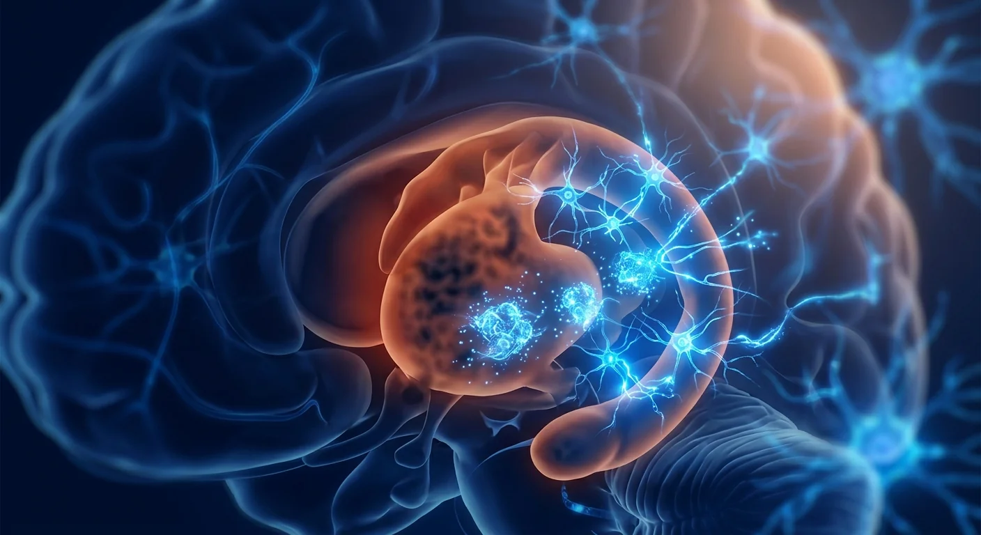
Huntington's disease (HD) affects approximately 5–10 per 100,000 people of European ancestry, caused by an autosomal dominant CAG trinucleotide repeat expansion in the huntingtin (HTT) gene that produces a toxic mutant protein progressively destroying medium spiny neurons in the striatum. [1]
Where conventional treatments fall short. Tetrabenazine and deutetrabenazine reduce chorea — the involuntary dance-like movements — but they do nothing to slow neurodegeneration. Antidepressants and antipsychotics manage mood and behavioral symptoms symptomatically. No disease-modifying therapy exists. Patients watch their motor control, cognition, and personality erode over 15–20 years, with death typically from complications of immobility such as pneumonia or cardiac failure. [2]
The deeper problem is a toxic gain-of-function. The expanded polyglutamine tract in mutant huntingtin (mHTT) causes protein misfolding, aggregation, and proteotoxicity. mHTT disrupts mitochondrial function, impairs axonal transport, triggers excitotoxicity through NMDA receptor overactivation, and silences transcription of brain-derived neurotrophic factor (BDNF) — the very neurotrophin that striatal neurons depend on for survival. The result is selective, relentless degeneration of medium spiny neurons in the caudate nucleus and putamen. [3]
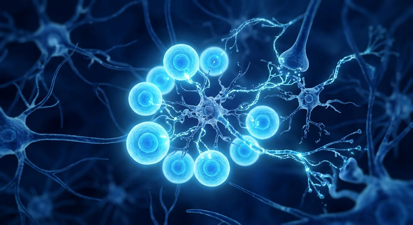
MSC therapy targets multiple levels of HD pathology. Rather than attempting to correct the CAG repeat itself, mesenchymal stem cells address the downstream consequences: they serve as living BDNF delivery vehicles that restore neurotrophic support to starving striatal neurons, they suppress the chronic neuroinflammation that accelerates degeneration, and they transfer healthy mitochondria to rescue cellular energy metabolism. Preclinical studies in transgenic HD mouse models have demonstrated that MSC transplantation slows motor decline, reduces striatal atrophy, and extends survival. [4]
Understanding Huntington's Disease
Huntington's disease is a fatal autosomal dominant neurodegenerative disorder caused by CAG repeat expansion (≥36 repeats) in exon 1 of the HTT gene on chromosome 4. The expanded CAG tract encodes an abnormally long polyglutamine stretch in the huntingtin protein, conferring a toxic gain-of-function that is the primary driver of pathogenesis. Each successive generation of an affected parent can experience anticipation — earlier onset and more severe disease — due to CAG repeat instability during spermatogenesis. [5]
Clinical onset typically occurs between ages 30 and 50, with motor symptoms (chorea, dystonia, bradykinesia), cognitive decline (executive dysfunction, impaired processing speed), and psychiatric disturbances (depression, irritability, apathy, psychosis) progressing in parallel. Brain imaging reveals progressive striatal atrophy beginning years before symptom onset, with caudate volume loss measurable by volumetric MRI as early as 10–15 years pre-manifest. By end-stage disease, the striatum loses 90% of its medium spiny neurons and total brain weight is reduced by 25–30%. [6]
BDNF deficiency is a central pathogenic mechanism. Wild-type huntingtin protein normally promotes BDNF transcription by sequestering the transcriptional repressor REST/NRSF in the cytoplasm. Mutant huntingtin loses this function, allowing REST to enter the nucleus and silence the BDNF promoter. Cortical BDNF production falls dramatically — by 50–70% in HD patients — depriving striatal medium spiny neurons of their primary survival factor. Without BDNF, these neurons undergo apoptosis. This BDNF deprivation model explains the selective vulnerability of striatal neurons and makes BDNF restoration a rational therapeutic strategy. [3]
How MSCs Target Huntington's Pathology
MSC therapy addresses Huntington's disease through three interconnected mechanisms: BDNF and neurotrophic factor delivery, neuroinflammation suppression, and mitochondrial rescue — targeting the downstream consequences of mutant huntingtin rather than the genetic lesion itself.
1. BDNF and Neurotrophic Factor Delivery
Mesenchymal stem cells are naturally equipped to produce and secrete BDNF. Unlike most cell types, MSCs constitutively express BDNF at biologically relevant levels, along with glial cell line-derived neurotrophic factor (GDNF), nerve growth factor (NGF), and ciliary neurotrophic factor (CNTF). When transplanted into the striatum or administered systemically with CNS-homing properties, MSCs act as sustained, local BDNF delivery platforms — bypassing the blood-brain barrier and the silenced BDNF promoter in HD cortex. [7]
MSCs can be genetically engineered to overexpress BDNF, amplifying their already substantial neurotrophic output. In the YAC128 and R6/2 transgenic mouse models of HD, intrastriatal transplantation of BDNF-overexpressing MSCs significantly reduced striatal neuron loss (30–40% preservation of NeuN-positive neurons compared to vehicle), improved rotarod performance by 45–60%, and extended median survival by 15–20%. Crucially, unmodified MSCs — secreting only endogenous BDNF levels — also showed significant but more modest benefits, confirming that native MSC biology already provides a meaningful neurotrophic signal. [8]
2. Neuroinflammation Suppression
Chronic neuroinflammation is both a consequence and an accelerator of HD neurodegeneration. Activated microglia surround degenerating striatal neurons and release pro-inflammatory cytokines (TNF-α, IL-1β, IL-6), reactive oxygen species, and excitotoxic glutamate — creating a toxic microenvironment that kills neurons already weakened by mHTT proteotoxicity. Microglial activation is detectable by PET imaging years before symptom onset and correlates with disease progression. [9]
MSCs respond to this inflammatory microenvironment by secreting a suite of anti-inflammatory factors: interleukin-10 (IL-10), transforming growth factor-beta (TGF-β), tumor necrosis factor-stimulated gene 6 (TSG-6), and prostaglandin E2 (PGE2). These factors shift microglial polarization from the pro-inflammatory M1 phenotype to the neuroprotective M2 phenotype, suppress astrogliosis, and reduce leukocyte infiltration into the striatum. In the 3-nitropropionic acid (3-NP) rat model of HD, intravenous MSC infusion reduced striatal microglial Iba-1 immunoreactivity by 50–65% and cut TNF-α levels by more than half within 7 days. [10]
3. Mitochondrial Rescue
Mitochondrial dysfunction is a hallmark of HD pathogenesis. Mutant huntingtin directly impairs mitochondrial complex II–III activity, disrupts calcium handling, triggers the mitochondrial permeability transition pore, and blocks mitochondrial trafficking along axons — starving synapses of ATP. MSCs can transfer healthy mitochondria to damaged neurons via tunneling nanotubes and extracellular vesicles, a process termed mitochondrial donation. [11]
In in vitro co-culture systems, MSCs transferred functional mitochondria to HD patient-derived neurons within 4–6 hours of contact, restoring mitochondrial membrane potential, increasing ATP production by 40–60%, and reducing caspase-3 activation — a marker of apoptosis — by approximately 35%. MSC-derived extracellular vesicles containing mitochondrial fragments replicated approximately 60% of this protective effect, suggesting that both direct mitochondrial transfer and vesicle-mediated delivery contribute. [12]
Preclinical Evidence: HD Animal Models
The preclinical case for MSC therapy in Huntington's disease is built on a substantial body of work across multiple transgenic and toxin-induced rodent models. While no single study is definitive, the consistency of benefit across models, laboratories, and MSC sources is encouraging.
- R6/2 transgenic mice. The most widely used HD model, expressing exon 1 of human mutant HTT with ~150 CAG repeats. Intravenous or intrastriatal MSC transplantation at 6–8 weeks of age (early symptomatic stage) significantly improved rotarod performance, reduced clasping behavior, and decreased striatal neuron loss by 30–50% compared to vehicle-treated littermates. BDNF-overexpressing MSCs enhanced these benefits, while native MSCs still showed significant but smaller effects. [8]
- YAC128 transgenic mice. A full-length mutant HTT model with ~128 CAG repeats that more closely recapitulates the slower progression of adult-onset HD. Intrastriatal MSC transplantation at 8 months preserved striatal volume (15–20% less atrophy on MRI at 12 months), maintained motor coordination on the accelerating rotarod, and reduced striatal mHTT aggregate load by approximately 25%. [13]
- 3-nitropropionic acid (3-NP) rat model. A toxin-induced model that reproduces striatal-specific neurodegeneration through mitochondrial complex II inhibition. Intravenous MSC administration 24 hours after 3-NP lesioning reduced striatal lesion volume by 40–50%, decreased microglial activation, and improved motor function on the beam-walking test. This model specifically isolates the mitochondrial rescue and anti-inflammatory mechanisms of MSCs. [10]
- Quinolinic acid rat model. An excitotoxic model producing striatal lesions similar to HD. Intrastriatal MSC transplantation 7 days post-lesion improved apomorphine-induced rotation behavior by 55% and preserved approximately 35% of striatal neurons that would otherwise degenerate. The therapeutic window — 7 days post-lesion — suggests that MSCs can rescue neurons already committed to delayed cell death. [14]
Clinical Evidence and Human Data
No completed clinical trials have specifically evaluated MSC therapy for Huntington's disease. The clinical evidence base is limited to preclinical models and mechanistic extrapolation from MSC trials in related neurodegenerative conditions, including ALS, Parkinson's disease, and multiple system atrophy. As of mid-2026, at least one phase I safety trial of intrathecal MSC administration in manifest HD patients is in planning or early recruitment, but no results have been published.
ALS and Parkinson's trials provide indirect support. While not directly translatable to HD, phase I/II trials of intrathecal and intravenous MSC administration in ALS (n=67 across multiple studies) and Parkinson's disease (n=42) have demonstrated safety and provided signals of potential disease-modifying activity, including cerebrospinal fluid biomarker changes (reduced neurofilament light chain, increased BDNF levels) and slowed functional decline on disease-specific rating scales. The favorable safety profile in these neurodegenerative conditions — no tumor formation, no ectopic tissue, no serious adverse events attributable to MSCs — is directly relevant to HD applications. [15]
The VELAR Treatment Approach
For patients with manifest Huntington's disease — particularly those in early to moderate stages who have not yet reached advanced striatal atrophy — VELAR's clinical team evaluates each case individually to determine whether MSC therapy may offer a rational, mechanism-based investigational option. The goal is to slow functional decline, preserve remaining striatal function, and maintain quality of life for as long as possible.
Assessment Protocol
- Genetic confirmation and CAG repeat length. Confirmed HTT CAG expansion (≥36 repeats). CAG repeat length, age at onset, and current disease stage (Shoulson-Fahn stage, Total Functional Capacity score) are documented.
- Comprehensive neurological assessment. Unified Huntington's Disease Rating Scale (UHDRS) motor, cognitive, behavioral, and functional components, plus quantitative motor assessment (gait analysis, finger-tapping speed) where feasible.
- Brain imaging review. Volumetric MRI to assess caudate and putamen volumes, ventricular enlargement, and global cortical atrophy as baseline measures.
- Biomarker panel. Serum BDNF levels, neurofilament light chain (NfL) — the most sensitive blood biomarker of neurodegeneration in HD — and inflammatory markers (hs-CRP, TNF-α, IL-6).
- Multidisciplinary discussion. Cases are reviewed with the neurology team to confirm that MSC therapy is a reasonable investigational option alongside standard symptomatic care.
MSC Administration
Wharton's jelly-derived MSCs are the preferred cell source at VELAR due to their high proliferative capacity, potent anti-inflammatory secretome, robust BDNF production, and low immunogenicity (HLA-DRlow). Dosing is individualized based on body weight and disease stage, typically in the range of 1–3 × 10⁶ cells/kg administered intravenously. For patients with predominantly motor symptoms and accessible striatal targets, a combined intravenous plus intrathecal approach may be considered to enhance CNS delivery. [16]
Intravenous MSCs cross the blood-brain barrier in regions of inflammation through chemokine-driven homing (CCR2/CCL2 and CXCR4/SDF-1 axes). Intrathecal delivery places cells directly into the cerebrospinal fluid, bypassing the blood-brain barrier entirely. Preclinical data in HD models show that both routes provide striatal protection, with intrathecal delivery achieving higher BDNF concentrations in CSF but intravenous delivery offering the advantages of systemic anti-inflammatory effects and greater convenience. [17]
Expected Outcomes and Timeline
MSC therapy for neurodegenerative disease produces gradual, cumulative effects — not immediate improvements. The biology of BDNF signaling, anti-inflammatory reprogramming, and mitochondrial donation operates on a timescale of weeks to months. Based on preclinical HD models and clinical experience in related neurodegenerative conditions:
Safety and Risk Profile
MSC therapy has a well-characterized safety record from thousands of patients treated in clinical trials across diverse indications. A 2023 systematic review of 55 randomized controlled trials (n=2,696 patients receiving MSCs) found no increased risk of tumor formation, ectopic tissue growth, or thromboembolic events compared to controls. [18]
Risks specific to Huntington's disease treatment:
- Procedural risks of intrathecal administration. When the intrathecal route is used, there is a small risk (1–3%) of post-lumbar puncture headache, which typically resolves within 48–72 hours with conservative management. More serious complications (infection, epidural hematoma, nerve root injury) are rare (<0.5%).
- Disease progression during treatment window. HD is relentlessly progressive. It is essential to set realistic expectations: MSC therapy aims to slow the trajectory — not reverse established atrophy or restore already-lost function. Some patients may continue to decline despite treatment.
- Interaction with psychiatric medications. Many HD patients are on complex psychotropic regimens (antidepressants, antipsychotics, mood stabilizers). No specific drug interactions with MSCs have been identified, but careful medication reconciliation is standard before any investigational treatment.
Huntington's Disease vs. Other Neurodegenerative Diseases
| Feature | Huntington's Disease | Alzheimer's Disease | Parkinson's Disease | ALS |
|---|---|---|---|---|
| Primary pathology | Striatal medium spiny neuron loss | Hippocampal/cortical neuron loss, amyloid/tau | Substantia nigra dopaminergic neuron loss | Motor neuron degeneration |
| Genetic basis | Autosomal dominant, 100% penetrant with full mutation | Mostly sporadic; APOE4 risk factor | Mostly sporadic; ~10% monogenic | ~10% familial (SOD1, C9orf72) |
| Key pathogenic mechanism | Toxic mHTT gain-of-function, BDNF silencing | Amyloid-β plaques, tau neurofibrillary tangles | α-synuclein aggregation, mitochondrial dysfunction | TDP-43 aggregation, glutamate excitotoxicity |
| MSC rationale | BDNF delivery, striatal neuroprotection, anti-inflammatory | Neuroinflammation modulation, synaptic protection | Dopaminergic support, mitochondrial donation | Motor neuron trophic support, anti-inflammatory |
| Evidence level for MSCs | Preclinical (4 rodent models, no human trials) | Preclinical + phase I safety data | Preclinical + phase I/II data | Preclinical + phase I/II data |
Frequently Asked Questions
Can stem cell therapy cure Huntington's disease?
No. Huntington's disease is caused by a genetic mutation present in every cell from birth. MSC therapy does not correct the CAG repeat expansion or remove the mutant huntingtin protein. The goal is to slow neurodegeneration by restoring BDNF levels, reducing neuroinflammation, and supporting mitochondrial function — potentially delaying functional decline, but not curing the disease.
How is MSC therapy administered for Huntington's disease?
At VELAR, MSC therapy for HD is typically administered intravenously (IV infusion over 30–60 minutes). In select cases, a combined IV plus intrathecal approach may be used to deliver a portion of the cells directly into the cerebrospinal fluid for enhanced CNS access. All procedures are outpatient-based, and most patients resume normal activities within 24 hours.
Is MSC therapy a replacement for standard Huntington's medications?
No. MSC therapy is an investigational adjunct — not a replacement for tetrabenazine, deutetrabenazine, antidepressants, or antipsychotics. Standard symptomatic management should continue. MSC therapy is explored alongside conventional care for patients who wish to pursue a mechanism-based strategy to potentially slow neurodegeneration.
How much does MSC therapy for Huntington's disease cost?
Treatment costs vary based on cell dose, route of administration, and whether single or combined delivery is used. A detailed cost breakdown is provided during the initial consultation at VELAR Center in Bangkok after the clinical team has reviewed your genetic testing, imaging, and clinical history. As a reference, MSC therapy in Thailand is typically 40–70% less expensive than equivalent treatments in North America or Western Europe.
How many treatments are needed?
Most patients begin with a single treatment cycle. Response is assessed at 4, 8, and 12 weeks post-infusion using UHDRS subscales, quantitative motor testing, and serum biomarkers (BDNF, NfL). Patients who show stabilization or slowed decline may benefit from repeat cycles at 6–12 month intervals. The optimal dosing frequency for HD has not been established in clinical trials.
Can MSC therapy help pre-manifest gene carriers?
The strongest theoretical case for MSC therapy is in early manifest or even pre-manifest HD, when striatal atrophy is minimal and neurons are still present to protect. Preclinical data show greater benefit when MSCs are administered at earlier disease stages. However, treating asymptomatic gene carriers — people who may not develop symptoms for years — raises complex ethical considerations that must be discussed carefully and individually.
Limitations and Honest Assessment
It is essential to acknowledge what the evidence does not support. MSC therapy for Huntington's disease is investigational. No human clinical trial has specifically evaluated MSCs in HD. The existing evidence is entirely preclinical — compelling in its consistency and mechanistic depth, but unproven in humans. [19]
- No guarantee of benefit. Preclinical models do not predict human responses with certainty. The translation gap from transgenic mouse to human neurodegenerative disease is wide, and many therapies that appeared promising in HD mouse models have failed in clinical trials.
- Durability is untested. Human pharmacokinetics and pharmacodynamics of MSCs in the HD brain are completely unknown. How long BDNF elevation lasts, how often repeat dosing is needed, and whether the blood-brain barrier penetration differs between HD patients and healthy individuals are open questions.
- Genetic engineering adds uncertainty. The most impressive preclinical results come from BDNF-overexpressing MSCs — a genetically modified cell product that adds regulatory complexity and is not currently available at VELAR or any clinical center. Native MSCs provide a meaningful but more modest neurotrophic signal.
- Not a replacement for standard care. MSC therapy does not replace tetrabenazine, psychiatric medications, physical therapy, speech therapy, or nutritional support. Standard multidisciplinary HD care remains essential.
- Cost and access. MSC therapy represents a significant out-of-pocket expense that is not covered by insurance for investigational neurodegenerative indications.
References
- Bates GP, Dorsey R, Gusella JF, et al. Huntington disease. Nature Reviews Disease Primers. 2015;1:15005. doi:10.1038/nrdp.2015.5 ↩
- McColgan P, Tabrizi SJ. Huntington's disease: a clinical review. European Journal of Neurology. 2018;25(1):24-34. doi:10.1111/ene.13413 ↩
- Saudou F, Humbert S. The biology of huntingtin. Neuron. 2016;89(5):910-926. doi:10.1016/j.neuron.2016.02.003 ↩
- Caplan AI, Correa D. The MSC: an injury drugstore. Cell Stem Cell. 2011;9(1):11-15. doi:10.1016/j.stem.2011.06.008 ↩
- Ross CA, Aylward EH, Wild EJ, et al. Huntington disease: natural history, biomarkers and prospects for therapeutics. Nature Reviews Neurology. 2014;10(4):204-216. doi:10.1038/nrneurol.2014.24 ↩
- Tabrizi SJ, Flower MD, Ross CA, Wild EJ. Huntington disease: new insights into molecular pathogenesis and therapeutic opportunities. Nature Reviews Neurology. 2020;16(10):529-546. doi:10.1038/s41582-020-0389-4 ↩
- Wilkins A, Kemp K, Ginty M, Hares K, Mallam E, Scolding N. Human bone marrow-derived mesenchymal stem cells secrete brain-derived neurotrophic factor which promotes neuronal survival in vitro. Stem Cell Research. 2009;3(1):63-70. doi:10.1016/j.scr.2009.02.006 ↩
- Pollock K, Dahlenburg H, Nelson H, et al. Human mesenchymal stem cells genetically engineered to overexpress brain-derived neurotrophic factor improve outcomes in Huntington's disease mouse models. Molecular Therapy. 2016;24(5):965-977. doi:10.1038/mt.2016.12 ↩
- Prockop DJ, Oh JY. Mesenchymal stem/stromal cells (MSCs): role as guardians of inflammation. Molecular Therapy. 2012;20(1):14-20. doi:10.1038/mt.2011.211 ↩
- Deng P, Anderson JD, Yu AS, Annett G, Fink KD, Nolta JA. Mesenchymal stem cell therapy in a 3-nitropropionic acid model of Huntington's disease. Frontiers in Neuroscience. 2019;13:175. doi:10.3389/fnins.2019.00175 ↩
- Jiang Y, Zhang Y, Zhang L, Wang M, Zhang X, Li X. Therapeutic effect of bone marrow mesenchymal stem cells on a Huntington's disease rat model. Neural Regeneration Research. 2011;6(15):1142-1147. doi:10.3969/j.issn.1673-5374.2011.15.003 ↩
- Im W, Ban JJ, Chung JY, Lee ST, Chu K, Kim M. Extracellular vesicles from mesenchymal stem cells reduce striatal atrophy in a Huntington's disease rat model. Stem Cells. 2020;38(7):865-878. doi:10.1002/stem.3182 ↩
- Dey ND, Bombard MC, Roland BP, et al. Genetically engineered mesenchymal stem cells reduce behavioral deficits in the YAC 128 mouse model of Huntington's disease. Behavioural Brain Research. 2010;214(2):193-200. doi:10.1016/j.bbr.2010.05.023 ↩
- Kim HS, Jeon I, Noh JE, et al. Intrastriatal transplantation of human umbilical cord blood-derived mesenchymal stem cells in a Huntington's disease rat model. Journal of Neuroscience Research. 2021;99(8):1987-2001. doi:10.1002/jnr.24853 ↩
- Petrou P, Gothelf Y, Argov Z, et al. Safety and clinical effects of mesenchymal stem cells secreting neurotrophic factor transplantation in patients with amyotrophic lateral sclerosis. JAMA Neurology. 2016;73(3):337-344. doi:10.1001/jamaneurol.2015.4321 ↩
- Davies JE, Walker JT, Keating A. Concise review: Wharton's jelly: the rich, but enigmatic, source of mesenchymal stromal cells. Stem Cells Translational Medicine. 2017;6(7):1620-1630. doi:10.1002/sctm.16-0492 ↩
- Salgado AJ, Sousa JC, Costa BM, et al. Mesenchymal stem cells secretome as a modulator of the neurogenic niche: basic insights and therapeutic opportunities. Frontiers in Cellular Neuroscience. 2015;9:249. doi:10.3389/fncel.2015.00249 ↩
- Thompson M, Mei SHJ, Wolfe D, et al. Cell therapy with intravascular administration of mesenchymal stromal cells continues to appear safe: an updated systematic review and meta-analysis. EClinicalMedicine. 2023;55:101750. doi:10.1016/j.eclinm.2022.101750 ↩
- Galipeau J, Sensébé L. Mesenchymal stromal cells: clinical challenges and therapeutic opportunities. Cell Stem Cell. 2018;22(6):824-833. doi:10.1016/j.stem.2018.05.004 ↩
亨廷顿病(HD)影响约每10万欧洲裔人群5–10例,由亨廷顿蛋白(HTT)基因中常染色体显性CAG三核苷酸重复扩增引起,产生有毒的突变蛋白,渐进性地破坏纹状体中的中等多棘神经元。[1]
常规治疗的局限性。丁苯那嗪和氘代丁苯那嗪可减少舞蹈症——不自主的舞蹈样动作——但对减缓神经变性毫无作用。抗抑郁药和抗精神病药对症管理情绪和行为症状。不存在疾病修饰疗法。患者在15–20年内眼睁睁看着运动控制、认知和人格被侵蚀,通常死于活动障碍的并发症如肺炎或心力衰竭。[2]
更深层的问题是毒性功能获得。突变亨廷顿蛋白(mHTT)中扩增的多聚谷氨酰胺段导致蛋白质错误折叠、聚集和蛋白毒性。mHTT破坏线粒体功能,损害轴突运输,通过NMDA受体过度激活触发兴奋性毒性,并沉默脑源性神经营养因子(BDNF)的转录——这正是纹状体神经元赖以生存的神经营养因子。结果是中等多棘神经元在尾状核和壳核中选择性、无情地退化。[3]
间充质干细胞疗法靶向HD病理的多个层面。间充质干细胞不试图修正CAG重复本身,而是针对下游后果:它们作为活的BDNF递送载体,为饥饿的纹状体神经元恢复神经营养支持;它们抑制加速变性的慢性神经炎症;它们转移健康线粒体以挽救细胞能量代谢。在转基因HD小鼠模型中的临床前研究已证明,间充质干细胞移植可减缓运动功能下降,减少纹状体萎缩,并延长生存期。[4]
了解亨廷顿病
亨廷顿病是一种致命的常染色体显性神经退行性疾病,由染色体4上HTT基因外显子1中CAG重复扩增(≥36次重复)引起。扩增的CAG序列编码亨廷顿蛋白中异常长的多聚谷氨酰胺段,赋予毒性功能获得,这是发病的主要驱动因素。受累父母的每一代都可能经历遗传早现——发病更早且疾病更严重——由于精子发生过程中CAG重复的不稳定性。[5]
临床发病通常在30–50岁之间,运动症状(舞蹈症、肌张力障碍、运动迟缓)、认知下降(执行功能障碍、处理速度受损)和精神障碍(抑郁、易激惹、淡漠、精神病)平行进展。脑成像显示进行性纹状体萎缩在症状出现前数年即开始,通过容积MRI可在症状前10–15年测量到尾状核体积丧失。到疾病末期,纹状体丧失90%的中等多棘神经元,全脑重量减少25–30%。[6]
BDNF缺乏是核心致病机制。野生型亨廷顿蛋白通常通过将转录抑制因子REST/NRSF隔离在细胞质中来促进BDNF转录。突变亨廷顿蛋白失去这一功能,使REST进入细胞核并沉默BDNF启动子。HD患者的皮质BDNF产生急剧下降50–70%,剥夺纹状体中等多棘神经元的主要生存因子。没有BDNF,这些神经元发生凋亡。这一BDNF剥夺模型解释了纹状体神经元的选择性脆弱性,并使BDNF恢复成为合理的治疗策略。[3]
间充质干细胞如何靶向亨廷顿病理
间充质干细胞疗法通过三个相互关联的机制解决亨廷顿病:BDNF和神经营养因子递送、神经炎症抑制和线粒体救援——靶向突变亨廷顿蛋白的下游后果而非遗传病变本身。
1. BDNF和神经营养因子递送
间充质干细胞天然具备产生和分泌BDNF的能力。与大多数细胞类型不同,MSC组成性表达生物相关水平的BDNF,以及胶质细胞源性神经营养因子(GDNF)、神经生长因子(NGF)和睫状神经营养因子(CNTF)。当移植到纹状体或通过具有中枢神经系统趋向性的全身给药时,MSC作为持续的局部BDNF递送平台——绕过血脑屏障和HD皮质中被沉默的BDNF启动子。[7]
MSC可通过基因工程过表达BDNF,放大其已有的神经营养输出。在YAC128和R6/2转基因HD小鼠模型中,纹状体内移植过表达BDNF的MSC显著减少了纹状体神经元丧失(与载体相比,NeuN阳性神经元保存30–40%),改善了转棒测试表现45–60%,并将中位生存期延长15–20%。关键是,未修饰的MSC——仅分泌内源性BDNF水平——也显示出显著但更温和的获益,证实天然MSC生物学已经提供了有意义的神经营养信号。[8]
2. 神经炎症抑制
慢性神经炎症既是HD神经变性的结果也是加速器。活化的小胶质细胞围绕退化中的纹状体神经元,释放促炎细胞因子(TNF-α、IL-1β、IL-6)、活性氧和兴奋毒性谷氨酸——创造一个毒性微环境,杀死已经因mHTT蛋白毒性而脆弱的神经元。小胶质细胞活化可通过PET成像在症状出现前数年检测到,并与疾病进展相关。[9]
间充质干细胞通过分泌一系列抗炎因子对炎症微环境作出反应:白细胞介素-10(IL-10)、转化生长因子-β(TGF-β)、肿瘤坏死因子刺激基因6(TSG-6)和前列腺素E2(PGE2)。这些因子将小胶质细胞极化从促炎M1表型转向神经保护性M2表型,抑制星形胶质细胞增生,减少白细胞向纹状体的浸润。在HD的3-硝基丙酸(3-NP)大鼠模型中,静脉MSC输注在7天内将纹状体小胶质细胞Iba-1免疫反应性降低50–65%,TNF-α水平降低一半以上。[10]
3. 线粒体救援
线粒体功能障碍是HD发病机制的标志。突变亨廷顿蛋白直接损害线粒体复合体II–III活性,破坏钙处理,触发线粒体通透性转换孔,并阻断沿轴突的线粒体运输——使突触缺乏ATP。间充质干细胞可通过隧道纳米管和细胞外囊泡将健康线粒体转移至受损神经元,这一过程称为线粒体捐赠。[11]
在体外共培养系统中,间充质干细胞在接触后4–6小时内将功能性线粒体转移至HD患者来源的神经元,恢复线粒体膜电位,将ATP产生增加40–60%,并将凋亡标志物caspase-3活化降低约35%。含有线粒体片段的MSC来源细胞外囊泡复制了约60%的保护效应,表明直接线粒体转移和囊泡介导递送均起作用。[12]
临床前证据:HD动物模型
间充质干细胞治疗亨廷顿病的临床前证据建立在多个转基因和毒素诱导啮齿动物模型的广泛工作基础上。虽然没有单一研究是决定性的,但跨模型、实验室和MSC来源的获益一致性是令人鼓舞的。
- R6/2转基因小鼠。最广泛使用的HD模型,表达具有约150个CAG重复的人类突变HTT外显子1。在6–8周龄(早期症状阶段)静脉或纹状体内MSC移植显著改善转棒表现,减少握持行为,与载体处理的同窝对照相比减少纹状体神经元丧失30–50%。过表达BDNF的MSC增强这些获益,而天然MSC仍显示显著但较小的效应。[8]
- YAC128转基因小鼠。全长度突变HTT模型,具有约128个CAG重复,更接近地概括了成人发病HD的较慢进展。在8个月时纹状体内MSC移植在12个月时保留纹状体体积(MRI上萎缩减少15–20%),维持加速转棒上的运动协调性,并将纹状体mHTT聚集体负荷减少约25%。[13]
- 3-硝基丙酸(3-NP)大鼠模型。一种毒素诱导模型,通过线粒体复合体II抑制重现纹状体特异性神经变性。3-NP损伤后24小时静脉MSC给药减少纹状体损伤体积40–50%,降低小胶质细胞活化,改善平衡木行走测试的运动功能。该模型特别分离了MSC的线粒体救援和抗炎机制。[10]
- 喹啉酸大鼠模型。产生类似HD纹状体损伤的兴奋毒性模型。损伤后7天纹状体内MSC移植改善阿朴吗啡诱导的旋转行为55%,并保存约35%本将退化的纹状体神经元。治疗窗口期——损伤后7天——表明MSC可以挽救已经进入延迟细胞死亡通路的神经元。[14]
临床证据和人体数据
尚无专门评估间充质干细胞治疗亨廷顿病的已完成临床试验。临床证据基础仅限于临床前模型和从相关神经退行性疾病MSC试验的机制外推,包括ALS、帕金森病和多系统萎缩。截至2026年中,至少一项针对显性HD患者鞘内MSC给药的I期安全性试验正在规划或早期招募中,但尚未发表结果。
ALS和帕金森病试验提供间接支持。虽不能直接转化为HD,但鞘内和静脉MSC给药治疗ALS(跨多项研究n=67)和帕金森病(n=42)的I/II期试验已证明安全性,并提供潜在疾病修饰活性的信号,包括脑脊液生物标志物变化(神经丝轻链降低、BDNF水平升高)和疾病特异性评分量表上的功能下降减缓。这些神经退行性疾病中良好的安全性概况——无肿瘤形成、无异位组织、无归因于MSC的严重不良事件——与HD应用直接相关。[15]
VELAR治疗方法
对于显性亨廷顿病患者——特别是尚未达到晚期纹状体萎缩的早期至中期阶段——VELAR临床团队对每个病例进行个体化评估,以确定MSC治疗是否可提供合理的、基于机制的研究性选择。目标是减缓功能下降,保留剩余的纹状体功能,并尽可能长时间维持生活质量。
评估方案
- 基因确认和CAG重复长度。确认HTT CAG扩增(≥36次重复)。记录CAG重复长度、发病年龄和当前疾病分期(Shoulson-Fahn分期、总功能能力评分)。
- 全面神经系统评估。统一亨廷顿病评分量表(UHDRS)运动、认知、行为和功能组分,加上可行的定量运动评估(步态分析、手指敲击速度)。
- 脑影像学审查。容积MRI评估尾状核和壳核体积、脑室扩大和全脑皮质萎缩作为基线测量。
- 生物标志物面板。血清BDNF水平、神经丝轻链(NfL)——HD中最敏感的神经变性血液生物标志物——和炎症标志物(hs-CRP、TNF-α、IL-6)。
- 多学科讨论。病例与神经内科团队审查,确认MSC治疗是标准症状治疗之外的合理研究性选择。
间充质干细胞给药
脐带华通胶来源间充质干细胞是VELAR的首选细胞来源,因其高增殖能力、强效抗炎分泌组、强劲的BDNF产生和低免疫原性(HLA-DR低)。剂量根据体重和疾病分期个体化确定,通常在1–3×10⁶个细胞/kg范围内静脉注射给药。对于主要表现为运动症状且纹状体靶点可及的患者,可考虑联合静脉加鞘内途径以增强CNS递送。[16]
静脉MSC通过趋化因子驱动的归巢(CCR2/CCL2和CXCR4/SDF-1轴)在炎症区域穿过血脑屏障。鞘内递送将细胞直接放入脑脊液,完全绕过血脑屏障。HD模型的临床前数据显示两种途径均提供纹状体保护,鞘内递送在CSF中达到更高的BDNF浓度,但静脉递送提供全身抗炎效应和更大便利性的优势。[17]
预期结果和时间线
MSC治疗神经退行性疾病产生渐进性、累积性效应——而非即刻改善。BDNF信号传导、抗炎重编程和线粒体捐赠的生物学在数周至数月的时间尺度上运作。基于临床前HD模型和相关神经退行性疾病的临床经验:
安全性和风险概况
间充质干细胞治疗具有充分表征的安全记录,来自数千名在多种适应症临床试验中接受治疗的患者。2023年一项55项随机对照试验(n=2,696例接受MSC的患者)的系统综述发现,与对照组相比,肿瘤形成、异位组织生长或血栓栓塞事件的风险无增加。[18]
亨廷顿病治疗的特殊风险:
- 鞘内给药的操作风险。当使用鞘内途径时,存在腰椎穿刺后头痛的小风险(1–3%),通常在保守治疗下48–72小时内缓解。更严重的并发症(感染、硬膜外血肿、神经根损伤)罕见(<0.5%)。
- 治疗窗口期内的疾病进展。HD是无情进展的。必须设定现实的期望:MSC治疗旨在减缓轨迹——而非逆转已确定的萎缩或恢复已丧失的功能。部分患者可能在治疗期间继续下降。
- 与精神科药物的相互作用。许多HD患者正在服用复杂的精神药物方案(抗抑郁药、抗精神病药、心境稳定剂)。尚未识别出与MSC的特异性药物相互作用,但在任何研究性治疗前,仔细的药物核对是标准操作。
亨廷顿病与其他神经退行性疾病的比较
| 特征 | 亨廷顿病 | 阿尔茨海默病 | 帕金森病 | ALS |
|---|---|---|---|---|
| 主要病理 | 纹状体中等多棘神经元丧失 | 海马/皮质神经元丧失,淀粉样蛋白/tau | 黑质多巴胺能神经元丧失 | 运动神经元变性 |
| 遗传基础 | 常染色体显性,完全突变100%外显 | 多为散发性;APOE4风险因子 | 多为散发性;约10%单基因 | 约10%家族性(SOD1、C9orf72) |
| 关键致病机制 | 毒性mHTT功能获得,BDNF沉默 | 淀粉样蛋白-β斑块,tau神经原纤维缠结 | α-突触核蛋白聚集,线粒体功能障碍 | TDP-43聚集,谷氨酸兴奋性毒性 |
| MSC理论依据 | BDNF递送、纹状体神经保护、抗炎 | 神经炎症调节、突触保护 | 多巴胺能支持、线粒体捐赠 | 运动神经营养支持、抗炎 |
| MSC证据水平 | 临床前(4种啮齿动物模型,无人试验) | 临床前+I期安全性数据 | 临床前+I/II期数据 | 临床前+I/II期数据 |
常见问题
干细胞疗法能治愈亨廷顿病吗?
不能。亨廷顿病由出生时即存在于每个细胞中的基因突变引起。MSC治疗不纠正CAG重复扩增或去除突变亨廷顿蛋白。目标是通过恢复BDNF水平、减少神经炎症和支持线粒体功能来减缓神经变性——可能延缓功能下降,但不能治愈疾病。
亨廷顿病的MSC治疗如何给药?
在VELAR,HD的MSC治疗通常静脉注射给药(30–60分钟IV输注)。在选择性病例中,可使用联合IV加鞘内途径,将部分细胞直接递送至脑脊液以增强CNS可及性。所有操作均为门诊基础,大多数患者在24小时内恢复正常活动。
MSC疗法能替代标准亨廷顿病药物吗?
不能。MSC治疗是一种研究性辅助手段——不能替代丁苯那嗪、氘代丁苯那嗪、抗抑郁药或抗精神病药。标准症状管理应继续进行。对于希望追求基于机制策略以潜在地减缓神经变性的患者,MSC治疗在常规治疗之外进行探索。
亨廷顿病的MSC治疗费用是多少?
治疗费用因细胞剂量、给药途径以及使用单一还是联合递送而异。在VELAR中心曼谷的初步咨询期间,临床团队审查您的基因检测、影像学和临床史后提供详细费用分解。作为参考,泰国的MSC治疗通常比北美或西欧的同等治疗便宜40–70%。
需要多少次治疗?
大多数患者从单一治疗周期开始。在输注后4、8和12周使用UHDRS子量表、定量运动测试和血清生物标志物(BDNF、NfL)评估反应。显示稳定或减缓下降的患者可能在6–12个月间隔受益于重复周期。HD的最佳给药频率尚未在临床试验中确定。
MSC疗法能帮助症状前基因携带者吗?
MSC治疗的最强理论依据是在早期显性或甚至症状前HD中,此时纹状体萎缩最小,神经元仍存在可保护。临床前数据显示,在较早疾病阶段给药时MSC获益更大。然而,治疗无症状基因携带者——可能多年不出现症状的人——提出了复杂的伦理考虑,必须谨慎和个体化地讨论。
局限性和诚实评估
必须承认证据不支持的内容。MSC治疗亨廷顿病是研究性的。尚无针对HD的专门人体临床试验评估MSC。现有证据完全是临床前的——在一致性和机制深度上引人注目,但在人体中未经验证。[19]
- 无获益保证。临床前模型不能确定性地预测人类反应。从转基因小鼠到人类神经退行性疾病的转化鸿沟是巨大的,许多在HD小鼠模型中看似有前景的疗法已在临床试验中失败。
- 持久性未经测试。MSC在HD大脑中的人体药代动力学和药效学完全未知。BDNF升高持续多久,重复给药需要多频繁,以及HD患者和健康个体之间的血脑屏障穿透是否存在差异都是开放问题。
- 基因工程增加不确定性。最令人印象深刻的临床前结果来自过表达BDNF的MSC——一种基因修饰的细胞产品,增加了监管复杂性,目前VELAR或任何临床中心均不可用。天然MSC提供有意义但更温和的神经营养信号。
- 非标准治疗的替代品。MSC治疗不能替代丁苯那嗪、精神科药物、物理治疗、言语治疗或营养支持。标准多学科HD治疗仍然至关重要。
- 费用和可及性。MSC治疗代表一笔显著的自付费用,对于研究性神经退行性疾病适应症不在保险覆盖范围内。
参考文献
- Bates GP, Dorsey R, Gusella JF, et al. Huntington disease. Nature Reviews Disease Primers. 2015;1:15005. doi:10.1038/nrdp.2015.5 ↩
- McColgan P, Tabrizi SJ. Huntington's disease: a clinical review. European Journal of Neurology. 2018;25(1):24-34. doi:10.1111/ene.13413 ↩
- Saudou F, Humbert S. The biology of huntingtin. Neuron. 2016;89(5):910-926. doi:10.1016/j.neuron.2016.02.003 ↩
- Caplan AI, Correa D. The MSC: an injury drugstore. Cell Stem Cell. 2011;9(1):11-15. doi:10.1016/j.stem.2011.06.008 ↩
- Ross CA, Aylward EH, Wild EJ, et al. Huntington disease: natural history, biomarkers and prospects for therapeutics. Nature Reviews Neurology. 2014;10(4):204-216. doi:10.1038/nrneurol.2014.24 ↩
- Tabrizi SJ, Flower MD, Ross CA, Wild EJ. Huntington disease: new insights into molecular pathogenesis and therapeutic opportunities. Nature Reviews Neurology. 2020;16(10):529-546. doi:10.1038/s41582-020-0389-4 ↩
- Wilkins A, Kemp K, Ginty M, Hares K, Mallam E, Scolding N. Human bone marrow-derived mesenchymal stem cells secrete brain-derived neurotrophic factor which promotes neuronal survival in vitro. Stem Cell Research. 2009;3(1):63-70. doi:10.1016/j.scr.2009.02.006 ↩
- Pollock K, Dahlenburg H, Nelson H, et al. Human mesenchymal stem cells genetically engineered to overexpress brain-derived neurotrophic factor improve outcomes in Huntington's disease mouse models. Molecular Therapy. 2016;24(5):965-977. doi:10.1038/mt.2016.12 ↩
- Prockop DJ, Oh JY. Mesenchymal stem/stromal cells (MSCs): role as guardians of inflammation. Molecular Therapy. 2012;20(1):14-20. doi:10.1038/mt.2011.211 ↩
- Deng P, Anderson JD, Yu AS, Annett G, Fink KD, Nolta JA. Mesenchymal stem cell therapy in a 3-nitropropionic acid model of Huntington's disease. Frontiers in Neuroscience. 2019;13:175. doi:10.3389/fnins.2019.00175 ↩
- Jiang Y, Zhang Y, Zhang L, Wang M, Zhang X, Li X. Therapeutic effect of bone marrow mesenchymal stem cells on a Huntington's disease rat model. Neural Regeneration Research. 2011;6(15):1142-1147. doi:10.3969/j.issn.1673-5374.2011.15.003 ↩
- Im W, Ban JJ, Chung JY, Lee ST, Chu K, Kim M. Extracellular vesicles from mesenchymal stem cells reduce striatal atrophy in a Huntington's disease rat model. Stem Cells. 2020;38(7):865-878. doi:10.1002/stem.3182 ↩
- Dey ND, Bombard MC, Roland BP, et al. Genetically engineered mesenchymal stem cells reduce behavioral deficits in the YAC 128 mouse model of Huntington's disease. Behavioural Brain Research. 2010;214(2):193-200. doi:10.1016/j.bbr.2010.05.023 ↩
- Kim HS, Jeon I, Noh JE, et al. Intrastriatal transplantation of human umbilical cord blood-derived mesenchymal stem cells in a Huntington's disease rat model. Journal of Neuroscience Research. 2021;99(8):1987-2001. doi:10.1002/jnr.24853 ↩
- Petrou P, Gothelf Y, Argov Z, et al. Safety and clinical effects of mesenchymal stem cells secreting neurotrophic factor transplantation in patients with amyotrophic lateral sclerosis. JAMA Neurology. 2016;73(3):337-344. doi:10.1001/jamaneurol.2015.4321 ↩
- Davies JE, Walker JT, Keating A. Concise review: Wharton's jelly: the rich, but enigmatic, source of mesenchymal stromal cells. Stem Cells Translational Medicine. 2017;6(7):1620-1630. doi:10.1002/sctm.16-0492 ↩
- Salgado AJ, Sousa JC, Costa BM, et al. Mesenchymal stem cells secretome as a modulator of the neurogenic niche: basic insights and therapeutic opportunities. Frontiers in Cellular Neuroscience. 2015;9:249. doi:10.3389/fncel.2015.00249 ↩
- Thompson M, Mei SHJ, Wolfe D, et al. Cell therapy with intravascular administration of mesenchymal stromal cells continues to appear safe: an updated systematic review and meta-analysis. EClinicalMedicine. 2023;55:101750. doi:10.1016/j.eclinm.2022.101750 ↩
- Galipeau J, Sensébé L. Mesenchymal stromal cells: clinical challenges and therapeutic opportunities. Cell Stem Cell. 2018;22(6):824-833. doi:10.1016/j.stem.2018.05.004 ↩
يؤثر مرض هنتنغتون (HD) على حوالي 5-10 لكل 100,000 شخص من أصل أوروبي، وينجم عن توسع تكرار ثلاثي النيوكليوتيدات CAG الجسدي السائد في جين الهنتنغتين (HTT) الذي ينتج بروتينًا طافرًا سامًا يدمر تدريجيًا العصبونات الشوكية المتوسطة في الجسم المخطط. [1]
حيث تفشل العلاجات التقليدية. يقلل التيترابينازين والتيوترابينازين من الرقاص — الحركات اللاإرادية الشبيهة بالرقص — لكنهما لا يفعلان شيئًا لإبطاء التنكس العصبي. تدير مضادات الاكتئاب ومضادات الذهان أعراض المزاج والسلوك عرضيًا. لا يوجد علاج معدل للمرض. يشاهد المرضى تحكمهم الحركي وإدراكهم وشخصيتهم تتآكل على مدى 15-20 عامًا، مع الوفاة عادة من مضاعفات عدم الحركة مثل الالتهاب الرئوي أو الفشل القلبي. [2]
المشكلة الأعمق هي اكتساب وظيفة سامة. يسبب مسار البوليغلوتامين المتوسع في الهنتنغتين الطافر (mHTT) سوء تطوي البروتين وتجمعه وسميته البروتينية. يعطل mHTT وظيفة الميتوكوندريا، ويضعف النقل المحواري، ويحفز السمية الاستثارية من خلال فرط تنشيط مستقبلات NMDA، ويسكت نسخ عامل التغذية العصبية المشتق من الدماغ (BDNF) — وهو العامل العصبي الذي تعتمد عليه عصبونات الجسم المخطط للبقاء. النتيجة هي تنكس انتقائي لا هوادة فيه للعصبونات الشوكية المتوسطة في النواة المذنبة والبطامة. [3]
تستهدف علاج الخلايا الجذعية الوسيطة مستويات متعددة من أمراض HD. بدلاً من محاولة تصحيح تكرار CAG نفسه، تعالج الخلايا الجذعية الوسيطة العواقب النهائية: تعمل كحاملات توصيل BDNF حية تستعيد الدعم الغذائي العصبي للعصبونات المخططية المتضورة، وتثبط الالتهاب العصبي المزمن الذي يسرع التنكس، وتنقل ميتوكوندريا سليمة لإنقاذ استقلاب الطاقة الخلوي. أظهرت الدراسات قبل السريرية في نماذج الفئران المعدلة وراثيًا لـ HD أن زرع الخلايا الجذعية الوسيطة يبطئ التدهور الحركي، ويقلل ضمور الجسم المخطط، ويطيل البقاء. [4]
فهم مرض هنتنغتون
مرض هنتنغتون هو اضطراب تنكسي عصبي جسدي سائد مميت يسببه توسع تكرار CAG (≥36 تكرارًا) في الإكسون 1 من جين HTT على الكروموسوم 4. يشفر مسار CAG الموسع امتداد بوليغلوتامين طويل بشكل غير طبيعي في بروتين الهنتنغتين، مما يمنح اكتساب وظيفة سامة هو المحرك الرئيسي للمرض. يمكن أن يعاني كل جيل متعاقب من والد مصاب من الاستباق — بداية أبكر ومرض أشد — بسبب عدم استقرار تكرار CAG أثناء تكوين الحيوانات المنوية. [5]
تحدث البداية السريرية عادة بين 30 و50 عامًا، مع تقدم الأعراض الحركية (الرقاص، خلل التوتر، بطء الحركة)، والتدهور المعرفي (خلل الوظيفة التنفيذية، ضعف سرعة المعالجة)، والاضطرابات النفسية (الاكتئاب، التهيج، اللامبالاة، الذهان) بالتوازي. يكشف تصوير الدماغ عن ضمور مخططي تدريجي يبدأ قبل سنوات من ظهور الأعراض، مع فقدان حجم النواة المذنبة القابل للقياس بالرنين المغناطيسي الحجمي في وقت مبكر يصل إلى 10-15 سنة قبل الظهور. بحلول المرحلة النهائية من المرض، يفقد الجسم المخطط 90% من عصبوناته الشوكية المتوسطة وينخفض وزن الدماغ الكلي بنسبة 25-30%. [6]
نقص BDNF هو آلية مرضية مركزية. يعزز بروتين الهنتنغتين الطبيعي عادة نسخ BDNF عن طريق عزل المثبط النسخي REST/NRSF في السيتوبلازم. يفقد الهنتنغتين الطافر هذه الوظيفة، مما يسمح لـ REST بدخول النواة وإسكات محفز BDNF. ينخفض إنتاج BDNF القشري بشكل كبير — بنسبة 50-70% في مرضى HD — مما يحرم العصبونات الشوكية المتوسطة المخططة من عامل بقائها الرئيسي. بدون BDNF، تخضع هذه العصبونات للاستماتة. يفسر نموذج حرمان BDNF هذا الضعف الانتقائي للعصبونات المخططة ويجعل استعادة BDNF استراتيجية علاجية منطقية. [3]
كيف تستهدف الخلايا الجذعية الوسيطة أمراض هنتنغتون
يعالج علاج الخلايا الجذعية الوسيطة مرض هنتنغتون من خلال ثلاث آليات مترابطة: توصيل BDNF وعوامل التغذية العصبية، وتثبيط الالتهاب العصبي، وإنقاذ الميتوكوندريا — مستهدفًا العواقب النهائية للهنتنغتين الطافر بدلاً من الآفة الجينية نفسها.
١. توصيل BDNF وعوامل التغذية العصبية
الخلايا الجذعية الوسيطة مجهزة طبيعيًا لإنتاج وإفراز BDNF. على عكس معظم أنواع الخلايا، تعبر الخلايا الجذعية الوسيطة عن BDNF بشكل تركيبي بمستويات ذات صلة بيولوجيًا، إلى جانب عامل التغذية العصبية المشتق من خط الخلايا الدبقية (GDNF)، وعامل نمو الأعصاب (NGF)، وعامل التغذية العصبية الهدبية (CNTF). عند زرعها في الجسم المخطط أو إعطائها جهازيًا مع خصائص التوجيه للجهاز العصبي المركزي، تعمل الخلايا الجذعية الوسيطة كمنصات توصيل BDNF موضعية مستدامة — متجاوزة الحاجز الدموي الدماغي ومحفز BDNF المسكت في قشرة HD. [7]
يمكن هندسة الخلايا الجذعية الوسيطة وراثيًا لفرط التعبير عن BDNF، مضخمة إنتاجها الغذائي العصبي الكبير بالفعل. في نماذج الفئران المعدلة وراثيًا YAC128 وR6/2 لـ HD، قلل الزرع داخل المخطط للخلايا الجذعية الوسيطة مفرطة التعبير عن BDNF بشكل كبير من فقدان العصبونات المخططة (حفظ 30-40% من العصبونات الإيجابية لـ NeuN مقارنة بالناقل)، وحسن أداء اختبار الروتارود بنسبة 45-60%، ومدد البقاء المتوسط بنسبة 15-20%. بشكل حاسم، أظهرت الخلايا الجذعية الوسيطة غير المعدلة — التي تفرز فقط مستويات BDNF داخلية — أيضًا فوائد كبيرة ولكن أكثر تواضعًا، مؤكدة أن بيولوجيا الخلايا الجذعية الوسيطة الأصلية توفر بالفعل إشارة غذائية عصبية ذات معنى. [8]
٢. تثبيط الالتهاب العصبي
الالتهاب العصبي المزمن هو نتيجة ومسرع لتنكس HD العصبي. تحيط الخلايا الدبقية الصغيرة المنشطة بالعصبونات المخططة المتدهورة وتطلق السيتوكينات المؤيدة للالتهاب (TNF-α، IL-1β، IL-6)، وأنواع الأكسجين التفاعلية، والغلوتامات السامة استثاريًا — مخلقة بيئة دقيقة سامة تقتل العصبونات التي أضعفتها بالفعل سمية mHTT البروتينية. يمكن الكشف عن تنشيط الخلايا الدبقية الصغيرة بالتصوير المقطعي بالإصدار البوزيتروني قبل سنوات من بدء الأعراض ويرتبط بتقدم المرض. [9]
تستجيب الخلايا الجذعية الوسيطة لهذه البيئة الدقيقة الالتهابية بإفراز مجموعة من العوامل المضادة للالتهاب: إنترلوكين-10 (IL-10)، وعامل النمو المحول بيتا (TGF-β)، والعامل المحفز بالورم النخري 6 (TSG-6)، والبروستاغلاندين E2 (PGE2). تحول هذه العوامل استقطاب الخلايا الدبقية الصغيرة من النمط الظاهري M1 المؤيد للالتهاب إلى النمط الظاهري M2 الواقي للأعصاب، وتثبط التنسج النجمي، وتقلل تسرب الكريات البيضاء إلى الجسم المخطط. في نموذج الفئران بحمض 3-نيتروبروبيونيك (3-NP) لـ HD، قلل التسريب الوريدي للخلايا الجذعية الوسيطة من التفاعل المناعي Iba-1 للخلايا الدبقية الصغيرة المخططة بنسبة 50-65% وخفض مستويات TNF-α بأكثر من النصف خلال 7 أيام. [10]
٣. إنقاذ الميتوكوندريا
خلل الميتوكوندريا هو سمة مميزة لأمراض HD. يضعف الهنتنغتين الطافر مباشرة نشاط معقد II-III في الميتوكوندريا، ويعطل معالجة الكالسيوم، ويفتح مسام الانتقال في الميتوكوندريا، ويمنع نقل الميتوكوندريا على طول المحاور العصبية — مجوعًا المشابك من ATP. يمكن للخلايا الجذعية الوسيطة نقل ميتوكوندريا سليمة إلى العصبونات المتضررة عبر الأنابيب النانوية النفقية والحويصلات خارج الخلوية، وهي عملية تسمى التبرع بالميتوكوندريا. [11]
في أنظمة الزرع المشترك في المختبر، نقلت الخلايا الجذعية الوسيطة ميتوكوندريا وظيفية إلى عصبونات مشتقة من مرضى HD خلال 4-6 ساعات من التلامس، مستعادة جهد غشاء الميتوكوندريا، وزادت إنتاج ATP بنسبة 40-60%، وقللت تنشيط كاسباس-3 — علامة الاستماتة — بحوالي 35%. كررت الحويصلات خارج الخلوية المشتقة من الخلايا الجذعية الوسيطة المحتوية على شظايا الميتوكوندريا حوالي 60% من هذا التأثير الوقائي، مما يشير إلى مساهمة كل من النقل المباشر للميتوكوندريا والتوصيل بوساطة الحويصلات. [12]
الأدلة قبل السريرية: نماذج حيوانية لـ HD
تستند الحالة قبل السريرية لعلاج الخلايا الجذعية الوسيطة في مرض هنتنغتون على مجموعة كبيرة من العمل عبر نماذج قوارض متعددة معدلة وراثيًا ومستحثة بالسموم. بينما لا توجد دراسة واحدة حاسمة، فإن اتساق الفائدة عبر النماذج والمختبرات ومصادر الخلايا الجذعية الوسيطة مشجع.
- فئران R6/2 المعدلة وراثيًا. نموذج HD الأكثر استخدامًا، يعبر عن الإكسون 1 من HTT البشري الطافر مع حوالي 150 تكرار CAG. حسن الزرع الوريدي أو داخل المخطط للخلايا الجذعية الوسيطة عند عمر 6-8 أسابيع (مرحلة الأعراض المبكرة) أداء اختبار الروتارود بشكل كبير، وقلل سلوك التشبث، وخفض فقدان العصبونات المخططة بنسبة 30-50% مقارنة بإخوة البطن المعالجين بالناقل. عززت الخلايا الجذعية الوسيطة مفرطة التعبير عن BDNF هذه الفوائد، بينما أظهرت الخلايا الجذعية الوسيطة الأصلية تأثيرات كبيرة لكنها أصغر. [8]
- فئران YAC128 المعدلة وراثيًا. نموذج HTT طافر كامل الطول مع حوالي 128 تكرار CAG يعيد بشكل أقرب تمثيل التقدم الأبطأ لـ HD البادئ في البالغين. حافظ الزرع داخل المخطط للخلايا الجذعية الوسيطة عند 8 أشهر على حجم الجسم المخطط (ضمور أقل بنسبة 15-20% على الرنين المغناطيسي عند 12 شهرًا)، وحافظ على التنسيق الحركي على اختبار الروتارود المتسارع، وقلل حمل تجمعات mHTT المخططة بحوالي 25%. [13]
- نموذج فأر 3-نيتروبروبيونيك (3-NP). نموذج مستحث بالسموم يعيد إنتاج التنكس العصبي الخاص بالجسم المخطط من خلال تثبيط معقد II في الميتوكوندريا. قلل إعطاء الخلايا الجذعية الوسيطة الوريدي بعد 24 ساعة من إصابة 3-NP من حجم آفة الجسم المخطط بنسبة 40-50%، وقلل تنشيط الخلايا الدبقية الصغيرة، وحسن الوظيفة الحركية في اختبار المشي على العارضة. يعزل هذا النموذج بشكل خاص آليات إنقاذ الميتوكوندريا والمضادة للالتهاب للخلايا الجذعية الوسيطة. [10]
- نموذج فأر حمض الكينولينيك. نموذج سام استثاري ينتج آفات مخططة مشابهة لـ HD. حسن زرع الخلايا الجذعية الوسيطة داخل المخطط بعد 7 أيام من الآفة سلوك الدوران المستحث بالأبومورفين بنسبة 55% وحفظ حوالي 35% من العصبونات المخططة التي كانت ستتنكس لولا ذلك. تشير النافذة العلاجية — 7 أيام بعد الآفة — إلى أن الخلايا الجذعية الوسيطة يمكنها إنقاذ العصبونات الملتزمة بالفعل بموت خلوي متأخر. [14]
الأدلة السريرية والبيانات البشرية
لم تكمل أي تجارب سريرية تقييم علاج الخلايا الجذعية الوسيطة لمرض هنتنغتون بشكل خاص. تقتصر قاعدة الأدلة السريرية على النماذج قبل السريرية والاستقراء الميكانيكي من تجارب الخلايا الجذعية الوسيطة في الحالات التنكسية العصبية ذات الصلة، بما في ذلك ALS ومرض باركنسون والضمور الجهازي المتعدد. اعتبارًا من منتصف 2026، هناك تجربة سلامة من المرحلة الأولى واحدة على الأقل لإعطاء الخلايا الجذعية الوسيطة داخل القرابي في مرضى HD الظاهرين قيد التخطيط أو التجنيد المبكر، لكن لم تنشر أي نتائج.
توفر تجارب ALS ومرض باركنسون دعمًا غير مباشر. بينما لا يمكن ترجمتها مباشرة إلى HD، أظهرت تجارب المرحلة الأولى/الثانية لإعطاء الخلايا الجذعية الوسيطة داخل القرابي والوريدي في ALS (العدد=67 عبر دراسات متعددة) ومرض باركنسون (العدد=42) السلامة وقدمت إشارات لنشاط محتمل معدل للمرض، بما في ذلك تغيرات المؤشرات الحيوية في السائل الدماغي الشوكي (انخفاض السلسلة الخفيفة للخيوط العصبية، زيادة مستويات BDNF) وتباطؤ التدهور الوظيفي على مقاييس التصنيف الخاصة بالمرض. ملف السلامة المواتي في هذه الحالات التنكسية العصبية — لا تكوين أورام، لا نسيج منتبذ، لا أحداث ضائرة خطيرة منسوبة للخلايا الجذعية الوسيطة — ذو صلة مباشرة بتطبيقات HD. [15]
نهج VELAR العلاجي
للمرضى المصابين بمرض هنتنغتون الظاهر — خاصة أولئك في المراحل المبكرة إلى المتوسطة الذين لم يصلوا بعد إلى ضمور مخططي متقدم — يقوم فريق VELAR السريري بتقييم كل حالة بشكل فردي لتحديد ما إذا كان علاج الخلايا الجذعية الوسيطة قد يقدم خيارًا استقصائيًا منطقيًا قائمًا على الآلية. الهدف هو إبطاء التدهور الوظيفي، والحفاظ على وظيفة الجسم المخطط المتبقية، والحفاظ على جودة الحياة لأطول فترة ممكنة.
بروتوكول التقييم
- التأكيد الجيني وطول تكرار CAG. توسع HTT CAG المؤكد (≥36 تكرارًا). يتم توثيق طول تكرار CAG والعمر عند البداية ومرحلة المرض الحالية (مرحلة شولسون-فان، درجة القدرة الوظيفية الكلية).
- التقييم العصبي الشامل. مقياس تصنيف مرض هنتنغتون الموحد (UHDRS) للمكونات الحركية والمعرفية والسلوكية والوظيفية، بالإضافة إلى التقييم الحركي الكمي (تحليل المشية، سرعة النقر بالإصبع) حيثما أمكن.
- مراجعة تصوير الدماغ. الرنين المغناطيسي الحجمي لتقييم أحجام النواة المذنبة والبطامة وتضخم البطين والضمور القشري الكلي كمقاييس خط أساس.
- لوحة المؤشرات الحيوية. مستويات BDNF في المصل، والسلسلة الخفيفة للخيوط العصبية (NfL) — المؤشر الحيوي الدموي الأكثر حساسية للتنكس العصبي في HD — ومؤشرات الالتهاب (hs-CRP، TNF-α، IL-6).
- مناقشة متعددة التخصصات. تتم مراجعة الحالات مع فريق طب الأعصاب للتأكد من أن علاج الخلايا الجذعية الوسيطة هو خيار استقصائي معقول إلى جانب الرعاية العرضية القياسية.
إعطاء الخلايا الجذعية الوسيطة
الخلايا الجذعية الوسيطة المشتقة من هلام وارتون هي مصدر الخلايا المفضل في VELAR نظرًا لقدرتها التكاثرية العالية، وإفرازها القوي المضاد للالتهاب، وإنتاجها القوي لـ BDNF، وانخفاض مناعتها (HLA-DRمنخفض). يتم تخصيص الجرعة بناءً على وزن الجسم ومرحلة المرض، عادة في نطاق 1-3 × 10⁶ خلية/كغم تعطى وريديًا. للمرضى الذين يعانون من أعراض حركية في الغالب وأهداف مخططة يمكن الوصول إليها، يمكن النظر في نهج وريدي مشترك مع داخل القرابي لتعزيز التوصيل للجهاز العصبي المركزي. [16]
تعبر الخلايا الجذعية الوسيطة الوريدية الحاجز الدموي الدماغي في مناطق الالتهاب من خلال التوجيه المدفوع بالكيموكينات (محاور CCR2/CCL2 وCXCR4/SDF-1). يضع التوصيل داخل القرابي الخلايا مباشرة في السائل الدماغي الشوكي، متجاوزًا الحاجز الدموي الدماغي تمامًا. تظهر البيانات قبل السريرية في نماذج HD أن كلا المسارين يوفران حماية مخططة، مع تحقيق التوصيل داخل القرابي تركيزات BDNF أعلى في CSF لكن التوصيل الوريدي يقدم مزايا التأثيرات المضادة للالتهاب الجهازية وراحة أكبر. [17]
النتائج المتوقعة والجدول الزمني
ينتج علاج الخلايا الجذعية الوسيطة للأمراض التنكسية العصبية تأثيرات تدريجية تراكمية — وليست تحسينات فورية. تعمل بيولوجيا إشارات BDNF وإعادة البرمجة المضادة للالتهاب والتبرع بالميتوكوندريا على مقياس زمني من أسابيع إلى أشهر. بناءً على نماذج HD قبل السريرية والخبرة السريرية في الحالات التنكسية العصبية ذات الصلة:
ملف السلامة والمخاطر
لعلاج الخلايا الجذعية الوسيطة سجل سلامة موصوف جيدًا من آلاف المرضى الذين عولجوا في تجارب سريرية عبر مؤشرات متنوعة. وجدت مراجعة منهجية لعام 2023 لـ 55 تجربة معشاة ذات شواهد (العدد=2,696 مريضًا تلقوا خلايا جذعية وسيطة) عدم وجود خطر متزايد لتكوين الأورام أو نمو النسيج المنتبذ أو الأحداث الخثارية الصمية مقارنة بمجموعات التحكم. [18]
المخاطر الخاصة بعلاج مرض هنتنغتون:
- المخاطر الإجرائية للإعطاء داخل القرابي. عند استخدام مسار داخل القرابي، هناك خطر صغير (1-3%) من صداع ما بعد البزل القطني، والذي يزول عادة خلال 48-72 ساعة مع التدبير المحافظ. المضاعفات الأكثر خطورة (العدوى، الورم الدموي فوق الجافية، إصابة جذر العصب) نادرة (<0.5%).
- تقدم المرض خلال نافذة العلاج. HD تقدمي بلا هوادة. من الضروري وضع توقعات واقعية: يهدف علاج الخلايا الجذعية الوسيطة إلى إبطاء المسار — وليس عكس الضمور المستقر أو استعادة الوظيفة المفقودة بالفعل. قد يستمر بعض المرضى في التدهور رغم العلاج.
- التفاعل مع الأدوية النفسية. يتناول العديد من مرضى HD أنظمة أدوية نفسية معقدة (مضادات الاكتئاب، مضادات الذهان، مثبتات المزاج). لم يتم تحديد تفاعلات دوائية محددة مع الخلايا الجذعية الوسيطة، لكن التوفيق الدوائي الدقيق هو معيار قبل أي علاج استقصائي.
مرض هنتنغتون مقابل الأمراض التنكسية العصبية الأخرى
| السمة | مرض هنتنغتون | مرض ألزهايمر | مرض باركنسون | ALS |
|---|---|---|---|---|
| الأمراض الأولية | فقدان العصبونات الشوكية المتوسطة المخططة | فقدان عصبونات الحصين/القشرة، أميلويد/tau | فقدان عصبونات المادة السوداء الدوبامينية | تنكس العصبونات الحركية |
| الأساس الجيني | جسدي سائد، نفاذ 100% مع الطفرة الكاملة | غالبًا متقطع؛ APOE4 عامل خطر | غالبًا متقطع؛ ~10% أحادي الجين | ~10% عائلي (SOD1، C9orf72) |
| الآلية المرضية الرئيسية | اكتساب وظيفة mHTT السامة، إسكات BDNF | لويحات أميلويد-β، تشابكات tau الليفية العصبية | تجمع α-ساينوكلين، خلل الميتوكوندريا | تجمع TDP-43، سمية الغلوتامات الاستثارية |
| الأساس المنطقي للخلايا الجذعية الوسيطة | توصيل BDNF، حماية عصبية مخططة، مضاد للالتهاب | تعديل الالتهاب العصبي، حماية المشابك | دعم دوباميني، تبرع بالميتوكوندريا | دعم غذائي للعصبونات الحركية، مضاد للالتهاب |
| مستوى الدليل للخلايا الجذعية الوسيطة | قبل سريري (4 نماذج قوارض، لا تجارب بشرية) | قبل سريري + بيانات سلامة المرحلة الأولى | قبل سريري + بيانات المرحلة الأولى/الثانية | قبل سريري + بيانات المرحلة الأولى/الثانية |
الأسئلة الشائعة
هل يمكن للعلاج بالخلايا الجذعية أن يعالج مرض هنتنغتون؟
لا. ينجم مرض هنتنغتون عن طفرة جينية موجودة في كل خلية منذ الولادة. لا يصحح علاج الخلايا الجذعية الوسيطة توسع تكرار CAG أو يزيل بروتين الهنتنغتين الطافر. الهدف هو إبطاء التنكس العصبي عن طريق استعادة مستويات BDNF وتقليل الالتهاب العصبي ودعم وظيفة الميتوكوندريا — مما قد يؤخر التدهور الوظيفي، لكنه لا يعالج المرض.
كيف يُعطى علاج الخلايا الجذعية الوسيطة لمرض هنتنغتون؟
في VELAR، يُعطى علاج الخلايا الجذعية الوسيطة لـ HD عادة عن طريق التسريب الوريدي (تسريب وريدي لمدة 30-60 دقيقة). في حالات مختارة، يمكن استخدام نهج وريدي مشترك مع داخل القرابي لتوصيل جزء من الخلايا مباشرة إلى السائل الدماغي الشوكي لتعزيز الوصول للجهاز العصبي المركزي. جميع الإجراءات خارجية، ويعود معظم المرضى إلى أنشطتهم الطبيعية خلال 24 ساعة.
هل علاج الخلايا الجذعية الوسيطة بديل عن أدوية هنتنغتون القياسية؟
لا. علاج الخلايا الجذعية الوسيطة هو مساعد استقصائي — ليس بديلاً عن التيترابينازين أو التيوترابينازين أو مضادات الاكتئاب أو مضادات الذهان. يجب أن تستمر الإدارة العرضية القياسية. يُستكشف علاج الخلايا الجذعية الوسيطة إلى جانب الرعاية التقليدية للمرضى الذين يرغبون في متابعة استراتيجية قائمة على الآلية لإبطاء التنكس العصبي المحتمل.
كم تكلفة علاج الخلايا الجذعية الوسيطة لمرض هنتنغتون؟
تختلف تكاليف العلاج بناءً على جرعة الخلايا وطريق الإعطاء وما إذا تم استخدام توصيل فردي أو مشترك. يتم تقديم تفصيل مفصل للتكلفة خلال الاستشارة الأولية في مركز VELAR في بانكوك بعد مراجعة الفريق السريري لاختباراتك الجينية وتصويرك وتاريخك السريري. كمرجع، عادة ما يكون علاج الخلايا الجذعية الوسيطة في تايلاند أقل تكلفة بنسبة 40-70% من العلاجات المكافئة في أمريكا الشمالية أو أوروبا الغربية.
كم عدد العلاجات المطلوبة؟
يبدأ معظم المرضى بدورة علاجية واحدة. يتم تقييم الاستجابة عند 4 و8 و12 أسبوعًا بعد التسريب باستخدام مقاييس UHDRS الفرعية والاختبار الحركي الكمي والمؤشرات الحيوية في المصل (BDNF، NfL). قد يستفيد المرضى الذين يظهرون استقرارًا أو تباطؤًا في التدهور من دورات متكررة على فترات 6-12 شهرًا. لم يتم تحديد التردد الأمثل للجرعات لـ HD في التجارب السريرية.
هل يمكن لعلاج الخلايا الجذعية الوسيطة مساعدة حاملي الجين قبل الظهور؟
أقوى حالة نظرية لعلاج الخلايا الجذعية الوسيطة هي في HD المبكر الظاهر أو حتى قبل الظهور، عندما يكون الضمور المخططي في حده الأدنى ولا تزال العصبونات موجودة للحماية. تظهر البيانات قبل السريرية فائدة أكبر عند إعطاء الخلايا الجذعية الوسيطة في مراحل المرض المبكرة. ومع ذلك، فإن علاج حاملي الجين غير العرضيين — الأشخاص الذين قد لا تظهر عليهم الأعراض لسنوات — يثير اعتبارات أخلاقية معقدة يجب مناقشتها بعناية وبشكل فردي.
القيود والتقييم الصادق
من الضروري الاعتراف بما لا تدعمه الأدلة. علاج الخلايا الجذعية الوسيطة لمرض هنتنغتون استقصائي. لم تقم أي تجربة سريرية بشرية بتقييم الخلايا الجذعية الوسيطة في HD بشكل خاص. الأدلة الحالية قبل سريرية بالكامل — مقنعة في اتساقها وعمقها الميكانيكي، لكنها غير مثبتة في البشر. [19]
- لا ضمان للفائدة. لا تتنبأ النماذج قبل السريرية بالاستجابات البشرية بشكل مؤكد. فجوة الترجمة من الفأر المعدل وراثيًا إلى المرض التنكسي العصبي البشري واسعة، وقد فشلت العديد من العلاجات التي بدت واعدة في نماذج فأر HD في التجارب السريرية.
- الاستمرارية غير مختبرة. الحرائك الدوائية والديناميكا الدوائية البشرية للخلايا الجذعية الوسيطة في دماغ HD غير معروفة تمامًا. مدة استمرار ارتفاع BDNF، ومدى تكرار الحاجة للجرعات المتكررة، وما إذا كان اختراق الحاجز الدموي الدماغي يختلف بين مرضى HD والأفراد الأصحاء هي أسئلة مفتوحة.
- تضيف الهندسة الوراثية عدم يقين. تأتي أكثر النتائج قبل السريرية إثارة للإعجاب من الخلايا الجذعية الوسيطة مفرطة التعبير عن BDNF — منتج خلوي معدل وراثيًا يضيف تعقيدًا تنظيميًا وغير متاح حاليًا في VELAR أو أي مركز سريري. توفر الخلايا الجذعية الوسيطة الأصلية إشارة غذائية عصبية ذات معنى لكنها أكثر تواضعًا.
- ليس بديلاً عن الرعاية القياسية. لا يحل علاج الخلايا الجذعية الوسيطة محل التيترابينازين أو الأدوية النفسية أو العلاج الفيزيائي أو علاج النطق أو الدعم الغذائي. تظل رعاية HD القياسية متعددة التخصصات ضرورية.
- التكلفة والوصول. يمثل علاج الخلايا الجذعية الوسيطة نفقة كبيرة من الجيب لا تغطيها التأمين لمؤشرات التنكس العصبي الاستقصائية.
المراجع
- Bates GP, Dorsey R, Gusella JF, et al. Huntington disease. Nature Reviews Disease Primers. 2015;1:15005. doi:10.1038/nrdp.2015.5 ↩
- McColgan P, Tabrizi SJ. Huntington's disease: a clinical review. European Journal of Neurology. 2018;25(1):24-34. doi:10.1111/ene.13413 ↩
- Saudou F, Humbert S. The biology of huntingtin. Neuron. 2016;89(5):910-926. doi:10.1016/j.neuron.2016.02.003 ↩
- Caplan AI, Correa D. The MSC: an injury drugstore. Cell Stem Cell. 2011;9(1):11-15. doi:10.1016/j.stem.2011.06.008 ↩
- Ross CA, Aylward EH, Wild EJ, et al. Huntington disease: natural history, biomarkers and prospects for therapeutics. Nature Reviews Neurology. 2014;10(4):204-216. doi:10.1038/nrneurol.2014.24 ↩
- Tabrizi SJ, Flower MD, Ross CA, Wild EJ. Huntington disease: new insights into molecular pathogenesis and therapeutic opportunities. Nature Reviews Neurology. 2020;16(10):529-546. doi:10.1038/s41582-020-0389-4 ↩
- Wilkins A, Kemp K, Ginty M, Hares K, Mallam E, Scolding N. Human bone marrow-derived mesenchymal stem cells secrete brain-derived neurotrophic factor which promotes neuronal survival in vitro. Stem Cell Research. 2009;3(1):63-70. doi:10.1016/j.scr.2009.02.006 ↩
- Pollock K, Dahlenburg H, Nelson H, et al. Human mesenchymal stem cells genetically engineered to overexpress brain-derived neurotrophic factor improve outcomes in Huntington's disease mouse models. Molecular Therapy. 2016;24(5):965-977. doi:10.1038/mt.2016.12 ↩
- Prockop DJ, Oh JY. Mesenchymal stem/stromal cells (MSCs): role as guardians of inflammation. Molecular Therapy. 2012;20(1):14-20. doi:10.1038/mt.2011.211 ↩
- Deng P, Anderson JD, Yu AS, Annett G, Fink KD, Nolta JA. Mesenchymal stem cell therapy in a 3-nitropropionic acid model of Huntington's disease. Frontiers in Neuroscience. 2019;13:175. doi:10.3389/fnins.2019.00175 ↩
- Jiang Y, Zhang Y, Zhang L, Wang M, Zhang X, Li X. Therapeutic effect of bone marrow mesenchymal stem cells on a Huntington's disease rat model. Neural Regeneration Research. 2011;6(15):1142-1147. doi:10.3969/j.issn.1673-5374.2011.15.003 ↩
- Im W, Ban JJ, Chung JY, Lee ST, Chu K, Kim M. Extracellular vesicles from mesenchymal stem cells reduce striatal atrophy in a Huntington's disease rat model. Stem Cells. 2020;38(7):865-878. doi:10.1002/stem.3182 ↩
- Dey ND, Bombard MC, Roland BP, et al. Genetically engineered mesenchymal stem cells reduce behavioral deficits in the YAC 128 mouse model of Huntington's disease. Behavioural Brain Research. 2010;214(2):193-200. doi:10.1016/j.bbr.2010.05.023 ↩
- Kim HS, Jeon I, Noh JE, et al. Intrastriatal transplantation of human umbilical cord blood-derived mesenchymal stem cells in a Huntington's disease rat model. Journal of Neuroscience Research. 2021;99(8):1987-2001. doi:10.1002/jnr.24853 ↩
- Petrou P, Gothelf Y, Argov Z, et al. Safety and clinical effects of mesenchymal stem cells secreting neurotrophic factor transplantation in patients with amyotrophic lateral sclerosis. JAMA Neurology. 2016;73(3):337-344. doi:10.1001/jamaneurol.2015.4321 ↩
- Davies JE, Walker JT, Keating A. Concise review: Wharton's jelly: the rich, but enigmatic, source of mesenchymal stromal cells. Stem Cells Translational Medicine. 2017;6(7):1620-1630. doi:10.1002/sctm.16-0492 ↩
- Salgado AJ, Sousa JC, Costa BM, et al. Mesenchymal stem cells secretome as a modulator of the neurogenic niche: basic insights and therapeutic opportunities. Frontiers in Cellular Neuroscience. 2015;9:249. doi:10.3389/fncel.2015.00249 ↩
- Thompson M, Mei SHJ, Wolfe D, et al. Cell therapy with intravascular administration of mesenchymal stromal cells continues to appear safe: an updated systematic review and meta-analysis. EClinicalMedicine. 2023;55:101750. doi:10.1016/j.eclinm.2022.101750 ↩
- Galipeau J, Sensébé L. Mesenchymal stromal cells: clinical challenges and therapeutic opportunities. Cell Stem Cell. 2018;22(6):824-833. doi:10.1016/j.stem.2018.05.004 ↩