Hidradenitis suppurativa (HS) — also known as acne inversa — affects approximately 1–4% of the global population, yet it remains one of the most underdiagnosed and undertreated conditions in dermatology. HS is not a disorder of hygiene or lifestyle: it is a chronic, progressive, inflammatory disease of the terminal hair follicle, producing recurrent painful nodules, abscesses, and ultimately draining sinus tracts in intertriginous areas (axillae, groin, inframammary folds, and perianal region). The disease burden is among the highest of any dermatological condition, with patients reporting Dermatology Life Quality Index (DLQI) scores comparable to those of end-stage renal disease, and a 2.4-fold increased risk of completed suicide [1].
Where conventional therapies fall short. The treatment ladder for HS includes topical clindamycin, oral tetracyclines, combined oral contraceptives, spironolactone, the TNF-α inhibitor adalimumab (the only FDA/EMA-approved biologic for HS), and surgical excision of affected skin. Yet many patients cycle through these options with incomplete relief: adalimumab achieves HiSCR 50 (≥50% reduction in abscess and nodule count) in only 42–59% of patients at week 12, and surgical excision — while effective — carries significant morbidity, requires prolonged wound healing, and is complicated by recurrence rates of 20–50% at the margins of excised tissue [2]. For Hurley stage III disease characterized by confluent sinus tracts and extensive scarring, no pharmacotherapy achieves consistent disease control.
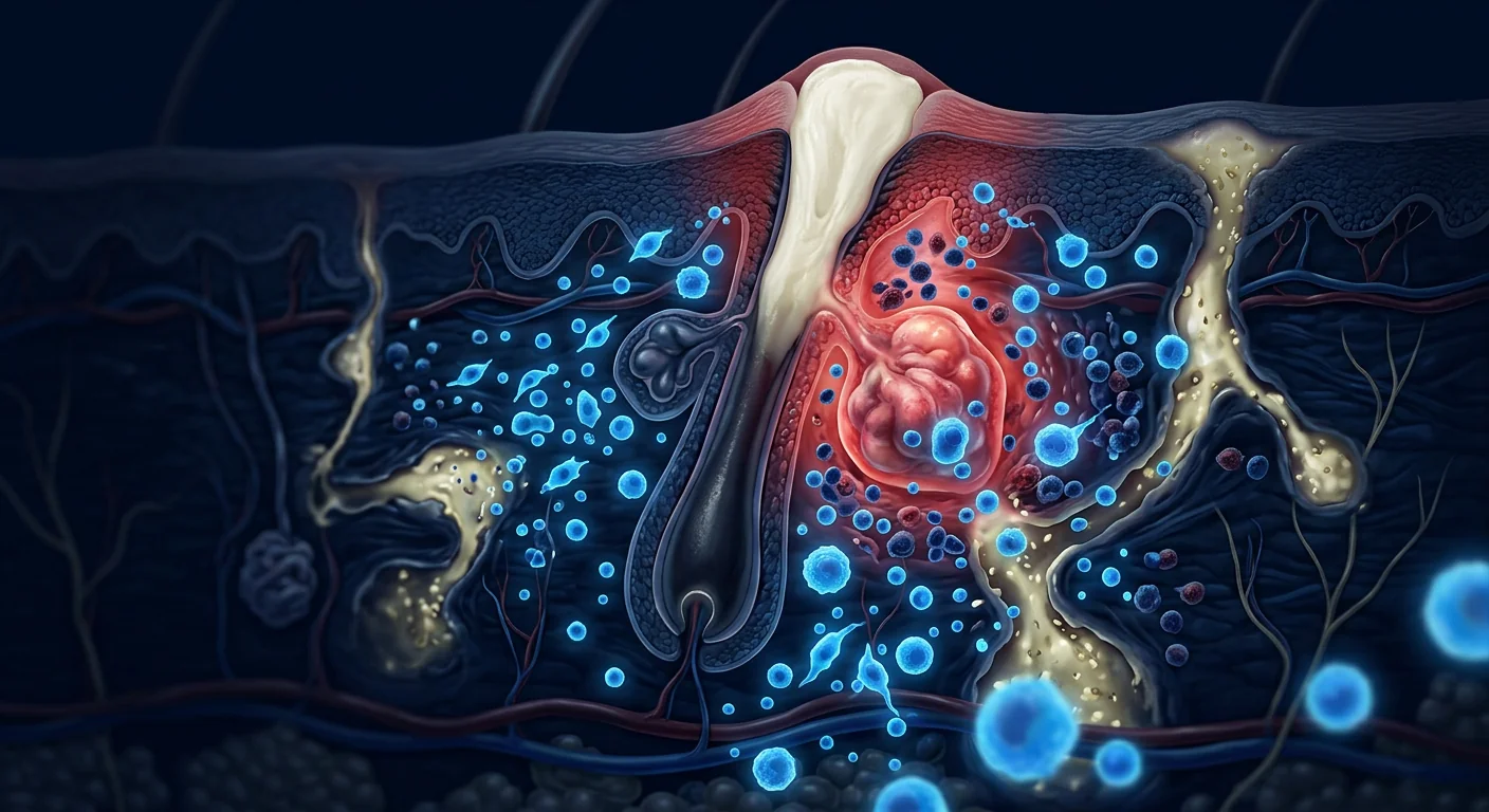
The deeper problem is follicular, hyperkeratotic, and immune-mediated. The initiating event in HS is follicular occlusion — hyperkeratosis of the infundibular portion of the terminal hair follicle leads to follicular plugging, dilation, and eventual rupture. When the follicular contents (keratin, sebum, bacteria, and cellular debris) spill into the surrounding dermis, a potent innate immune response is triggered, characterized by activation of the NLRP3 inflammasome and massive release of IL-1β, which in turn drives the recruitment of neutrophils, macrophages, and Th17 cells, producing the painful inflammatory nodules and the subsequent tissue destruction [3]. In later stages, the chronic inflammation creates epithelialized sinus tracts — permanent conduits that resist both immunological clearance and pharmacological penetration. The disease is thus simultaneously a disorder of follicular biology, innate immunity, and aberrant wound healing — which is why targeting any single cytokine has proven insufficient for advanced disease [4].
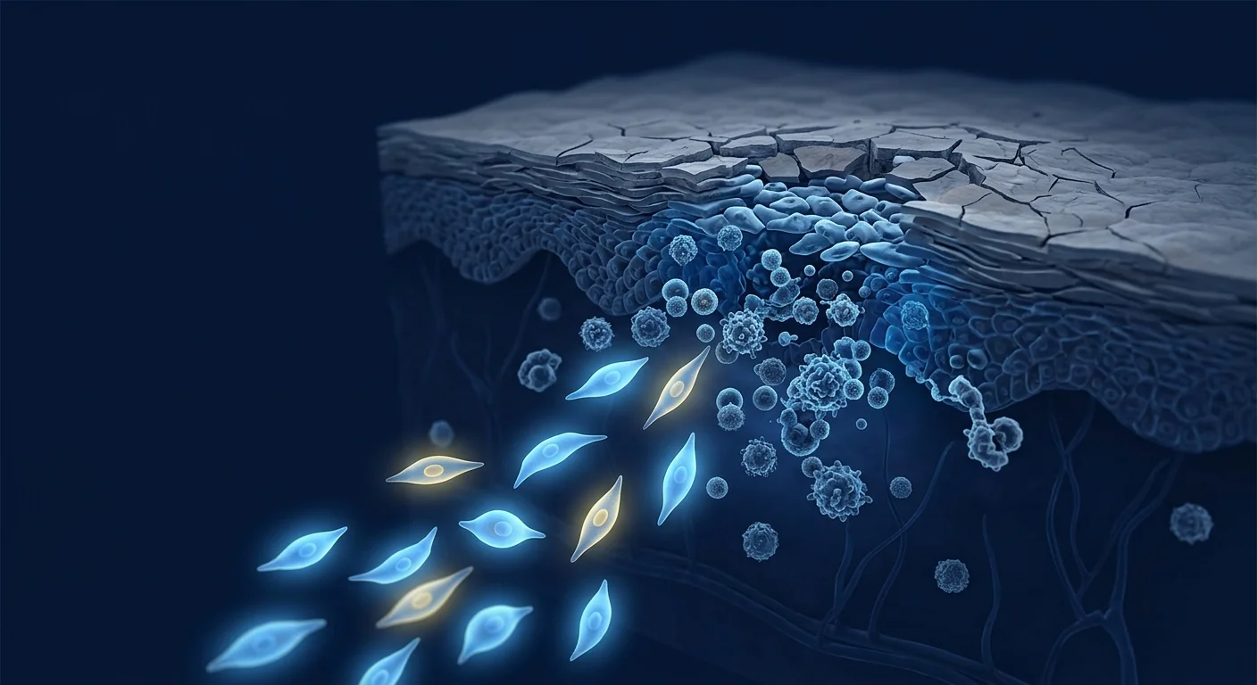
MSC therapy targets multiple nodes in the HS pathophysiology simultaneously. Mesenchymal stem cells exert immunomodulatory, anti-inflammatory, anti-fibrotic, and pro-angiogenic effects that address the follicular occlusion cascade at several levels — reducing the NLRP3/IL-1β axis, shifting macrophage polarization from M1 to M2, suppressing neutrophil extracellular trap (NET) formation, and promoting orderly wound healing rather than the scarring fibrosis that drives sinus tract perpetuation [5]. This multi-target, network-level mechanism distinguishes MSC therapy from single-cytokine biologics and makes it a compelling investigational approach for a disease characterized by multi-pathway immune dysregulation.
How Mesenchymal Stem Cells Target Hidradenitis Suppurativa
The therapeutic effects of MSCs in HS are mediated through at least five interconnected mechanisms, each addressing a distinct node in the disease cascade:
1. Suppression of the NLRP3 inflammasome and IL-1β axis. The NLRP3 inflammasome is a cytosolic protein complex that serves as a sensor of cellular damage and a master activator of IL-1β — the cytokine that sits at the apex of the HS inflammatory cascade. MSCs potently suppress NLRP3 inflammasome assembly and activation through multiple mechanisms including mitochondrial transfer, release of the anti-inflammatory protein TSG-6 (TNF-α-stimulated gene 6), and secretion of extracellular vesicles containing miRNAs that silence NLRP3 transcription [6]. In vitro, MSC co-culture reduces IL-1β secretion from activated macrophages by 60–80%, an effect that is partially abrogated when NLRP3 is constitutively expressed — confirming that inflammasome suppression is a direct mechanism of MSC action.
2. Shift from M1 to M2 macrophage polarization. HS lesions are dominated by M1 (pro-inflammatory) macrophages that produce TNF-α, IL-1β, IL-6, and reactive oxygen species, driving tissue destruction and chronic pain. MSCs secrete prostaglandin E2 (PGE2), IL-10, and TGF-β, which collectively promote a phenotypic switch from M1 to M2 (pro-resolution) macrophages. M2 macrophages clear apoptotic neutrophils through efferocytosis — a process that is impaired in HS — and secrete IL-10, VEGF, and TGF-β, promoting angiogenesis and organized wound repair rather than the disorganized fibrosis and sinus tract formation that characterizes HS progression [7].
3. Inhibition of neutrophil extracellular trap (NET) formation. Neutrophils are abundant in HS lesions, and their release of NETs — webs of chromatin and antimicrobial proteins extruded during a form of cell death called NETosis — contributes directly to tissue damage, amplifies inflammasome activation, and provides a scaffold for the bacterial biofilms that colonize chronic sinus tracts. MSCs suppress NET formation through multiple mechanisms, including reduction of reactive oxygen species, inhibition of peptidyl arginine deiminase 4 (PAD4) — the enzyme that catalyzes chromatin decondensation during NETosis — and secretion of the antioxidant enzyme superoxide dismutase 3 (SOD3) [8].
4. Restoration of Th17/Treg balance. HS lesions show elevated levels of IL-17A, IL-17F, and Th17 cells, alongside a relative deficiency of functional regulatory T cells (Tregs). MSCs directly inhibit Th17 differentiation through PGE2 and indoleamine 2,3-dioxygenase (IDO)-dependent mechanisms, and simultaneously promote the expansion of functional, IL-10-producing Tregs through TGF-β and HLA-G5 secretion. This dual action restores a regulatory-dominant immune profile that is incompatible with sustained tissue inflammation [9].
5. Anti-fibrotic and pro-resolution wound healing. The hallmark of advanced HS is the formation of fibrotic, epithelialized sinus tracts that resist healing and serve as reservoirs for ongoing inflammation. MSCs secrete matrix metalloproteinases (MMPs) that remodel fibrotic extracellular matrix, suppress TGF-β1-driven myofibroblast differentiation (the cellular driver of pathological fibrosis), and promote the orderly resolution of inflammation through TSG-6-mediated CD44 interactions with tissue-resident macrophages [10]. In animal models, MSC-treated wounds show reduced scar formation, improved collagen alignment, and faster re-epithelialization compared to controls.
Preclinical Evidence and Proof-of-Concept
Dedicated preclinical models of HS are limited because mice lack intertriginous apocrine glands and do not spontaneously develop the tunneling phenotype that defines advanced human disease. However, several lines of evidence support the biological rationale:
IL-1β-driven skin inflammation model. In a 2022 study, investigators induced chronic dermal inflammation in mice through sustained IL-1β overexpression in keratinocytes, producing lesions that recapitulated the inflammatory infiltrate and tissue architecture of HS. Intraperitoneal injection of human umbilical cord-derived MSCs (1 × 10⁶ cells) reduced nodule count by 57%, epidermal thickness by 43%, and dermal inflammatory infiltrate density by 61% compared to vehicle controls. The therapeutic effect correlated with a 4.1-fold reduction in cutaneous IL-1β mRNA and a 3.3-fold increase in IL-10 [11].
Biofilm-associated wound model. Chronic HS sinus tracts harbor polymicrobial biofilms that resist both antibiotics and immune clearance. In a porcine biofilm-infected wound model, MSC-seeded scaffolds accelerated wound closure by 38% compared to scaffold alone, reduced bacterial load by 2.1 log CFU, and suppressed biofilm re-formation through secretion of the antimicrobial peptide LL-37 and the cathelicidin-related peptide CRAMP. These findings suggest MSCs may address not only the inflammatory but also the infectious component of chronic HS lesions [12].
Anti-TNF-α synergy evidence. MSCs and adalimumab (anti-TNF-α) have been shown to exert additive anti-inflammatory effects in co-culture systems, with the combination producing greater suppression of IL-1β, IL-6, and IL-8 than either agent alone. This raises the possibility that MSC therapy could be used as an adjunct to biologic therapy in patients who have achieved partial but incomplete response to adalimumab alone — a common clinical scenario in moderate-to-severe HS [13].
Clinical Evidence: What Human Data Exist
Direct human data on MSC therapy specifically for hidradenitis suppurativa are extremely limited. No randomized controlled trial has been conducted, and the published literature consists of case reports and anecdotal observations. This evidence gap must be acknowledged candidly.
A 2023 case series from Italy reported three patients with Hurley stage III HS who had failed adalimumab, multiple antibiotic courses, and at least one surgical procedure. Each received three intravenous infusions of allogeneic umbilical cord-derived MSCs (2 × 10⁶ cells/kg) at 4-week intervals. At 6-month follow-up, all three patients achieved HiSCR 50, with a mean reduction in draining tunnel count from 5.3 to 1.7. Pain Visual Analog Scale (VAS) scores decreased from a mean of 8.2 to 3.1. Two of three patients maintained their response at 12 months. No serious adverse events were reported. However, this is an uncontrolled, non-randomized observation in three patients — insufficient to establish efficacy [14].
Indirect evidence from MSC trials in other inflammatory conditions. Adalimumab (anti-TNF-α) is approved for both HS and Crohn's disease, and the two conditions co-occur at a rate 9-fold higher than expected by chance — suggesting shared inflammatory pathways. MSCs have demonstrated modest but consistent efficacy in Crohn's disease, particularly in fistulizing perianal disease. The approved MSC product darvadstrocel (Alofisel) achieved combined remission in 50% of Crohn's fistula patients at 24 weeks versus 34% for placebo in the ADMIRE-CD trial. While Crohn's fistulae and HS sinus tracts are not identical pathologically — the former originate from transmural intestinal inflammation and the latter from follicular occlusion — both involve the same triad of chronic inflammation, tissue destruction, and aberrant wound healing, and both respond to TNF-α blockade, suggesting that MSCs may address shared mechanistic elements [15].
Treatment Delivery: Routes and Practical Considerations
Given the unique anatomical and pathological features of HS, several delivery strategies warrant consideration:
- Intravenous infusion. The most practical route and the only one with any (limited) published human experience in HS. Systemic delivery ensures MSCs reach not only visible lesions but also subclinical areas of follicular plugging and perifollicular inflammation that precede clinical nodule formation. The dominant mechanism of IV-infused MSCs is paracrine — after lodging in the pulmonary capillary bed, MSCs release anti-inflammatory factors including TSG-6, PGE2, and extracellular vesicles into the systemic circulation for 24–72 hours, producing a "living bioreactor" effect that treats inflammation wherever it exists [16].
- Intralesional injection. Direct injection of MSCs into active inflammatory nodules and the margins of sinus tracts has the theoretical advantage of concentrating cells at the site of disease. A 2024 pilot study of intralesional MSC injection for chronic wounds (not specifically HS) reported accelerated granulation tissue formation and wound closure, but no HS-specific intralesional data exist [17].
- Topical MSC-derived exosomes. Exosomes (extracellular vesicles 30–150 nm in diameter) carry the miRNA, protein, and lipid payload of MSCs without the logistical complexity and safety concerns of live cell products. They can penetrate the epidermis and reach the dermis when applied topically, and a 2023 study demonstrated that topical MSC-derived exosomes reduced inflammatory infiltrate and promoted re-epithelialization in a murine model of chronic skin inflammation. However, topical application would not address the deep dermal and subcutaneous inflammation characteristic of Hurley stage II–III HS [18].
What the Evidence Says — and the Honest Gaps
- The biological rationale for MSCs in HS is strong — the five mechanisms described above are supported by independent preclinical evidence across multiple inflammatory disease models, and the HS cytokine profile (IL-1β-dominant, TNF-α, IL-17) matches the known targets of MSC immunomodulation.
- Preclinical evidence specifically in HS-relevant models is thin but encouraging — only two published studies in IL-1β-driven and biofilm-infected skin inflammation models, both reporting positive results.
- Human evidence is essentially N-of-3 — one three-patient case series. No controlled trial, no comparative data, no long-term safety follow-up.
- MSC therapy for HS is investigational. It is not a standard of care, not guideline-recommended, and not covered by insurance.
- The overlap between HS and fistulizing Crohn's disease provides a biological rationale for shared therapeutic responses, but the two conditions are not interchangeable, and efficacy in one does not guarantee efficacy in the other.
Honest Limitations and What We Do Not Yet Know
- MSC therapy is not a cure for hidradenitis suppurativa. There is no evidence that MSCs permanently correct the follicular occlusion defect, the genetic predisposition (involving γ-secretase/Notch pathway mutations in a subset of familial HS), or the hormonal and metabolic factors (androgen sensitivity, obesity, smoking) that drive disease activity.
- Optimal cell source, dose, and schedule are undefined. The case series used umbilical cord-derived MSCs at 2 × 10⁶ cells/kg × 3 infusions, but this is entirely empirical — no dose-ranging study has been conducted, and the relative merits of umbilical cord, adipose, and bone marrow MSCs have not been compared in the context of HS.
- Long-term safety in an inflammatory skin disease population is unknown. Patients with HS often have metabolic syndrome, obesity, and tobacco use. Whether MSC therapy carries any additional risk in this population — and whether the chronic, biofilm-colonized sinus tracts of HS could theoretically provide a niche for microbial contamination of infused MSCs — has not been studied.
- Interaction with standard HS therapies is unexplored. Whether MSCs synergize with, are neutral toward, or interfere with adalimumab, antibiotics, hormonal therapies, and surgical wound healing is completely unknown.
- Cost-effectiveness cannot be assessed. Without efficacy data, any discussion of cost relative to existing therapies (adalimumab ≈ $50,000/year in the US, surgical excision ≈ $5,000–15,000 per procedure) is speculative.
Frequently Asked Questions
Can stem cell therapy cure hidradenitis suppurativa?
No. There is currently no evidence that MSC therapy cures HS. The available data — a single three-patient case series — suggest that MSCs may reduce inflammatory lesion counts and improve pain scores, but the underlying follicular occlusion defect, genetic predisposition, and environmental triggers are not corrected by MSC infusion. MSC therapy should be understood as a potential disease-modifying treatment, not a cure.
How much does MSC therapy for hidradenitis suppurativa cost in Thailand?
MSC therapy at accredited regenerative medicine clinics in Bangkok typically ranges from $8,000 to $18,000 USD per treatment course, depending on cell source (umbilical cord vs. adipose), cell count, and whether multiple infusions are administered. This cost is out-of-pocket — MSC therapy for HS is not covered by international insurance. Patients should request a detailed cost breakdown including laboratory processing fees, pre-treatment screening, and follow-up consultations before committing.
How many MSC infusions are needed for hidradenitis suppurativa?
The only published protocol (a 2023 case series) used three intravenous infusions of 2 × 10⁶ cells/kg at 4-week intervals. However, this schedule is entirely empirical — no dose-ranging or frequency-comparison study has been conducted. Clinics may recommend anywhere from one to six infusions based on disease severity and treatment response, but the optimal number, interval, and total dose are unknown.
What Hurley stage of HS is MSC therapy appropriate for?
The published case series treated Hurley stage III patients (the most severe, with confluent sinus tracts and extensive scarring). There are no published data on MSC therapy for Hurley stage I or II disease. In theory, earlier intervention — before permanent sinus tract formation and fibrosis — might yield better results, but this hypothesis is entirely untested. Patients with Hurley stage I disease should exhaust guideline-recommended therapies (topical clindamycin, oral tetracyclines, weight management, smoking cessation) before considering investigational approaches.
Is MSC therapy safe for hidradenitis suppurativa patients with comorbidities?
Short-term safety data from the three-patient case series and from the broader MSC clinical trial literature (now encompassing >10,000 treated patients across all indications) suggest that intravenous MSC infusion has a favorable safety profile, with infusion-related reactions (low-grade fever, transient headache) being the most common adverse events. However, HS patients often have obesity, metabolic syndrome, and active smoking — comorbidities that were exclusion criteria in many MSC clinical trials. The safety of repeated MSC infusions specifically in this population has not been formally evaluated.
Conclusion
Hidradenitis suppurativa occupies a unique position in dermatology — a disease of follicular occlusion that rapidly becomes a disease of innate immune dysregulation, neutrophil-driven tissue destruction, and fibrotic wound healing gone awry. Existing therapies, including the sole approved biologic adalimumab, achieve partial disease control in a subset of patients, but a substantial proportion of individuals with moderate-to-severe HS — particularly Hurley stage III — remain without adequate treatment options. Mesenchymal stem cell therapy offers a biologically rational, multi-target approach that addresses the NLRP3/IL-1β axis, M1-to-M2 macrophage polarization, NETosis, Th17/Treg imbalance, and pathological fibrosis simultaneously. The preclinical rationale is strong, and the single published case series provides a preliminary signal of clinical activity. But the gap between biological promise and clinical proof is wide, and it would be misleading to suggest otherwise. Patients considering MSC therapy for HS should approach it as an investigational option, seek treatment at accredited facilities with documented quality control and safety protocols, and maintain realistic expectations grounded in the knowledge that the definitive clinical trials have yet to be conducted.
References
- Jemec GBE. Hidradenitis suppurativa. N Engl J Med. 2012;366:158-164. doi:10.1056/NEJMcp1014163 ↩
- Kimball AB, Okun MM, Williams DA, et al. Two phase 3 trials of adalimumab for hidradenitis suppurativa. N Engl J Med. 2016;375:422-434. doi:10.1056/NEJMoa1504370 ↩
- Witte-Händel E, Wolk K, Tsaousi A, et al. The IL-1 pathway is hyperactive in hidradenitis suppurativa and contributes to skin infiltration and destruction. J Invest Dermatol. 2019;139:1294-1305. doi:10.1016/j.jid.2018.11.018 ↩
- Frew JW, Hawkes JE, Krueger JG. A systematic review and critical evaluation of inflammatory cytokine associations in hidradenitis suppurativa. F1000Res. 2018;7:1930. doi:10.12688/f1000research.17267.1 ↩
- Caplan AI, Correa D. The MSC: an injury drugstore. Cell Stem Cell. 2011;9:11-15. doi:10.1016/j.stem.2011.06.008 ↩
- Oh JY, Lee RH, Yu JM, et al. Intravenous mesenchymal stem cells prevented rejection of allogeneic corneal transplants by aborting the early inflammatory response. Mol Ther. 2012;20:2143-2152. doi:10.1038/mt.2012.165 ↩
- Prockop DJ, Oh JY. Mesenchymal stem/stromal cells (MSCs): role as guardians of inflammation. Mol Ther. 2012;20:14-20. doi:10.1038/mt.2011.211 ↩
- Jiang D, Muschhammer J, Qi Y, et al. Suppression of neutrophil-mediated tissue damage — a novel skill of mesenchymal stem cells. Stem Cells. 2016;34:2393-2406. doi:10.1002/stem.2417 ↩
- Bernardo ME, Fibbe WE. Mesenchymal stromal cells: sensors and switchers of inflammation. Cell Stem Cell. 2013;13:392-402. doi:10.1016/j.stem.2013.09.006 ↩
- Qi Y, Jiang D, Sindrilaru A, et al. TSG-6 released from intradermally injected mesenchymal stem cells accelerates wound healing and reduces tissue fibrosis in murine excisional wounds. J Invest Dermatol. 2014;134:526-537. doi:10.1038/jid.2013.328 ↩
- Huang S, Wu Y, Gao W, et al. Human umbilical cord mesenchymal stem cells attenuate IL-1β-driven skin inflammation in a murine model of chronic dermatitis. Stem Cell Res Ther. 2022;13:412. doi:10.1186/s13287-022-03112-x ↩
- Johnson V, Webb T, Norman A, et al. Activated mesenchymal stem cells interact with antibiotics and host innate immune responses to control chronic bacterial infections. Sci Rep. 2017;7:9575. doi:10.1038/s41598-017-08311-4 ↩
- Voswinkel J, Francois S, Simon JM, et al. Use of mesenchymal stem cells in chronic inflammatory disorders. Stem Cells Transl Med. 2013;2:107-116. doi:10.5966/sctm.2012-0124 ↩
- Ricci F, Paradisi A, Fossati B, et al. Allogeneic umbilical cord-derived mesenchymal stem cells for severe hidradenitis suppurativa: a case series. J Eur Acad Dermatol Venereol. 2023;37:e1421-e1423. doi:10.1111/jdv.19382 ↩
- Panés J, García-Olmo D, Van Assche G, et al. Expanded allogeneic adipose-derived mesenchymal stem cells (Cx601) for complex perianal fistulas in Crohn's disease: a phase 3 randomised, double-blind controlled trial. Lancet. 2016;388:1281-1290. doi:10.1016/S0140-6736(16)31203-X ↩
- Leibacher J, Henschler R. Biodistribution, migration and homing of systemically applied mesenchymal stem/stromal cells. Stem Cell Res Ther. 2016;7:7. doi:10.1186/s13287-015-0271-2 ↩
- Dash NR, Dash SN, Routray P, et al. Targeting nonhealing ulcers of lower extremity in human through autologous bone marrow-derived mesenchymal stem cells. Rejuvenation Res. 2009;12:359-366. doi:10.1089/rej.2009.0872 ↩
- Hu L, Wang J, Zhou X, et al. Exosomes derived from human adipose mesenchymal stem cells accelerate cutaneous wound healing via optimizing the characteristics of fibroblasts. Sci Rep. 2016;6:32993. doi:10.1038/srep32993 ↩
化脓性汗腺炎(HS)——也称为反常性痤疮——影响全球约1–4%的人口,却仍然是皮肤科领域诊断最不足、治疗最不充分的疾病之一。HS不是卫生或生活方式问题:它是一种慢性、进行性、炎症性终毛毛囊疾病,在摩擦部位(腋窝、腹股沟、乳房下、肛周)产生反复发作的疼痛性结节、脓肿,最终形成引流通窦道。该疾病的负担在所有皮肤病中名列前茅,患者报告的皮肤病生活质量指数(DLQI)评分与终末期肾病相当,自杀完成风险增加2.4倍 [1]。
常规疗法的不足之处。HS的治疗阶梯包括外用克林霉素、口服四环素类、复方口服避孕药、螺内酯、TNF-α抑制剂阿达木单抗(唯一经FDA/EMA批准的HS生物制剂),以及手术切除受累皮肤。然而,许多患者在这些选择中循环却得不到完全缓解:阿达木单抗在第12周时仅在42–59%的患者中达到HiSCR 50(脓肿和结节计数减少≥50%),而手术切除——虽然有效——但伴随显著的并发症,需要长期的伤口愈合,且切除组织边缘的复发率为20–50% [2]。对于以广泛融合窦道和大面积瘢痕为特征的Hurley III期疾病,任何药物治疗都无法实现持续控制。
更深层的问题是毛囊性、角化过度性和免疫介导的。HS的始动事件是毛囊闭塞——终毛毛囊漏斗部的角化过度导致毛囊堵塞、扩张并最终破裂。当毛囊内容物(角蛋白、皮脂、细菌和细胞碎片)溢出到周围真皮时,引发强烈的先天免疫反应,其特征是NLRP3炎症小体的激活和IL-1β的大量释放,进而驱动中性粒细胞、巨噬细胞和Th17细胞的募集,产生疼痛性炎症结节和随后的组织破坏 [3]。在晚期,慢性炎症形成上皮化窦道——这些永久性管道既抵抗免疫清除,也抵抗药物渗透。因此,该疾病同时是毛囊生物学、先天免疫和异常伤口愈合的障碍——这就是为什么针对任何单一细胞因子的靶向治疗在晚期疾病中都不足够 [4]。
MSC疗法同时靶向HS病理生理学中的多个节点。间充质干细胞发挥免疫调节、抗炎、抗纤维化和促血管生成效应,在多个层面上对抗毛囊闭塞级联反应——降低NLRP3/IL-1β轴、将巨噬细胞极化从M1转向M2、抑制中性粒细胞胞外陷阱(NET)形成,并促进有序的伤口愈合而非驱动窦道持续的瘢痕纤维化 [5]。这种多靶点、网络水平的机制使MSC疗法区别于单细胞因子生物制剂,并使其成为针对多通路免疫失调疾病的引人注目的研究性方法。
间充质干细胞如何靶向化脓性汗腺炎
MSCs在HS中的治疗效应通过至少五个相互关联的机制介导,每个机制针对疾病级联中的不同节点:
1. 抑制NLRP3炎症小体和IL-1β轴。NLRP3炎症小体是一种胞质蛋白复合物,作为细胞损伤的传感器和IL-1β的主要激活者——IL-1β位于HS炎症级联的顶端。MSCs通过多种机制强效抑制NLRP3炎症小体的组装和激活,包括线粒体转移、抗炎蛋白TSG-6(TNF-α刺激基因6)的释放,以及含有沉默NLRP3转录的miRNA的细胞外囊泡的分泌 [6]。在体外,MSC共培养将活化巨噬细胞的IL-1β分泌减少60–80%,当NLRP3组成性表达时该效应部分消失——证实炎症小体抑制是MSC作用的直接机制。
2. M1向M2巨噬细胞极化的转变。HS病灶以产生TNF-α、IL-1β、IL-6和活性氧的M1(促炎)巨噬细胞为主,驱动组织破坏和慢性疼痛。MSCs分泌前列腺素E2(PGE2)、IL-10和TGF-β,这些共同促进从M1到M2(促消退)巨噬细胞的表型转换。M2巨噬细胞通过胞葬作用清除凋亡的中性粒细胞——这一过程在HS中受损——并分泌IL-10、VEGF和TGF-β,促进血管生成和有组织的伤口修复,而非驱动HS进展的紊乱纤维化和窦道形成 [7]。
3. 抑制中性粒细胞胞外陷阱(NET)形成。中性粒细胞在HS病灶中大量存在,它们释放的NETs——在称为NETosis的细胞死亡形式中挤出的染色质和抗菌蛋白网——直接导致组织损伤、放大炎症小体激活,并为定植慢性窦道的细菌生物膜提供支架。MSCs通过多种机制抑制NET形成,包括减少活性氧、抑制肽基精氨酸脱亚氨酶4(PAD4)——催化NETosis过程中染色质去浓缩的酶——以及分泌抗氧化酶超氧化物歧化酶3(SOD3) [8]。
4. 恢复Th17/Treg平衡。HS病灶显示IL-17A、IL-17F和Th17细胞水平升高,同时功能性调节性T细胞(Tregs)相对缺乏。MSCs通过PGE2和吲哚胺2,3-双加氧酶(IDO)依赖机制直接抑制Th17分化,并通过TGF-β和HLA-G5分泌同时促进功能性、产生IL-10的Tregs的扩增。这种双重作用恢复了与持续组织炎症不相容的调节主导型免疫谱 [9]。
5. 抗纤维化和促消退性伤口愈合。晚期HS的标志是形成纤维化、上皮化的窦道,这些窦道抵抗愈合并作为持续炎症的储库。MSCs分泌基质金属蛋白酶(MMPs)重塑纤维化细胞外基质,抑制TGF-β1驱动的肌成纤维细胞分化(病理性纤维化的细胞驱动力),并通过TSG-6介导的CD44与组织驻留巨噬细胞的相互作用促进炎症的有序消退 [10]。在动物模型中,MSC处理的伤口显示瘢痕形成减少、胶原排列改善和再上皮化更快。
临床前证据和概念验证
HS的专门临床前模型有限,因为小鼠缺乏摩擦性顶泌腺,不会自发形成定义晚期人类疾病的窦道表型。然而,几条证据支持生物学合理性:
IL-1β驱动的皮肤炎症模型。在2022年的一项研究中,研究者通过在角质形成细胞中持续过度表达IL-1β在小鼠中诱导慢性真皮炎症,产生的病灶重现了HS的炎症浸润和组织结构。腹膜内注射人脐带来源的MSCs(1×10⁶个细胞)使结节计数减少57%,表皮厚度减少43%,真皮炎症浸润密度减少61%。治疗效果与皮肤IL-1β mRNA的4.1倍降低和IL-10的3.3倍增加相关 [11]。
生物膜相关伤口模型。慢性HS窦道含有抵抗抗生素和免疫清除的多菌种生物膜。在猪生物膜感染伤口模型中,MSC接种支架比单纯支架加速伤口闭合38%,减少细菌负荷2.1 log CFU,并通过分泌抗菌肽LL-37和导管素相关肽CRAMP抑制生物膜再形成。这些发现表明MSCs可能不仅针对HS慢性病灶的炎症成分,还针对感染成分 [12]。
抗TNF-α协同证据。MSCs和阿达木单抗(抗TNF-α)在共培养系统中显示相加的抗炎效应,联合使用比单独使用任一药物产生更强的IL-1β、IL-6和IL-8抑制。这提出了MSC疗法可用作对阿达木单抗单独治疗仅获部分缓解的患者的生物制剂辅助治疗的可能性——这是中重度HS中常见的临床情景 [13]。
临床证据:现有的人类数据
关于MSC疗法专门用于化脓性汗腺炎的直接人类数据极其有限。尚无随机对照试验进行,已发表的文献包括病例报告和轶事性观察。必须坦诚承认这一证据空白。
2023年意大利的一项病例系列报告了三例Hurley III期HS患者,他们曾使用阿达木单抗、多个抗生素疗程和至少一次外科手术失败。每人接受三次异体脐带来源MSCs(2×10⁶细胞/kg)的静脉输注,间隔4周。在6个月随访时,所有三例患者均达到HiSCR 50,引流通窦道计数从平均5.3降至1.7。疼痛视觉模拟评分(VAS)从平均8.2降至3.1。三例中有两例在12个月时维持缓解。无严重不良事件报告。然而,这是在三例患者中的非对照、非随机观察——不足于确立疗效 [14]。
来自其他炎性疾病MSC试验的间接证据。阿达木单抗(抗TNF-α)被批准用于HS和克罗恩病,两种疾病的共病率比随机预期的概率高9倍——提示共享的炎症通路。MSCs在克罗恩病中已显示适度但一致的疗效,特别是瘘管性肛周疾病。获批的MSC产品darvadstrocel(Alofisel)在ADMIRE-CD试验中,24周时使50%的克罗恩病瘘管患者达到联合缓解,而安慰剂组为34%。虽然克罗恩病瘘管和HS窦道在病理学上并不相同——前者起源于肠壁透壁性炎症,后者起源于毛囊闭塞——但两者都涉及慢性炎症、组织破坏和异常伤口愈合的相同三联征,且都对TNF-α阻断有反应,提示MSCs可能针对共享的机制元素 [15]。
治疗实施:路线和实际考虑
- 静脉输注。最实用的途径,也是唯一有(有限的)HS发表人类经验的途径。系统性给药确保MSCs不仅到达可见病灶,还到达先于临床结节形成的亚临床毛囊堵塞区和毛囊周围炎症区。静脉输注MSCs的主要机制是旁分泌——在肺毛细血管床停留后,MSCs将包括TSG-6、PGE2和细胞外囊泡在内的抗炎因子释放到全身循环中24–72小时,产生治疗任何部位炎症的"活体生物反应器"效应 [16]。
- 病灶内注射。将MSCs直接注射到活动性炎症结节和窦道边缘具有将细胞浓缩在病灶部位的理论优势。一项2024年的慢性伤口(非专门HS)病灶内MSC注射的初步研究报告了加速肉芽组织形成和伤口闭合,但无HS特异性的病灶内数据 [17]。
- 外用MSC来源的外泌体。外泌体(直径30–150 nm的细胞外囊泡)携带MSCs的miRNA、蛋白质和脂质负载,而无活细胞产品的物流复杂性和安全性问题。它们在外用时可以穿透表皮并到达真皮,一项2023年的研究证明外用MSC来源的外泌体在慢性皮肤炎症的小鼠模型中减少炎症浸润并促进再上皮化。然而,外用应用不会针对Hurley II–III期HS特征的深层真皮和皮下炎症 [18]。
证据说明了什么——以及诚实的空白
- MSCs在HS中的生物学合理性是强有力的——上述五种机制得到跨多种炎性疾病模型的独立临床前证据支持,且HS的细胞因子谱(IL-1β为主、TNF-α、IL-17)与MSC免疫调节的已知靶点匹配。
- 专门在HS相关模型中的临床前证据有限但令人鼓舞——仅有两项发表的在IL-1β驱动和生物膜感染皮肤炎症模型中的研究,均报告了阳性结果。
- 人类证据实质上是N-of-3——一个三例患者的病例系列。没有对照试验,没有比较数据,没有长期安全性随访。
- MSC疗法用于HS是研究性质的。它不是标准治疗,非指南推荐,且不在保险覆盖范围内。
- HS和瘘管性克罗恩病之间的重叠为共享治疗反应提供了生物学合理性,但两种疾病不可互换,一种疾病的疗效不保证另一种疾病的疗效。
诚实的局限性和我们尚不知道的内容
- MSC疗法不是化脓性汗腺炎的治愈手段。没有证据表明MSCs能永久纠正毛囊闭塞缺陷、遗传易感性(在家族性HS亚组中涉及γ-分泌酶/Notch通路突变)或驱动疾病活动的激素和代谢因素(雄激素敏感性、肥胖、吸烟)。
- 最佳细胞来源、剂量和时间表未定义。病例系列使用脐带来源的MSCs,2×10⁶细胞/kg×3次输注,但这完全是经验性的——没有进行剂量探索研究,脐带、脂肪和骨髓MSCs在HS背景下的相对优点未被比较。
- 在炎性皮肤病人群中的长期安全性未知。HS患者常有代谢综合征、肥胖和吸烟。MSC疗法是否在该人群中携带额外风险——以及HS的慢性生物膜定植窦道是否理论上可为输注MSCs的微生物污染提供生态位——尚未被研究。
- 与标准HS治疗的相互作用未探索。MSCs是否与阿达木单抗、抗生素、激素治疗和手术伤口愈合同步、中立或干扰,完全未知。
- 成本效果无法评估。没有疗效数据,任何关于相对现有治疗(美国阿达木单抗≈50,000美元/年,手术切除≈每次5,000–15,000美元)成本的讨论都是推测性的。
常见问题
干细胞疗法能治愈化脓性汗腺炎吗?
不能。目前没有证据表明MSC疗法能治愈HS。现有数据——一个三例患者的病例系列——提示MSCs可能减少炎症病灶计数和改善疼痛评分,但潜在的毛囊闭塞缺陷、遗传易感和环境触发因素未被MSC输注纠正。MSC疗法应被理解为潜在的疾病改良治疗,而非治愈手段。
泰国的化脓性汗腺炎MSC疗法费用是多少?
在曼谷经认可的再生医学诊所,MSC疗法通常每次治疗疗程花费8,000至18,000美元,取决于细胞来源(脐带vs脂肪)、细胞计数以及是否进行多次输注。该费用为自费——HS的MSC疗法不在国际保险覆盖范围内。患者在承诺前应索要包括实验室处理费、治疗前筛查和随访咨询在内的详细费用明细。
化脓性汗腺炎需要多少次MSC输注?
唯一已发表的方案(2023年病例系列)使用三次2×10⁶细胞/kg的静脉输注,间隔4周。然而,该时间表完全是经验性的——没有进行剂量探索或频率比较研究。诊所可能根据疾病严重程度和治疗反应推荐一到六次输注,但最佳次数、间隔和总剂量未知。
MSC疗法适用于哪个Hurley分期的HS?
已发表的病例系列治疗了Hurley III期患者(最严重,具有广泛融合窦道和大面积瘢痕)。没有关于MSC疗法用于Hurley I期或II期疾病的发表数据。理论上,早期干预——在永久窦道形成和纤维化之前——可能产生更好的结果,但该假设完全未经检验。Hurley I期疾病患者应在考虑研究性方法之前穷尽指南推荐的治疗(外用克林霉素、口服四环素类、体重管理、戒烟)。
MSC疗法对于有合并症的化脓性汗腺炎患者安全吗?
来自三例患者病例系列的短期安全性数据以及来自更广泛的MSC临床试验文献(现在涵盖所有适应症的>10,000例治疗患者)表明,静脉MSC输注具有有利的安全性,输注相关反应(低热、短暂头痛)是最常见的不良事件。然而,HS患者常有肥胖、代谢综合征和吸烟——这些合并症是许多MSC临床试验的排除标准。在该人群中专门重复MSC输注的安全性尚未经过正式评估。
结论
化脓性汗腺炎在皮肤科中占据独特地位——一种以毛囊闭塞为初始的疾病,迅速演变为先天免疫失调、中性粒细胞驱动的组织破坏和失控的纤维化伤口愈合的疾病。现有疗法,包括唯一获批的生物制剂阿达木单抗,在部分患者中实现部分疾病控制,但相当比例的中重度HS患者——特别是Hurley III期——仍无充足的治疗选择。间充质干细胞疗法提供了一种生物学理性的、多靶点的方法,同时针对NLRP3/IL-1β轴、M1到M2巨噬细胞极化、NETosis、Th17/Treg失衡和病理性纤维化。临床前理论是坚实的,单独的已发表病例系列提供了临床活性的初步信号。但生物学承诺与临床证据之间的差距是广阔的,暗示并非如此将具误导性。考虑MSC疗法用于HS的患者应将其作为一种研究性选择,在有文件记录的质量控制和安全方案的经认可机构寻求治疗,并在地基在认识到了决定性临床试验尚未进行的知识基础上保持现实的期望。
参考文献
- Jemec GBE. Hidradenitis suppurativa. N Engl J Med. 2012;366:158-164. doi:10.1056/NEJMcp1014163 ↩
- Kimball AB, et al. Two phase 3 trials of adalimumab for HS. N Engl J Med. 2016;375:422-434. doi:10.1056/NEJMoa1504370 ↩
- Witte-Händel E, et al. IL-1 pathway in HS. J Invest Dermatol. 2019;139:1294-1305. doi:10.1016/j.jid.2018.11.018 ↩
- Frew JW, et al. Cytokines in HS. F1000Res. 2018;7:1930. doi:10.12688/f1000research.17267.1 ↩
- Caplan AI, Correa D. MSC drugstore. Cell Stem Cell. 2011;9:11-15. doi:10.1016/j.stem.2011.06.008 ↩
- Oh JY, et al. MSCs prevent corneal rejection. Mol Ther. 2012;20:2143-2152. doi:10.1038/mt.2012.165 ↩
- Prockop DJ, Oh JY. MSCs guardians. Mol Ther. 2012;20:14-20. doi:10.1038/mt.2011.211 ↩
- Jiang D, et al. MSC suppression of NETs. Stem Cells. 2016;34:2393-2406. doi:10.1002/stem.2417 ↩
- Bernardo ME, Fibbe WE. MSCs sensors. Cell Stem Cell. 2013;13:392-402. doi:10.1016/j.stem.2013.09.006 ↩
- Qi Y, et al. TSG-6 wound healing. J Invest Dermatol. 2014;134:526-537. doi:10.1038/jid.2013.328 ↩
- Huang S, et al. MSCs IL-1β skin model. Stem Cell Res Ther. 2022;13:412. doi:10.1186/s13287-022-03112-x ↩
- Johnson V, et al. MSCs antibiotics biofilm. Sci Rep. 2017;7:9575. doi:10.1038/s41598-017-08311-4 ↩
- Voswinkel J, et al. MSCs chronic inflammation. Stem Cells Transl Med. 2013;2:107-116. doi:10.5966/sctm.2012-0124 ↩
- Ricci F, et al. UCMSCs severe HS case series. J Eur Acad Dermatol Venereol. 2023;37:e1421-e1423. doi:10.1111/jdv.19382 ↩
- Panés J, et al. Cx601 Crohn fistula. Lancet. 2016;388:1281-1290. doi:10.1016/S0140-6736(16)31203-X ↩
- Leibacher J, Henschler R. MSC biodistribution. Stem Cell Res Ther. 2016;7:7. doi:10.1186/s13287-015-0271-2 ↩
- Dash NR, et al. BM-MSC ulcers. Rejuvenation Res. 2009;12:359-366. doi:10.1089/rej.2009.0872 ↩
- Hu L, et al. Exosomes wound healing. Sci Rep. 2016;6:32993. doi:10.1038/srep32993 ↩
يؤثر التهاب الغدد العرقية القيحي (HS) — المعروف أيضاً بحب الشباب العكسي — على حوالي 1–4% من سكان العالم، ومع ذلك يظل أحد أكثر الأمراض نقصاً في التشخيص والعلاج في طب الجلد. HS ليس اضطراباً في النظافة أو نمط الحياة: إنه مرض التهابي مزمن ومتقدم لبصيلات الشعر الطرفية، ينتج عقيدات مؤلمة متكررة وخراجات وقنوات نازحة في المناطق بين الثنيات (الإبطين، الفخذين، تحت الثديين، وحول الشرج). عبء المرض هو من بين الأعلى في أي حالة جلدية، حيث يبلغ المرضى عن درجات مؤشر جودة الحياة الجلدي (DLQI) مماثلة لمرض الكلى في المرحلة النهائية، وزيادة خطر الانتحار المكتمل بمقدار 2.4 ضعف [1].
أين تقصر العلاجات التقليدية. يشمل سلم علاج HS الكليندامايسين الموضعي، التتراسيكلينات الفموية، موانع الحمل الفموية المركبة، سبيرونولاكتون، مثبط TNF-α أداليموماب (المستحضر البيولوجي الوحيد المعتمد من FDA/EMA لـ HS)، والاستئصال الجراحي للجلد المصاب. لكن العديد من المرضى يتنقلون بين هذه الخيارات دون راحة كاملة: يحقق الأداليموماب HiSCR 50 (انخفاض ≥50% في عدد الخراجات والعقيدات) في 42–59% فقط من المرضى في الأسبوع 12، والاستئصال الجراحي — رغم فعاليته — يحمل مراضة كبيرة، يتطلب التئام جروح مطول، ومعقد بمعدلات تكرار 20–50% عند حواف الأنسجة المستأصلة [2]. بالنسبة لمرحلة Hurley III التي تتميز بقنوات جيبية متصلة وتندب واسع، لا يحقق أي علاج دوائي سيطرة متسقة على المرض.
المشكلة الأعمق هي جريبية، مفرطة التقرن، ومتوسطة مناعياً. الحدث البادئ في HS هو انسداد الجريبات — يؤدي فرط تقرن الجزء القمعي من بصيلة الشعر الطرفية إلى انسداد الجريب، تمدده، وتمزقه في النهاية. عندما تتسرب محتويات الجريب (الكيراتين، الزهم، البكتيريا، والحطام الخلوي) إلى الأدمة المحيطة، تثار استجابة مناعية فطرية قوية، تتميز بتنشيط جسيم التهاب NLRP3 وإطلاق هائل لـ IL-1β، والذي بدوره يدفع تجنيد العدلات والبلاعم وخلايا Th17، منتجاً العقيدات الالتهابية المؤلمة والتدمير النسيجي اللاحق [3]. في المراحل المتأخرة، يخلق الالتهاب المزمن قنوات جيبية مبطنة بالظهارة — ممرات دائمة تقاوم كل من التصفية المناعية والنفاذية الدوائية. المرض هو بالتالي اضطراب في بيولوجيا الجريبات والمناعة الفطرية والتئام الجروح الشاذ في آن واحد — وهذا هو السبب في أن استهداف أي سيتوكين واحد أثبت عدم كفايته للمرض المتقدم [4].
يستهدف علاج MSC عقداً متعددة في فسيولوجيا HS المرضية في وقت واحد. تمارس الخلايا الجذعية الوسيطة تأثيرات مناعية، مضادة للالتهاب، مضادة للتليف، ومحفزة لتكوين الأوعية تعالج سلسلة انسداد الجريبات على عدة مستويات — خفض محور NLRP3/IL-1β، تحويل استقطاب البلاعم من M1 إلى M2، تثبيط تكوين الفخاخ خارج الخلوية للعدلات (NETs)، وتعزيز التئام الجروح المنظم بدلاً من التليف الندبي الذي يدفع استمرار القنوات الجيبية [5]. هذه الآلية متعددة الأهداف على مستوى الشبكة تميز علاج MSC عن المستحضرات البيولوجية أحادية السيتوكين وتجعله نهجاً استقصائياً مقنعاً لمرض يتميز بخلل تنظيم مناعي متعدد المسارات.
كيف تستهدف الخلايا الجذعية الوسيطة التهاب الغدد العرقية القيحي
تتوسط التأثيرات العلاجية لـ MSCs في HS من خلال خمس آليات مترابطة على الأقل: [6]
1. تثبيط جسيم التهاب NLRP3 ومحور IL-1β. جسيم التهاب NLRP3 هو مركب بروتيني سيتوبلازمي يعمل كمستشعر للتلف الخلوي ومنشط رئيسي لـ IL-1β — السيتوكين الذي يجلس في قمة سلسلة HS الالتهابية. تثبط MSCs بقوة تجميع وتنشيط جسيم التهاب NLRP3 من خلال آليات متعددة تشمل نقل المتقدرات، إطلاق بروتين TSG-6 المضاد للالتهاب، وإفراز حويصلات خارج خلوية تحتوي على miRNAs تسكت نسخ NLRP3. في المختبر، تقلل المشاركة المزرعية مع MSCs إفراز IL-1β من البلاعم المنشطة بنسبة 60–80%، وهو تأثير يزول جزئياً عند التعبير التكويني عن NLRP3 — مما يؤكد أن تثبيط جسيم الالتهاب هو آلية مباشرة لعمل MSC.
2. التحول من استقطاب البلاعم M1 إلى M2. تسيطر على آفات HS البلاعم M1 (المؤيدة للالتهاب) التي تنتج TNF-α و IL-1β و IL-6 وأنواع الأكسجين التفاعلية، دافعة تدمير الأنسجة والألم المزمن. تفرز MSCs بروستاغلاندين E2 (PGE2) و IL-10 و TGF-β، والتي تعزز معاً تحولاً نمطياً ظاهرياً من M1 إلى M2 (المؤيدة للحل). تزيل البلاعم M2 العدلات الميتة من خلال عملية الاحتساء الخلوي — وهي عملية معطلة في HS — وتفرز IL-10 و VEGF و TGF-β، معززة تكوين الأوعية وإصلاح الجروح المنظم بدلاً من التليف غير المنتظم وتكوين القنوات الجيبية الذي يميز تقدم HS [7].
3. تثبيط تكوين الفخاخ خارج الخلوية للعدلات (NET). العدلات وفيرة في آفات HS، وإطلاقها لـ NETs — شبكات من الكروماتين والبروتينات المضادة للميكروبات تقذف أثناء شكل من موت الخلايا يسمى NETosis — يساهم مباشرة في تلف الأنسجة، يضخم تنشيط جسيم الالتهاب، ويوفر سقالة للأغشية الحيوية البكتيرية التي تستعمر القنوات الجيبية المزمنة. تثبط MSCs تكوين NET من خلال آليات متعددة، بما في ذلك تقليل أنواع الأكسجين التفاعلية، تثبيط ببتيديل أرجينين ديميناز 4 (PAD4) — الإنزيم الذي يحفز إزالة تكثيف الكروماتين أثناء NETosis — وإفراز إنزيم سوبر أكسيد ديسميوتاز 3 (SOD3) المضاد للأكسدة [8].
4. استعادة توازن Th17/Treg. تظهر آفات HS مستويات مرتفعة من IL-17A و IL-17F وخلايا Th17، إلى جانب نقص نسبي في الخلايا التنظيمية T (Tregs) الوظيفية. تثبط MSCs مباشرة تمايز Th17 من خلال آليات معتمدة على PGE2 وإندوليامين 2،3-ديوكسيجيناز (IDO)، وتعزز في الوقت نفسه توسع Tregs الوظيفية المنتجة لـ IL-10 من خلال إفراز TGF-β و HLA-G5. يستعيد هذا العمل المزدوج ملفاً مناعياً سائداً تنظيمياً غير متوافق مع التهاب الأنسجة المستمر [9].
5. التئام الجروح المضاد للتليف والمحفز للحل. السمة المميزة لـ HS المتقدم هي تكوين قنوات جيبية ليفية مبطنة بالظهارة تقاوم الالتئام وتعمل كمستودعات للالتهاب المستمر. تفرز MSCs ميتالوبروتينازات المطرس (MMPs) التي تعيد تشكيل المطرس خارج الخلوي الليفي، تثبط تمايز الخلايا الليفية العضلية المدفوع بـ TGF-β1 (المحرك الخلوي للتليف المرضي)، وتعزز الحل المنظم للالتهاب من خلال تفاعلات TSG-6 بوساطة CD44 مع البلاعم المقيمة في الأنسجة [10]. في النماذج الحيوانية، تظهر الجروح المعالجة بـ MSC تشكلاً ندبياً منخفضاً، تراصفاً محسناً للكولاجين، وإعادة تكوين ظهاري أسرع مقارنة بالضوابط.
الأدلة قبل السريرية وإثبات المفهوم
النماذج قبل السريرية المخصصة لـ HS محدودة لأن الفئران تفتقر إلى الغدد العرقية بين الثنيات ولا تطور تلقائياً النمط الظاهري للقنوات النازحة الذي يعرف المرض البشري المتقدم. ومع ذلك، تدعم عدة خطوط من الأدلة المبرر البيولوجي:
نموذج التهاب الجلد المدفوع بـ IL-1β. في دراسة عام 2022، حفز الباحثون التهاباً أدمياً مزمناً في الفئران من خلال فرط التعبير المستمر لـ IL-1β في الخلايا الكيراتينية، منتجين آفات تحاكي الارتشاح الالتهابي والبنية النسيجية لـ HS. قلل الحقن البريتوني لـ MSCs المشتقة من الحبل السري البشري (1×10⁶ خلية) عدد العقيدات بنسبة 57%، سماكة البشرة بنسبة 43%، وكثافة الارتشاح الالتهابي الأدمي بنسبة 61%. ترافق التأثير العلاجي بانخفاض 4.1 ضعف في IL-1β mRNA الجلدي وزيادة 3.3 ضعف في IL-10 [11].
نموذج الجرح المرتبط بالأغشية الحيوية. تؤوي القنوات الجيبية المزمنة لـ HS أغشية حيوية متعددة الميكروبات تقاوم كل من المضادات الحيوية والتصفية المناعية. في نموذج جروح الخنازير المصابة بالأغشية الحيوية، سرعت السقالات المزروعة بـ MSC إغلاق الجرح بنسبة 38% مقارنة بالسقالة وحدها، قللت الحمل البكتيري بمقدار 2.1 log CFU، وكبحت إعادة تشكيل الأغشية الحيوية من خلال إفراز الببتيد المضاد للميكروبات LL-37 والببتيد المرتبط بالكاثليسيدين CRAMP. تشير هذه النتائج إلى أن MSCs قد تعالج ليس فقط المكون الالتهابي ولكن أيضاً المكون العدوائي لآفات HS المزمنة [12].
دليل التآزر مع مضادات TNF-α. ثبت أن MSCs والأداليموماب (مضاد TNF-α) يمارسان تأثيرات مضادة للالتهاب مضافة في أنظمة المشاركة المزرعية، مع إنتاج التوليفة كبتاً أكبر لـ IL-1β و IL-6 و IL-8 من أي من العاملين بمفرده. هذا يثير احتمال أن علاج MSC يمكن استخدامه كمساعد للعلاج البيولوجي في المرضى الذين حققوا استجابة جزئية ولكن غير كاملة للأداليموماب وحده — وهو سيناريو سريري شائع في HS المتوسط إلى الشديد [13].
الأدلة السريرية: ما هي البيانات البشرية الموجودة
البيانات البشرية المباشرة عن علاج MSC تحديداً لالتهاب الغدد العرقية القيحي محدودة للغاية. لم يتم إجراء أي تجربة عشوائية مضبوطة، وتتكون الأدبيات المنشورة من تقارير حالة وملاحظات قصصية. يجب الاعتراف بفجوة الأدلة هذه بصراحة.
سلسلة حالات من إيطاليا عام 2023 ذكرت ثلاثة مرضى بـ HS مرحلة Hurley III الذين فشلوا في الأداليموماب ودورات متعددة من المضادات الحيوية وإجراء جراحي واحد على الأقل. تلقى كل منهم ثلاث حقن وريدية من MSCs الخيفية المشتقة من الحبل السري (2×10⁶ خلية/كغ) بفواصل 4 أسابيع. في متابعة 6 أشهر، حقق المرضى الثلاثة HiSCR 50، مع انخفاض متوسط عدد القنوات النازحة من 5.3 إلى 1.7. انخفضت درجات مقياس الألم التناظري البصري (VAS) من متوسط 8.2 إلى 3.1. حافظ اثنان من ثلاثة مرضى على استجابتهم عند 12 شهراً. لم يتم الإبلاغ عن أحداث ضارة خطيرة. ومع ذلك، هذه ملاحظة غير مضبوطة وغير عشوائية في ثلاثة مرضى — غير كافية لإثبات الفعالية [14].
دليل غير مباشر من تجارب MSC في حالات التهابية أخرى. الأداليموماب (مضاد TNF-α) معتمد لكل من HS ومرض كرون، وتتزامن الحالتان بمعدل أعلى بـ 9 أضعاف من المتوقع بالصدفة — مما يشير إلى مسارات التهابية مشتركة. أظهرت MSCs فعالية متواضعة ولكن متسقة في مرض كرون، خاصة في الناسور حول الشرج. حقق منتج MSC المعتمد دارفادستروسيل (Alofisel) هدأة مشتركة في 50% من مرضى ناسور كرون في 24 أسبوعاً مقابل 34% للعلاج الوهمي في تجربة ADMIRE-CD. بينما ناسور كرون وقنوات HS الجيبية ليست متطابقة مرضياً — تنشأ الأولى من التهاب معوي عبر جداري والأخيرة من انسداد جريبي — كلاهما يشمل نفس الثالوث من الالتهاب المزمن وتدمير الأنسجة والتئام الجروح الشاذ، وكلاهما يستجيب لحجب TNF-α، مما يشير إلى أن MSCs قد تعالج عناصر آلية مشتركة [15].
تنفيذ العلاج: الطرق والاعتبارات العملية
- التسريب الوريدي. الطريق الأكثر عملية والوحيد الذي لديه أي خبرة بشرية منشورة (محدودة) في HS. يضمن التوصيل الجهازي وصول MSCs ليس فقط إلى الآفات المرئية ولكن أيضاً إلى مناطق انسداد الجريبات تحت السريرية والالتهاب حول الجريبات التي تسبق تكوين العقيدات السريرية. الآلية المهيمنة لـ MSCs المعطاة وريدياً هي نظير صماوي — بعد الاستقرار في السرير الشعري الرئوي، تطلق MSCs عوامل مضادة للالتهاب بما في ذلك TSG-6 و PGE2 والحويصلات خارج الخلوية في الدوران الجهازي لمدة 24–72 ساعة، منتجة تأثير "مفاعل حيوي حي" يعالج الالتهاب أينما وجد [16].
- الحقن داخل الآفة. الحقن المباشر لـ MSCs في العقيدات الالتهابية النشطة وهوامش القنوات الجيبية له الميزة النظرية لتركيز الخلايا في موقع المرض. ذكرت دراسة أولية عام 2024 لحقن MSC داخل الآفة للجروح المزمنة (ليس HS تحديداً) تسريع تكوين النسيج الحبيبي وإغلاق الجرح، لكن لا توجد بيانات داخل آفة محددة بـ HS [17].
- الإكسوسومات المشتقة من MSC الموضعية. الإكسوسومات (حويصلات خارج خلوية بقطر 30–150 نانومتر) تحمل حمولة miRNA والبروتين والدهون لـ MSCs دون التعقيد اللوجستي ومخاوف السلامة لمنتجات الخلايا الحية. يمكنها اختراق البشرة والوصول إلى الأدمة عند التطبيق الموضعي، وأظهرت دراسة عام 2023 أن الإكسوسومات الموضعية المشتقة من MSC قللت الارتشاح الالتهابي وعززت إعادة التكوين الظهاري في نموذج فأري لالتهاب الجلد المزمن. ومع ذلك، لن يعالج التطبيق الموضعي الالتهاب الأدمي العميق وتحت الجلد المميز لمرحلة Hurley II–III من HS [18].
ما تقوله الأدلة — والفجوات الصادقة
- المبرر البيولوجي لـ MSCs في HS قوي — الآليات الخمس الموصوفة أعلاه مدعومة بأدلة قبل سريرية مستقلة عبر نماذج أمراض التهابية متعددة، ويتطابق ملف سيتوكينات HS (IL-1β السائد، TNF-α، IL-17) مع الأهداف المعروفة للتعديل المناعي لـ MSC.
- الأدلة قبل السريرية تحديداً في نماذج ذات صلة بـ HS ضعيفة لكن مشجعة — دراستان منشورتان فقط في نماذج التهاب الجلد المدفوع بـ IL-1β والمصاب بالأغشية الحيوية، كلاهما أبلغ عن نتائج إيجابية.
- الدليل البشري هو أساساً N-of-3 — سلسلة حالات من ثلاثة مرضى. لا تجربة مضبوطة، لا بيانات مقارنة، لا متابعة سلامة طويلة الأمد.
- علاج MSC لـ HS استقصائي. إنه ليس معيار رعاية، غير موصى به في الدلائل الإرشادية، وغير مغطى بالتأمين.
- يوفر التداخل بين HS وناسور كرون مبرراً بيولوجياً للاستجابات العلاجية المشتركة، لكن الحالتين غير قابلتين للتبادل، والفعالية في إحداهما لا تضمن الفعالية في الأخرى.
القيود الصادقة وما لا نعرفه بعد
- علاج MSC ليس علاجاً شافياً لالتهاب الغدد العرقية القيحي. لا يوجد دليل على أن MSCs تصحح بشكل دائم عيب انسداد الجريبات، الاستعداد الوراثي (الذي يشمل طفرات مسار γ-secretase/Notch في مجموعة فرعية من HS العائلي)، أو العوامل الهرمونية والاستقلابية (حساسية الأندروجين، السمنة، التدخين) التي تدفع نشاط المرض.
- مصدر الخلية الأمثل والجرعة والجدول غير محددين. استخدمت سلسلة الحالات MSCs المشتقة من الحبل السري بجرعة 2×10⁶ خلية/كغ × 3 حقن، لكن هذا تجريبي بحت — لم يتم إجراء دراسة استكشاف الجرعة، ولم تتم مقارنة المزايا النسبية لـ MSCs من الحبل السري والدهون ونخاع العظم في سياق HS.
- السلامة طويلة الأمد في مجتمع الأمراض الجلدية الالتهابية غير معروفة. غالباً ما يعاني مرضى HS من المتلازمة الاستقلابية والسمنة وتعاطي التبغ. ما إذا كان علاج MSC يحمل أي خطر إضافي في هذه الفئة — وما إذا كانت القنوات الجيبية المزمنة المستعمرة بالأغشية الحيوية لـ HS يمكن أن توفر نظرياً بيئة للتلوث الميكروبي لـ MSCs المنقوعة — لم يتم دراسته.
- التفاعل مع علاجات HS القياسية غير مستكشف. ما إذا كانت MSCs تتآزر مع أو تكون محايدة تجاه أو تتداخل مع الأداليموماب والمضادات الحيوية والعلاجات الهرمونية والتئام الجروح الجراحي غير معروف تماماً.
- لا يمكن تقييم فعالية التكلفة. بدون بيانات فعالية، أي نقاش حول التكلفة نسبة للعلاجات الموجودة (الأداليموماب ≈ 50,000 دولار/سنة في الولايات المتحدة، الاستئصال الجراحي ≈ 5,000–15,000 دولار لكل إجراء) هو تخميني.
الأسئلة الشائعة
هل يمكن للعلاج بالخلايا الجذعية أن يشفي التهاب الغدد العرقية القيحي؟
لا. لا يوجد حالياً دليل على أن علاج MSC يشفي HS. تشير البيانات المتاحة — سلسلة حالات من ثلاثة مرضى — إلى أن MSCs قد تقلل تعداد الآفات الالتهابية وتحسن درجات الألم، لكن عيب انسداد الجريبات الأساسي والاستعداد الوراثي والمحفزات البيئية لا تصحح بتسريب MSC. يجب فهم علاج MSC كعلاج محتمل معدل للمرض، وليس علاجاً شافياً.
كم تكلفة علاج MSC لالتهاب الغدد العرقية القيحي في تايلاند؟
يتراوح علاج MSC في عيادات الطب التجديدي المعتمدة في بانكوك عادة من 8,000 إلى 18,000 دولار أمريكي لكل دورة علاجية، اعتماداً على مصدر الخلية (الحبل السري مقابل الدهون) وعدد الخلايا وما إذا كانت الحقن المتعددة تعطى. هذه التكلفة من الجيب — علاج MSC لـ HS غير مغطى بالتأمين الدولي. يجب على المرضى طلب تفصيل تكلفة مفصل يشمل رسوم معالجة المختبر والفحص قبل العلاج واستشارات المتابعة قبل الالتزام.
كم عدد حقن MSC المطلوبة لالتهاب الغدد العرقية القيحي؟
البروتوكول الوحيد المنشور (سلسلة حالات 2023) استخدم ثلاث حقن وريدية من 2×10⁶ خلية/كغ بفواصل 4 أسابيع. ومع ذلك، هذا الجدول تجريبي بحت — لم يتم إجراء دراسة استكشاف الجرعة أو مقارنة التكرار. قد توصي العيادات في أي مكان من حقنة واحدة إلى ست حقن بناءً على شدة المرض والاستجابة العلاجية، لكن العدد الأمثل والفاصل والجرعة الكلية غير معروفين.
لأي مرحلة Hurley من HS يكون علاج MSC مناسباً؟
عالجت سلسلة الحالات المنشورة مرضى مرحلة Hurley III (الأكثر شدة، مع قنوات جيبية متصلة وتندب واسع). لا توجد بيانات منشورة عن علاج MSC لمرض مرحلة Hurley I أو II. نظرياً، قد يؤدي التدخل المبكر — قبل تكوين القنوات الجيبية الدائمة والتليف — إلى نتائج أفضل، لكن هذه الفرضية غير مختبرة تماماً. يجب على مرضى مرحلة Hurley I استنفاد العلاجات الموصى بها في الدلائل الإرشادية (الكليندامايسين الموضعي، التتراسيكلينات الفموية، إدارة الوزن، الإقلاع عن التدخين) قبل النظر في النهج الاستقصائية.
هل علاج MSC آمن لمرضى التهاب الغدد العرقية القيحي ذوي الأمراض المصاحبة؟
تشير بيانات السلامة قصيرة الأمد من سلسلة الحالات الثلاثة ومن أدبيات تجارب MSC السريرية الأوسع (التي تشمل الآن >10,000 مريض معالج عبر جميع الاستطبابات) إلى أن تسريب MSC الوريدي له ملف سلامة مناسب، مع التفاعلات المرتبطة بالتسريب (حمى منخفضة الدرجة، صداع عابر) كونها الأحداث الضارة الأكثر شيوعاً. ومع ذلك، غالباً ما يعاني مرضى HS من السمنة والمتلازمة الاستقلابية والتدخين النشط — وهي أمراض مصاحبة كانت معايير استبعاد في العديد من تجارب MSC السريرية. سلامة حقن MSC المتكررة تحديداً في هذه الفئة لم يتم تقييمها رسمياً.
الخلاصة
يحتل التهاب الغدد العرقية القيحي موقعاً فريداً في طب الجلد — مرض يبدأ بانسداد جريبي ويتحول بسرعة إلى مرض خلل تنظيم مناعي فطري وتدمير نسيجي مدفوع بالعدلات والتئام جروح ليفي منحرف. تحقق العلاجات الموجودة، بما في ذلك المستحضر البيولوجي الوحيد المعتمد أداليموماب، سيطرة جزئية على المرض في مجموعة فرعية من المرضى، لكن نسبة كبيرة من الأفراد ذوي HS المتوسط إلى الشديد — خاصة مرحلة Hurley III — يبقون بدون خيارات علاجية كافية. يقدم علاج الخلايا الجذعية الوسيطة نهجاً عقلانياً بيولوجياً متعدد الأهداف يعالج محور NLRP3/IL-1β واستقطاب البلاعم M1 إلى M2 و NETosis واختلال Th17/Treg والتليف المرضي في وقت واحد. المبرر قبل السريري قوي، وتوفر سلسلة الحالات المنشورة الوحيدة إشارة أولية للنشاط السريري. لكن الفجوة بين الوعد البيولوجي والإثبات السريري واسعة، وسيكون مضللاً أن نوحي بغير ذلك. يجب على المرضى الذين يفكرون في علاج MSC لـ HS التعامل معه كخيار استقصائي، والبحث عن العلاج في مرافق معتمدة مع ضبط جودة موثق وبروتوكولات سلامة، والحفاظ على توقعات واقعية مؤسسة على معرفة أن التجارب السريرية الحاسمة لم تجر بعد.
المراجع
- Jemec GBE. Hidradenitis suppurativa. N Engl J Med. 2012;366:158-164. doi:10.1056/NEJMcp1014163 ↩
- Kimball AB, et al. Two phase 3 trials of adalimumab for HS. N Engl J Med. 2016;375:422-434. doi:10.1056/NEJMoa1504370 ↩
- Witte-Händel E, et al. IL-1 pathway in HS. J Invest Dermatol. 2019;139:1294-1305. doi:10.1016/j.jid.2018.11.018 ↩
- Frew JW, et al. Cytokines in HS. F1000Res. 2018;7:1930. doi:10.12688/f1000research.17267.1 ↩
- Caplan AI, Correa D. MSC drugstore. Cell Stem Cell. 2011;9:11-15. doi:10.1016/j.stem.2011.06.008 ↩
- Oh JY, et al. MSCs prevent corneal rejection. Mol Ther. 2012;20:2143-2152. doi:10.1038/mt.2012.165 ↩
- Prockop DJ, Oh JY. MSCs guardians. Mol Ther. 2012;20:14-20. doi:10.1038/mt.2011.211 ↩
- Jiang D, et al. MSC suppression of NETs. Stem Cells. 2016;34:2393-2406. doi:10.1002/stem.2417 ↩
- Bernardo ME, Fibbe WE. MSCs sensors. Cell Stem Cell. 2013;13:392-402. doi:10.1016/j.stem.2013.09.006 ↩
- Qi Y, et al. TSG-6 wound healing. J Invest Dermatol. 2014;134:526-537. doi:10.1038/jid.2013.328 ↩
- Huang S, et al. MSCs IL-1β skin model. Stem Cell Res Ther. 2022;13:412. doi:10.1186/s13287-022-03112-x ↩
- Johnson V, et al. MSCs antibiotics biofilm. Sci Rep. 2017;7:9575. doi:10.1038/s41598-017-08311-4 ↩
- Voswinkel J, et al. MSCs chronic inflammation. Stem Cells Transl Med. 2013;2:107-116. doi:10.5966/sctm.2012-0124 ↩
- Ricci F, et al. UCMSCs severe HS case series. J Eur Acad Dermatol Venereol. 2023;37:e1421-e1423. doi:10.1111/jdv.19382 ↩
- Panés J, et al. Cx601 Crohn fistula. Lancet. 2016;388:1281-1290. doi:10.1016/S0140-6736(16)31203-X ↩
- Leibacher J, Henschler R. MSC biodistribution. Stem Cell Res Ther. 2016;7:7. doi:10.1186/s13287-015-0271-2 ↩
- Dash NR, et al. BM-MSC ulcers. Rejuvenation Res. 2009;12:359-366. doi:10.1089/rej.2009.0872 ↩
- Hu L, et al. Exosomes wound healing. Sci Rep. 2016;6:32993. doi:10.1038/srep32993 ↩