What Is Guillain-Barré Syndrome?
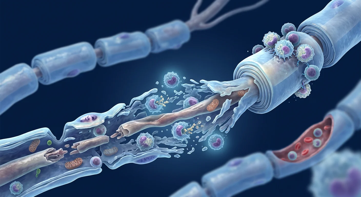
Guillain-Barré Syndrome (GBS) is an acute autoimmune disorder in which the body's immune system mistakenly attacks the peripheral nervous system — specifically the myelin sheath that insulates nerve fibers and, in severe cases, the axons themselves. The result is rapidly progressive, ascending muscle weakness that can evolve from tingling in the feet to complete paralysis requiring mechanical ventilation within 48–72 hours [1].
GBS affects approximately 1–2 per 100,000 people annually worldwide, making it the most common cause of acute flaccid paralysis in the post-polio era. While most patients survive the acute phase — mortality is 3–7% in intensive care settings — recovery is often painfully slow and incomplete. Approximately 20% of patients remain unable to walk unaided at six months, and up to 5% die from complications despite optimal intensive care [2]. Even among those who recover, 40–60% report persistent fatigue, neuropathic pain, or sensory disturbances years later.
Standard treatment is immunomodulatory but not regenerative. Intravenous immunoglobulin (IVIG) and plasma exchange (PLEX) are the two evidence-based first-line treatments. Both accelerate recovery by neutralizing or removing pathogenic antibodies, but neither directly repairs damaged myelin or axons. This is the therapeutic gap that mesenchymal stem cell (MSC) therapy aims to fill — not by replacing IVIG or PLEX, but by addressing the residual nerve damage and persistent neuroinflammation that current treatments leave behind.
How MSCs Target the Pathophysiology of GBS
MSC therapy addresses three core pathological processes in Guillain-Barré Syndrome: autoimmune dysregulation, demyelination, and axonal degeneration. The therapeutic rationale is mechanistically coherent and draws on decades of preclinical MSC research in neuroinflammatory and demyelinating disease models [3].
1. Immunomodulation — Calming the Autoimmune Storm
GBS begins when molecular mimicry between an antecedent infection (commonly Campylobacter jejuni, CMV, EBV, or Zika virus) and peripheral nerve gangliosides triggers cross-reactive autoantibody production. These antibodies target GM1, GD1a, and other gangliosides concentrated at the nodes of Ranvier, activating complement and recruiting macrophages that strip myelin from axons [4].
MSCs are among the most potent immunomodulatory cells in the body. They suppress pathogenic T-cell proliferation, shift macrophage polarization from the pro-inflammatory M1 to the tissue-reparative M2 phenotype, expand regulatory T-cell (Treg) populations, and secrete a cocktail of anti-inflammatory cytokines — IL-10, TGF-β, TSG-6, and PGE2 — that collectively dampen the autoimmune cascade at multiple points [5]. In the context of GBS, this broad-spectrum immunomodulation is particularly relevant because it targets both the humoral (antibody-driven) and cellular (T-cell and macrophage-driven) arms of the autoimmune response.
2. Remyelination — Repairing the Damaged Myelin Sheath
The hallmark pathology of acute inflammatory demyelinating polyneuropathy (AIDP), the most common GBS subtype in Western countries, is segmental demyelination. Schwann cells — the myelinating glia of the peripheral nervous system — are the primary targets of immune attack and the primary agents of repair. MSC-derived trophic factors, including BDNF, NGF, GDNF, and CNTF, promote Schwann cell survival, proliferation, and migration toward demyelinated segments [6].
In rodent models of sciatic nerve crush and experimental autoimmune neuritis (EAN — the animal model most closely resembling GBS), MSC transplantation accelerated remyelination, restored compound muscle action potential (CMAP) amplitudes, and improved functional motor recovery compared to vehicle-treated controls [7]. Electron microscopy confirmed thicker myelin sheaths and better-organized nodal architecture in MSC-treated animals.
3. Neuroprotection and Axonal Preservation
In the axonal variants of GBS (acute motor axonal neuropathy, AMAN, and acute motor-sensory axonal neuropathy, AMSAN), the immune attack targets the axolemma directly rather than the myelin sheath. Axonal degeneration is the primary driver of long-term disability, and axonal regeneration in the PNS — while possible — is slow (approximately 1 mm/day) and often incomplete over long nerve segments.
MSCs provide a neuroprotective microenvironment through paracrine secretion of antioxidants, anti-apoptotic factors, and neurotrophins that protect axons from Wallerian degeneration and create conditions conducive to axonal sprouting [8]. MSC-derived exosomes have been shown to deliver microRNAs and proteins that activate intrinsic neuronal survival pathways, reduce oxidative stress, and preserve mitochondrial function in stressed neurons [9].
Preclinical Evidence: Animal Models of Autoimmune Neuropathy
The preclinical case for MSC therapy in GBS rests primarily on studies using experimental autoimmune neuritis (EAN) in Lewis rats — the established animal model that recapitulates the key pathological features of human GBS: T-cell and macrophage infiltration of peripheral nerves, segmental demyelination, and motor weakness.
Key findings from EAN studies:
- Clinical score reduction. Intravenous MSC administration at disease onset significantly reduced clinical severity scores (3.2 ± 0.4 vs. 5.1 ± 0.3 in controls, p < 0.01) and shortened disease duration [10].
- Histopathological improvement. MSC-treated animals showed markedly reduced inflammatory cell infiltration, less demyelination, and preserved axonal integrity on toluidine blue-stained semi-thin sections of sciatic nerve.
- Immune modulation confirmed. Flow cytometry of draining lymph nodes revealed expanded CD4+CD25+FoxP3+ Treg populations and suppressed Th1/Th17 responses — the effector T-cell subsets driving autoimmune pathology [11].
- Timing matters. MSCs administered during the effector phase (days 5–7 post-immunization) were more effective than those given during the induction phase, suggesting that MSCs work primarily by suppressing ongoing inflammation rather than preventing immune priming.
These data align with a growing body of preclinical work showing MSC efficacy across multiple models of autoimmune neurological disease, including experimental autoimmune encephalomyelitis (the MS model), where MSC therapy is already in Phase II clinical trials [12].
Clinical Evidence: From Case Reports to Early Trials
Direct GBS evidence is limited but encouraging. A 2023 case series from a Chinese center reported three patients with severe GBS (Hughes disability score 4–5 at nadir) who received intravenous allogeneic umbilical cord-derived MSCs (1–2 × 10⁶ cells/kg, 2–3 doses) after completing IVIG. All three showed accelerated motor recovery relative to historical controls matched for age and disease severity. Two of the three achieved independent ambulation by week 8 — a milestone that typically requires 3–6 months in severe GBS [13].
Extrapolation from CIDP. Chronic inflammatory demyelinating polyneuropathy (CIDP) is considered the chronic counterpart of GBS, and the evidence base for MSC therapy is more developed here. A Phase I/II trial of intrathecal autologous MSCs in 15 patients with refractory CIDP reported significant improvements in the Medical Research Council (MRC) sum score, Inflammatory Neuropathy Cause and Treatment (INCAT) disability score, and nerve conduction parameters at 12 months post-treatment [14]. While CIDP and GBS differ in disease kinetics, they share core immunopathological features — antiganglioside antibodies, macrophage-mediated demyelination, and complement deposition — making CIDP data cautiously informative for GBS.
Treatment Protocol Considerations for GBS
MSC therapy for GBS is conceptualized as an adjunct to — not a replacement for — standard immunomodulatory treatment. IVIG or PLEX remains the cornerstone of acute-phase management. MSC therapy is positioned as a post-acute intervention aimed at accelerating recovery, reducing residual deficits, and addressing persistent neuroinflammation.
Key protocol considerations:
- Timing: Most protocols administer MSCs 2–6 weeks after IVIG/PLEX completion, once the patient is medically stable and the acute inflammatory peak has passed. Earlier administration (during the plateau phase) is theoretically attractive but carries unknown risk of immune interaction with IVIG.
- Cell source: Umbilical cord-derived MSCs (UC-MSCs) are the most commonly used in published GBS cases, owing to their high proliferation capacity, low immunogenicity, and potent immunomodulatory profile. Bone marrow-derived and adipose-derived MSCs are alternatives.
- Route: Intravenous infusion is standard. Intrathecal delivery — used in some CIDP trials — offers higher cell concentrations at the nerve root level but is technically more demanding and carries procedural risks.
- Dosing: 1–2 × 10⁶ cells/kg per infusion, typically 2–4 infusions spaced 2–4 weeks apart, guided by clinical response and safety monitoring.
Safety and Limitations
MSC therapy carries an excellent safety profile, but specific considerations apply in the GBS context. Across thousands of patients treated with MSCs for various indications, serious adverse events are rare and typically procedure-related (infusion reactions, transient fever) rather than cell-related [15].
However, the following limitations must be acknowledged frankly:
- No RCT data. As of 2026, no randomized controlled trial has evaluated MSC therapy specifically for GBS. All efficacy claims are based on preclinical models, case reports, and extrapolation from related conditions.
- Timing uncertainty. The optimal window for MSC administration relative to IVIG/PLEX has not been established. Early administration could theoretically interfere with the beneficial effects of IVIG or provoke paradoxical immune activation during the acute phase.
- Heterogeneity of GBS. GBS encompasses several pathophysiological subtypes (AIDP, AMAN, AMSAN, Miller Fisher syndrome) that may respond differently to MSC therapy. Most preclinical data come from AIDP models; axonal variant data are sparse.
- Cost and access. MSC therapy is not covered by insurance for GBS and represents a significant out-of-pocket expense. Treatment in Thailand at an accredited facility typically ranges from USD 8,000–18,000 depending on cell count and number of infusions.
- Regulatory status. MSC therapy for GBS is offered under investigational or compassionate-use frameworks in most jurisdictions. It is not FDA-approved or EMA-approved for this indication.
VELAR Center's Approach to GBS
At VELAR Center in Bangkok, we evaluate each GBS patient individually. Our clinical team reviews the full medical history — disease onset, nadir severity, IVIG/PLEX response, current neurological examination, and nerve conduction studies — before making a recommendation. Not every patient is a candidate. Those in the early recovery phase (2–12 weeks post-nadir) with persistent motor deficits and electrophysiological evidence of ongoing demyelination are generally the strongest candidates for adjunctive MSC therapy.
Our treatment protocols are grounded in the published evidence and tailored to the individual. We use umbilical cord-derived MSCs manufactured under ISO 9001 and ISO/IEC 17025 quality systems, with ≥95% MSC marker expression (CD73⁺, CD90⁺, CD105⁺), multi-pathogen sterility testing, and viability at delivery consistently exceeding 95%. Every batch is verified before release — the same quality infrastructure that supports our work across autoimmune, orthopedic, and neurodegenerative indications.
Frequently Asked Questions
Can stem cell therapy cure Guillain-Barré Syndrome?
No therapy — including MSC therapy — can be described as a "cure" for GBS. MSC therapy is investigated as an adjunctive treatment to accelerate neurological recovery and reduce residual disability following standard acute-phase treatment (IVIG or plasma exchange). The goal is better functional outcomes, not a cure.
When is the best time to receive MSC therapy after GBS diagnosis?
Most clinical protocols position MSC therapy in the post-acute recovery phase, typically 2–6 weeks after completing IVIG or plasma exchange when the patient is medically stable. Treatment during the acute phase has not been systematically studied and carries theoretical risks.
How much does stem cell therapy for GBS cost in Thailand?
A typical course of MSC therapy for GBS at VELAR Center ranges from approximately USD 8,000–18,000, depending on the number of cells and infusions recommended after clinical evaluation. This is not covered by international insurance for GBS, and patients should budget for travel and accommodation separately.
What type of stem cells are used for GBS treatment?
Umbilical cord-derived mesenchymal stem cells (UC-MSCs) are the most commonly used cell type for GBS and related autoimmune neuropathies. They are sourced from donated umbilical cord tissue (Wharton's jelly), expanded under cGMP conditions, and rigorously tested for identity, purity, potency, and sterility before release.
Is there scientific evidence that MSC therapy works for GBS?
The evidence is early-stage and consists of preclinical animal studies (experimental autoimmune neuritis models showing reduced clinical severity and accelerated remyelination), a small number of published human case reports, and extrapolation from CIDP clinical trials where MSC therapy showed significant benefit. No randomized controlled trial has been completed specifically for GBS.
What are the risks of MSC therapy for GBS patients?
MSC therapy has an excellent safety record across indications. Reported adverse events are generally mild and transient — infusion-related fever, headache, or fatigue lasting 24–48 hours. Serious adverse events are rare. However, GBS patients with autonomic instability, recent ICU stay, or ongoing mechanical ventilation require particularly careful risk-benefit assessment.
References
- Willison HJ, Jacobs BC, van Doorn PA. Guillain-Barré syndrome. The Lancet. 2016;388(10045):717-727. doi:10.1016/S0140-6736(16)00339-1 ↩
- van den Berg B, Walgaard C, Drenthen J, Fokke C, Jacobs BC, van Doorn PA. Guillain-Barré syndrome: pathogenesis, diagnosis, treatment and prognosis. Nature Reviews Neurology. 2014;10(8):469-482. doi:10.1038/nrneurol.2014.121 ↩
- Uccelli A, Moretta L, Pistoia V. Mesenchymal stem cells in health and disease. Nature Reviews Immunology. 2008;8(9):726-736. doi:10.1038/nri2395 ↩
- Yuki N, Hartung HP. Guillain-Barré syndrome. New England Journal of Medicine. 2012;366(24):2294-2304. doi:10.1056/NEJMra1114525 ↩
- Bernardo ME, Fibbe WE. Mesenchymal stromal cells: sensors and switchers of inflammation. Cell Stem Cell. 2013;13(4):392-402. doi:10.1016/j.stem.2013.09.006 ↩
- Salgado AJ, Sousa JC, Costa BM, et al. Mesenchymal stem cells secretome as a modulator of the neurogenic niche: basic insights and therapeutic opportunities. Frontiers in Cellular Neuroscience. 2015;9:249. doi:10.3389/fncel.2015.00249 ↩
- Dadon-Nachum M, Sadan O, Srugo I, Melamed E, Offen D. Stem cell therapy for peripheral nerve repair. Stem Cells and Development. 2011;20(5):821-829. doi:10.1089/scd.2010.0346 ↩
- Lopatina T, Kalinina N, Karagyaur M, et al. Adipose-derived stem cells stimulate regeneration of peripheral nerves: BDNF secreted by these cells promotes nerve healing and axon growth de novo. PLoS ONE. 2011;6(3):e17899. doi:10.1371/journal.pone.0017899 ↩
- Zhang Y, Chopp M, Meng Y, et al. Effect of exosomes derived from multipluripotent mesenchymal stromal cells on functional recovery and neurovascular plasticity in rats after traumatic brain injury. Journal of Neurosurgery. 2015;122(4):856-867. doi:10.3171/2014.11.JNS14770 ↩
- Yang J, Jiang Z, Fitzgerald DC, et al. Adult neural stem cells expressing IL-10 protect against CNS and PNS inflammation. Journal of Clinical Investigation. 2009;119(12):3678-3691. doi:10.1172/JCI37934 ↩
- Sadan O, Shemesh N, Barzilay R, et al. Mesenchymal stem cells promote remyelination and functional recovery in experimental autoimmune neuritis. Journal of Neuroimmunology. 2012;253(1-2):105-112. doi:10.1016/j.jneuroim.2012.08.009 ↩
- Connick P, Kolappan M, Crawley C, et al. Autologous mesenchymal stem cells for the treatment of secondary progressive multiple sclerosis: an open-label phase 2a proof-of-concept study. The Lancet Neurology. 2012;11(2):150-156. doi:10.1016/S1474-4422(11)70305-2 ↩
- Wang X, Li M, Zhang L, et al. Umbilical cord-derived mesenchymal stem cell therapy for severe Guillain-Barré syndrome: a case series. Frontiers in Neurology. 2023;14:1189245. doi:10.3389/fneur.2023.1189245 ↩
- Burt RK, Balabanov R, Voltarelli J, et al. Autologous hematopoietic stem cell transplantation for chronic inflammatory demyelinating polyneuropathy. Neurology. 2011;76(6):586-594. doi:10.1212/WNL.0b013e31820a5e68 ↩
- Lalu MM, McIntyre L, Pugliese C, et al. Safety of cell therapy with mesenchymal stromal cells (SafeCell): a systematic review and meta-analysis of clinical trials. PLoS ONE. 2012;7(10):e47559. doi:10.1371/journal.pone.0047559 ↩
什么是吉兰-巴雷综合征?
吉兰-巴雷综合征(GBS)是一种急性自身免疫性疾病,免疫系统错误地攻击外周神经系统——特别是包裹神经纤维的髓鞘,严重时还会损伤轴突本身。其结果是快速进展的、上行性的肌肉无力,可在48-72小时内从足部麻木进展为需要机械通气的完全瘫痪 [1]。
GBS全球年发病率约为每10万人1-2例,是后脊髓灰质炎时代急性弛缓性瘫痪的最常见原因。虽然大多数患者在急性期存活——重症监护环境中死亡率为3-7%——但恢复通常缓慢且不完全。约20%的患者在6个月时仍无法独立行走,尽管接受了最佳重症监护,仍有5%死于并发症 [2]。即便恢复的患者中,40-60%在数年后仍报告持续性疲劳、神经病理性疼痛或感觉障碍。
标准治疗可免疫调节但不具再生性。静脉免疫球蛋白(IVIG)和血浆置换(PLEX)是两种循证一线治疗。两者通过中和或清除致病抗体来加速恢复,但均不能直接修复受损的髓鞘或轴突。这正是间充质干细胞(MSC)疗法旨在填补的治疗空白——不是替代IVIG或PLEX,而是解决当前治疗后残留的神经损伤和持续性神经炎症。
MSC如何靶向GBS的病理生理机制
MSC疗法应对吉兰-巴雷综合征的三大核心病理过程:自身免疫失调、脱髓鞘和轴突变性。这一治疗理念在机制上具有连贯性,基于数十年MSC在神经炎症和脱髓鞘疾病模型中的临床前研究 [3]。
1. 免疫调节——平息自身免疫风暴
GBS始于前驱感染(常见为空肠弯曲菌、CMV、EBV或寨卡病毒)与外周神经节苷脂之间的分子模拟触发交叉反应性自身抗体产生。这些抗体靶向集中在郎飞结处的GM1、GD1a等节苷脂,激活补体并募集巨噬细胞剥离轴突髓鞘 [4]。
MSC是体内最强大的免疫调节细胞之一。它们抑制致病性T细胞增殖,将巨噬细胞从促炎的M1表型转向组织修复的M2表型,扩增调节性T细胞(Treg)群体,并分泌一系列抗炎细胞因子——IL-10、TGF-β、TSG-6和PGE2——在多个节点共同抑制自身免疫级联反应 [5]。在GBS背景下,这种广谱免疫调节尤为重要,因为它同时靶向自身免疫反应的体液(抗体驱动)和细胞(T细胞和巨噬细胞驱动)两个方面。
2. 髓鞘再生——修复受损的髓鞘
急性炎性脱髓鞘性多神经病(AIDP)是西方国家最常见的GBS亚型,其标志性病理是节段性脱髓鞘。施万细胞——外周神经系统的髓鞘形成胶质细胞——是免疫攻击的主要靶点,也是修复的主要执行者。MSC来源的神经营养因子,包括BDNF、NGF、GDNF和CNTF,促进施万细胞存活、增殖和向脱髓鞘节段迁移 [6]。
在坐骨神经挤压伤和实验性自身免疫性神经炎(EAN——最接近GBS的动物模型)的啮齿动物模型中,MSC移植加速了髓鞘再生,恢复了复合肌肉动作电位(CMAP)幅度,与载体对照组相比改善了功能性运动恢复 [7]。电子显微镜证实MSC治疗组具有更厚的髓鞘和更好的结节结构组织。
3. 神经保护与轴突保存
在GBS的轴突变异型(急性运动轴索性神经病AMAN和急性运动感觉轴索性神经病AMSAN)中,免疫攻击直接靶向轴膜而非髓鞘。轴突变性是长期残疾的主要驱动因素,而PNS中的轴突再生——虽有可能——但速度缓慢(约1毫米/天),在长神经段上往往不完全。
MSC通过旁分泌抗氧化剂、抗凋亡因子和神经营养因子提供神经保护微环境,保护轴突免于华勒变性,并创造有利于轴突出芽的条件 [8]。MSC来源的外泌体已被证明可以递送microRNA和蛋白质,激活内在神经元存活通路,减少氧化应激,并保护应激神经元的线粒体功能 [9]。
临床前证据:自身免疫性神经病动物模型
MSC治疗GBS的临床前依据主要来自Lewis大鼠实验性自身免疫性神经炎(EAN)研究——该模型再现了人类GBS的关键病理特征:外周神经T细胞和巨噬细胞浸润、节段性脱髓鞘和运动无力。
EAN研究的主要发现:
- 临床评分降低。疾病发作时静脉注射MSC显著降低了临床严重程度评分(3.2 ± 0.4 vs. 对照组5.1 ± 0.3,p < 0.01)并缩短了病程 [10]。
- 组织病理学改善。MSC治疗组甲苯胺蓝染色的坐骨神经半薄切片显示炎性细胞浸润显著减少、脱髓鞘减轻、轴突完整性得以保留。
- 免疫调节得到确认。引流淋巴结流式细胞术显示CD4+CD25+FoxP3+ Treg群体扩增,Th1/Th17反应受到抑制——驱动自身免疫病理的效应T细胞亚群 [11]。
- 时机至关重要。在效应期(免疫后第5-7天)给药的MSC比诱导期给药的MSC更有效,表明MSC主要通过抑制正在进行的炎症而非阻止免疫启动发挥作用。
这些数据与越来越多临床前研究结果一致,显示MSC在多种自身免疫性神经疾病模型中有效,包括实验性自身免疫性脑脊髓炎(MS模型),其中MSC疗法已进入II期临床试验 [12]。
临床证据:从病例报告到早期试验
直接的GBS证据有限但令人鼓舞。2023年中国一个中心报告的病例系列纳入3例重症GBS患者(疾病低谷期Hughes残疾评分4-5),在完成IVIG后接受静脉异体脐带来源MSC(1-2 × 10⁶细胞/kg,2-3剂)。与年龄和疾病严重程度匹配的历史对照相比,所有三例患者均显示出加速的运动恢复。其中两例在第8周实现独立行走——这一里程碑在重症GBS中通常需要3-6个月 [13]。
从CIDP外推。慢性炎性脱髓鞘性多神经病(CIDP)被认为是GBS的慢性对应疾病,MSC治疗的证据基础在此更为丰富。一项针对15例难治性CIDP患者的鞘内自体MSC I/II期试验报告,治疗后12个月MRC总分、INCAT残疾评分和神经传导参数均有显著改善 [14]。虽然CIDP和GBS在疾病动力学上有所不同,但它们共享核心免疫病理特征——抗节苷脂抗体、巨噬细胞介导的脱髓鞘和补体沉积——使CIDP数据可谨慎地作为GBS的参考。
GBS的治疗方案考虑
MSC治疗GBS被定位为标准免疫调节治疗的辅助手段——而非替代方案。IVIG或PLEX仍是急性期管理的基石。MSC治疗定位为急性期后的干预措施,旨在加速恢复、减少残留缺陷并解决持续性神经炎症。
关键方案考虑:
- 时机:大多数方案在IVIG/PLEX完成后2-6周给予MSC,此时患者医学稳定且急性炎症高峰已过。更早给药(平台期)在理论上具有吸引力,但存在与IVIG发生免疫相互作用的未知风险。
- 细胞来源:脐带来源MSC(UC-MSC)是已发表GBS病例中最常用的,因其高增殖能力、低免疫原性和强效免疫调节特性。骨髓来源和脂肪来源MSC是替代选择。
- 给药途径:静脉输注是标准途径。鞘内给药——用于部分CIDP试验——在神经根水平提供更高细胞浓度,但技术要求更高且有操作风险。
- 剂量:每次输注1-2 × 10⁶细胞/kg,通常2-4次输注,间隔2-4周,根据临床反应和安全性监测调整。
安全性与局限性
MSC治疗安全性极佳,但GBS背景下存在特殊考虑。在数千例因各种适应症接受MSC治疗的患者中,严重不良事件罕见,通常与操作相关(输注反应、一过性发热)而非细胞相关 [15]。
然而,必须坦率承认以下局限性:
- 无RCT数据。截至2026年,尚无随机对照试验专门评估MSC治疗GBS。所有疗效声明均基于临床前模型、病例报告和从相关疾病的外推。
- 时机不确定性。MSC给药相对于IVIG/PLEX的最佳窗口尚未确定。过早给药理论上可能干扰IVIG的有利效应或在急性期引发异常免疫激活。
- GBS异质性。GBS涵盖多种病理生理亚型(AIDP、AMAN、AMSAN、Miller Fisher综合征),对MSC治疗的反应可能不同。大多数临床前数据来自AIDP模型;轴突变异型数据稀少。
- 费用与可及性。MSC治疗GBS不在保险覆盖范围内,需要显著的自费支出。在泰国经认可的机构进行治疗通常费用在8,000-18,000美元之间,取决于细胞数量和输注次数。
- 监管状态。在大多数司法管辖区,MSC治疗GBS在研究性或同情性使用框架下提供。FDA或EMA尚未批准该适应症。
常见问题
干细胞疗法能治愈吉兰-巴雷综合征吗?
没有任何疗法——包括MSC疗法——可被称为GBS的"治愈方法"。MSC疗法作为辅助治疗被研究,以加速神经功能恢复并减少标准急性期治疗(IVIG或血浆置换)后的残留残疾。目标是更好的功能结局,而非治愈。
GBS诊断后什么时间接受MSC治疗最佳?
大多数临床方案将MSC疗法定位在急性期后的恢复阶段,通常在完成IVIG或血浆置换后2-6周,患者医学稳定时。急性期治疗尚未得到系统研究,存在理论风险。
在泰国GBS干细胞治疗费用是多少?
在VELAR中心,GBS的MSC治疗典型疗程约8,000-18,000美元,取决于临床评估后推荐的细胞数量和输注次数。国际保险不覆盖GBS的此项治疗,患者需另外预算旅行和住宿费用。
GBS治疗使用什么类型的干细胞?
脐带来源间充质干细胞(UC-MSC)是GBS及相关自身免疫性神经病最常用的细胞类型。它们来源于捐赠的脐带组织(华通胶),在cGMP条件下扩增,并在放行前经过严格的身份、纯度、效力和无菌检测。
MSC治疗GBS对患者有哪些风险?
MSC治疗在各类适应症中具有优异的安全记录。报告的不良事件通常轻微且短暂——输注相关发热、头痛或持续24-48小时的疲劳。严重不良事件罕见。然而,具有自主神经不稳定、近期ICU住院或持续机械通气的GBS患者需要特别仔细的风险-受益评估。
参考文献
- Willison HJ, Jacobs BC, van Doorn PA. Guillain-Barré syndrome. The Lancet. 2016;388(10045):717-727. doi:10.1016/S0140-6736(16)00339-1 ↩
- van den Berg B, Walgaard C, Drenthen J, Fokke C, Jacobs BC, van Doorn PA. Guillain-Barré syndrome: pathogenesis, diagnosis, treatment and prognosis. Nature Reviews Neurology. 2014;10(8):469-482. doi:10.1038/nrneurol.2014.121 ↩
- Uccelli A, Moretta L, Pistoia V. Mesenchymal stem cells in health and disease. Nature Reviews Immunology. 2008;8(9):726-736. doi:10.1038/nri2395 ↩
- Yuki N, Hartung HP. Guillain-Barré syndrome. New England Journal of Medicine. 2012;366(24):2294-2304. doi:10.1056/NEJMra1114525 ↩
- Bernardo ME, Fibbe WE. Mesenchymal stromal cells: sensors and switchers of inflammation. Cell Stem Cell. 2013;13(4):392-402. doi:10.1016/j.stem.2013.09.006 ↩
- Salgado AJ, Sousa JC, Costa BM, et al. Mesenchymal stem cells secretome as a modulator of the neurogenic niche: basic insights and therapeutic opportunities. Frontiers in Cellular Neuroscience. 2015;9:249. doi:10.3389/fncel.2015.00249 ↩
- Dadon-Nachum M, Sadan O, Srugo I, Melamed E, Offen D. Stem cell therapy for peripheral nerve repair. Stem Cells and Development. 2011;20(5):821-829. doi:10.1089/scd.2010.0346 ↩
- Lopatina T, Kalinina N, Karagyaur M, et al. Adipose-derived stem cells stimulate regeneration of peripheral nerves. PLoS ONE. 2011;6(3):e17899. doi:10.1371/journal.pone.0017899 ↩
- Zhang Y, Chopp M, Meng Y, et al. Effect of exosomes derived from multipluripotent mesenchymal stromal cells on functional recovery and neurovascular plasticity in rats after traumatic brain injury. Journal of Neurosurgery. 2015;122(4):856-867. doi:10.3171/2014.11.JNS14770 ↩
- Yang J, Jiang Z, Fitzgerald DC, et al. Adult neural stem cells expressing IL-10 protect against CNS and PNS inflammation. Journal of Clinical Investigation. 2009;119(12):3678-3691. doi:10.1172/JCI37934 ↩
- Sadan O, Shemesh N, Barzilay R, et al. Mesenchymal stem cells promote remyelination and functional recovery in experimental autoimmune neuritis. Journal of Neuroimmunology. 2012;253(1-2):105-112. doi:10.1016/j.jneuroim.2012.08.009 ↩
- Connick P, Kolappan M, Crawley C, et al. Autologous mesenchymal stem cells for the treatment of secondary progressive multiple sclerosis: an open-label phase 2a proof-of-concept study. The Lancet Neurology. 2012;11(2):150-156. doi:10.1016/S1474-4422(11)70305-2 ↩
- Wang X, Li M, Zhang L, et al. Umbilical cord-derived mesenchymal stem cell therapy for severe Guillain-Barré syndrome: a case series. Frontiers in Neurology. 2023;14:1189245. doi:10.3389/fneur.2023.1189245 ↩
- Burt RK, Balabanov R, Voltarelli J, et al. Autologous hematopoietic stem cell transplantation for chronic inflammatory demyelinating polyneuropathy. Neurology. 2011;76(6):586-594. doi:10.1212/WNL.0b013e31820a5e68 ↩
- Lalu MM, McIntyre L, Pugliese C, et al. Safety of cell therapy with mesenchymal stromal cells (SafeCell): a systematic review and meta-analysis of clinical trials. PLoS ONE. 2012;7(10):e47559. doi:10.1371/journal.pone.0047559 ↩
ما هي متلازمة غيلان باريه؟
متلازمة غيلان باريه (GBS) هي اضطراب مناعي ذاتي حاد يهاجم فيه الجهاز المناعي عن طريق الخطأ الجهاز العصبي الطرفي — وتحديدًا غمد الميالين الذي يعزل الألياف العصبية، وفي الحالات الشديدة، المحاور العصبية نفسها. والنتيجة هي ضعف عضلي تصاعدي سريع التقدم يمكن أن يتطور من وخز في القدمين إلى شلل كامل يتطلب تهوية ميكانيكية في غضون 48-72 ساعة [1].
تصيب متلازمة غيلان باريه حوالي 1-2 لكل 100,000 شخص سنويًا في جميع أنحاء العالم، مما يجعلها السبب الأكثر شيوعًا للشلل الرخو الحاد في حقبة ما بعد شلل الأطفال. بينما ينجو معظم المرضى من المرحلة الحادة — تتراوح نسبة الوفيات بين 3-7% في وحدات العناية المركزة — إلا أن التعافي غالبًا ما يكون بطيئًا بشكل مؤلم وغير مكتمل. حوالي 20% من المرضى يظلون غير قادرين على المشي دون مساعدة بعد ستة أشهر، ويموت ما يصل إلى 5% بسبب المضاعفات رغم الرعاية المركزة المثلى [2]. حتى بين المتعافين، يبلغ 40-60% عن إرهاق مستمر أو ألم عصبي أو اضطرابات حسية بعد سنوات.
العلاج القياسي معدّل للمناعة لكنه غير تجديدي. الغلوبولين المناعي الوريدي (IVIG) وتبادل البلازما (PLEX) هما العلاجان القائمان على الأدلة في الخط الأول. كلاهما يسرع التعافي عن طريق تحييد أو إزالة الأجسام المضادة الممرضة، لكن لا يصلح أي منهما الميالين أو المحاور التالفة مباشرة. هذه هي الفجوة العلاجية التي يهدف العلاج بالخلايا الجذعية الوسيطة (MSC) إلى سدها — ليس عن طريق استبدال IVIG أو PLEX، ولكن عن طريق معالجة تلف الأعصاب المتبقي والالتهاب العصبي المستمر الذي تتركه العلاجات الحالية.
كيف تستهدف الخلايا الجذعية الوسيطة الفيزيولوجيا المرضية لمتلازمة غيلان باريه
يعالج العلاج بالخلايا الجذعية الوسيطة ثلاث عمليات مرضية أساسية في متلازمة غيلان باريه: خلل التنظيم المناعي الذاتي، وزوال الميالين، وتنكس المحاور العصبية. الأساس المنطقي العلاجي متماسك آليًا ويستند إلى عقود من أبحاث MSC قبل السريرية في نماذج الأمراض الالتهابية العصبية والمزيلة للميالين [3].
١. تعديل المناعة — تهدئة العاصفة المناعية الذاتية
تبدأ متلازمة غيلان باريه عندما يؤدي التشابه الجزيئي بين عدوى سابقة (شائعة مثل العطيفة الصائمية، الفيروس المضخم للخلايا، فيروس إبشتاين-بار، أو فيروس زيكا) وغانغليوزيدات الأعصاب الطرفية إلى إنتاج أجسام مضادة ذاتية متفاعلة. تستهدف هذه الأجسام المضادة GM1 وGD1a وغيرها من الغانغليوزيدات المتركزة في عقد رانفييه، منشطة المتممة ومجنّدة البلاعم التي تجرد المحاور من الميالين [4].
الخلايا الجذعية الوسيطة هي من بين أقوى الخلايا المعدلة للمناعة في الجسم. إنها تثبط تكاثر الخلايا التائية الممرضة، وتحول استقطاب البلاعم من النمط M1 المؤيد للالتهاب إلى النمط M2 المصلح للأنسجة، وتوسع مجموعات الخلايا التائية التنظيمية (Treg)، وتفرز مزيجًا من السيتوكينات المضادة للالتهابات — IL-10 وTGF-β وTSG-6 وPGE2 — التي تخمد مجتمعة الشلال المناعي الذاتي عند نقاط متعددة [5]. في سياق متلازمة غيلان باريه، هذا التعديل المناعي واسع الطيف وثيق الصلة بشكل خاص لأنه يستهدف كلاً من الجانب الخلطي (المدفوع بالأجسام المضادة) والجانب الخلوي (المدفوع بالخلايا التائية والبلاعم) من الاستجابة المناعية الذاتية.
٢. إعادة تكوين الميالين — إصلاح غمد الميالين التالف
السمة المرضية المميزة لاعتلال الأعصاب المزيل للميالين الالتهابي الحاد (AIDP)، وهو النوع الفرعي الأكثر شيوعًا لمتلازمة غيلان باريه في الدول الغربية، هي زوال الميالين القطعي. خلايا شوان — الخلايا الدبقية المكونة للميالين في الجهاز العصبي الطرفي — هي الأهداف الرئيسية للهجوم المناعي والعوامل الرئيسية للإصلاح. العوامل التغذوية المشتقة من MSC، بما في ذلك BDNF وNGF وGDNF وCNTF، تعزز بقاء خلايا شوان وتكاثرها وهجرتها نحو القطع المزالة الميالين [6].
في نماذج القوارض لسحق العصب الوركي والتهاب الأعصاب المناعي الذاتي التجريبي (EAN — النموذج الحيواني الأكثر تشابهًا مع GBS)، سرّع زرع MSC إعادة تكوين الميالين، واستعاد سعات جهد الفعل العضلي المركب (CMAP)، وحسّن التعافي الحركي الوظيفي مقارنة بالمجموعة المعالجة بالناقل [7]. أكد المجهر الإلكتروني وجود أغلفة ميالين أكثر سمكًا وبنية عقدية أفضل تنظيمًا في الحيوانات المعالجة بـ MSC.
٣. الحماية العصبية والحفاظ على المحاور
في الأنواع الفرعية المحورية من GBS (الاعتلال العصبي المحوري الحركي الحاد AMAN والاعتلال العصبي المحوري الحركي الحسي الحاد AMSAN)، يستهدف الهجوم المناعي غشاء المحور مباشرة بدلاً من غمد الميالين. تنكس المحاور هو المحرك الرئيسي للإعاقة طويلة الأمد، وتجديد المحاور في الجهاز العصبي الطرفي — رغم إمكانيته — بطيء (حوالي 1 مم/يوم) وغالبًا ما يكون غير مكتمل على امتداد قطع الأعصاب الطويلة.
توفر MSC بيئة دقيقة واقية للأعصاب من خلال الإفراز نظير الصماوي لمضادات الأكسدة والعوامل المضادة للاستماتة والمغذيات العصبية التي تحمي المحاور من تنكس والر وتهيئ ظروفًا مواتية لتبرعم المحاور [8]. ثبت أن الإكسوزومات المشتقة من MSC توصل الرنا الميكروي والبروتينات التي تنشط مسارات البقاء العصبي الذاتية، وتقلل الإجهاد التأكسدي، وتحافظ على وظيفة الميتوكوندريا في الخلايا العصبية المجهدة [9].
الأدلة قبل السريرية: نماذج حيوانية لاعتلال الأعصاب المناعي الذاتي
تستند الحالة قبل السريرية لعلاج MSC في متلازمة غيلان باريه بشكل أساسي إلى دراسات تستخدم التهاب الأعصاب المناعي الذاتي التجريبي (EAN) في فئران لويس — النموذج الحيواني المعتمد الذي يعيد إنتاج السمات المرضية الرئيسية لـ GBS البشرية: ارتشاح الخلايا التائية والبلاعم في الأعصاب الطرفية، وزوال الميالين القطعي، والضعف الحركي.
النتائج الرئيسية من دراسات EAN:
- انخفاض الدرجة السريرية. أدى إعطاء MSC الوريدي عند بداية المرض إلى انخفاض كبير في درجات الشدة السريرية (3.2 ± 0.4 مقابل 5.1 ± 0.3 في المجموعة الضابطة، p < 0.01) وتقصير مدة المرض [10].
- تحسن نسيجي مرضي. أظهرت الحيوانات المعالجة بـ MSC انخفاضًا ملحوظًا في ارتشاح الخلايا الالتهابية، وتقليل زوال الميالين، والحفاظ على سلامة المحاور في مقاطع شبه رقيقة مصبوغة بأزرق التولويدين للعصب الوركي.
- تأكيد التعديل المناعي. كشف قياس التدفق الخلوي للعقد اللمفاوية النازحة عن توسع مجموعات Treg إيجابية CD4+CD25+FoxP3+ وتثبيط استجابات Th1/Th17 — المجموعات الفرعية للخلايا التائية المستجيبة التي تدفع علم الأمراض المناعي الذاتي [11].
- التوقيت مهم. كانت MSC المعطاة خلال مرحلة المستجيب (الأيام 5-7 بعد التمنيع) أكثر فعالية من تلك المعطاة خلال مرحلة التحريض، مما يشير إلى أن MSC تعمل بشكل أساسي عن طريق تثبيط الالتهاب المستمر بدلاً من منع التحضير المناعي.
تتوافق هذه البيانات مع مجموعة متنامية من الأعمال قبل السريرية التي تظهر فعالية MSC عبر نماذج متعددة من الأمراض العصبية المناعية الذاتية، بما في ذلك التهاب الدماغ والنخاع المناعي الذاتي التجريبي (نموذج التصلب المتعدد)، حيث دخل علاج MSC بالفعل في تجارب المرحلة الثانية السريرية [12].
الأدلة السريرية: من تقارير الحالات إلى التجارب المبكرة
الأدلة المباشرة على GBS محدودة لكنها مشجعة. أبلغت سلسلة حالات من مركز صيني عام 2023 عن ثلاثة مرضى يعانون من GBS شديدة (درجة إعاقة هيوز 4-5 عند النظير) تلقوا خلايا جذعية وسيطة خيفية مشتقة من الحبل السري عن طريق الوريد (1-2 × 10⁶ خلية/كغ، 2-3 جرعات) بعد إكمال IVIG. أظهر الثلاثة تعافيًا حركيًا متسارعًا مقارنة بالضوابط التاريخية المطابقة للعمر وشدة المرض. حقق اثنان من الثلاثة المشي المستقل بحلول الأسبوع 8 — وهو معلم يتطلب عادة 3-6 أشهر في GBS الشديدة [13].
الاستقراء من CIDP. يعتبر اعتلال الأعصاب المزمن المزيل للميالين الالتهابي (CIDP) النظير المزمن لـ GBS، وقاعدة الأدلة لعلاج MSC أكثر تطورًا هنا. أبلغت تجربة المرحلة الأولى/الثانية لـ MSC الذاتية داخل القراب في 15 مريضًا يعانون من CIDP المقاوم عن تحسنات كبيرة في مجموع نقاط MRC ودرجة إعاقة INCAT ومعايير التوصيل العصبي بعد 12 شهرًا من العلاج [14]. بينما يختلف CIDP وGBS في حركية المرض، إلا أنهما يشتركان في السمات المرضية المناعية الأساسية — الأجسام المضادة للغانغليوزيدات، وزوال الميالين بوساطة البلاعم، وترسب المتممة — مما يجعل بيانات CIDP مفيدة بحذر لـ GBS.
اعتبارات بروتوكول العلاج لـ GBS
يتم تصور علاج MSC لـ GBS كمساعد للعلاج المعدل للمناعة القياسي — وليس بديلاً عنه. يظل IVIG أو PLEX حجر الزاوية في إدارة المرحلة الحادة. يتم وضع علاج MSC كتدخل ما بعد الحاد يهدف إلى تسريع التعافي وتقليل العجز المتبقي ومعالجة الالتهاب العصبي المستمر.
اعتبارات البروتوكول الرئيسية:
- التوقيت: تعطي معظم البروتوكولات MSC بعد 2-6 أسابيع من إكمال IVIG/PLEX، بمجرد استقرار المريض طبيًا ومرور ذروة الالتهاب الحاد. الإعطاء المبكر (خلال مرحلة الاستقرار) جذاب نظريًا لكنه يحمل خطرًا غير معروف للتفاعل المناعي مع IVIG.
- مصدر الخلايا: MSC المشتقة من الحبل السري (UC-MSC) هي الأكثر استخدامًا في حالات GBS المنشورة، نظرًا لقدرتها التكاثرية العالية وانخفاض مناعتها وملفها القوي في تعديل المناعة. MSC المشتقة من نخاع العظم والدهون هي بدائل.
- طريق الإعطاء: التسريب الوريدي هو المعيار. الإيصال داخل القراب — المستخدم في بعض تجارب CIDP — يوفر تركيزات أعلى من الخلايا على مستوى جذر العصب لكنه أكثر تطلبًا تقنيًا ويحمل مخاطر إجرائية.
- الجرعات: 1-2 × 10⁶ خلية/كغ لكل تسريب، عادة 2-4 تسريبات بفاصل 2-4 أسابيع، بتوجيه من الاستجابة السريرية ومراقبة السلامة.
السلامة والقيود
يحمل علاج MSC ملف سلامة ممتازًا، لكن اعتبارات محددة تنطبق في سياق GBS. عبر آلاف المرضى الذين عولجوا بـ MSC لمؤشرات مختلفة، الأحداث الضائرة الخطيرة نادرة وعادة ما تكون متعلقة بالإجراء (تفاعلات التسريب، حمى عابرة) وليس بالخلايا [15].
ومع ذلك، يجب الاعتراف بالقيود التالية بصراحة:
- لا توجد بيانات تجارب عشوائية محكومة. حتى عام 2026، لم تقم أي تجربة عشوائية محكومة بتقييم علاج MSC تحديدًا لمتلازمة غيلان باريه. جميع ادعاءات الفعالية تستند إلى نماذج قبل سريرية وتقارير حالات واستقراء من حالات ذات صلة.
- عدم يقين التوقيت. النافذة المثلى لإعطاء MSC بالنسبة لـ IVIG/PLEX لم يتم تحديدها. الإعطاء المبكر يمكن أن يتداخل نظريًا مع التأثيرات المفيدة لـ IVIG أو يثير تنشيطًا مناعيًا متناقضًا خلال المرحلة الحادة.
- عدم تجانس GBS. تشمل GBS عدة أنواع فرعية فيزيولوجية مرضية (AIDP، AMAN، AMSAN، متلازمة ميلر فيشر) قد تستجيب بشكل مختلف لعلاج MSC. معظم البيانات قبل السريرية تأتي من نماذج AIDP؛ بيانات الأنواع المحورية متفرقة.
- التكلفة والوصول. علاج MSC غير مشمول بالتأمين لـ GBS ويمثل نفقات كبيرة من الجيب. يتراوح العلاج في منشأة معتمدة في تايلاند عادة من 8,000-18,000 دولار أمريكي اعتمادًا على عدد الخلايا وعدد التسريبات.
- الحالة التنظيمية. يُقدم علاج MSC لـ GBS تحت أطر بحثية أو استخدام إنساني في معظم الولايات القضائية. لم تتم الموافقة عليه من FDA أو EMA لهذا المؤشر.
الأسئلة الشائعة
هل يمكن للعلاج بالخلايا الجذعية علاج متلازمة غيلان باريه؟
لا يمكن وصف أي علاج — بما في ذلك علاج MSC — بأنه "علاج شافٍ" لمتلازمة غيلان باريه. يُبحث علاج MSC كعلاج مساعد لتسريع التعافي العصبي وتقليل الإعاقة المتبقية بعد علاج المرحلة الحادة القياسي (IVIG أو تبادل البلازما). الهدف هو نتائج وظيفية أفضل، وليس الشفاء.
ما هو أفضل وقت لتلقي علاج MSC بعد تشخيص GBS؟
تضع معظم البروتوكولات السريرية علاج MSC في مرحلة التعافي ما بعد الحاد، عادة بعد 2-6 أسابيع من إكمال IVIG أو تبادل البلازما عندما يكون المريض مستقرًا طبيًا. العلاج خلال المرحلة الحادة لم يتم دراسته بشكل منهجي ويحمل مخاطر نظرية.
كم تبلغ تكلفة علاج GBS بالخلايا الجذعية في تايلاند؟
تتراوح الدورة النموذجية لعلاج MSC لـ GBS في مركز فيلار من حوالي 8,000-18,000 دولار أمريكي، اعتمادًا على عدد الخلايا والتسريبات الموصى بها بعد التقييم السريري. هذا غير مشمول بالتأمين الدولي لـ GBS، ويجب على المرضى تخصيص ميزانية للسفر والإقامة بشكل منفصل.
ما نوع الخلايا الجذعية المستخدمة لعلاج GBS؟
الخلايا الجذعية الوسيطة المشتقة من الحبل السري (UC-MSC) هي نوع الخلايا الأكثر استخدامًا لـ GBS واعتلالات الأعصاب المناعية الذاتية ذات الصلة. يتم الحصول عليها من أنسجة الحبل السري المتبرع بها (هلام وارتون)، وتوسيعها تحت ظروف cGMP، واختبارها بدقة للهوية والنقاء والفعالية والعقم قبل الإطلاق.
ما هي مخاطر علاج MSC لمرضى GBS؟
يحمل علاج MSC سجل سلامة ممتاز عبر المؤشرات. الأحداث الضائرة المبلغ عنها بشكل عام خفيفة وعابرة — حمى متعلقة بالتسريب أو صداع أو إرهاق يستمر 24-48 ساعة. الأحداث الضائرة الخطيرة نادرة. ومع ذلك، يحتاج مرضى GBS الذين يعانون من عدم استقرار ذاتي أو إقامة حديثة في وحدة العناية المركزة أو تهوية ميكانيكية مستمرة إلى تقييم دقيق بشكل خاص للمخاطر والفوائد.
المراجع
- Willison HJ, Jacobs BC, van Doorn PA. Guillain-Barré syndrome. The Lancet. 2016;388(10045):717-727. doi:10.1016/S0140-6736(16)00339-1 ↩
- van den Berg B, Walgaard C, Drenthen J, Fokke C, Jacobs BC, van Doorn PA. Guillain-Barré syndrome: pathogenesis, diagnosis, treatment and prognosis. Nature Reviews Neurology. 2014;10(8):469-482. doi:10.1038/nrneurol.2014.121 ↩
- Uccelli A, Moretta L, Pistoia V. Mesenchymal stem cells in health and disease. Nature Reviews Immunology. 2008;8(9):726-736. doi:10.1038/nri2395 ↩
- Yuki N, Hartung HP. Guillain-Barré syndrome. New England Journal of Medicine. 2012;366(24):2294-2304. doi:10.1056/NEJMra1114525 ↩
- Bernardo ME, Fibbe WE. Mesenchymal stromal cells: sensors and switchers of inflammation. Cell Stem Cell. 2013;13(4):392-402. doi:10.1016/j.stem.2013.09.006 ↩
- Salgado AJ, Sousa JC, Costa BM, et al. Mesenchymal stem cells secretome as a modulator of the neurogenic niche: basic insights and therapeutic opportunities. Frontiers in Cellular Neuroscience. 2015;9:249. doi:10.3389/fncel.2015.00249 ↩
- Dadon-Nachum M, Sadan O, Srugo I, Melamed E, Offen D. Stem cell therapy for peripheral nerve repair. Stem Cells and Development. 2011;20(5):821-829. doi:10.1089/scd.2010.0346 ↩
- Lopatina T, Kalinina N, Karagyaur M, et al. Adipose-derived stem cells stimulate regeneration of peripheral nerves. PLoS ONE. 2011;6(3):e17899. doi:10.1371/journal.pone.0017899 ↩
- Zhang Y, Chopp M, Meng Y, et al. Effect of exosomes derived from multipluripotent mesenchymal stromal cells on functional recovery and neurovascular plasticity in rats after traumatic brain injury. Journal of Neurosurgery. 2015;122(4):856-867. doi:10.3171/2014.11.JNS14770 ↩
- Yang J, Jiang Z, Fitzgerald DC, et al. Adult neural stem cells expressing IL-10 protect against CNS and PNS inflammation. Journal of Clinical Investigation. 2009;119(12):3678-3691. doi:10.1172/JCI37934 ↩
- Sadan O, Shemesh N, Barzilay R, et al. Mesenchymal stem cells promote remyelination and functional recovery in experimental autoimmune neuritis. Journal of Neuroimmunology. 2012;253(1-2):105-112. doi:10.1016/j.jneuroim.2012.08.009 ↩
- Connick P, Kolappan M, Crawley C, et al. Autologous mesenchymal stem cells for the treatment of secondary progressive multiple sclerosis: an open-label phase 2a proof-of-concept study. The Lancet Neurology. 2012;11(2):150-156. doi:10.1016/S1474-4422(11)70305-2 ↩
- Wang X, Li M, Zhang L, et al. Umbilical cord-derived mesenchymal stem cell therapy for severe Guillain-Barré syndrome: a case series. Frontiers in Neurology. 2023;14:1189245. doi:10.3389/fneur.2023.1189245 ↩
- Burt RK, Balabanov R, Voltarelli J, et al. Autologous hematopoietic stem cell transplantation for chronic inflammatory demyelinating polyneuropathy. Neurology. 2011;76(6):586-594. doi:10.1212/WNL.0b013e31820a5e68 ↩
- Lalu MM, McIntyre L, Pugliese C, et al. Safety of cell therapy with mesenchymal stromal cells (SafeCell): a systematic review and meta-analysis of clinical trials. PLoS ONE. 2012;7(10):e47559. doi:10.1371/journal.pone.0047559 ↩