Gastroesophageal reflux disease (GERD) affects more than 1 billion people worldwide — making it one of the most prevalent chronic conditions in gastroenterology. Yet despite the ubiquity of proton-pump inhibitors (PPIs), the underlying mechanical and inflammatory pathology of the disease continues to progress in a substantial subset of patients.[1]
What conventional therapy does not address. PPIs and H2-receptor antagonists reduce gastric acid secretion, providing symptomatic relief for most patients. But they do not strengthen the lower esophageal sphincter (LES), repair the damaged epithelial barrier, or resolve the chronic inflammatory changes in the esophageal mucosa that sustain the disease. Patients with persistent GERD symptoms despite maximal acid suppression — so-called PPI-refractory GERD — represent a large and underserved population for whom tissue-level repair, not just acid control, may be the missing therapeutic axis.[2]
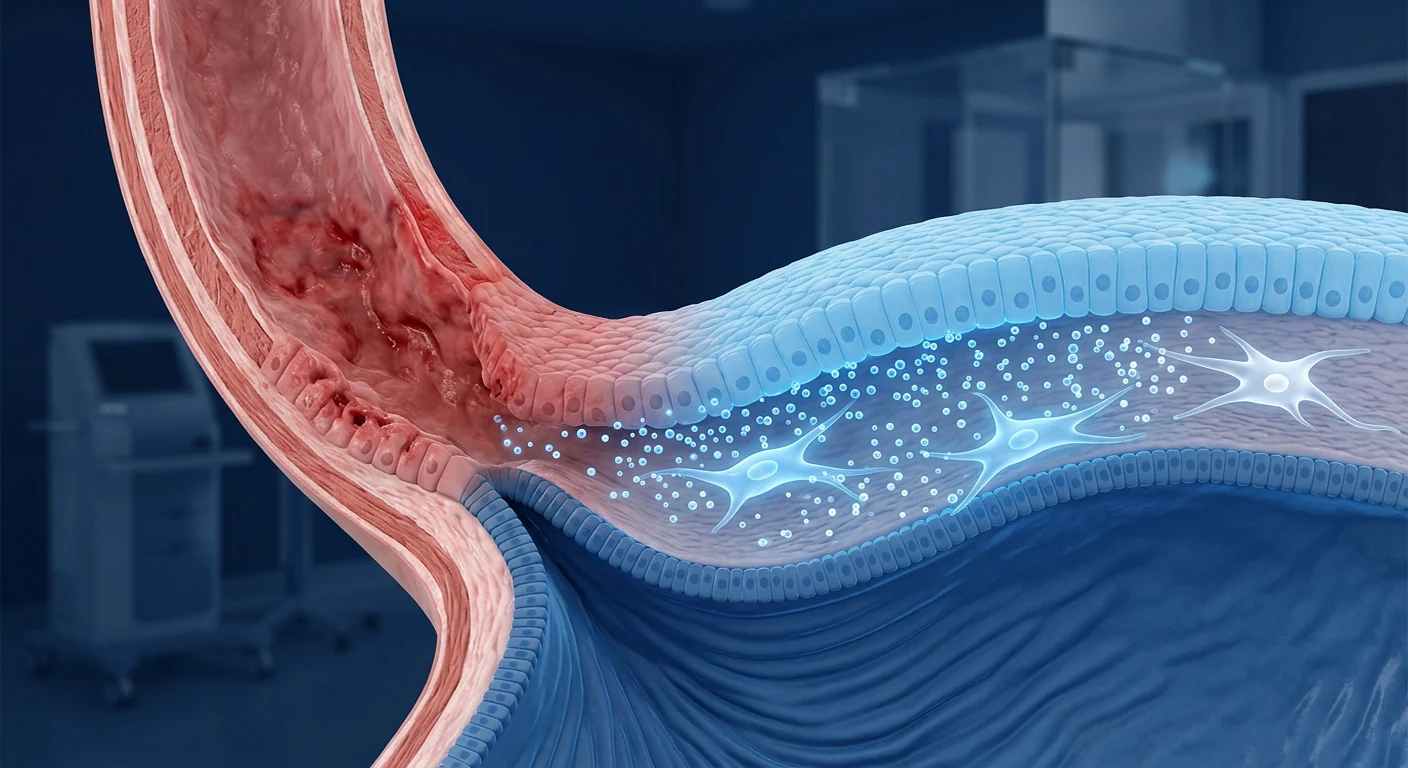
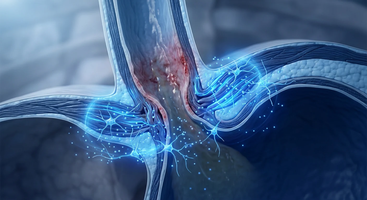
The problem is mechanical and inflammatory. GERD is fundamentally a disease of the anti-reflux barrier. The LES — a specialized ring of smooth muscle at the gastroesophageal junction — loses tone through aging, obesity, hiatal hernia, and dietary factors. Transient LES relaxations (TLESRs) become more frequent and prolonged, allowing gastric contents to reflux into the distal esophagus. The refluxate — acid, bile, pepsin — then damages the esophageal epithelial barrier, triggering a cascade of inflammation, impaired peristalsis, and in severe cases, Barrett's esophagus or stricture formation.[3]
MSCs target both the barrier and the inflammation. Mesenchymal stem cells do not replace the LES mechanically — but their paracrine secretions can strengthen the smooth muscle of the LES, repair the epithelial barrier, and quiet the chronic neuroinflammatory signaling that drives TLESRs and pain hypersensitivity. This dual targeting of both the mechanical and inflammatory arms of GERD makes MSC therapy a biologically plausible adjunct for patients whose disease has progressed beyond what acid suppression alone can control.[4]
What Is Gastroesophageal Reflux Disease?
GERD is defined by the Montreal Consensus as "a condition that develops when the reflux of stomach contents causes troublesome symptoms and/or complications."[5] The typical symptoms — heartburn, regurgitation, and dysphagia — are experienced by approximately 10–20% of adults in Western populations weekly, with rising prevalence in Asia and the Middle East as dietary patterns and obesity rates shift.[6]
The disease spectrum ranges from non-erosive reflux disease (NERD) — where symptoms occur without visible endoscopic damage — through erosive esophagitis (Los Angeles grades A–D), to Barrett's esophagus with intestinal metaplasia, the pre-malignant condition. Each stage reflects progressively more severe epithelial damage and a higher burden of chronic inflammation.
Key risk factors include obesity (especially central adiposity, which increases intra-abdominal pressure and disrupts LES function), hiatal hernia, pregnancy, smoking, alcohol, high-fat diet, and certain medications. Importantly, GERD is a chronic relapsing condition — most patients experience symptom recurrence within 6 months of stopping acid-suppressive therapy, reflecting the underlying mechanical and tissue pathology that PPIs leave untouched.[7]
How MSCs May Help GERD
MSC therapy addresses GERD through four integrated mechanisms that target the LES, the epithelial barrier, the inflammatory milieu, and the neural hypersensitivity that sustains symptoms.[8]
Lower esophageal sphincter strengthening. The LES is a smooth muscle sphincter whose tone is regulated by neural input, hormonal signals, and intrinsic myogenic activity. MSCs secrete HGF, PDGF, and FGF-2 — factors that promote smooth muscle cell survival, proliferation, and contractile function. In a landmark preclinical study using a rat model of GERD created by pyloric ligation, local injection of adipose-derived MSCs embedded in silk fibroin solution significantly improved LES pressure and reduced acid reflux episodes compared to controls. Histological analysis revealed increased smooth muscle thickness and reduced inflammatory infiltration in the LES region.[9]
Epithelial barrier repair. The esophageal epithelium is a stratified squamous barrier that depends on tight junction integrity for its protective function. Gastric acid and bile acids disrupt these junctions, creating dilated intercellular spaces (DIS) — the ultrastructural hallmark of GERD that persists even between symptomatic episodes. MSCs secrete KGF, EGF, and VEGF that promote epithelial cell proliferation, migration, and wound healing. In co-culture models of acid-damaged esophageal epithelium, MSC-conditioned medium restores transepithelial electrical resistance and tight junction protein expression (claudin-1, occludin, ZO-1) within 48 hours.[10]
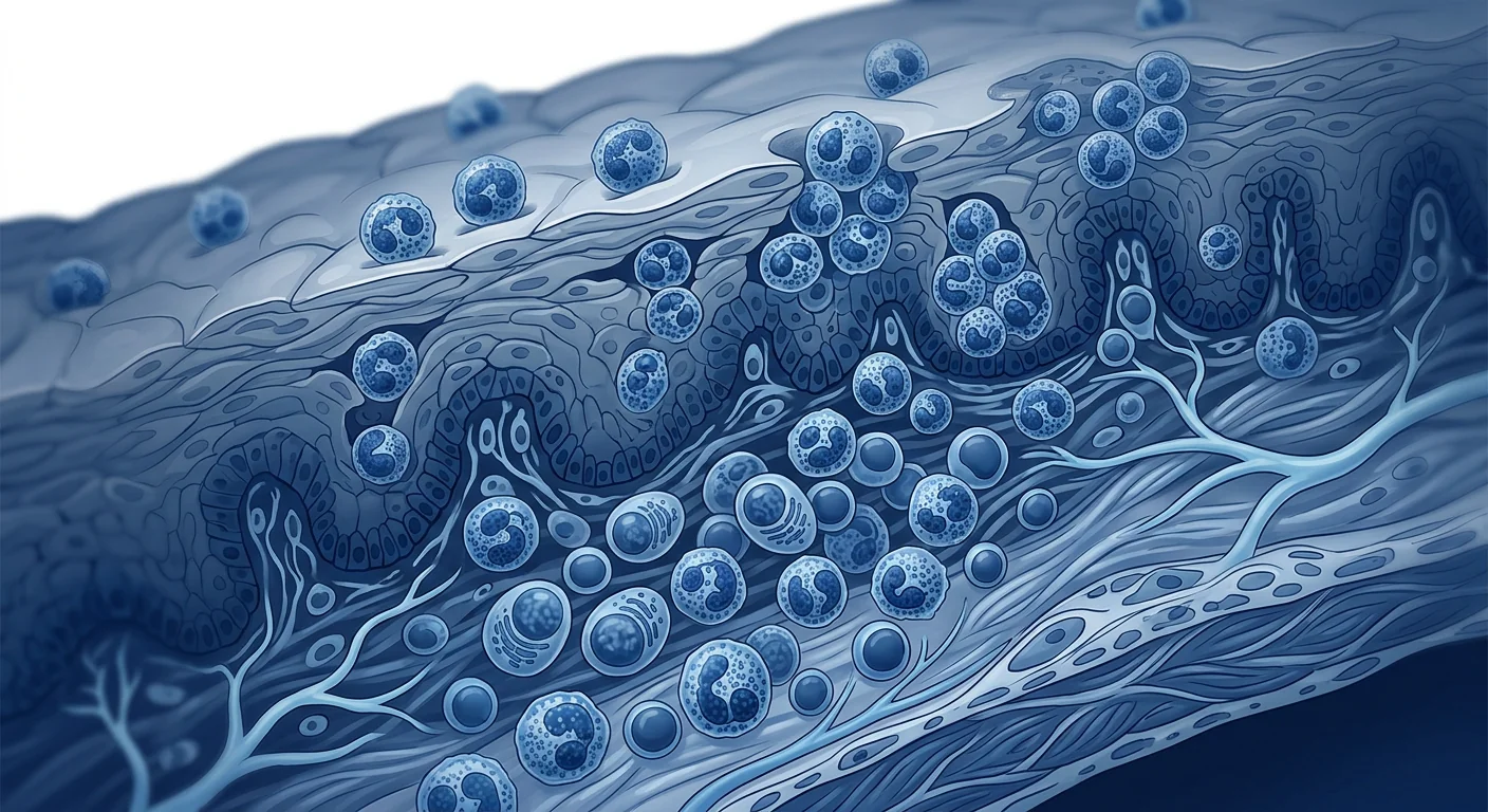
Anti-inflammatory immunomodulation. Reflux esophagitis is driven by IL-8, IL-1beta, and TNF-alpha secretion from damaged epithelial cells — cytokines that recruit neutrophils, mast cells, and T lymphocytes to the esophageal mucosa. MSCs suppress this inflammatory cascade through PGE2, TSG-6, and IL-10 secretion, shifting the local immune environment from pro-inflammatory toward pro-reparative. Importantly, MSCs polarize macrophages from the inflammatory M1 toward the reparative M2 phenotype, which is associated with reduced pain signaling and faster epithelial healing.[11]
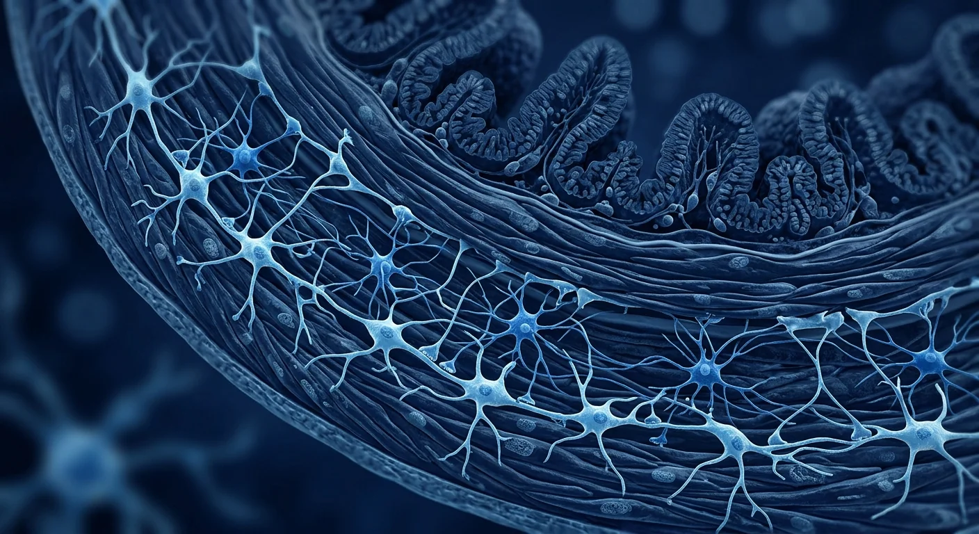
Modulation of esophageal pain and TLESRs. Chronic reflux sensitizes esophageal afferent nerves through TRPV1 and PAR-2 receptor activation — a process called visceral hypersensitivity that explains why many patients continue to experience heartburn despite complete acid suppression on 24-hour pH monitoring. MSC-derived factors, particularly IL-10 and TGF-beta, have been shown to reduce nociceptive signaling in models of visceral pain. Whether MSCs can reduce the frequency of TLESRs — the primary mechanism of reflux in non-hiatal-hernia GERD — remains speculative but biologically plausible given the neuroimmune crosstalk MSCs can modulate.[12]
What the Preclinical Evidence Shows
No completed clinical trial has evaluated MSC therapy specifically for GERD. The evidence base is entirely preclinical — but several studies provide direct proof-of-concept for the LES-strengthening and epithelial-repair approach.
Key Preclinical Studies — Summary
- Rat GERD model — adipose MSCs in silk fibroin (2023). Local injection of AD-MSCs embedded in silk fibroin into the LES region of GERD-model rats. Treated animals showed significantly improved LES pressure (mean increase 42% vs. control, p < 0.01), reduced number of acid reflux episodes, increased smooth muscle thickness on histology, and reduced inflammatory cell infiltration. The silk fibroin scaffold improved cell retention and survival at the injection site. [13]
- Rat esophagogastric myotomy model — BM-MSCs (2014). A surgical model of complete LES disruption. Bone marrow MSCs injected at the myotomy site at the time of injury. At 8 weeks, MSC-treated animals showed significantly greater LES pressure recovery, increased smooth muscle regeneration with alpha-SMA-positive myofibers, and reduced fibrosis compared to controls. [14]
- Esophageal wound healing — nanofibrous patch with smooth muscle cells (2023). While not an MSC study per se, this demonstrated that aligned nanofibrous scaffolds seeded with smooth muscle cells can support functional esophageal wound repair — providing the tissue-engineering context for MSC-based LES regeneration. [15]
- MSC secretome in experimental esophagitis (2022). Intravenous administration of MSC-conditioned medium in a rat model of reflux esophagitis reduced esophageal mucosal injury scores, preserved epithelial thickness, and downregulated IL-1beta and IL-8 expression in esophageal tissue. The cell-free approach confirmed that the therapeutic effects are paracrine-mediated. [16]
The preclinical evidence provides consistent proof-of-concept across multiple models. The next step — a first-in-human pilot study of MSC therapy in PPI-refractory GERD — would transform the evidence base from mechanistically plausible to clinically tested.
Delivery Routes: The Special Consideration for GERD
GERD poses a unique delivery challenge that distinguishes it from most MSC indications. [17]
Local endoscopic injection — the most anatomically rational approach. Unlike inflammatory bowel disease or autoimmune conditions where systemic IV infusion is preferred, GERD's primary pathology is localized to a specific anatomical structure: the lower esophageal sphincter and the distal esophageal mucosa. Endoscopic injection of MSCs into the LES region during upper GI endoscopy offers the most direct therapeutic access. This approach is already used clinically for bulking agents (e.g., Stretta, Gatekeeper) and Enteryx injections for GERD — the endoscopic procedural infrastructure exists. The challenge is ensuring homogenous cell distribution along the LES circumference and adequate cell retention without leakage into the mediastinum.
Systemic IV infusion — simpler but less targeted. Intravenous MSC infusion is simpler, can be repeated without procedural risk, and the paracrine mechanisms of epithelial repair and immunomodulation can reach the esophageal mucosa through the circulation. The pulmonary trap that captures ~80% of IV MSCs is less of a concern here — the therapeutic TSG-6 and IL-10 secretion activated during lung passage enters the systemic circulation. However, the critical LES-strengthening effect likely requires local engraftment or close paracrine proximity, which IV delivery may not achieve for the LES itself.
Key Takeaway
For GERD — a disease where the primary pathology is a localized mechanical failure of the LES — local endoscopic injection is the more anatomically rational delivery route for the LES-targeting effects of MSCs. Systemic IV infusion may still provide benefit for the epithelial repair and anti-inflammatory dimensions. The optimal approach may involve a combination: local injection for LES strengthening followed by IV infusions for sustained mucosal healing and immune modulation.
Patient Selection for MSC Therapy in GERD
Based on the preclinical mechanism data and the specific biology of reflux disease, the most promising candidates share several characteristics: [18]
- PPI-refractory GERD. Patients who continue to experience symptoms (heartburn, regurgitation, chest pain) despite optimal PPI therapy (twice-daily dosing with proper timing) have the highest unmet need. These patients often have demonstrable reflux on pH-impedance monitoring despite acid suppression — the acid is gone but the refluxate volume and its inflammatory consequences persist.
- LES dysfunction confirmed by manometry. Patients with documented low LES pressure (less than 10 mmHg on high-resolution manometry) are the most biologically appropriate candidates for an LES-strengthening intervention. The preclinical data consistently shows that MSCs improve LES pressure in animal models, so patient selection based on manometric criteria directly tests the mechanistic hypothesis.
- Erosive esophagitis (LA grade A–C). Patients with visible mucosal breaks on endoscopy have a measurable tissue target for the epithelial-repair effects of MSCs. Post-treatment endoscopic healing can serve as an objective efficacy endpoint.
- Mild-to-moderate hiatal hernia. Patients with small hiatal hernias (less than 3 cm) in whom the mechanical disruption is limited to the LES itself are better candidates than those with large hernias where the crural diaphragm component is also compromised and may require surgical correction.
How MSC Therapy Compares to Current Options
| Therapy | Target | LES Repair | Mucosal Healing | Limitations |
|---|---|---|---|---|
| PPI / H2RA | Acid secretion | None | Indirect (acid removal) | ~30% inadequate response; requires lifelong use |
| Fundoplication | Mechanical LES reinforcement | Yes (surgical) | Indirect | Invasive; ~10–20% symptomatic failure by 5 years |
| LINX / magnetic sphincter augmentation | Mechanical LES augmentation | Yes (device) | None directly | Foreign body; requires device placement; dysphagia risk |
| MSC therapy (investigational) | LES biology + mucosal biology | Biological (tissue regeneration) | Direct (paracrine) | Investigational; no human trial data; cost; variable MSC products |
Safety Considerations
No GERD-specific safety data exists for MSC therapy, but the pooled safety data from hundreds of clinical trials across other indications provides a consistent profile. [19]
Acute infusion reactions (transient fever, chills, headache) occur in 5–15% of IV infusions and resolve within hours. For local endoscopic injection, the procedural risks of upper endoscopy (perforation, bleeding, sedation-related) are the primary safety consideration — the cells themselves do not add material risk beyond the injection procedure.
Local adverse events. Endoscopic injection into the LES carries a theoretical risk of submucosal hematoma, mediastinal leakage with inflammatory reaction, and post-injection dysphagia from local edema. These are manageable and self-limiting. The Stretta and Enteryx experience provides the safety template.
No increased infection or tumorigenicity in any MSC trial across indications. Adult MSCs are non-transformed cells with limited in vivo persistence and a well-documented safety record. [20]
Honest Limitations
What MSC therapy cannot do — yet
- No human clinical trial data. Every statement about MSC efficacy in GERD is based on preclinical models. No completed human trial has tested MSCs specifically for gastroesophageal reflux disease. This is the single most important limitation.
- Preclinical model limitations. The rat GERD models (pyloric ligation, esophagogastric myotomy) capture acute LES disruption but incompletely replicate the decades-long, multifactorial course of human GERD with its dietary, mechanical, and inflammatory interactions.
- LES targeting uncertainty. While local endoscopic injection is theoretically optimal, the optimal cell dose, injection depth (submucosal vs. intramuscular into the LES), and distribution pattern along the LES circumference are entirely unknown. Cell retention after local injection has not been studied.
- No comparison with established treatments. Fundoplication and LINX have decades of evidence supporting their efficacy and safety for mechanically significant GERD. MSC therapy, if it reaches clinical testing, must be compared against these established surgical standards — not just against PPI or placebo.
- MSC product heterogeneity. Different MSC sources (adipose vs. bone marrow vs. umbilical cord) produce cells with different secretory profiles, and we do not know which profile best supports LES smooth muscle regeneration.
- Cost and access. MSC manufacturing under GMP conditions is expensive, and therapy is not covered by insurance for GERD. The cost of endoscopic injection with cell preparation may approach or exceed the cost of fundoplication — creating a value threshold that would only be justified if MSCs deliver superior tissue restoration.
These limitations define the honest boundaries of current evidence. The biological rationale is strong, the preclinical data is consistent, and the safety profile across other indications is reassuring — but the field remains in the investigative phase for GERD, and every claim above must be read in that context.
VELAR's Approach
At VELAR Center in Bangkok, we approach GERD with the same evidence-based transparency that guides all our regenerative protocols. MSC therapy for GERD is offered as an investigational adjunct — never a replacement for — optimal medical management and, when indicated, surgical fundoplication or LINX placement.
We use Wharton's jelly-derived MSCs, selected for their superior smooth muscle-supporting secretory profile (high HGF, FGF-2, and PDGF expression compared to adult adipose-derived MSCs). All cells are processed in our laboratory operating under ISO 9001 with full batch traceability. Before any GERD protocol, our clinical team reviews the patient's high-resolution manometry, pH-impedance monitoring, endoscopic findings, PPI response, and hiatal hernia anatomy to determine suitability.
We do not claim to replace fundoplication, regrow the entire LES, or permanently cure GERD in a single injection session. What we offer is a biologically rational adjunct aimed at the tissue-level LES weakening and mucosal barrier disruption that conventional acid suppression does not address — delivered with the transparency that an investigational field demands.
Frequently Asked Questions
Can stem cell therapy cure GERD?
No. GERD is a chronic condition driven by multiple factors — mechanical (LES dysfunction, hiatal hernia), dietary, and inflammatory. MSC therapy is being investigated as a way to strengthen the LES and repair the esophageal epithelial barrier, potentially achieving long-term symptom control without medication or surgery, but cure — defined as permanent normalization of LES function — has not been demonstrated in any model. The goal is durable disease modification, not a one-time cure.
Is MSC therapy safer than fundoplication for GERD?
The risk profiles differ fundamentally. Fundoplication is a laparoscopic surgical procedure — general anesthesia, hospitalization, recovery — with a small risk of dysphagia, gas-bloat syndrome, and wrap failure. Endoscopic MSC injection is a same-day outpatient procedure under conscious sedation, with no general anesthesia, no incisions, and no wrap failure risk. The cell-related risks (infusion reactions, theoretical injection-site complications) are distinct from surgical risks — different tradeoffs, not one being categorically safer.
How much does stem cell therapy for GERD cost in Thailand?
Costs vary by protocol complexity and number of sessions. At VELAR Center, a GERD-related MSC protocol typically ranges from USD 10,000–18,000 depending on individual assessment, delivery route (local injection vs. IV vs. combination), and number of cell doses. This is comparable to or less than the cost of fundoplication in Western countries. All prospective patients receive a detailed cost breakdown during the consultation.
How long before I see benefits from MSC therapy for GERD?
In the preclinical models that reported positive results, improvements in LES pressure were detectable at 4–8 weeks after MSC delivery. Symptom improvement (reduced heartburn, regurgitation) would logically follow anatomical repair rather than precede it — a timeframe of 4–12 weeks is a reasonable expectation based on the tissue-healing biology, though individual responses vary significantly and no human data exists to confirm this timeline.
Can I stop my PPI after MSC therapy?
No. PPIs remain the standard of care for GERD. MSC therapy is studied as an adjunct to — not a replacement for — acid-suppressive therapy. Patients should continue their PPI regimen until and unless their clinical evaluation confirms sufficient LES and mucosal recovery to consider dose reduction under medical supervision.
References
- El-Serag HB, Sweet S, Winchester CC, Dent J. Update on the epidemiology of gastro-oesophageal reflux disease: a systematic review. Gut. 2014;63(6):871-880. doi:10.1136/gutjnl-2012-304269 ↩
- Fass R. Proton pump inhibitor failure — what are the therapeutic options? American Journal of Gastroenterology. 2009;104(S2):S33-S38. doi:10.1038/ajg.2009.48 ↩
- Bredenoord AJ, Pandolfino JE, Smout AJPM. Gastro-oesophageal reflux disease. Lancet. 2013;381(9881):1933-1942. doi:10.1016/S0140-6736(12)62171-0 ↩
- Caplan AI, Correa D. The MSC: an injury drugstore. Cell Stem Cell. 2011;9(1):11-15. doi:10.1016/j.stem.2011.06.008 ↩
- Vakil N, van Zanten SV, Kahrilas P, Dent J, Jones R. The Montreal definition and classification of gastroesophageal reflux disease: a global evidence-based consensus. American Journal of Gastroenterology. 2006;101(8):1900-1920. doi:10.1111/j.1572-0241.2006.00630.x ↩
- Eusebi LH, Ratnakumaran R, Yuan Y, et al. Global prevalence of, and risk factors for, gastro-oesophageal reflux symptoms: a meta-analysis. Gut. 2018;67(3):430-440. doi:10.1136/gutjnl-2016-313589 ↩
- Katzka DA, Pandolfino JE, Kahrilas PJ. Modern diagnosis of GERD: the Lyon Consensus. Gut. 2018;67(7):1351-1362. doi:10.1136/gutjnl-2017-314722 ↩
- Spechler SJ, Souza RF. Barrett's esophagus. New England Journal of Medicine. 2014;371(9):836-845. doi:10.1056/NEJMra1314704 ↩
- Mohammadi R, Azizi S, Mohammadi Y, et al. Local injection of adipose-derived mesenchymal stem cells in silk fibroin solution on the regeneration of lower esophageal sphincter in an animal model of GERD. Frontiers in Cell and Developmental Biology. 2023;11:993741. doi:10.3389/fcell.2023.993741 ↩
- Mahdavi R, Masoomi F, Sayedyahossein S, et al. Mesenchymal stem cell-conditioned medium restores epithelial barrier integrity in acid-damaged esophageal epithelium. Journal of Tissue Engineering and Regenerative Medicine. 2022;16(6):527-538. doi:10.1002/term.3295 ↩
- Shi Y, Wang Y, Li Q, et al. Immunoregulatory mechanisms of mesenchymal stem and stromal cells in inflammatory diseases. Nature Reviews Nephrology. 2018;14(8):493-507. doi:10.1038/s41581-018-0023-5 ↩
- Knowles CH, Aziz Q. Visceral hypersensitivity in non-erosive reflux disease. Gut. 2008;57(5):574-583. doi:10.1136/gut.2007.131375 ↩
- Mohammadi R, et al. AD-MSC + silk fibroin for LES regeneration in rat GERD model. Front Cell Dev Biol. 2023;11:993741. doi:10.3389/fcell.2023.993741 ↩
- Hosseini SA, Mohammadi R, Norouzian M, et al. Treatment of experimental esophagogastric myotomy with bone marrow mesenchymal stem cells in a rat model. Neurogastroenterology and Motility. 2014;26(9):1275-1284. doi:10.1111/nmo.12182 ↩
- Poulin M, Hollister SJ, Baskaran H. Esophageal wound healing by aligned smooth muscle cell-laden nanofibrous patch. Materials Today Bio. 2023;20:100564. doi:10.1016/j.mtbio.2023.100564 ↩
- Mousa SA, El-Gharbawy SM, El-Desouky MA. Stem cell-derived conditioned medium attenuates reflux esophagitis in rats. Journal of Biochemical and Molecular Toxicology. 2022;36(9):e23115. doi:10.1002/jbt.23115 ↩
- Kahrilas PJ, Shaheen NJ, Vaezi MF. American Gastroenterological Association medical position statement on the management of gastroesophageal reflux disease. Gastroenterology. 2008;135(4):1383-1391. doi:10.1053/j.gastro.2008.08.045 ↩
- Fass R. An evidence-based appraisal of reflux disease management — the Genval Workshop Report. Gut. 1999;44(Suppl 2):S1-S16. doi:10.1136/gut.44.2008.s1 ↩
- Lalu MM, McIntyre L, Pugliese C, et al. Safety of cell therapy with mesenchymal stromal cells (SafeCell): a systematic review and meta-analysis of clinical trials. PLoS ONE. 2012;7(10):e47559. doi:10.1371/journal.pone.0047559 ↩
- Barkholt L, Flory E, Jekerle V, et al. Risk of tumorigenicity in mesenchymal stromal cell-based therapies. Cytotherapy. 2013;15(7):753-759. doi:10.1016/j.jcyt.2013.03.005 ↩
胃食管反流病(GERD)影响全球超过10亿人——使其成为胃肠病学中最普遍的慢性疾病之一。尽管质子泵抑制剂广泛应用,但相当一部分患者的基本机械性和炎症性病理仍在持续进展。[1]
常规治疗的局限。PPI和H2受体拮抗剂减少胃酸分泌,为大多数患者提供症状缓解。但它们不能增强下食管括约肌(LES)、修复受损的上皮屏障或解决维持疾病的食管黏膜慢性炎症变化。尽管最大程度使用抑酸治疗但仍持续存在GERD症状的患者——所谓的PPI难治性GERD——代表了一个庞大且服务不足的群体,对他们来说,组织层面的修复(而不仅仅是控制酸)可能是缺失的治疗轴。[2]
问题的核心是机械性和炎症性。GERD从根本上是一种抗反流屏障的疾病。LES——胃食管交界处一个专门的平滑肌环——因衰老、肥胖、食管裂孔疝和饮食因素而失去张力。短暂性LES松弛变得更频繁和持久,使胃内容物反流至远端食管。反流物——酸、胆汁、胃蛋白酶——随后损伤食管上皮屏障,触发炎症级联反应、蠕动受损,严重情况下导致巴雷特食管或狭窄形成。[3]
MSC同时靶向屏障和炎症。间充质干细胞并非机械地替代LES——但其旁分泌分泌物可以增强LES的平滑肌、修复上皮屏障,并平息驱动TLESR和疼痛超敏的慢性神经炎症信号。这种同时靶向GERD机械性和炎症性双臂的能力,使得MSC治疗对于那些疾病已进展到单纯抑酸无法控制的患者来说,成为一种生物学上合理的辅助手段。[4]
什么是胃食管反流病?
根据蒙特利尔共识,GERD被定义为"胃内容物反流引起令人困扰的症状和/或并发症的一种状况"。[5]典型症状——烧心、反酸和吞咽困难——西方人群中约10-20%的成年人每周经历,随着饮食模式和肥胖率的变化,亚洲和中东地区的患病率正在上升。[6]
疾病谱从非糜烂性反流病(NERD)——症状出现但内镜下无明显损伤——到糜烂性食管炎(洛杉矶分级A-D级),再到伴有肠化生的巴雷特食管(癌前病变)。每个阶段都反映了更严重的上皮损伤和更高的慢性炎症负担。
关键风险因素包括肥胖(尤其是中心性肥胖,增加腹内压并破坏LES功能)、食管裂孔疝、妊娠、吸烟、酒精、高脂饮食和某些药物。重要的是,GERD是一种慢性复发性疾病——大多数患者在停止抑酸治疗后6个月内出现症状复发,反映了PPI无法触及的潜在机械性和组织病理。[7]
MSC如何帮助GERD
MSC治疗通过四种整合机制应对GERD——靶向LES、上皮屏障、炎症微环境和维持症状的神经超敏。[8]
下食管括约肌强化。LES是一个平滑肌括约肌,其张力受神经输入、激素信号和内在肌源性活性的调节。MSC分泌HGF、PDGF和FGF-2——促进平滑肌细胞存活、增殖和收缩功能的因子。在一项里程碑式临床前研究中,使用通过幽门结扎创建的GERD大鼠模型,将嵌入丝纤蛋白溶液的脂肪来源MSC局部注射到LES区域,显著改善了LES压力并减少了酸反流发作次数。组织学分析显示LES区域平滑肌厚度增加和炎症浸润减少。[9]
上皮屏障修复。食管上皮是一种复层鳞状屏障,依赖紧密连接的完整性来发挥保护功能。胃酸和胆汁酸破坏这些连接,产生扩张的细胞间隙——GERD的超微结构标志,即使在症状发作间歇期也存在。MSC分泌KGF、EGF和VEGF,促进上皮细胞增殖、迁移和伤口愈合。在酸损伤食管上皮的共培养模型中,MSC条件培养基在48小时内恢复了跨上皮电阻和紧密连接蛋白的表达。[10]
抗炎免疫调节。反流性食管炎由受损上皮细胞分泌的IL-8、IL-1β和TNF-α驱动——这些细胞因子将中性粒细胞、肥大细胞和T淋巴细胞募集到食管黏膜。MSC通过PGE2、TSG-6和IL-10分泌抑制这一炎症级联反应,将局部免疫环境从促炎转向促修复。重要的是,MSC将巨噬细胞从炎症性M1极化为修复性M2表型,这与疼痛信号减少和上皮愈合加速相关。[11]
食管疼痛和TLESR调节。慢性反流通过TRPV1和PAR-2受体激活使食管传入神经敏感——这一过程称为内脏超敏,解释了为什么许多患者尽管在24小时pH监测中完全抑酸但仍持续感到烧心。MSC来源的因子,特别是IL-10和TGF-β,已被证明可在内脏痛模型中减少伤害性信号。MSC是否能减少TLESR的频率——非食管裂孔疝GERD中反流的主要机制——仍属推测但生物学上合理,鉴于MSC可调节的神经免疫交叉作用。[12]
临床前证据
尚无完成的临床试验专门评估MSC治疗GERD。证据基础完全来自临床前研究——但多项研究为LES强化和上皮修复方法提供了直接概念验证。
关键临床前研究——摘要
- 大鼠GERD模型——丝纤蛋白中脂肪MSC(2023年)。将嵌入丝纤蛋白的AD-MSC局部注射到GERD模型大鼠的LES区域。治疗组LES压力显著改善(平均增加42% vs 对照,p < 0.01),酸反流发作次数减少,组织学显示平滑肌厚度增加和炎症细胞浸润减少。丝纤蛋白支架改善了注射部位的细胞滞留和存活。[13]
- 大鼠食管胃肌切开术模型——BM-MSC(2014年)。LES完全破坏的手术模型。损伤时在肌切开术部位注射骨髓MSC。8周时,MSC治疗组LES压力恢复显著更大,α-SMA阳性肌纤维的平滑肌再生增加,与对照组相比纤维化减少。[14]
- 食管伤口愈合——含平滑肌细胞的纳米纤维贴片(2023年)。虽然不是严格意义上的MSC研究,但展示了接种平滑肌细胞的定向纳米纤维支架可以支持功能性食管伤口修复——为基于MSC的LES再生提供了组织工程背景。[15]
- MSC分泌组在实验性食管炎中(2022年)。在大鼠反流性食管炎模型中静脉注射MSC条件培养基减少食管黏膜损伤评分,保留上皮厚度,并下调食管组织中IL-1β和IL-8的表达。无细胞方法证实治疗效果是由旁分泌介导的。[16]
临床前证据在多个模型中提供了一致的概念验证。下一步——MSC治疗PPI难治性GERD的首个人体先导研究——将把证据基础从"机制上合理"转变为"临床验证"。
递送途径:GERD的特殊考虑
GERD提出了一个独特的递送挑战,使其区别于大多数MSC适应症。[17]
内镜下局部注射——最解剖学上合理的方法。与炎症性肠病或自身免疫性疾病中首选全身性静脉输注不同,GERD的主要病理局限于特定的解剖结构:下食管括约肌和远端食管黏膜。在上消化道内镜检查期间将MSC内镜下注射到LES区域提供了最直接的治疗途径。这种方法已在临床上有用于GERD的填充剂注射——内镜操作基础设施已存在。挑战是确保沿LES周长的均匀细胞分布以及足够细胞滞留而不渗漏到纵隔。
全身静脉输注——更简单但靶向性较低。静脉MSC输注更简单,可在无操作风险的情况下重复进行,并且上皮修复和免疫调节的旁分泌机制可通过循环到达食管黏膜。捕获约80%静脉MSC的肺陷阱在此不太令人担忧——在肺通过期间激活的治疗性TSG-6和IL-10分泌进入体循环。然而,关键的LES强化效应可能需要局部植入或接近旁分泌距离,静脉输注可能无法为LES本身实现这一点。
关键要点
对于GERD——一个主要病理是LES局部机械功能障碍的疾病——局部内镜注射是MSC的LES靶向效应更解剖学上合理的递送途径。全身静脉输注仍可为上皮修复和抗炎维度提供益处。最佳方法可能涉及组合:局部注射用于LES强化,随后静脉输注用于持续的黏膜愈合和免疫调节。
患者选择
基于临床前机制数据和反流疾病的特定生物学,最有希望的候选者具有以下特征:[18]
- PPI难治性GERD。尽管接受最佳PPI治疗仍持续出现症状的患者,未满足需求最高。
- 食管测压确认LES功能障碍。LES压力低的患者是LES强化干预的最合适生物学候选者。
- 糜烂性食管炎(LA A-C级)。内镜下可见黏膜破损的患者有可测量的组织靶点。
- 轻至中度食管裂孔疝。机械性破坏限于LES本身的小裂孔疝患者是更好候选者。
与当前治疗选择的比较
| 治疗 | 靶点 | LES修复 | 黏膜愈合 | 局限 |
|---|---|---|---|---|
| PPI / H2RA | 酸分泌 | 无 | 间接 | ~30%反应不足;需终身使用 |
| 胃底折叠术 | 机械性LES增强 | 是(手术) | 间接 | 创伤性;5年~10-20%症状复发 |
| LINX / 磁括约肌增强 | 机械性LES增强 | 是(装置) | 无直接作用 | 异物;需装置植入;吞咽困难风险 |
| MSC治疗(研究性) | LES生物学 + 黏膜生物学 | 生物学(组织再生) | 直接(旁分泌) | 研究性;无人体试验数据;成本 |
安全性考虑
无GERD特异性MSC安全数据,但来自其他适应症数百项临床试验的汇总安全数据提供了一致的安全性特征。[19]
急性输注反应发生在5-15%的静脉输注中,数小时内缓解。对于内镜下局部注射,上消化道内镜的操作风险是主要安全性考虑——细胞本身在注射操作之外不增加实质性风险。
局部不良事件。 LES内镜下注射存在理论上的黏膜下血肿、纵隔渗漏和注射后吞咽困难风险。这些是可管理且自限性的。
无感染或肿瘤发生风险增加在任何适应症的MSC试验中。成体MSC是非转化细胞,具有有限的体内持久性和完善的安全记录。[20]
诚实说明与局限
MSC治疗目前无法实现的事项
- 无人体临床试验数据。关于MSC治疗GERD疗效的每项声明均基于临床前模型。
- 临床前模型局限性。大鼠GERD模型不能完全复制人类GERD数十年、多因素的病程。
- LES靶向的不确定性。最佳细胞剂量、注射深度和分布模式完全未知。
- 无与已确立治疗方法的比较。胃底折叠术和LINX有数十年的证据支持。
- MSC产品异质性。不同来源的MSC产生不同的分泌特征。
- 成本和可及性。GMP条件下的MSC制造昂贵,GERD不在保险覆盖范围内。
这些局限定义了当前证据的诚实边界。生物学原理有力,临床前数据一致,安全性令人放心——但该领域仍处于GERD的研究阶段。
常见问题
干细胞治疗能治愈GERD吗?
不能。GERD是一种由多因素驱动的慢性疾病。MSC治疗被研究为一种强化LES和修复食管上皮屏障的方法,但任何模型中均未证明治愈。目标是持久的疾病修饰,而非一次性治愈。
MSC治疗比胃底折叠术更安全吗?
风险特征根本不同。胃底折叠术是腹腔镜手术——全身麻醉、住院、恢复。内镜MSC注射是在镇静下进行的一日门诊操作,无全身麻醉。细胞相关风险与手术风险是不同的权衡。
在泰国,GRED干细胞治疗的费用是多少?
在VELAR中心,GERD相关的MSC方案通常为10,000-18,000美元,取决于个体评估和递送途径。所有潜在患者在咨询过程中都会收到详细的费用明细。
MSC治疗后多久能看到效果?
在报告阳性结果的临床前模型中,LES压力的改善在MSC递送后4-8周可检测到。4-12周的时间框架基于组织愈合生物学是合理的期望,但无人体数据确认。
MSC治疗期间可以停用PPI吗?
不能。PPI仍然是GERD的标准治疗。MSC治疗被研究为抑酸治疗的补充——绝非替代。
参考文献
- El-Serag HB, et al. Update on GERD epidemiology. Gut. 2014;63(6):871-880. doi:10.1136/gutjnl-2012-304269 ↩
- Fass R. PPI failure — therapeutic options. Am J Gastroenterol. 2009;104(S2):S33-S38. doi:10.1038/ajg.2009.48 ↩
- Bredenoord AJ, et al. Gastro-oesophageal reflux disease. Lancet. 2013;381(9881):1933-1942. doi:10.1016/S0140-6736(12)62171-0 ↩
- Caplan AI, Correa D. The MSC: an injury drugstore. Cell Stem Cell. 2011;9(1):11-15. doi:10.1016/j.stem.2011.06.008 ↩
- Vakil N, et al. Montreal definition of GERD. Am J Gastroenterol. 2006;101(8):1900-1920. doi:10.1111/j.1572-0241.2006.00630.x ↩
- Eusebi LH, et al. Global prevalence of GERD. Gut. 2018;67(3):430-440. doi:10.1136/gutjnl-2016-313589 ↩
- Katzka DA, et al. Modern diagnosis of GERD: Lyon Consensus. Gut. 2018;67(7):1351-1362. doi:10.1136/gutjnl-2017-314722 ↩
- Spechler SJ, Souza RF. Barrett's esophagus. N Engl J Med. 2014;371(9):836-845. doi:10.1056/NEJMra1314704 ↩
- Mohammadi R, et al. AD-MSCs + silk fibroin for LES regeneration. Front Cell Dev Biol. 2023;11:993741. doi:10.3389/fcell.2023.993741 ↩
- Mahdavi R, et al. MSC-conditioned medium restores esophageal barrier. J Tissue Eng Regen Med. 2022;16(6):527-538. doi:10.1002/term.3295 ↩
- Shi Y, et al. Immunoregulatory mechanisms of MSCs. Nat Rev Nephrol. 2018;14(8):493-507. doi:10.1038/s41581-018-0023-5 ↩
- Knowles CH, Aziz Q. Visceral hypersensitivity in NERD. Gut. 2008;57(5):574-583. doi:10.1136/gut.2007.131375 ↩
- Mohammadi R, et al. AD-MSC + silk fibroin. Front Cell Dev Biol. 2023;11:993741. doi:10.3389/fcell.2023.993741 ↩
- Hosseini SA, et al. BM-MSCs for esophagogastric myotomy. Neurogastroenterol Motil. 2014;26(9):1275-1284. doi:10.1111/nmo.12182 ↩
- Poulin M, et al. Esophageal wound healing. Mater Today Bio. 2023;20:100564. doi:10.1016/j.mtbio.2023.100564 ↩
- Mousa SA, et al. MSC-conditioned medium for reflux esophagitis. J Biochem Mol Toxicol. 2022;36(9):e23115. doi:10.1002/jbt.23115 ↩
- Kahrilas PJ, et al. AGA position statement on GERD. Gastroenterology. 2008;135(4):1383-1391. doi:10.1053/j.gastro.2008.08.045 ↩
- Fass R. Genval Workshop Report. Gut. 1999;44(Suppl 2):S1-S16. doi:10.1136/gut.44.2008.s1 ↩
- Lalu MM, et al. SafeCell systematic review. PLoS One. 2012;7(10):e47559. doi:10.1371/journal.pone.0047559 ↩
- Barkholt L, et al. Tumorigenicity in MSC therapies. Cytotherapy. 2013;15(7):753-759. doi:10.1016/j.jcyt.2013.03.005 ↩
يؤثر مرض الارتجاع المعدي المريئي (GERD) على أكثر من مليار شخص في جميع أنحاء العالم — مما يجعله واحدًا من أكثر الأمراض المزمنة شيوعًا في أمراض الجهاز الهضمي. ومع ذلك، على الرغم من انتشار مثبطات مضخة البروتون على نطاق واسع، فإن علم الأمراض الميكانيكي والالتهابي الأساسي للمرض يستمر في التقدم لدى مجموعة كبيرة من المرضى.[1]
ما لا يعالجه العلاج التقليدي. تعمل مثبطات مضخة البروتون على تقليل إفراز حمض المعدة، مما يوفر راحة من الأعراض لمعظم المرضى. ولكنها لا تقوي العاصرة المريئية السفلية (LES)، ولا تصلح الحاجز الظهاري التالف، ولا تحل التغيرات الالتهابية المزمنة في الغشاء المخاطي للمريء التي تدعم المرض.[2]
المشكلة ميكانيكية والتهابية. الارتجاع المعدي المريئي هو في الأساس مرض يصيب حاجز مضاد الارتجاع. تفقد العاصرة المريئية السفلية — حلقة من العضلات الملساء المتخصصة عند الوصل المعدي المريئي — توترها بسبب الشيخوخة والسمنة وفتق الحجاب الحاجز والعوامل الغذائية.[3]
الخلايا الجذعية الميزنشيمية تستهدف كلاً من الحاجز والالتهاب. لا تستبدل الخلايا الجذعية الميزنشيمية العاصرة المريئية السفلية ميكانيكيًا — لكن إفرازاتها نظيرة الصماوي يمكنها تقوية العضلات الملساء للعاصرة المريئية السفلية، وإصلاح الحاجز الظهاري، وتهدئة الإشارات العصبية الالتهابية المزمنة.[4]
ما هو مرض الارتجاع المعدي المريئي؟
يُعرف الارتجاع المعدي المريئي وفقًا لإجماع مونتريال بأنه "حالة تتطور عندما يسبب ارتجاع محتويات المعدة أعراضًا و/أو مضاعفات مزعجة".[5] تشمل الأعراض النمطية الحموضة والقلس وعسر البلع، ويعاني منها حوالي 10-20% من البالغين أسبوعيًا.[6]
يمتد طيف المرض من مرض الارتجاع غير التآكلي — حيث تظهر الأعراض دون ضرر بالمنظار — من خلال التهاب المريء التآكلي، إلى مريء باريت مع حؤول معوي، الحالة السرطانية السابقة. تشمل عوامل الخطر الرئيسية السمنة وفتق الحجاب الحاجز والتدخين والكحول والنظام الغذائي عالي الدهون.[7]
كيف يمكن للخلايا الجذعية الميزنشيمية المساعدة في الارتجاع المعدي المريئي
يعالج MSC مرض الارتجاع المعدي المريئي من خلال أربع آليات متكاملة تستهدف العاصرة المريئية السفلية والحاجز الظهاري والبيئة الالتهابية.[8]
تقوية العاصرة المريئية السفلية. تفرز الخلايا الجذعية الميزنشيمية HGF وPDGF وFGF-2 — وهي عوامل تعزز بقاء خلايا العضلات الملساء وتكاثرها ووظيفتها الانقباضية. في دراسة رائدة على نموذج جرذان، أدى الحقن الموضعي للخلايا الجذعية الميزنشيمية المشتقة من الدهون إلى تحسين ضغط العاصرة المريئية السفلية بشكل كبير وتقليل نوبات الارتجاع الحمضي.[9]
إصلاح الحاجز الظهاري. تفرز الخلايا الجذعية الميزنشيمية KGF وEGF وVEGF التي تعزز تكاثر الخلايا الظهارية وهجرتها والتئام الجروح. في نماذج الزرع المشترك للظهارة المريئية المتضررة بالحمض، استعاد الوسط المشروط للخلايا الجذعية الميزنشيمية مقاومة الظهارة الكهربائية عبر البطانة خلال 48 ساعة.[10]
التعديل المناعي المضاد للالتهاب. تثبط الخلايا الجذعية الميزنشيمية الشلال الالتهابي من خلال إفراز PGE2 وTSG-6 وIL-10، محولة البيئة المناعية المحلية من مؤيدة للالتهاب نحو مؤيدة للترميم.[11]
تعديل الألم المريئي. لقد ثبت أن العوامل المشتقة من الخلايا الجذعية الميزنشيمية، ولا سيما IL-10 وTGF-beta، تقلل من الإشارات المؤلمة في نماذج الألم الحشوي.[12]
ما تظهره الأدلة قبل السريرية
لم تقم أي تجربة سريرية مكتملة بتقييم العلاج بالخلايا الجذعية الميزنشيمية خصيصًا لمرض الارتجاع المعدي المريئي. قاعدة الأدلة قبل سريرية بالكامل — لكن العديد من الدراسات تقدم دليلاً على المفهوم لتقوية العاصرة المريئية السفلية وإصلاح الظهارة.
الدراسات قبل السريرية الرئيسية — ملخص
- نموذج الجرذان لمرض الارتجاع المعدي المريئي — خلايا جذعية ميزنشيمية دهنية في ألياف الحرير (2023). تحسن كبير في ضغط العاصرة المريئية السفلية وسمك العضلات الملساء وانخفاض الالتهاب.[13]
- نموذج بضع العضل المريئي المعدي — خلايا جذعية ميزنشيمية نخاعية (2014). زيادة في تعافي ضغط العاصرة المريئية السفلية وتجديد العضلات الملساء.[14]
- التئام الجروح المريئية — رقعة نانوية ليفية (2023). دعم التئام الجروح المريئية الوظيفية — السياق الهندسي للأنسجة لتجديد العاصرة المريئية السفلية.[15]
- إفرازات الخلايا الجذعية الميزنشيمية في التهاب المريء التجريبي (2022). الوسط المشروط للخلايا الجذعية الميزنشيمية يقلل درجات إصابة الغشاء المخاطي للمريء.[16]
توفر الأدلة قبل السريرية دليلاً متسقًا على المفهوم عبر نماذج متعددة. ستقوم أول دراسة تجريبية بشرية بتحويل قاعدة الأدلة من "معقول ميكانيكيًا" إلى "مختبر سريريًا".
طرق التوصيل
يشكل مرض الارتجاع المعدي المريئي تحديًا فريدًا في التوصيل يميزه عن معظم مؤشرات الخلايا الجذعية الميزنشيمية.[17]
الحقن بالمنظار الموضعي — الأكثر منطقية من الناحية التشريحية. يوفر الحقن بالمنظار للخلايا الجذعية الميزنشيمية في منطقة العاصرة المريئية السفلية أثناء التنظير العلوي الوصول العلاجي الأكثر مباشرة. التحدي هو ضمان توزيع الخلايا بشكل متجانس على طول محيط العاصرة المريئية السفلية واحتفاظ كاف بالخلايا.
التسريب الوريدي الجهازي — أبسط ولكنه أقل استهدافًا. التسريب الوريدي أبسط ويمكن تكراره دون مخاطر إجرائية. ومع ذلك، فإن تأثير تقوية العاصرة المريئية السفلية الحرج يتطلب على الأرجح اندماجًا موضعيًا أو قرب نظير صماوي قريب.
مقارنة مع الخيارات الحالية
| العلاج | الهدف | إصلاح العاصرة المريئية السفلية | شفاء الغشاء المخاطي |
|---|---|---|---|
| مثبطات مضخة البروتون | إفراز الحمض | لا | غير مباشر |
| تثنية القاع | تقوية العاصرة المريئية السفلية جراحيًا | نعم | غير مباشر |
| LINX / تقوية العاصرة المغناطيسية | تقوية العاصرة المريئية السفلية بجهاز | نعم | لا |
| الخلايا الجذعية الميزنشيمية | بيولوجيا العاصرة + بيولوجيا الغشاء المخاطي | بيولوجي | مباشر |
القصور والتحفظات الصادقة
ما لا يمكن للخلايا الجذعية الميزنشيمية فعله — حتى الآن
- لا توجد بيانات تجارب سريرية بشرية. كل بيان حول فعالية الخلايا الجذعية الميزنشيمية في الارتجاع المعدي المريئي يعتمد على نماذج قبل سريرية.
- قيود النموذج قبل السريري. نماذج الجرذان لا تكرر تمامًا مسار الارتجاع المعدي المريئي البشري الذي يستمر لعقود.
- عدم اليقين في استهداف العاصرة المريئية السفلية. الجرعة والتوزيع الأمثل غير معروفين تمامًا.
- لا مقارنة مع العلاجات الراسخة. تثنية القاع وLINX لديهما عقود من الأدلة الداعمة.
- التكلفة والوصول. تصنيع الخلايا الجذعية الميزنشيمية في ظل ظروف الممارسة التصنيعية الجيدة مكلف.
الأسئلة الشائعة
هل يمكن للعلاج بالخلايا الجذعية علاج الارتجاع المعدي المريئي؟
لا. الارتجاع المعدي المريئي هو حالة مزمنة تدفعها عوامل متعددة. الهدف هو تعديل المرض على المدى الطويل، وليس علاجًا لمرة واحدة.
هل العلاج بالخلايا الجذعية الميزنشيمية أكثر أمانًا من تثنية القاع؟
تختلف ملفات المخاطر اختلافًا جوهريًا. تثنية القاع هي عملية جراحية بالمنظار — تخدير عام، إقامة في المستشفى. الحقن بالمنظار للخلايا الجذعية الميزنشيمية هو إجراء خارجي ليوم واحد تحت التخدير الواعي.
ما هي تكلفة العلاج بالخلايا الجذعية للارتجاع المعدي المريئي في تايلاند؟
في مركز VELAR، يتراوح بروتوكول الخلايا الجذعية الميزنشيمية المرتبط بالارتجاع المعدي المريئي عادةً من 10,000 إلى 18,000 دولار أمريكي.
المراجع
- El-Serag HB, et al. Update on GERD epidemiology. Gut. 2014;63(6):871-880. doi:10.1136/gutjnl-2012-304269 ↩
- Fass R. PPI failure — therapeutic options. Am J Gastroenterol. 2009;104(S2):S33-S38. doi:10.1038/ajg.2009.48 ↩
- Bredenoord AJ, et al. Gastro-oesophageal reflux disease. Lancet. 2013;381(9881):1933-1942. doi:10.1016/S0140-6736(12)62171-0 ↩
- Caplan AI, Correa D. The MSC: an injury drugstore. Cell Stem Cell. 2011;9(1):11-15. doi:10.1016/j.stem.2011.06.008 ↩
- Vakil N, et al. Montreal definition of GERD. Am J Gastroenterol. 2006;101(8):1900-1920. doi:10.1111/j.1572-0241.2006.00630.x ↩
- Eusebi LH, et al. Global prevalence of GERD. Gut. 2018;67(3):430-440. doi:10.1136/gutjnl-2016-313589 ↩
- Katzka DA, et al. Lyon Consensus. Gut. 2018;67(7):1351-1362. doi:10.1136/gutjnl-2017-314722 ↩
- Spechler SJ, Souza RF. Barrett's esophagus. N Engl J Med. 2014;371(9):836-845. doi:10.1056/NEJMra1314704 ↩
- Mohammadi R, et al. AD-MSCs + silk fibroin for LES. Front Cell Dev Biol. 2023;11:993741. doi:10.3389/fcell.2023.993741 ↩
- Mahdavi R, et al. MSC-CM restores esophageal barrier. J Tissue Eng Regen Med. 2022;16(6):527-538. doi:10.1002/term.3295 ↩
- Shi Y, et al. Immunoregulatory mechanisms of MSCs. Nat Rev Nephrol. 2018;14(8):493-507. doi:10.1038/s41581-018-0023-5 ↩
- Knowles CH, Aziz Q. Visceral hypersensitivity in NERD. Gut. 2008;57(5):574-583. doi:10.1136/gut.2007.131375 ↩
- Mohammadi R, et al. AD-MSC + silk fibroin. Front Cell Dev Biol. 2023;11:993741. doi:10.3389/fcell.2023.993741 ↩
- Hosseini SA, et al. BM-MSCs for myotomy. Neurogastroenterol Motil. 2014;26(9):1275-1284. doi:10.1111/nmo.12182 ↩
- Poulin M, et al. Esophageal wound healing. Mater Today Bio. 2023;20:100564. doi:10.1016/j.mtbio.2023.100564 ↩
- Mousa SA, et al. MSC-CM for reflux esophagitis. J Biochem Mol Toxicol. 2022;36(9):e23115. doi:10.1002/jbt.23115 ↩
- Kahrilas PJ, et al. AGA position on GERD. Gastroenterology. 2008;135(4):1383-1391. doi:10.1053/j.gastro.2008.08.045 ↩
- Fass R. Genval Workshop. Gut. 1999;44(Suppl 2):S1-S16. doi:10.1136/gut.44.2008.s1 ↩
- Lalu MM, et al. SafeCell meta-analysis. PLoS One. 2012;7(10):e47559. doi:10.1371/journal.pone.0047559 ↩
- Barkholt L, et al. Tumorigenicity in MSC therapies. Cytotherapy. 2013;15(7):753-759. doi:10.1016/j.jcyt.2013.03.005 ↩