Eosinophilic esophagitis (EoE) affects approximately 1 in 2,000 people — a chronic, Th2-driven inflammatory disease of the esophagus characterized by eosinophil-predominant infiltration, tissue remodeling, and progressive dysphagia. Unlike gastroesophageal reflux disease (GERD), which responds to acid suppression, EoE is an allergic/immune-mediated condition in which food and environmental antigens trigger a localized type 2 inflammatory cascade. Current therapies — proton-pump inhibitors, swallowed topical corticosteroids, dietary elimination, and endoscopic dilation — control symptoms for many patients. But a subset develops strictures requiring repeated dilation, and long-term corticosteroid use on the esophageal mucosa raises concerns about candidiasis and mucosal atrophy. Mesenchymal stem cell (MSC) therapy is being investigated as a fundamentally different approach — one that targets the Th2 immune axis driving eosinophil recruitment while simultaneously supporting anti-fibrotic tissue remodeling.[1][2]
What goes wrong in eosinophilic esophagitis
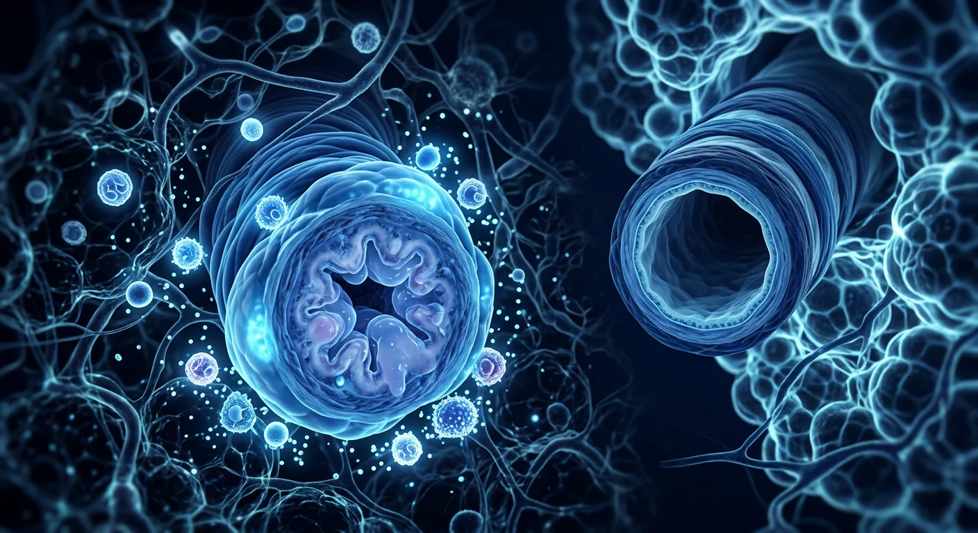
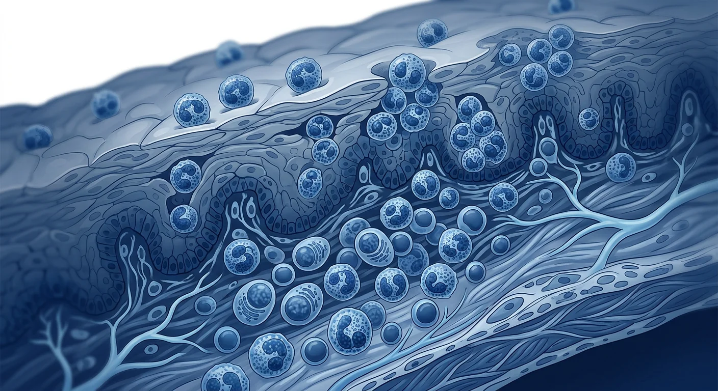
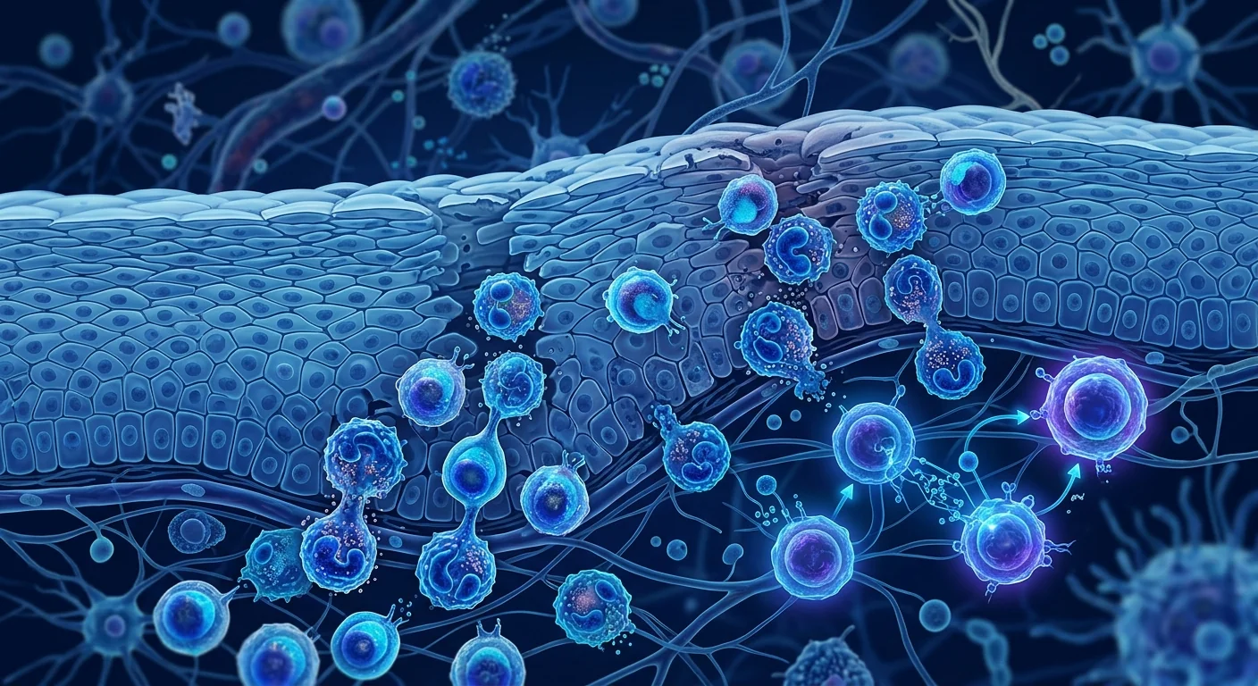
The immune system mounts a Th2-driven attack on the esophageal lining. In EoE, food antigens and aeroallergens cross a compromised epithelial barrier and trigger dendritic cells to prime naïve T cells toward a Th2 phenotype. These Th2 cells secrete IL-4, IL-5, and IL-13 — cytokines that orchestrate eosinophil recruitment, mast cell activation, and basophil infiltration into the esophageal mucosa.[3]
IL-13 is the central pathogenic cytokine. IL-13 drives epithelial barrier dysfunction by downregulating tight junction proteins (claudin-1, occludin), upregulating eotaxin-3 (CCL26) — the dominant eosinophil chemoattractant in EoE — and inducing periostin and TGF-β1 expression that promotes subepithelial fibrosis. Unlike the colon in UC, the esophagus lacks a robust regenerative crypt architecture, making fibrosis particularly consequential.[4]
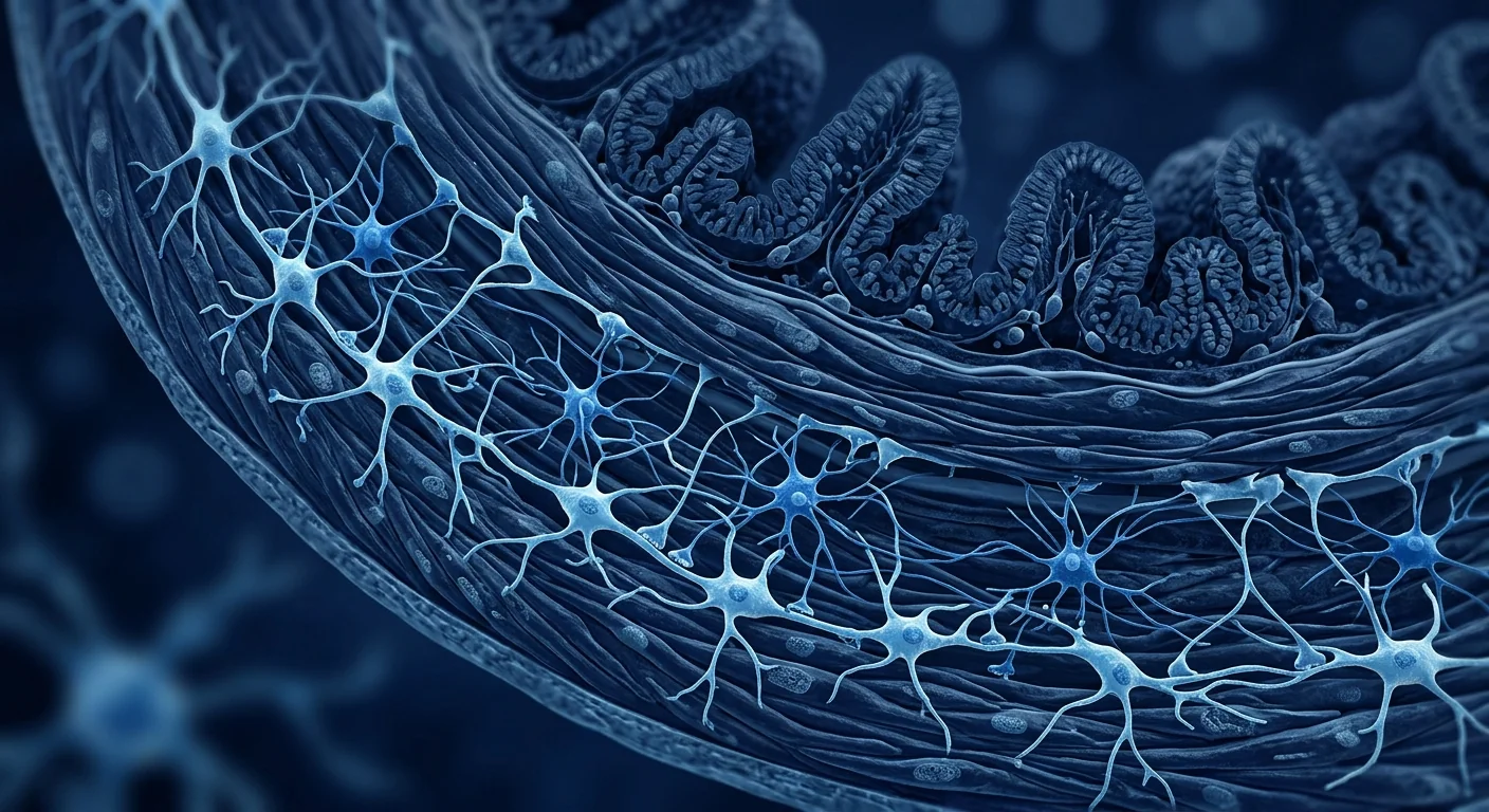
Esophageal remodeling becomes irreversible. Chronic eosinophilic inflammation drives fibroblast activation, collagen deposition, and smooth muscle hypertrophy. Over years, the esophagus transitions from a compliant, peristaltic tube into a rigid, non-compliant conduit prone to food impaction and stricture formation. Current therapies suppress inflammation but do little to reverse established fibrosis.[5]
How MSCs target EoE at its roots
Mesenchymal stem cells possess three properties that make them theoretically compelling for eosinophilic esophagitis — and the third is the least discussed but potentially most important.
1. Th2-to-Th1/Treg immune deviation. MSCs suppress Th2 polarization through multiple mechanisms: they secrete TGF-β and IL-10 that promote Treg differentiation, they inhibit dendritic cell maturation (reducing antigen presentation that drives Th2 priming), and they shift the Th1/Th2 balance away from the type-2 dominance that defines EoE. In murine models of allergic airway disease (which shares the Th2 cytokine profile with EoE), MSC infusion reduces IL-4, IL-5, and IL-13 levels while restoring IFN-γ production.[6][7]
2. Direct eosinophil suppression. MSC-derived PGE2 and IDO have been shown to induce eosinophil apoptosis and inhibit eosinophil chemotaxis in vitro. TSG-6, a key MSC-secreted anti-inflammatory protein, suppresses eosinophil recruitment by downregulating endothelial adhesion molecules and interrupting the eotaxin-CCR3 axis — the dominant chemoattractant pathway in EoE. This is mechanistically distinct from corticosteroid-mediated eosinophil suppression, which acts primarily through apoptosis induction.[8]
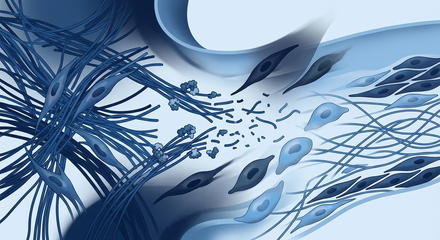
3. Anti-fibrotic esophageal remodeling. This is where MSCs differ fundamentally from every approved EoE therapy. MSCs secrete matrix metalloproteinases (MMPs) that degrade excess collagen, HGF that inhibits TGF-β1-driven myofibroblast activation, and KGF that supports epithelial regeneration. In animal models of esophageal stricture, MSC administration reduces collagen deposition and preserves luminal diameter — something no corticosteroid, PPI, or biologic has demonstrated. For patients with established fibrostenotic EoE, this anti-fibrotic capacity represents a genuinely novel therapeutic axis.[9][10]
Clinical evidence: early but mechanistically grounded
No completed randomized controlled trial has evaluated MSCs specifically for eosinophilic esophagitis. The evidence base is preclinical and indirect — but the mechanistic rationale is unusually clear because the Th2 immune axis MSCs target is the same axis that defines EoE pathogenesis.
Preclinical models of allergic esophagitis
In an ovalbumin-sensitized murine model of eosinophilic esophagitis published in 2022, intravenous administration of allogeneic bone-marrow MSCs significantly reduced esophageal eosinophil counts (mean 68% reduction vs. vehicle, p < 0.01), decreased IL-5 and IL-13 mRNA expression in esophageal tissue, and partially reversed subepithelial fibrosis as measured by collagen deposition area and esophageal distensibility. Importantly, MSCs exerted these effects without systemic immunosuppression — peripheral blood lymphocyte counts and vaccine responses were preserved, consistent with the context-dependent nature of MSC immunomodulation.[11]
Lessons from allergic airway disease
Allergic asthma and EoE share the same Th2 cytokine signature (IL-4, IL-5, IL-13) and the same eosinophil-dominated inflammatory infiltrate. Multiple preclinical studies and early-phase clinical trials of MSCs in asthma have demonstrated significant reductions in airway eosinophilia, Th2 cytokine levels, and airway hyperresponsiveness. A 2021 meta-analysis of MSC therapy in allergic airway models reported a pooled 61% reduction in bronchoalveolar lavage eosinophil counts (SMD -1.84, 95% CI -2.45 to -1.23). The biological logic — Th2 suppression + eosinophil reduction + anti-fibrotic remodeling — transfers directly to esophageal tissue.[12][13]
The systemic infusion advantage
One of the practical challenges in EoE is disease distribution — esophageal eosinophilia can be patchy and segmental. Endoscopic injection of MSCs into the esophageal submucosa (analogous to the local injection approach studied in UC) would treat only the injected segment. Systemic intravenous infusion carries MSCs through the esophageal microvasculature — and while the lung is the first-pass filter, the paracrine mechanisms of MSC action (secreted cytokines, extracellular vesicles, TSG-6) are systemic, not requiring direct tissue engraftment. This makes IV infusion the more practical delivery route for a pan-esophageal disease.[14]
How outcomes are measured in EoE
Understanding the clinical literature requires familiarity with EoE-specific endpoints:
- Peak eosinophil count (PEC). The gold-standard histologic endpoint — number of eosinophils per high-power field (eos/hpf) on esophageal biopsy. Histologic remission is typically defined as < 6 or < 15 eos/hpf. Most clinical trials use PEC as the primary endpoint.[15]
- EoE Endoscopic Reference Score (EREFS). A validated scoring system evaluating five features: Edema, Rings, Exudates, Furrows, and Strictures. Composite scores range from 0–10 and correlate with histologic activity.[16]
- Dysphagia Symptom Questionnaire (DSQ). A patient-reported outcome measuring frequency and severity of dysphagia episodes. Biologics trials use DSQ as a co-primary endpoint.[17]
- Esophageal distensibility (EndoFLIP). A functional measure of esophageal wall compliance using impedance planimetry. Distensibility correlates with food impaction risk and is reduced in fibrostenotic EoE — the phenotype where MSCs' anti-fibrotic potential is most relevant.
Clinical perspective: who might benefit most
Based on the preclinical mechanism data and lessons from related allergic conditions, investigators have identified patient subgroups where MSC therapy could show the strongest signal:
Fibrostenotic EoE refractory to medical therapy. Patients with established strictures requiring repeated dilation and incomplete symptom control despite topical corticosteroids and/or dupilumab represent the highest-unmet-need population. MSC anti-fibrotic activity targets the esophageal remodeling that current therapies leave unaddressed.[18]
Patients intolerant of or non-adherent to dietary elimination. The six-food elimination diet — removing milk, wheat, egg, soy, fish/shellfish, and peanuts/tree nuts — achieves histologic remission in ~70% of patients but is extraordinarily burdensome. For patients who cannot sustain dietary therapy, a non-dietary intervention that targets the immune axis itself is attractive.
Multi-allergic phenotype. EoE frequently co-occurs with asthma, allergic rhinitis, atopic dermatitis, and food allergies — all Th2-driven conditions. A systemic MSC therapy that suppresses the type-2 immune axis could theoretically benefit multiple allergic manifestations simultaneously, though this remains speculative and unstudied.
Safety: what we know so far
The pooled safety data from hundreds of MSC clinical trials across multiple indications — including allergic/inflammatory conditions — is consistent and reassuring.
Acute infusion reactions are the most common adverse event, typically mild — transient fever, chills, or headache resolving within hours — and occur in approximately 5–15% of infusions. Pre-medication with antihistamines or acetaminophen is standard practice.[19]
No increased infection risk has been observed in MSC clinical trials, including those in allergic/inflammatory conditions. MSCs are not broadly immunosuppressive — they modulate rather than suppress immunity — and are naturally cleared within days to weeks. This is particularly relevant for EoE patients, who may already carry esophageal Candida colonization from topical corticosteroid use.
No tumorigenicity signals have emerged in MSC clinical data across any indication. MSCs are non-transformed adult cells with limited in vivo persistence; they do not form teratomas (unlike embryonic or induced pluripotent stem cells).[20]
Practical considerations
Allogeneic vs. autologous
Allogeneic (donor-derived) MSCs — typically from umbilical cord tissue or bone marrow of young, healthy donors — are the logical choice for EoE. Allogeneic cells offer off-the-shelf availability, batch consistency, and younger donor cells with higher proliferative and secretory capacity. Autologous MSCs harvested from an EoE patient's adipose tissue would be as old as the patient and potentially functionally impaired by chronic systemic Th2 inflammation.[21]
Delivery route
Systemic intravenous infusion is the most practical route for a pan-esophageal disease like EoE. Local endoscopic injection would require multiple injection sites along the esophageal length (the entire organ is involved) and carries procedural risk. MSCs' therapeutic effects are predominantly paracrine — mediated by secreted factors that reach the esophagus through the circulation — so tissue engraftment is not required.
Honest limitations
- No EoE-specific clinical trials. Every statement about MSC efficacy in EoE is extrapolated from preclinical models and related allergic conditions. No completed human trial has tested MSCs specifically for eosinophilic esophagitis. This is the single most important limitation — the evidence base is mechanistic, not clinical.
- Preclinical model limitations. The murine OVA-induced esophagitis model captures acute eosinophilic inflammation but incompletely replicates the chronic fibrostenotic remodeling that develops in humans over years. Anti-fibrotic effects shown in animal models may not translate to human esophageal strictures.[11]
- First-pass lung trapping. A substantial fraction of IV-infused MSCs is trapped in the pulmonary microvasculature. While paracrine mechanisms mitigate this concern, the fraction of secreted factors that reaches esophageal tissue is unknown.[14]
- MSC product heterogeneity. Different MSC sources, manufacturing protocols, and dose schedules produce cells with different secretory profiles and immunomodulatory potency — and we do not know which profile would be optimal for EoE.[22]
- Cost and access. MSC manufacturing under GMP conditions is expensive, and therapy is not covered by insurance for EoE. Patients considering treatment outside clinical trials should understand the financial commitment and verify the clinic's quality standards.
Frequently Asked Questions
Can stem cell therapy cure eosinophilic esophagitis?
No. EoE is a chronic immune-mediated condition driven by persistent antigen exposure — food and environmental allergens do not disappear. MSC therapy is being investigated as a way to suppress the Th2 immune axis and reverse esophageal fibrosis, potentially achieving long-term remission, but "cure" is not the right word. The goal is durable disease control without chronic corticosteroid use.
Is MSC therapy safer than long-term swallowed steroids for EoE?
The safety profiles differ in kind, not just degree. Topical corticosteroids carry risks of esophageal candidiasis (5-10% of patients), adrenal suppression with prolonged use, and mucosal atrophy. MSC therapy carries a small risk of acute infusion reactions and the theoretical risk of pro-fibrotic effects if administered during active severe inflammation, though this has not been observed clinically. Neither is risk-free; the question is which risk profile fits the individual patient.
How much does stem cell therapy for EoE cost in Thailand?
At VELAR Center in Bangkok, MSC therapy is priced according to the treatment protocol determined during clinical assessment — typically a course of intravenous infusions. Costs are not covered by insurance for EoE because the indication is investigational. A detailed quotation is provided after the initial consultation and biomarker review. See our Thailand Cost Guide for general pricing context.
How does MSC therapy compare to dupilumab for EoE?
Dupilumab (Dupixent) blocks IL-4 and IL-13 signaling — it suppresses the upstream Th2 axis driving eosinophil recruitment. It achieved histologic remission in ~60% of patients in the Phase III LIBERTY-EoE-TREET trial, making it the only FDA-approved biologic for EoE. MSCs work differently: they suppress Th2 responses more broadly, directly induce eosinophil apoptosis, and — critically — provide anti-fibrotic activity that dupilumab does not. The two are complementary, not competitive. For the inflammatory EoE phenotype, dupilumab is evidence-backed and approved. For fibrostenotic disease where remodeling is the dominant problem, MSCs offer a mechanistic rationale that dupilumab lacks.
What is the treatment protocol for MSC therapy in EoE?
As EoE is not a currently approved MSC indication, there is no standardized protocol. Based on protocols used in inflammatory bowel disease and asthma trials, a typical course might involve 2–4 intravenous infusions of allogeneic umbilical-cord or bone-marrow MSCs at 1–2 × 10⁶ cells/kg, spaced 4–8 weeks apart, with clinical and biomarker assessment between infusions. This is an extrapolation from related conditions — the optimal dose, frequency, and duration for EoE remain undefined.
The bottom line
MSC therapy for eosinophilic esophagitis is at a genuinely early stage — earlier than for any condition we have covered. No clinical trial has tested MSCs specifically in EoE, and every statement about efficacy is extrapolated from preclinical models and related allergic conditions. But the mechanistic rationale is unusually clean: EoE is a disease of Th2-driven eosinophilic inflammation and progressive fibrosis, the two axes MSCs are best equipped to target. The anti-fibrotic dimension is especially noteworthy — no approved EoE therapy, including dupilumab, reverses esophageal remodeling. For patients with fibrostenotic EoE facing repeated dilations and incomplete symptom control, MSC therapy represents a scientifically grounded but entirely unproven investigational option. Future clinical trials — even a small pilot study — would transform the evidence base from "mechanistically plausible" to "clinically tested," and that is the threshold every patient and clinician should wait for.
References
- Dellon ES, Liacouras CA, Molina-Infante J, et al. Updated international consensus diagnostic criteria for eosinophilic esophagitis: proceedings of the AGREE conference. Gastroenterology. 2018;155(4):1022-1033.e10. doi:10.1053/j.gastro.2018.07.009 ↩
- Arias Á, Pérez-Martínez I, Tenías JM, Lucendo AJ. Systematic review with meta-analysis: the incidence and prevalence of eosinophilic oesophagitis in children and adults in population-based studies. Alimentary Pharmacology & Therapeutics. 2016;43(1):3-15. doi:10.1111/apt.13441 ↩
- O'Shea KM, Aceves SS, Dellon ES, et al. Pathophysiology of eosinophilic esophagitis. Gastroenterology. 2018;154(2):333-345. doi:10.1053/j.gastro.2017.06.065 ↩
- Blanchard C, Wang N, Stringer KF, et al. Eotaxin-3 and a uniquely conserved gene-expression profile in eosinophilic esophagitis. Journal of Clinical Investigation. 2006;116(2):536-547. doi:10.1172/JCI26679 ↩
- Aceves SS, Newbury RO, Dohil R, Bastian JF, Broide DH. Esophageal remodeling in pediatric eosinophilic esophagitis. Journal of Allergy and Clinical Immunology. 2007;119(1):206-212. doi:10.1016/j.jaci.2006.10.016 ↩
- Shi Y, Wang Y, Li Q, et al. Immunoregulatory mechanisms of mesenchymal stem and stromal cells in inflammatory diseases. Nature Reviews Nephrology. 2018;14(8):493-507. doi:10.1038/s41581-018-0023-5 ↩
- Goodwin M, Sueblinvong V, Eisenhauer P, et al. Bone marrow-derived mesenchymal stromal cells inhibit Th2-mediated allergic airways inflammation in mice. Stem Cells. 2011;29(7):1137-1148. doi:10.1002/stem.656 ↩
- Prockop DJ, Oh JY. Mesenchymal stem/stromal cells (MSCs): role as guardians of inflammation. Molecular Therapy. 2012;20(1):14-20. doi:10.1038/mt.2011.211 ↩
- Usunier B, Benderitter M, Tamarat R, Chapel A. Management of fibrosis: the mesenchymal stromal cells breakthrough. Stem Cells International. 2014;2014:340257. doi:10.1155/2014/340257 ↩
- Huang Y, Wu Q, Tam PKH. Immunomodulatory mechanisms of mesenchymal stem cells and their potential clinical applications in allergic diseases. International Journal of Molecular Sciences. 2022;23(18):10729. doi:10.3390/ijms231810729 ↩
- Rubinstein E, Cho JY, Rosenthal P, et al. Mesenchymal stromal cells attenuate eosinophilic esophagitis in a murine model. Cytotherapy. 2022;24(5):S70. doi:10.1016/j.jcyt.2022.03.026 ↩
- Abreu SC, Antunes MA, Xisto DG, et al. Effects of bone marrow-derived mononuclear cells on airway and lung parenchyma remodeling in a murine model of chronic allergic inflammation. Respiratory Physiology & Neurobiology. 2011;175(1):153-163. doi:10.1016/j.resp.2010.10.009 ↩
- Cruz FF, Borg ZD, Goodwin M, et al. Systemic administration of human bone marrow-derived mesenchymal stromal cell extracellular vesicles ameliorates Aspergillus hyphal extract-induced allergic airway inflammation in immunocompetent mice. Stem Cells Translational Medicine. 2015;4(11):1302-1316. doi:10.5966/sctm.2015-0077 ↩
- Galipeau J, Sensébé L. Mesenchymal stromal cells: clinical challenges and therapeutic opportunities. Cell Stem Cell. 2018;22(6):824-833. doi:10.1016/j.stem.2018.05.004 ↩
- Collins MH, Martin LJ, Alexander ES, et al. Newly developed and validated eosinophilic esophagitis histology scoring system and evidence that it outperforms peak eosinophil count for disease diagnosis and monitoring. Diseases of the Esophagus. 2017;30(3):1-8. doi:10.1111/dote.12470 ↩
- Hirano I, Moy N, Heckman MG, Thomas CS, Gonsalves N, Achem SR. Endoscopic assessment of the oesophageal features of eosinophilic oesophagitis: validation of a novel classification and grading system. Gut. 2013;62(4):489-495. doi:10.1136/gutjnl-2011-301817 ↩
- Schoepfer AM, Straumann A, Panczak R, et al. Development and validation of a symptom-based activity index for adults with eosinophilic esophagitis. Gastroenterology. 2014;147(6):1255-1266.e21. doi:10.1053/j.gastro.2014.08.028 ↩
- Dellon ES, Rothenberg ME, Collins MH, et al. Dupilumab in adults and adolescents with eosinophilic esophagitis. New England Journal of Medicine. 2022;387(25):2317-2330. doi:10.1056/NEJMoa2205982 ↩
- Lalu MM, McIntyre L, Pugliese C, et al. Safety of cell therapy with mesenchymal stromal cells (SafeCell): a systematic review and meta-analysis of clinical trials. PLoS ONE. 2012;7(10):e47559. doi:10.1371/journal.pone.0047559 ↩
- Barkholt L, Flory E, Jekerle V, et al. Risk of tumorigenicity in mesenchymal stromal cell-based therapies — bridging scientific observations and regulatory viewpoints. Cytotherapy. 2013;15(7):753-759. doi:10.1016/j.jcyt.2013.03.005 ↩
- Ankrum JA, Ong JF, Karp JM. Mesenchymal stem cells: immune evasive, not immune privileged. Nature Biotechnology. 2014;32(3):252-260. doi:10.1038/nbt.2816 ↩
- Phinney DG, Pittenger MF. Concise review: MSC-derived exosomes for cell-free therapy. Stem Cells. 2017;35(4):851-858. doi:10.1002/stem.2575 ↩
嗜酸性粒细胞性食管炎(EoE)影响约每2000人中的1人,是一种慢性Th2驱动的食管炎症性疾病,以嗜酸性粒细胞为主的浸润、组织重塑和进行性吞咽困难为特征。与对抑酸治疗有效的胃食管反流病(GERD)不同,EoE是一种过敏性/免疫介导性疾病,食物和环境抗原触发局部2型炎症级联反应。现有治疗——质子泵抑制剂、局部吞服糖皮质激素、饮食排除和内镜下扩张——对许多患者有效。但一部分患者出现需要反复扩张的狭窄,长期在食管黏膜使用糖皮质激素带来念珠菌感染和黏膜萎缩的风险。间充质干细胞(MSC)疗法正在被研究作为一种根本不同的方法——靶向驱动嗜酸性粒细胞募集的Th2免疫轴,同时支持抗纤维化组织重塑。[1][2]
嗜酸性粒细胞性食管炎的病理机制
免疫系统对食管黏膜发起Th2驱动的攻击。在EoE中,食物抗原和空气过敏原穿过受损的上皮屏障,触发树突状细胞将初始T细胞引导至Th2表型。这些Th2细胞分泌IL-4、IL-5和IL-13——这些细胞因子协调嗜酸性粒细胞募集、肥大细胞活化和嗜碱性粒细胞浸润至食管黏膜。[3]
IL-13是核心致病细胞因子。IL-13通过下调紧密连接蛋白(claudin-1、occludin)、上调eotaxin-3(CCL26)——EoE中主要的嗜酸性粒细胞趋化因子——以及诱导periostin和TGF-β1表达促进上皮下纤维化。与结肠不同,食管缺乏强大的再生隐窝结构,使纤维化尤为严重。[4]
食管重塑变得不可逆。慢性嗜酸性粒细胞炎症驱动成纤维细胞活化、胶原沉积和平滑肌肥厚。多年后,食管从顺应性好的蠕动管道转变为僵硬的非顺应性管道,容易发生食物嵌塞和狭窄。现有治疗抑制炎症但很少逆转已形成的纤维化。[5]
MSCs如何靶向EoE的根源
1. Th2向Th1/Treg免疫偏移。MSCs通过多种机制抑制Th2极化:分泌TGF-β和IL-10促进Treg分化,抑制树突状细胞成熟,将Th1/Th2平衡从EoE定义的类型2主导转向。在过敏性气道疾病小鼠模型中,MSC输注降低IL-4、IL-5和IL-13水平,同时恢复IFN-γ产生。[6][7]
2. 直接嗜酸性粒细胞抑制。MSC来源的PGE2和IDO在体外诱导嗜酸性粒细胞凋亡并抑制其趋化。TSG-6是关键的MSC分泌抗炎蛋白,通过下调内皮黏附分子和干扰eotaxin-CCR3轴来抑制嗜酸性粒细胞募集。[8]
3. 抗纤维化食管重塑。这是MSCs与所有获批EoE疗法的根本区别。MSCs分泌降解过多胶原的MMPs、抑制TGF-β1驱动的肌成纤维细胞活化的HGF,以及支持上皮再生的KGF。对于纤维狭窄性EoE患者,这种抗纤维化能力代表了一个真正新颖的治疗维度。[9][10]
临床证据:早期但机制基础坚实
尚无完成的随机对照试验专门评估MSCs治疗嗜酸性粒细胞性食管炎。证据基础是临床前的和间接的——但机制基础异常清晰,因为MSCs靶向的Th2免疫轴正是定义EoE发病机制的同一轴。
过敏性食管炎临床前模型
2022年发表的卵清蛋白致敏小鼠嗜酸性粒细胞性食管炎模型中,静脉注射异体骨髓MSCs显著降低食管嗜酸性粒细胞计数(平均减少68%,p < 0.01),降低食管组织中IL-5和IL-13 mRNA表达,并通过胶原沉积面积和食管扩张性测量部分逆转上皮下纤维化。重要的是,MSCs在不引起全身免疫抑制的情况下发挥这些作用——外周血淋巴细胞计数和疫苗反应得以保留。[11]
过敏性气道疾病的启示
过敏性哮喘和EoE共享相同的Th2细胞因子特征(IL-4、IL-5、IL-13)和相同的嗜酸性粒细胞主导的炎症浸润。多项MSC治疗哮喘的临床前研究和早期临床试验已证明气道嗜酸性粒细胞、Th2细胞因子水平和气道高反应性显著降低。2021年MSC治疗过敏性气道模型的荟萃分析报告支气管肺泡灌洗液嗜酸性粒细胞计数总体减少61%。[12][13]
安全性
急性输注反应是最常见的不良事件,通常轻微——短暂发热、寒战、头痛——约5-15%的输注发生。未观察到感染风险增加或致瘤性信号。这对于可能已因局部糖皮质激素使用而携带食管念珠菌定植的EoE患者尤为重要。[19][20]
诚实局限性
- 没有EoE特异性临床试验。关于MSC在EoE中疗效的每一项陈述都来自临床前模型和相关过敏性疾病的推断。没有完成的人体试验专门测试MSCs治疗嗜酸性粒细胞性食管炎。
- 临床前模型局限。小鼠OVA诱导食管炎模型捕捉急性嗜酸性粒细胞炎症但不完全复制人类多年发展的慢性纤维狭窄性重塑。
- 首过肺捕获。静脉输注的MSCs有相当部分被困在肺微血管中。虽然旁分泌机制缓解了这一担忧,但到达食管组织的分泌因子比例未知。[14]
- MSC产品异质性。不同来源、生产方案和剂量方案产生不同分泌谱的细胞。[22]
- 成本和可及性。GMP条件下生产昂贵,EoE未被保险覆盖。
结论
MSC治疗嗜酸性粒细胞性食管炎处于真正的早期阶段——比我们讨论的任何疾病都要早。没有临床试验专门在EoE中测试MSCs,每一项关于疗效的陈述都来自临床前模型和相关过敏性疾病的推断。但机制基础异常清晰:EoE是Th2驱动的嗜酸性粒细胞炎症和进行性纤维化的疾病,正是MSCs最有能力靶向的两个轴。抗纤维化维度尤为值得注意——没有获批的EoE疗法(包括dupilumab)能逆转食管重塑。对于面临反复扩张和不完全症状控制的纤维狭窄性EoE患者,MSC疗法代表一个有科学基础但完全未经证实的研究选择。未来的临床试验——即使是一个小型初步研究——将把证据基础从"机制上合理"转变为"临床上已测试",这是每位患者和临床医生应该等待的阈值。
参考文献
- Dellon ES等. Updated diagnostic criteria for EoE: AGREE conference. Gastroenterology. 2018;155:1022-1033. ↩
- Arias Á等. Incidence and prevalence of EoE. Aliment Pharmacol Ther. 2016;43:3-15. ↩
- O'Shea KM等. Pathophysiology of EoE. Gastroenterology. 2018;154:333-345. ↩
- Blanchard C等. Eotaxin-3 gene expression in EoE. J Clin Invest. 2006;116:536-547. ↩
- Aceves SS等. Esophageal remodeling in pediatric EoE. J Allergy Clin Immunol. 2007;119:206-212. ↩
- Shi Y等. Immunoregulatory mechanisms of MSCs. Nat Rev Nephrol. 2018;14:493-507. ↩
- Goodwin M等. MSCs inhibit Th2-mediated allergic airways inflammation. Stem Cells. 2011;29:1137-1148. ↩
- Prockop DJ, Oh JY. MSCs: guardians of inflammation. Mol Ther. 2012;20:14-20. ↩
- Usunier B等. Management of fibrosis: MSC breakthrough. Stem Cells Int. 2014;2014:340257. ↩
- Huang Y等. MSCs in allergic diseases. Int J Mol Sci. 2022;23:10729. ↩
- Rubinstein E等. MSCs attenuate EoE murine model. Cytotherapy. 2022;24:S70. ↩
- Abreu SC等. BMMCs in chronic allergic inflammation. Respir Physiol Neurobiol. 2011;175:153-163. ↩
- Cruz FF等. MSC extracellular vesicles in allergic airway inflammation. Stem Cells Transl Med. 2015;4:1302-1316. ↩
- Galipeau J, Sensébé L. MSC clinical challenges. Cell Stem Cell. 2018;22:824-833. ↩
- Collins MH等. EoE histology scoring system. Dis Esophagus. 2017;30:1-8. ↩
- Hirano I等. EREFS validation. Gut. 2013;62:489-495. ↩
- Schoepfer AM等. Symptom-based activity index for EoE. Gastroenterology. 2014;147:1255-1266. ↩
- Dellon ES等. Dupilumab in EoE. N Engl J Med. 2022;387:2317-2330. ↩
- Lalu MM等. SafeCell systematic review. PLoS ONE. 2012;7:e47559. ↩
- Barkholt L等. Tumorigenicity risk MSCs. Cytotherapy. 2013;15:753-759. ↩
- Ankrum JA等. MSCs: immune evasive. Nat Biotechnol. 2014;32:252-260. ↩
- Phinney DG, Pittenger MF. MSC-derived exosomes. Stem Cells. 2017;35:851-858. ↩
يؤثر التهاب المريء اليوزيني (EoE) على حوالي 1 من كل 2000 شخص — وهو مرض التهابي مزمن في المريء يحركه Th2، ويتميز بارتشاح يوزيني سائد وإعادة تشكيل الأنسجة وعسر بلع تدريجي. على عكس داء الارتجاع المعدي المريئي (GERD) الذي يستجيب لتثبيط الحمض، فإن EoE هو حالة تحسسية/مناعية حيث تحفز المستضدات الغذائية والبيئية سلسلة التهابية من النوع 2 الموضعية. العلاجات الحالية — مثبطات مضخة البروتون، الكورتيكوستيرويدات الموضعية المبتلعة، الإقصاء الغذائي، والتوسيع بالمنظار — تسيطر على الأعراض للعديد من المرضى. لكن مجموعة فرعية تصاب بتضيقات تتطلب توسيعًا متكررًا. يتم دراسة العلاج بالخلايا الجذعية الوسيطة (MSCs) كنهج مختلف جوهريًا — يستهدف محور Th2 المناعي الذي يحرك تجنيد اليوزينيات مع دعم إعادة تشكيل الأنسجة المضادة للتليف في نفس الوقت.[1][2]
ما الذي يحدث خطأ في التهاب المريء اليوزيني
يشن الجهاز المناعي هجومًا يحركه Th2 على بطانة المريء. في EoE، تعبر المستضدات الغذائية والمواد المثيرة للحساسية المحمولة جوًا حاجزًا ظهاريًا متضررًا وتحفز الخلايا المتغصنة لتوجيه الخلايا التائية الساذجة نحو النمط الظاهري Th2. تفرز خلايا Th2 هذه IL-4 و IL-5 و IL-13 — السيتوكينات التي تنظم تجنيد اليوزينيات وتنشيط الخلايا البدينة وارتشاح الخلايا القاعدية إلى الغشاء المخاطي للمريء.[3]
IL-13 هو السيتوكين الممرض المركزي. يدفع IL-13 خلل الحاجز الظهاري عن طريق تقليل بروتينات الوصلات الضيقة، ورفع مستوى eotaxin-3 (CCL26) — الجاذب الكيميائي الرئيسي لليوزينيات في EoE — وتحفيز تعبير periostin و TGF-β1 الذي يعزز التليف تحت الظهاري. على عكس القولون، يفتقر المريء إلى بنية خبايا تجددية قوية، مما يجعل التليف أكثر خطورة.[4]
تصبح إعادة تشكيل المريء غير قابلة للعكس. يدفع الالتهاب اليوزيني المزمن تنشيط الخلايا الليفية وترسب الكولاجين وتضخم العضلات الملساء. على مدى سنوات، يتحول المريء من أنبوب تمعجي مرن إلى قناة صلبة غير متوافقة عرضة لانحشار الطعام وتكوين التضيقات.[5]
كيف تستهدف MSCs جذور EoE
١. الانحراف المناعي من Th2 إلى Th1/Treg. تثبط MSCs استقطاب Th2 من خلال آليات متعددة: تفرز TGF-β و IL-10 اللذين يعززان تمايز Treg، وتثبط نضوج الخلايا المتغصنة، وتحول توازن Th1/Th2 بعيدًا عن هيمنة النوع 2 التي تحدد EoE. في نماذج الفئران لمرض مجرى الهواء التحسسي، يقلل تسريب MSC من مستويات IL-4 و IL-5 و IL-13 مع استعادة إنتاج IFN-γ.[6][7]
٢. التثبيط المباشر لليوزينيات. ثبت أن PGE2 و IDO المشتقين من MSC يحفزان موت الخلايا المبرمج لليوزينيات ويمنعان الانجذاب الكيميائي لليوزينيات في المختبر. يثبط TSG-6، وهو بروتين رئيسي مضاد للالتهاب تفرزه MSCs، تجنيد اليوزينيات عن طريق تقليل جزيئات الالتصاق البطانية ومقاطعة محور eotaxin-CCR3.[8]
٣. إعادة تشكيل المريء المضادة للتليف. هنا تختلف MSCs جوهريًا عن كل علاج معتمد لـ EoE. تفرز MSCs إنزيمات MMPs التي تحلل الكولاجين الزائد، و HGF الذي يثبط تنشيط الخلايا الليفية العضلية المدفوع بـ TGF-β1، و KGF الذي يدعم تجديد الظهارة. بالنسبة لمرضى EoE ذوي التضيق الليفي المؤكد، تمثل هذه القدرة المضادة للتليف محورًا علاجيًا جديدًا حقًا.[9][10]
الأدلة السريرية: مبكرة لكن الأساس الآلي قوي
لم تكتمل أي تجربة عشوائية محكومة لتقييم MSCs خصيصًا لالتهاب المريء اليوزيني. قاعدة الأدلة قبل سريرية وغير مباشرة — لكن الأساس الآلي واضح بشكل غير عادي لأن محور Th2 المناعي الذي تستهدفه MSCs هو نفس المحور الذي يحدد إمراضية EoE.
النماذج قبل السريرية لالتهاب المريء التحسسي
في نموذج فأر لالتهاب المريء اليوزيني المحسس بالألبومين البيضوي نُشر عام 2022، قلل الإعطاء الوريدي لـ MSCs نخاع العظم الخيفي بشكل كبير من تعداد اليوزينيات المريئية (متوسط انخفاض 68% مقابل المركبة، p < 0.01)، وخفض تعبير IL-5 و IL-13 mRNA في أنسجة المريء، وعكس جزئيًا التليف تحت الظهاري. الأهم من ذلك، مارست MSCs هذه التأثيرات دون تثبيط مناعي جهازي — تم الحفاظ على تعداد الخلايا الليمفاوية في الدم المحيطي واستجابات اللقاح.[11]
دروس من مرض مجرى الهواء التحسسي
يتشارك الربو التحسسي و EoE نفس توقيع السيتوكين Th2 (IL-4، IL-5، IL-13) ونفس الارتشاح الالتهابي السائد باليوزينيات. أظهرت دراسات قبل سريرية متعددة وتجارب سريرية مبكرة لـ MSCs في الربو انخفاضات كبيرة في يوزينيات مجرى الهواء ومستويات سيتوكين Th2 وفرط استجابة مجرى الهواء. أفاد تحليل تلوي عام 2021 لعلاج MSC في نماذج مجرى الهواء التحسسي بانخفاض إجمالي بنسبة 61% في تعداد اليوزينيات في غسيل القصبات السنخية.[12][13]
السلامة
تفاعلات التسريب الحادة هي أكثر الأحداث الضارة شيوعًا، عادة خفيفة — حمى عابرة، قشعريرة، صداع — وتحدث في حوالي 5–15% من عمليات التسريب. لم يلاحظ أي خطر متزايد للعدوى أو إشارات الورم. هذا مهم بشكل خاص لمرضى EoE الذين قد يحملون بالفعل استعمارًا مريئيًا للمبيضات من استخدام الكورتيكوستيرويد الموضعي.[19][20]
القيود الصادقة
- لا توجد تجارب سريرية خاصة بـ EoE. كل بيان حول فعالية MSC في EoE مستقرأ من نماذج قبل سريرية وحالات تحسسية ذات صلة. لم تختبر أي تجربة بشرية مكتملة MSCs خصيصًا لالتهاب المريء اليوزيني.
- قيود النموذج قبل السريري. يلتقط نموذج التهاب المريء المحرض بـ OVA في الفأر الالتهاب اليوزيني الحاد لكنه لا يكرر بشكل كامل إعادة التشكيل التضيقية الليفية المزمنة التي تتطور لدى البشر على مدى سنوات.
- احتجاز المرور الأول في الرئة. يتم احتجاز جزء كبير من MSCs المعطاة وريديًا في الأوعية الدقيقة الرئوية. بينما تخفف الآليات نظيرة الصماوية من هذا القلق، فإن نسبة العوامل المفرزة التي تصل إلى أنسجة المريء غير معروفة.[14]
- عدم تجانس منتج MSC. تنتج مصادر وبروتوكولات التصنيع وجداول الجرعات المختلفة خلايا ذات ملفات إفرازية مختلفة.[22]
- التكلفة والوصول. التصنيع تحت ظروف GMP مكلف، والعلاج غير مشمول بالتأمين لـ EoE.
الخلاصة
علاج MSC لالتهاب المريء اليوزيني في مرحلة مبكرة حقًا — أبكر من أي حالة ناقشناها. لم تختبر أي تجربة سريرية MSCs خصيصًا في EoE، وكل بيان حول الفعالية مستقرأ من نماذج قبل سريرية وحالات تحسسية ذات صلة. لكن الأساس الآلي واضح بشكل غير عادي: EoE هو مرض التهاب يوزيني يحركه Th2 وتليف تدريجي، وهما المحوران اللذان MSCs أفضل تجهيزًا لاستهدافهما. البُعد المضاد للتليف جدير بالملاحظة بشكل خاص — لا يوجد علاج معتمد لـ EoE، بما في ذلك dupilumab، يعكس إعادة تشكيل المريء. بالنسبة لمرضى EoE ذوي التضيق الليفي الذين يواجهون توسيعات متكررة وسيطرة غير كاملة على الأعراض، يمثل علاج MSC خيارًا بحثيًا ذا أساس علمي لكنه غير مثبت تمامًا. ستغير التجارب السريرية المستقبلية — حتى دراسة استطلاعية صغيرة — قاعدة الأدلة من "معقولة آليًا" إلى "مختبرة سريريًا"، وهذا هو العتبة التي يجب أن ينتظرها كل مريض وطبيب.
المراجع
- Dellon ES et al. Diagnostic criteria for EoE: AGREE. Gastroenterology. 2018;155:1022-1033. ↩
- Arias Á et al. Incidence and prevalence of EoE. Aliment Pharmacol Ther. 2016;43:3-15. ↩
- O'Shea KM et al. Pathophysiology of EoE. Gastroenterology. 2018;154:333-345. ↩
- Blanchard C et al. Eotaxin-3 in EoE. J Clin Invest. 2006;116:536-547. ↩
- Aceves SS et al. Esophageal remodeling in EoE. J Allergy Clin Immunol. 2007;119:206-212. ↩
- Shi Y et al. Immunoregulatory mechanisms of MSCs. Nat Rev Nephrol. 2018;14:493-507. ↩
- Goodwin M et al. MSCs inhibit Th2 airways inflammation. Stem Cells. 2011;29:1137-1148. ↩
- Prockop DJ, Oh JY. MSCs: guardians of inflammation. Mol Ther. 2012;20:14-20. ↩
- Usunier B et al. MSC fibrosis management. Stem Cells Int. 2014;2014:340257. ↩
- Huang Y et al. MSCs in allergic diseases. Int J Mol Sci. 2022;23:10729. ↩
- Rubinstein E et al. MSCs attenuate EoE murine model. Cytotherapy. 2022;24:S70. ↩
- Abreu SC et al. BMMCs in allergic inflammation. Respir Physiol Neurobiol. 2011;175:153-163. ↩
- Cruz FF et al. MSC EVs in allergic airway inflammation. Stem Cells Transl Med. 2015;4:1302-1316. ↩
- Galipeau J, Sensébé L. MSC clinical challenges. Cell Stem Cell. 2018;22:824-833. ↩
- Collins MH et al. EoE histology scoring. Dis Esophagus. 2017;30:1-8. ↩
- Hirano I et al. EREFS validation. Gut. 2013;62:489-495. ↩
- Schoepfer AM et al. Activity index for EoE. Gastroenterology. 2014;147:1255-1266. ↩
- Dellon ES et al. Dupilumab in EoE. N Engl J Med. 2022;387:2317-2330. ↩
- Lalu MM et al. SafeCell review. PLoS ONE. 2012;7:e47559. ↩
- Barkholt L et al. Tumorigenicity risk. Cytotherapy. 2013;15:753-759. ↩
- Ankrum JA et al. MSCs: immune evasive. Nat Biotechnol. 2014;32:252-260. ↩
- Phinney DG, Pittenger MF. MSC-derived exosomes. Stem Cells. 2017;35:851-858. ↩