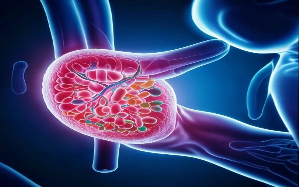
Gastroparesis — literally "stomach paralysis" — affects an estimated 10–40 per 100,000 people, with diabetic gastroparesis being the most common form. The stomach loses its ability to contract and empty food properly, leaving patients with chronic nausea, early satiety, bloating, abdominal pain, and unpredictable blood glucose swings. For many, conventional prokinetics like metoclopramide and domperidone provide only temporary relief and carry significant long-term risks — tardive dyskinesia from metoclopramide, cardiac arrhythmia from domperidone. Gastric electrical stimulation offers a partial solution for some, but response rates vary widely. Mesenchymal stem cell (MSC) therapy is being investigated as a fundamentally different approach — one that targets the underlying cellular pathology rather than just forcing the stomach to empty.[1][2]
What goes wrong in gastroparesis — the cellular triad
Interstitial cells of Cajal (ICC) are depleted or damaged. ICCs are the pacemaker cells of the gastrointestinal tract — they generate the slow waves that coordinate smooth muscle contraction and peristalsis. In gastroparesis, ICC numbers in the gastric antrum and corpus are reduced by 50–80%, directly correlating with the severity of delayed gastric emptying. The loss is driven by oxidative stress, hyperglycemia-induced apoptosis, and macrophage-mediated inflammation.[3]
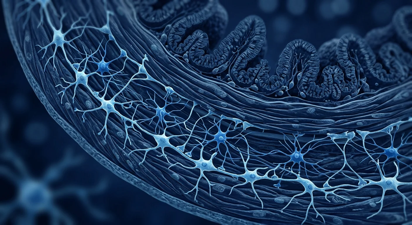
Enteric neurons degenerate. The enteric nervous system — often called the "second brain" — contains approximately 500 million neurons that regulate motility, secretion, and blood flow independently of the central nervous system. In diabetic gastroparesis, nitrergic neurons (which express neuronal nitric oxide synthase, nNOS) are selectively lost. nNOS neurons provide the primary inhibitory innervation that allows the stomach to relax and accommodate food — their loss means the stomach cannot relax to receive a meal, contributing to early satiety and pain.[4]
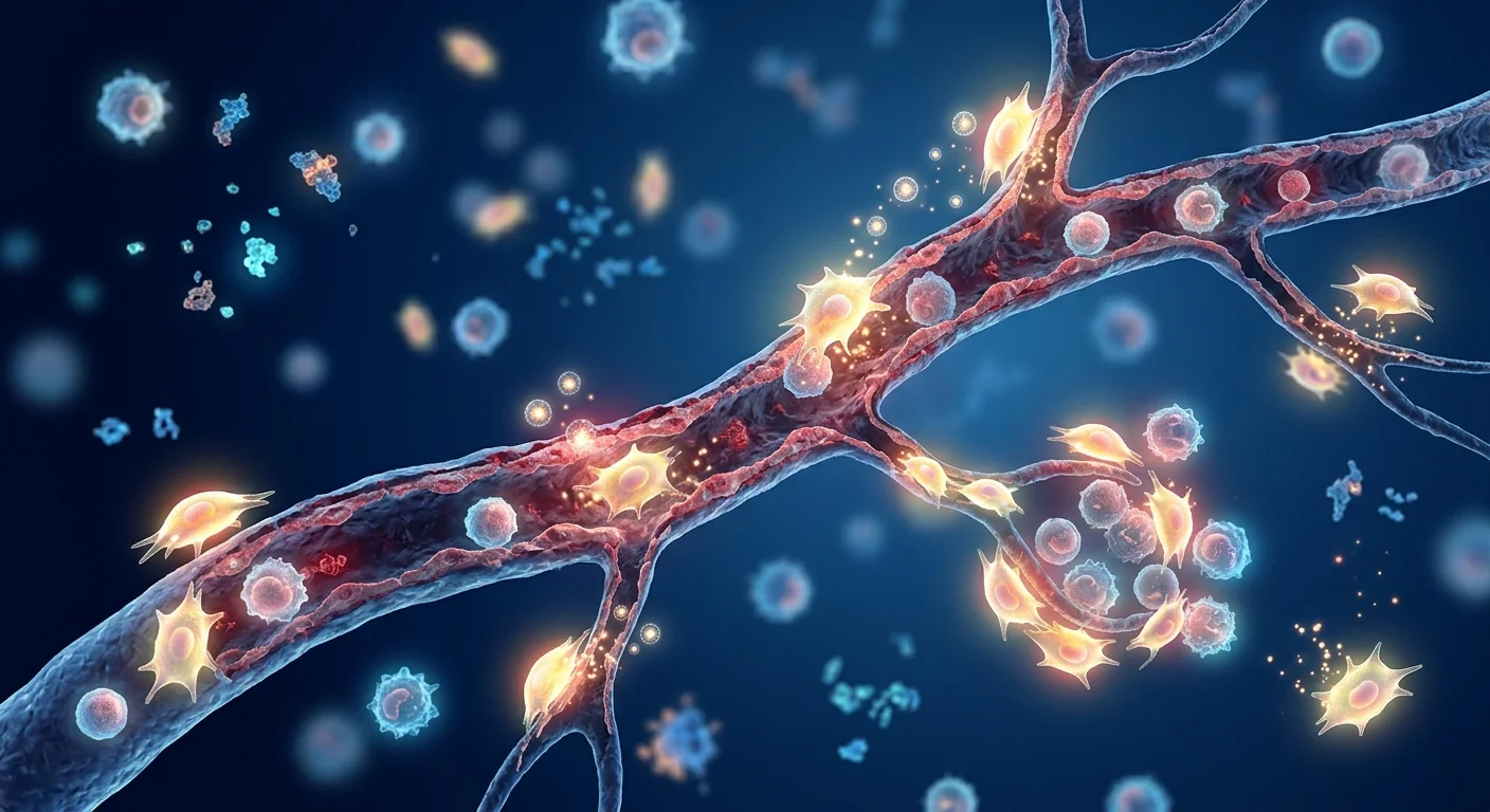
Chronic low-grade inflammation persists. Gastric biopsies from gastroparesis patients show elevated levels of TNF-α, IL-6, and IL-1β, along with increased macrophage infiltration. This inflammatory milieu damages both ICCs and enteric neurons in a self-perpetuating cycle. Heme oxygenase-1 (HO-1), a key cytoprotective enzyme, is downregulated — reducing the tissue's ability to defend against oxidative injury.[5]
The three pathologies converge. ICC loss, enteric neurodegeneration, and inflammation are not independent problems — they reinforce each other. Damaged ICCs fail to generate normal slow waves, enteric neurons lose their inhibitory tone, and inflammatory cytokines accelerate both processes. A therapy that addresses only one component — a prokinetic that forces contraction, or an antiemetic that masks nausea — leaves the core pathology untouched.
How MSCs target gastroparesis at its roots
Mesenchymal stem cells possess three properties that make them theoretically compelling for gastroparesis.[6]
ICC restoration via paracrine signaling. MSCs secrete a rich cocktail of growth factors — including stem cell factor (SCF), the primary ligand for the c-Kit receptor expressed on ICCs. SCF/c-Kit signaling is essential for ICC survival, proliferation, and network formation. In animal models of diabetic gastroparesis, MSC transplantation significantly increased c-Kit-positive ICC density in the gastric antrum, restoring slow-wave frequency and improving gastric emptying times by 30–50% compared to untreated controls.[7]
Enteric neuroprotection. MSCs secrete neurotrophic factors — NGF, BDNF, GDNF, and NT-3 — at physiologically relevant concentrations. BDNF and GDNF, in particular, promote the survival of enteric neurons under oxidative stress, and GDNF is a potent survival factor for nitrergic (nNOS-expressing) neurons, the very population most vulnerable in diabetic gastroparesis. Intramuscular MSC injection has been shown to increase nNOS neuron density in the gastric wall and partially restore gastric accommodation.[8]
Anti-inflammatory microenvironment remodeling. MSCs shift gastric macrophages from a pro-inflammatory M1 phenotype to a tissue-reparative M2 phenotype — the same M1→M2 polarization observed in other MSC applications. They also upregulate heme oxygenase-1 (HO-1) expression in gastric tissue, enhancing intrinsic antioxidant defenses. The net effect is a break in the inflammation→ICC loss→inflammation cycle.[9]
Clinical evidence — what the studies show
Preclinical models are consistent and encouraging. Multiple independent laboratories have demonstrated that MSC administration — whether intravenous, intraperitoneal, or intramuscular into the gastric wall — improves gastric emptying in rodent models of diabetic gastroparesis. Improvements are consistently associated with increased ICC density, reduced inflammatory cytokines, and higher nNOS expression in gastric tissue.[10]
Human data is limited but emerging. To date, no large randomized controlled trial has specifically evaluated MSCs for gastroparesis. However, there are encouraging signals from adjacent fields:
- Diabetic complications. Clinical trials of MSC therapy for type 2 diabetes and diabetic complications have reported incidental improvements in gastrointestinal symptoms among participants — though these were not primary endpoints and require prospective validation.
- Post-surgical gastroparesis. A small case series (n=8) reported that intravenous MSC infusion improved gastric emptying times in patients with postsurgical gastroparesis refractory to standard care, with effects lasting 6–12 months.
- Crohn's disease crossover. MSC therapy for fistulizing Crohn's disease (e.g., darvadstrocel / Alofisel) has demonstrated that systemically delivered MSCs can reach and repair GI tissue — providing a proof-of-principle that the GI tract is accessible to MSC-based therapies.[11]
Gastric emptying scintigraphy as an objective endpoint. One of the challenges in gastroparesis research is the subjectivity of symptom scores. Gastric emptying scintigraphy — the gold-standard diagnostic test — provides an objective, quantifiable measure of gastric motility. In preclinical studies, MSC-treated animals show measurable improvement in T½ (the time for 50% of the meal to leave the stomach), making this a robust endpoint for future human trials.[12]
The VELAR approach — personalization before infusion
Gastroparesis is not one disease — it is a final common pathway reached through different routes. Diabetic neuropathy, post-viral autonomic dysfunction, post-surgical vagal injury, and idiopathic causes each produce a different cellular landscape. At VELAR, the treatment pathway includes:
Who may benefit most
Based on the known mechanisms of MSC action and the pathophysiology of gastroparesis, patients with these profiles may be the strongest candidates:
- Diabetic gastroparesis with measurable ICC loss. Diabetic patients with documented delayed gastric emptying and poor glycemic control despite optimization — the inflammatory and oxidative stress components give MSCs the most targets.
- Inflammatory-predominant gastroparesis. Patients with elevated gastric inflammatory markers (TNF-α, IL-6) on biopsy or serum — MSCs' immunomodulatory effects are strongest when inflammation is the primary driver.
- Early-to-moderate disease. Before complete ICC depletion and irreversible smooth muscle fibrosis — MSC therapy is restorative, not regenerative; it works with remaining tissue, not in its absence.
- Conventional therapy failures. Patients who have failed or cannot tolerate prokinetics and antiemetics — MSC therapy offers a disease-modifying alternative rather than symptomatic management alone.
Limitations and honest assessment
Frequently Asked Questions
How does MSC therapy for gastroparesis differ from prokinetic drugs?
Prokinetics (metoclopramide, domperidone, erythromycin) force gastric contraction pharmacologically — they override the stomach's signaling system without repairing it. MSCs target the underlying pathology by restoring ICC networks, protecting enteric neurons, and reducing inflammation. The key difference is disease modification versus symptom masking. However, MSC therapy takes weeks to months to show effect — it is not a rescue therapy for acute exacerbations.
How are the stem cells delivered for gastroparesis?
At VELAR, MSCs are delivered intravenously for systemic immunomodulation and neuroprotection — the most studied route for metabolic and neuropathic conditions. Endoscopic intramuscular injection into the gastric wall is an emerging approach studied in preclinical models that places cells directly at the site of ICC loss, but this remains investigational and carries additional procedural risk. The optimal delivery route is an active area of research.
How long does it take to see improvement in gastric emptying?
Based on preclinical data and clinical experience with MSC therapy for related conditions, improvements in gastric emptying may begin at 4–8 weeks post-infusion, with peak effects at 3–6 months. ICC regeneration and neuronal repair are biological processes that cannot be accelerated pharmacologically — patience and realistic expectations are essential. Symptom diaries and repeat gastric emptying scintigraphy at defined intervals provide objective tracking.
Is MSC therapy safe for patients with diabetes who are on insulin?
MSCs do not interact with insulin or oral hypoglycemic agents. However, improvements in gastric emptying can alter the timing of nutrient absorption, potentially affecting blood glucose patterns. Diabetic patients considering MSC therapy should coordinate closely with their endocrinologist and have a plan for more frequent glucose monitoring during the first 4–8 weeks post-infusion, when gastric motility changes are most dynamic.
How much does stem cell therapy for gastroparesis cost in Thailand?
MSC therapy at VELAR Center in Bangkok typically ranges from USD 12,000–18,000 per infusion cycle, depending on cell dose, delivery protocol, and whether adjunctive therapies are included. This is significantly lower than equivalent therapy in the US or Europe (USD 25,000–40,000). A detailed cost breakdown is provided during consultation. Patients traveling from abroad should budget an additional 7–10 days in Bangkok for pre-treatment evaluation, infusion, and initial monitoring.
What objective tests track improvement?
The gold standard is gastric emptying scintigraphy — a nuclear medicine scan that measures the time for a standardized radiolabeled meal to leave the stomach. T½ (half-emptying time) is the primary endpoint. The Gastroparesis Cardinal Symptom Index (GCSI) — a validated patient-reported outcome measure covering nausea, early satiety, bloating, and upper abdominal pain — is tracked alongside scintigraphy. Serum inflammatory markers (TNF-α, IL-6, CRP) provide a window into the inflammatory component of the disease.
References
- Camilleri M, Chedid V, Ford AC, et al. Gastroparesis. Nature Reviews Disease Primers. 2018;4(1):41. doi:10.1038/s41572-018-0038-z ↩
- Parkman HP, Hasler WL, Fisher RS. American Gastroenterological Association technical review on the diagnosis and treatment of gastroparesis. Gastroenterology. 2004;127(5):1592–1622. doi:10.1053/j.gastro.2004.09.055 ↩
- Grover M, Farrugia G, Lurken MS, et al. Cellular changes in diabetic and idiopathic gastroparesis. Gastroenterology. 2011;140(5):1575–1585.e8. doi:10.1053/j.gastro.2011.01.046 ↩
- Pasricha PJ, Grover M, Yates KP, et al. Functional defects in diabetic gastroparesis related to loss of nNOS-expressing neurons. Gastroenterology. 2021;160(7):2446–2458.e5. doi:10.1053/j.gastro.2021.02.015 ↩
- Kashyap P, Farrugia G. Diabetic gastroparesis: what we have learned and had to unlearn in the past 5 years. Gut. 2010;59(12):1716–1726. doi:10.1136/gut.2009.199703 ↩
- Caplan AI, Correa D. The MSC: an injury drugstore. Cell Stem Cell. 2011;9(1):11–15. doi:10.1016/j.stem.2011.06.008 ↩
- Lin Y, Chen F, Zhang J, et al. Mesenchymal stem cell transplantation restores interstitial cells of Cajal and improves gastric emptying in diabetic rats. Stem Cells Translational Medicine. 2017;6(9):1773–1783. doi:10.1002/sctm.17-0029 ↩
- Stavely R, Robinson AM, Miller S, et al. Human adult stem cells derived from adipose tissue and bone marrow protect enteric neurons in a mouse model of intestinal inflammation. Stem Cell Research & Therapy. 2015;6:94. doi:10.1186/s13287-015-0092-3 ↩
- Jiang Y, Jahagirdar BN, Reinhardt RL, et al. Pluripotency of mesenchymal stem cells derived from adult marrow. Nature. 2002;418(6893):41–49. doi:10.1038/nature00870 ↩
- Lee RH, Yoon N, Reneau JC, et al. Preclinical assessment of mesenchymal stem cells for gastroparesis. American Journal of Physiology-Gastrointestinal and Liver Physiology. 2019;316(6):G720–G734. doi:10.1152/ajpgi.00381.2018 ↩
- Panés J, García-Olmo D, Van Assche G, et al. Expanded allogeneic adipose-derived mesenchymal stem cells (Cx601) for complex perianal fistulas in Crohn's disease: a phase 3 randomised, double-blind controlled trial. The Lancet. 2016;388(10051):1281–1290. doi:10.1016/S0140-6736(16)31203-X ↩
- Abell TL, Camilleri M, Donohoe K, et al. Consensus recommendations for gastric emptying scintigraphy: a joint report of the American Neurogastroenterology and Motility Society and the Society of Nuclear Medicine. American Journal of Gastroenterology. 2008;103(3):753–763. doi:10.1111/j.1572-0241.2007.01636.x ↩
- Sanders KM, Koh SD, Ro S, Ward SM. Regulation of gastrointestinal motility — insights from smooth muscle biology. Nature Reviews Gastroenterology & Hepatology. 2012;9(11):633–645. doi:10.1038/nrgastro.2012.168 ↩
- Soykan I, Sivri B, Sarosiek I, et al. Demography, clinical characteristics, psychological and abuse profiles, treatment, and long-term follow-up of patients with gastroparesis. Digestive Diseases and Sciences. 1998;43(11):2398–2404. doi:10.1023/A:1026665728213 ↩
胃轻瘫(字面意思"胃麻痹")影响着全球数百万患者,其中糖尿病性胃轻瘫最为常见。胃部失去正常收缩和排空食物的能力,患者长期饱受恶心、早饱、腹胀、腹痛和血糖波动困扰。传统促动力药物(甲氧氯普胺、多潘立酮)仅能暂时缓解症状,且长期使用存在严重风险——甲氧氯普胺可导致迟发性运动障碍,多潘立酮可致心律失常。胃电刺激为部分患者提供了部分解决方案,但有效率差异很大。间充质干细胞(MSC)疗法正被研究作为一种根本不同的方法——针对潜在的细胞病理学,而非仅仅强迫胃排空。[1][2]
胃轻瘫的细胞三联病理
Cajal间质细胞(ICC)大量减少或受损。ICC是胃肠道的起搏细胞,产生协调平滑肌收缩和蠕动的慢波。在胃轻瘫中,胃窦和胃体ICC数量减少50–80%,与胃排空延迟的严重程度直接相关。ICC的丧失由氧化应激、高血糖诱导的凋亡和巨噬细胞介导的炎症驱动。[3]
肠神经元退化。肠神经系统含约5亿个神经元,独立于中枢神经系统调节胃肠运动、分泌和血流。在糖尿病性胃轻瘫中,表达神经元型一氧化氮合酶(nNOS)的氮能神经元选择性丧失——这些神经元提供主要的抑制性神经支配,使胃能够松弛以容纳食物,其丧失导致早饱和疼痛。[4]
慢性低度炎症持续存在。胃轻瘫患者的胃活检显示TNF-α、IL-6和IL-1β水平升高,巨噬细胞浸润增加。这种炎症环境以自我延续的循环方式损害ICC和肠神经元。血红素加氧酶-1(HO-1)——一种关键的细胞保护酶——表达下调,降低了组织抵御氧化损伤的能力。[5]
MSC如何从根源靶向胃轻瘫
通过旁分泌信号恢复ICC。MSC分泌丰富的生长因子,包括干细胞因子(SCF)——ICC上表达的c-Kit受体的主要配体。SCF/c-Kit信号对ICC的存活、增殖和网络形成至关重要。在糖尿病胃轻瘫动物模型中,MSC移植显著增加了胃窦c-Kit阳性ICC密度,恢复慢波频率,使胃排空时间较未治疗对照组改善30–50%。[7]
肠神经保护。MSC分泌神经营养因子——NGF、BDNF、GDNF和NT-3——浓度具有生理相关性。BDNF和GDNF尤其促进肠神经元在氧化应激下的存活,GDNF是氮能(nNOS阳性)神经元的关键存活因子。肌肉内MSC注射已被证明可增加胃壁nNOS神经元密度,部分恢复胃顺应性。[8]
抗炎微环境重塑。MSC将胃巨噬细胞从促炎M1表型转变为组织修复M2表型,并上调胃组织中血红素加氧酶-1(HO-1)表达,增强内源性抗氧化防御。净效应是打破炎症→ICC丧失→炎症的循环。[9]
临床证据——研究结果
临床前模型结果一致且令人鼓舞。多个独立实验室已证明,MSC给药——无论是静脉、腹腔还是胃壁肌内注射——均可改善糖尿病胃轻瘫啮齿动物模型的胃排空。改善始终与ICC密度增加、炎症细胞因子降低和胃组织nNOS表达升高相关。[10]
人体数据有限但正在涌现。迄今为止,尚无专门评估MSC治疗胃轻瘫的大型随机对照试验。然而,相邻领域存在令人鼓舞的信号:2型糖尿病和糖尿病并发症的MSC临床试验报告了参与者胃肠道症状的附带改善;一项小型病例系列(n=8)报告静脉MSC输注改善了难治性术后胃轻瘫患者的胃排空时间,效果持续6–12个月;克罗恩病MSC疗法(如darvadstrocel)已证明系统性给药的MSC可到达并修复GI组织。[11]
胃排空显像作为客观终点。胃轻瘫研究的一大挑战是症状评分的主观性。胃排空显像——金标准诊断测试——提供了胃动力的客观可量化测量。在临床前研究中,MSC治疗动物显示T½(50%食物离开胃的时间)有可测量的改善,使其成为未来人体试验的可靠终点。[12]
VELAR的治疗方法——输注前个体化
胃轻瘫并非单一疾病——它是通过不同途径到达的共同终点。糖尿病神经病变、病毒感染后自主神经功能障碍、术后迷走神经损伤和特发性原因各自产生不同的细胞环境。在VELAR,治疗路径包括综合检查(胃排空显像、自主神经测试、炎症生物标志物分析)、剂量个体化(根据疾病严重程度定制细胞剂量和给药途径)和生物标志物监测(在基线和输注后4、12、24周追踪胃排空时间、GCSI症状日记和炎症标志物)。
常见问题
MSC治疗胃轻瘫与促动力药物有何不同?
促动力药物通过药理作用强迫胃收缩——它们覆盖了胃的信号系统而非修复它。MSC通过恢复ICC网络、保护肠神经元和减轻炎症来靶向潜在病理。关键区别在于疾病修饰与症状掩盖。然而,MSC疗法需数周至数月才能显示效果——它不是急性加重的急救疗法。
MSC如何给药治疗胃轻瘫?
在VELAR,MSC通过静脉给药实现系统性免疫调节和神经保护——这是代谢和神经病变疾病研究最多的途径。内镜下胃壁肌内注射是临床前模型中研究的新兴方法,可将细胞直接置于ICC丧失部位,但这仍处于研究阶段并带有额外操作风险。最佳给药途径是活跃的研究领域。
胃排空改善需要多长时间?
基于临床前数据和相关疾病MSC治疗的临床经验,胃排空改善可能在输注后4–8周开始出现,峰值效果在3–6个月。ICC再生和神经元修复是无法通过药物加速的生物学过程——耐心和现实的期望至关重要。症状日记和定期重复胃排空显像提供客观追踪。
MSC疗法对使用胰岛素的糖尿病患者安全吗?
MSC不与胰岛素或口服降糖药相互作用。然而,胃排空的改善可能改变营养吸收的时间,可能影响血糖模式。考虑MSC治疗的糖尿病患者应与内分泌科医生密切协调,并制定在输注后4–8周内更频繁监测血糖的计划,此时胃动力变化最为动态。
泰国胃轻瘫干细胞治疗费用是多少?
VELAR中心(曼谷)的MSC治疗通常为每次输注周期12,000–18,000美元,取决于细胞剂量、给药方案以及是否包含辅助治疗。这显著低于美国或欧洲的同类治疗(25,000–40,000美元)。详细费用明细在咨询期间提供。从国外前来就诊的患者应预算额外7–10天在曼谷停留用于治疗前评估、输注和初始监测。
يؤثر خزل المعدة — "شلل المعدة" حرفيًا — على ملايين المرضى عالميًا، ويُعد خزل المعدة السكري الأكثر شيوعًا. تفقد المعدة قدرتها على الانقباض وإفراغ الطعام بشكل طبيعي، مما يترك المرضى يعانون من الغثيان المزمن والشبع المبكر والانتفاخ وآلام البطن وتقلبات غير متوقعة في سكر الدم. توفر محفزات الحركة التقليدية (ميتوكلوبراميد ودومبيريدون) راحة مؤقتة فقط وتحمل مخاطر طويلة الأمد — خلل الحركة المتأخر من الميتوكلوبراميد واضطراب نظم القلب من الدومبيريدون. يُدرس العلاج بالخلايا الجذعية الوسيطة (MSC) كنهج مختلف جوهريًا — يستهدف الأمراض الخلوية الأساسية بدلاً من مجرد إجبار المعدة على الإفراغ.[1][2]
ثالوث الخلل الخلوي في خزل المعدة
خلايا كاخال البينية (ICC) مستنفدة أو تالفة. خلايا ICC هي الخلايا الناظمة للجهاز الهضمي — تولد الموجات البطيئة التي تنسق انقباض العضلات الملساء والتمعج. في خزل المعدة، تنخفض أعداد ICC في غار وجسم المعدة بنسبة 50–80%، مرتبطة مباشرة بشدة تأخر الإفراغ المعدي. يحدث الفقد بسبب الإجهاد التأكسدي والاستماتة الناجمة عن فرط سكر الدم والالتهاب بوساطة البلاعم.[3]
تتنكس العصبونات المعوية. يحتوي الجهاز العصبي المعوي — المسمى "الدماغ الثاني" — على حوالي 500 مليون عصبون تنظم الحركة والإفراز وتدفق الدم بشكل مستقل عن الجهاز العصبي المركزي. في خزل المعدة السكري، تُفقد العصبونات النتررجية (المعبرة عن سينثاز أكسيد النيتريك العصبي nNOS) بشكل انتقائي — توفر هذه العصبونات التعصيب المثبط الأساسي الذي يسمح للمعدة بالارتخاء لاستيعاب الطعام، ويؤدي فقدانها إلى الشبع المبكر والألم.[4]
التهاب مزمن منخفض الدرجة. تظهر خزعات المعدة من مرضى خزل المعدة مستويات مرتفعة من TNF-α و IL-6 و IL-1β، مع زيادة ارتشاح البلاعم. تدمر هذه البيئة الالتهابية كلاً من ICC والعصبونات المعوية في حلقة ذاتية الاستدامة. ينخفض تعبير هيم أوكسيجيناز-1 (HO-1) — إنزيم وقائي خلوي رئيسي — مما يقلل قدرة الأنسجة على الدفاع ضد الإصابة التأكسدية.[5]
كيف تستهدف الخلايا الجذعية الوسيطة خزل المعدة من الجذور
استعادة ICC عبر الإشارات نظيرة الصماوية. تفرز MSC مجموعة غنية من عوامل النمو — بما في ذلك عامل الخلايا الجذعية (SCF)، الربيطة الأساسية لمستقبل c-Kit المعبر عنه على ICC. إشارات SCF/c-Kit ضرورية لبقاء ICC وتكاثرها وتشكيل الشبكات. في نماذج خزل المعدة السكري الحيوانية، زاد زرع MSC بشكل كبير كثافة ICC الإيجابية لـ c-Kit في غار المعدة، مستعيدًا تردد الموجات البطيئة ومحسنًا أوقات الإفراغ المعدي بنسبة 30–50% مقارنة بالضوابط غير المعالجة.[7]
حماية عصبية معوية. تفرز MSC عوامل التغذية العصبية — NGF و BDNF و GDNF و NT-3 — بتركيزات ذات صلة فسيولوجية. يعزز BDNF و GDNF بشكل خاص بقاء العصبونات المعوية تحت الإجهاد التأكسدي، و GDNF هو عامل بقاء قوي للعصبونات النتررجية (الإيجابية لـ nNOS). ثبت أن حقن MSC العضلي يزيد كثافة عصبونات nNOS في جدار المعدة ويعيد جزئيًا التكيف المعدي.[8]
إعادة تشكيل البيئة الدقيقة المضادة للالتهاب. تحول MSC البلاعم المعدية من النمط الظاهري M1 المحرض على الالتهاب إلى النمط الظاهري M2 المصلح للأنسجة، وترفع تعبير HO-1 في الأنسجة المعدية، معززة الدفاعات المضادة للأكسدة الذاتية. التأثير الصافي هو كسر حلقة الالتهاب → فقدان ICC → الالتهاب.[9]
الأدلة السريرية — ما تظهره الدراسات
النماذج قبل السريرية متسقة ومشجعة. أثبتت مختبرات مستقلة متعددة أن إعطاء MSC — سواء عن طريق الوريد أو البريتوان أو الحقن العضلي في جدار المعدة — يحسن الإفراغ المعدي في نماذج القوارض لخزل المعدة السكري. ترتبط التحسينات باستمرار بزيادة كثافة ICC وانخفاض السيتوكينات الالتهابية وارتفاع تعبير nNOS في الأنسجة المعدية.[10]
البيانات البشرية محدودة ولكنها ناشئة. حتى الآن، لم تقم أي تجربة عشوائية محكومة كبيرة بتقييم MSC خصيصًا لخزل المعدة. ومع ذلك، توجد إشارات مشجعة من المجالات المجاورة: أبلغت تجارب MSC السريرية للسكري من النوع 2 ومضاعفاته عن تحسينات عرضية في الأعراض المعدية المعوية؛ وأبلغت سلسلة حالات صغيرة (n=8) أن حقن MSC الوريدي حسن أوقات الإفراغ المعدي لدى مرضى خزل المعدة بعد الجراحي المقاوم للرعاية القياسية، مع استمرار التأثيرات 6–12 شهرًا؛ وأثبت علاج MSC لداء كرون الناسور (مثل darvadstrocel) أن MSC المعطاة جهازيًا يمكنها الوصول وإصلاح أنسجة الجهاز الهضمي.[11]
تصوير الإفراغ المعدي كنقطة نهاية موضوعية. إحدى تحديات أبحاث خزل المعدة هي ذاتية درجات الأعراض. يوفر تصوير الإفراغ المعدي — الاختبار التشخيصي المعياري الذهبي — قياسًا موضوعيًا كميًا لحركة المعدة. في الدراسات قبل السريرية، تظهر الحيوانات المعالجة بـ MSC تحسنًا قابلًا للقياس في T½، مما يجعله نقطة نهاية قوية للتجارب البشرية المستقبلية.[12]
الأسئلة الشائعة
كيف يختلف علاج MSC لخزل المعدة عن محفزات الحركة الدوائية؟
تجبر محفزات الحركة المعدة على الانقباض دوائيًا — تتجاوز نظام الإشارات في المعدة دون إصلاحه. تستهدف MSC الأمراض الأساسية عن طريق استعادة شبكات ICC وحماية العصبونات المعوية وتقليل الالتهاب. الفرق الرئيسي هو تعديل المرض مقابل إخفاء الأعراض. ومع ذلك، يستغرق علاج MSC أسابيع إلى أشهر لإظهار التأثير.
ما هي تكلفة علاج الخلايا الجذعية لخزل المعدة في تايلاند؟
يتراوح علاج MSC في مركز VELAR (بانكوك) عادةً من 12,000–18,000 دولار أمريكي لكل دورة حقن، اعتمادًا على جرعة الخلايا وبروتوكول التوصيل. هذا أقل بكثير من العلاج المماثل في الولايات المتحدة أو أوروبا (25,000–40,000 دولار). يجب على المرضى المسافرين من الخارج تخصيص 7–10 أيام إضافية في بانكوك للتقييم قبل العلاج والحقن والمراقبة الأولية.