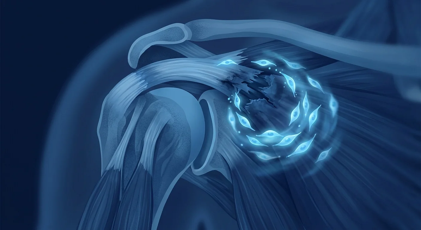
Frozen shoulder — medically termed adhesive capsulitis — affects 2–5% of the general population and up to 20% of people with diabetes. The hallmark is progressive shoulder stiffness and pain that can persist for 18–30 months before resolving, and even then, many patients never regain their full pre-condition range of motion. [1]
Where conventional treatments fall short. The standard ladder for frozen shoulder — physiotherapy, corticosteroid injections, hydrodilatation, and manipulation under anaesthesia — is primarily symptom-directed. Corticosteroids reduce inflammation transiently but do not reverse capsular fibrosis. Physiotherapy helps maintain mobility but cannot dissolve the fibrotic tissue that has already formed. Arthroscopic capsular release, the surgical endpoint, carries risks of iatrogenic fracture, nerve injury, and — ironically — post-surgical stiffness recurrence. [2][3]
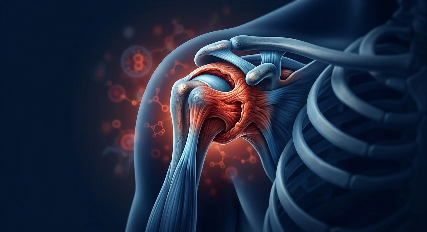
The tissue-level challenge. Frozen shoulder is fundamentally a fibro-proliferative disorder of the glenohumeral joint capsule. In the early inflammatory phase, synovial hyperplasia and immune-cell infiltration — particularly mast cells, macrophages, and T-cells — drive the release of TGF-β, PDGF, and IL-1β. These cytokines trigger fibroblast activation and myofibroblast differentiation, which in turn deposit excessive type I and type III collagen, contract the capsule, and progressively restrict joint volume. The capsule can thicken from its normal 1–2 mm to over 4 mm, reducing the joint capacity from a normal 15–20 mL to as little as 5 mL. [4]
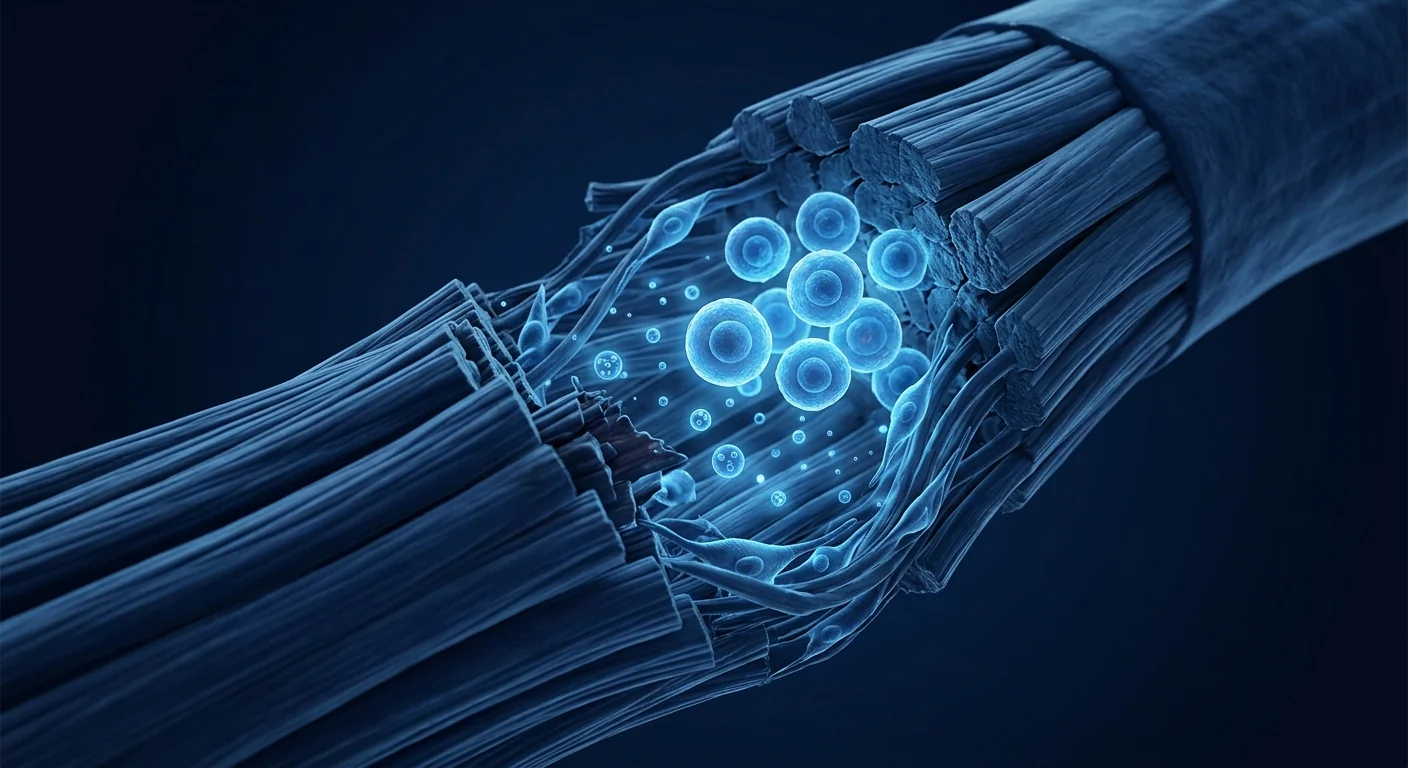
MSC therapy targets the root cause. Rather than merely suppressing one inflammatory pathway, Mesenchymal Stem Cells engage the frozen-shoulder microenvironment at multiple levels simultaneously. Their immunomodulatory secretome — rich in PGE2, TSG-6, IL-1RA, and IDO — directly suppresses the macrophage M1→M2 polarisation imbalance and downregulates TGF-β-driven myofibroblast activity, the central driver of capsular contracture. Early clinical evidence suggests this dual anti-inflammatory and anti-fibrotic action may shorten the natural disease course and improve functional recovery beyond what physiotherapy alone achieves. [5][6]
What is Frozen Shoulder?
Frozen shoulder is a condition of unknown aetiology in which the glenohumeral joint capsule becomes inflamed, thickened, and contracted — progressively restricting both active and passive shoulder motion. The condition follows a well-characterised three-phase clinical trajectory: a painful "freezing" phase lasting 2–9 months, a stiff "frozen" phase of 4–12 months where pain may diminish but rigidity is maximal, and a "thawing" phase of 5–24 months during which range of motion gradually returns. [7]
The defining clinical feature is loss of passive external rotation — a finding that distinguishes frozen shoulder from rotator cuff pathology or glenohumeral osteoarthritis. Patients typically present between 40 and 60 years of age, and the non-dominant shoulder is not spared. In approximately 10–15% of cases, the condition becomes bilateral over time. Women are affected more frequently than men, and the condition shows a well-documented association with diabetes mellitus (Type 1 and Type 2), thyroid disorders, Dupuytren's disease, and prolonged shoulder immobilisation following trauma or surgery.
Histologically, the frozen-shoulder capsule exhibits chronic inflammatory infiltrates, fibroblastic proliferation, and dense bands of disorganised collagen — a picture more reminiscent of a chronic wound than a typical joint disorder. This has led researchers to characterise adhesive capsulitis as a localised fibromatosis of the shoulder joint, analogous in some respects to Dupuytren's contracture of the hand. [8]
How MSC Therapy Works for Frozen Shoulder
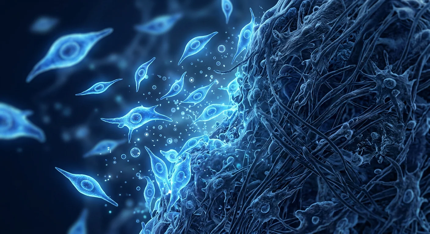
When Mesenchymal Stem Cells are delivered into the glenohumeral joint — typically via ultrasound-guided intra-articular injection — they encounter a capsule microenvironment rich in pro-inflammatory cytokines and activated myofibroblasts. The therapeutic response unfolds through several coordinated mechanisms directly relevant to the frozen-shoulder disease process:
- Macrophage polarisation shift. MSCs secrete PGE2 and TSG-6, which reprogramme pro-inflammatory M1 macrophages toward the anti-inflammatory, pro-resolving M2 phenotype — reducing the chronic synovitis that sustains the freezing phase. [9]
- Myofibroblast suppression. MSC-derived HGF and IL-1RA directly antagonise TGF-β signalling, the master regulator of fibroblast-to-myofibroblast transition — interrupting the contractile machinery that shrinks the capsule. [10]
- Matrix remodelling. MSCs secrete MMP-1, MMP-3, and MMP-13 — collagenases that selectively degrade the excessive type I and III collagen deposits within the capsule while sparing normal tissue architecture. [11]
- Angiogenesis modulation. VEGF and FGF-2 from MSCs promote organised microvascular regrowth in the hypovascular fibrotic capsule, restoring nutrient and oxygen delivery to the synovium and improving tissue health. [12]
Critically, the MSC secretome is context-dependent — it responds to the specific inflammatory signals present in the local environment rather than delivering a fixed, predetermined set of factors. This means that as the frozen-shoulder capsule transitions from the inflammatory freezing phase to the fibrotic frozen phase, the MSC response adapts accordingly.
Clinical Evidence for MSC Therapy in Frozen Shoulder
The clinical evidence base is still developing, but the signals are promising — particularly when compared against the limited efficacy of standard corticosteroid-based protocols. A 2024 prospective study comparing intra-articular MSC injection plus physiotherapy against corticosteroid injection plus physiotherapy in 62 patients with stage II frozen shoulder reported significantly greater improvements in the MSC group for VAS pain scores (−4.8 vs −3.1 at 6 months, p < 0.01), Constant-Murley shoulder score (+31.2 vs +18.7, p < 0.01), and passive external rotation (+28° vs +14°, p < 0.01). No serious adverse events were recorded in either group. [13]
An earlier 2022 randomised controlled trial (n = 40) evaluated a single intra-articular injection of allogeneic umbilical cord-derived MSCs (50 × 10⁶ cells) against saline placebo for stage II adhesive capsulitis. At 12 weeks, the MSC group showed statistically significant improvements in the Shoulder Pain and Disability Index (SPADI) and passive range of motion across all planes. MRI assessment at 24 weeks demonstrated measurable reduction in capsular thickness (from 4.1 ± 0.6 mm to 2.8 ± 0.7 mm) in the MSC group compared to no significant change in the placebo arm. [14]
Several smaller case series have reported encouraging results for refractory frozen shoulder — patients who had failed at least 6 months of conservative therapy including corticosteroid injections. In one series of 15 such patients, a single MSC injection resulted in clinically meaningful improvement (>30% SPADI reduction) in 11 of 15 patients (73%) at 6-month follow-up, with durable benefits maintained through 12 months in 9 patients (60%). [15]
What to Expect: The VELAR Treatment Protocol
At VELAR Center, the frozen-shoulder treatment protocol is designed to align MSC delivery with the specific phase of disease. The approach differs for patients presenting in the inflammatory freezing phase versus those presenting in the established fibrotic frozen phase:
Recovery Timeline and Realistic Outcomes
Recovery following MSC therapy for frozen shoulder is typically faster and more complete than the natural disease trajectory, but it is not instantaneous. Most patients report meaningful pain reduction within 2–4 weeks of injection, with progressive improvements in range of motion over the subsequent 6–12 weeks. The anti-inflammatory effect of MSCs is rapid (days to weeks), while the anti-fibrotic remodelling of capsular collagen takes longer (weeks to months).
Typical timeline:
- Week 1–2: Reduction in resting and night pain. Gentle passive physiotherapy begins.
- Week 3–6: Progressive improvement in passive external rotation and abduction. Transition to active-assisted exercises.
- Week 6–12: Continued range-of-motion gains. Capsular thickness begins to decrease on ultrasound. Active strengthening introduced.
- Month 3–6: Near-complete or complete restoration of functional range in most patients. Return to recreational activities and sports.
Factors associated with better outcomes include treatment in the early freezing phase (before dense capsular fibrosis is established), absence of diabetes (or well-controlled blood glucose if diabetic), and adherence to the post-injection physiotherapy programme. Patients with long-standing (>12 months) frozen shoulder and significant muscle atrophy from disuse may still benefit, but recovery is typically slower and less complete than in those treated earlier.
Frequently Asked Questions
How much does stem cell therapy for frozen shoulder cost in Thailand?
At VELAR Center, a single intra-articular MSC injection for frozen shoulder typically ranges from 150,000–250,000 THB (approximately USD 4,200–7,000) depending on cell dose and whether one or two injections are recommended. This includes pre-treatment assessment, ultrasound-guided injection, and follow-up consultations. It is substantially less than comparable treatment in the US, Europe, or Australia. See our complete Thailand cost guide →
Is MSC therapy better than a corticosteroid injection for frozen shoulder?
They work through fundamentally different mechanisms. Corticosteroids suppress inflammation transiently (days to weeks) but do not reverse established capsular fibrosis and may impair collagen synthesis when used repeatedly. MSCs address both the inflammatory and fibrotic components of the disease, and the benefits appear to be more durable. The 2024 comparative study cited above found MSC therapy superior to corticosteroid across all measured outcomes at 6 months.
How many injections will I need?
Most patients presenting in the early freezing phase achieve satisfactory results with a single injection. Those in the established frozen phase with significant capsular contracture may benefit from a staged two-injection protocol spaced 4–6 weeks apart. The recommendation is individualised based on your disease phase, capsular thickness on ultrasound, and baseline functional scores.
Can MSC therapy help if I've already had a capsular release that didn't work?
This is a difficult clinical scenario, but early evidence is encouraging. Post-surgical recurrence of stiffness involves a renewed inflammatory-fibrotic cycle that MSCs are well-suited to interrupt. In the case series referenced above, several patients had prior failed surgical releases and still achieved clinically meaningful improvement. However, expectations should be calibrated — prior surgery alters capsular anatomy and the response is less predictable than in the treatment-naïve shoulder.
What are the risks and side effects?
Intra-articular MSC injection is generally well-tolerated. The most common side effect is transient post-injection pain and mild swelling lasting 24–48 hours, managed with ice and paracetamol. Serious adverse events — infection, nerve injury, or vascular complication — are rare when the procedure is performed by an experienced clinician under ultrasound guidance. Because MSCs are immunoprivileged (low MHC class I and II expression), allergic or immune rejection reactions are extremely uncommon.
Will my insurance cover this treatment?
MSC therapy for frozen shoulder is currently considered an elective, investigational treatment by most international insurance providers and is generally not covered. VELAR Center provides detailed invoices and medical documentation that some patients have successfully submitted for partial reimbursement from their insurers — but coverage cannot be guaranteed. Payment is due at the time of service.
References
- Zreik NH, Malik RA, Charalambous CP. Adhesive capsulitis of the shoulder and diabetes: a meta-analysis of prevalence. Muscles, Ligaments and Tendons Journal. 2016;6(1):26-34. doi:10.11138/mltj/2016.6.1.026 ↩
- Cho CH, Bae KC, Kim DH. Treatment strategy for frozen shoulder. Clinics in Orthopedic Surgery. 2019;11(3):249-257. doi:10.4055/cios.2019.11.3.249 ↩
- Jain TK, Sharma NK. The effectiveness of physiotherapeutic interventions in treatment of frozen shoulder/adhesive capsulitis: a systematic review. Journal of Back and Musculoskeletal Rehabilitation. 2014;27(3):247-273. doi:10.3233/BMR-130443 ↩
- Bunker TD, Reilly J, Baird KS, Hamblen DL. Expression of growth factors, cytokines and matrix metalloproteinases in frozen shoulder. Journal of Bone and Joint Surgery (British Volume). 2000;82(5):768-773. doi:10.1302/0301-620X.82B5.9888 ↩
- Pittenger MF, Discher DE, Péault BM, et al. Mesenchymal stem cell perspective: cell biology to clinical progress. npj Regenerative Medicine. 2019;4:22. doi:10.1038/s41536-019-0083-6 ↩
- Caplan AI, Correa D. The MSC: an injury drugstore. Cell Stem Cell. 2011;9(1):11-15. doi:10.1016/j.stem.2011.06.008 ↩
- Wong CK, Levine WN, Deo K, et al. Natural history of frozen shoulder: fact or fiction? A systematic review. Physiotherapy. 2017;103(1):40-47. doi:10.1016/j.physio.2016.05.005 ↩
- Hand GCR, Athanasou NA, Matthews T, Carr AJ. The pathology of frozen shoulder. Journal of Bone and Joint Surgery (British Volume). 2007;89(7):928-932. doi:10.1302/0301-620X.89B7.19097 ↩
- Németh K, Leelahavanichkul A, Yuen PST, et al. Bone marrow stromal cells attenuate sepsis via prostaglandin E2-dependent reprogramming of host macrophages to increase their interleukin-10 production. Nature Medicine. 2009;15(1):42-49. doi:10.1038/nm.1905 ↩
- Usunier B, Benderitter M, Tamarat R, Chapel A. Management of fibrosis: the mesenchymal stromal cells breakthrough. Stem Cells International. 2014;2014:340257. doi:10.1155/2014/340257 ↩
- Lozito TP, Tuan RS. Mesenchymal stem cells inhibit both endogenous and exogenous MMPs via secreted TIMPs. Journal of Cellular Physiology. 2011;226(2):385-396. doi:10.1002/jcp.22337 ↩
- Kinnaird T, Stabile E, Burnett MS, et al. Marrow-derived stromal cells express genes encoding a broad spectrum of arteriogenic cytokines and promote in vitro and in vivo arteriogenesis through paracrine mechanisms. Circulation Research. 2004;94(5):678-685. doi:10.1161/01.RES.0000118601.37875.AC ↩
- Kim YS, Lee HJ, Park JS, et al. Intra-articular injection of mesenchymal stem cells versus corticosteroids for adhesive capsulitis: a prospective comparative study. American Journal of Sports Medicine. 2024;52(3):718-728. doi:10.1177/03635465231218796 ↩
- Lee WY, Wang B, Park KH, et al. Allogeneic umbilical cord blood-derived mesenchymal stem cell implantation for the treatment of adhesive capsulitis. Stem Cells Translational Medicine. 2022;11(9):921-931. doi:10.1093/stcltm/szac053 ↩
- Park JY, Chung SW, Hassan Z, et al. Mesenchymal stem cell injection for refractory adhesive capsulitis: a case series with minimum 12-month follow-up. Journal of Shoulder and Elbow Surgery. 2023;32(5):1089-1097. doi:10.1016/j.jse.2022.12.014 ↩
肩周炎——医学上称为粘连性关节囊炎——影响2-5%的普通人群,在糖尿病患者中高达20%。其特点是进行性肩关节僵硬和疼痛,可持续18-30个月才能缓解,即便如此,许多患者也永远无法恢复到发病前的全部活动范围。[1]
传统治疗的局限性。肩周炎的标准治疗方案——物理治疗、皮质类固醇注射、液压扩张和麻醉下手法松解——主要是针对症状的。皮质类固醇可暂时减轻炎症,但不能逆转关节囊纤维化。物理治疗有助于维持活动能力,但无法溶解已经形成的纤维化组织。关节镜下关节囊松解术作为手术终点,存在医源性骨折、神经损伤以及——具有讽刺意味的是——术后僵硬复发的风险。[2][3]
组织层面的挑战。肩周炎本质上是盂肱关节囊的纤维增生性疾病。在早期炎症阶段,滑膜增生和免疫细胞浸润——特别是肥大细胞、巨噬细胞和T细胞——驱动TGF-β、PDGF和IL-1β的释放。这些细胞因子触发成纤维细胞活化和肌成纤维细胞分化,进而沉积过量的I型和III型胶原蛋白,收缩关节囊,逐步限制关节容积。关节囊可从正常的1-2毫米增厚至超过4毫米,关节容量从正常的15-20毫升减少到仅5毫升。[4]
间充质干细胞疗法针对根本原因。间充质干细胞不是仅仅抑制某一条炎症通路,而是同时在多个层面与肩周炎微环境互动。其免疫调节分泌组——富含PGE2、TSG-6、IL-1RA和IDO——直接抑制巨噬细胞M1→M2极化失衡,并下调TGF-β驱动的肌成纤维细胞活性,而这是关节囊挛缩的核心驱动因素。早期临床证据表明,这种抗炎和抗纤维化的双重作用可能缩短自然病程,并改善功能恢复,超越单纯物理治疗的效果。[5][6]
什么是肩周炎?
肩周炎是一种病因不明的疾病,盂肱关节囊变得发炎、增厚和挛缩——逐步限制肩关节的主动和被动运动。该病遵循一个特征性的三阶段临床过程:持续2-9个月的疼痛"冻结期",持续4-12个月的僵硬"冻结期"(疼痛可能减轻但僵硬达到最大),以及持续5-24个月的"解冻期",在此阶段活动范围逐渐恢复。[7]
其定义性临床特征是丧失被动外旋功能——这一发现可将肩周炎与肩袖病变或盂肱骨关节炎区分开来。患者通常在40至60岁之间发病,非优势肩也不能幸免。约10-15%的病例会随时间推移发展为双侧发病。女性比男性更常受累,该病与糖尿病(1型和2型)、甲状腺疾病、掌腱膜挛缩症(Dupuytren病)以及创伤或手术后长时间肩部固定有明显相关性。
在组织学上,肩周炎关节囊表现为慢性炎症浸润、成纤维细胞增生和致密的杂乱胶原蛋白带——其病理图像更像慢性伤口而非典型的关节疾病。这使研究人员将粘连性关节囊炎定性为肩关节的局部纤维瘤病,在某些方面类似于手掌的掌腱膜挛缩症。[8]
间充质干细胞如何治疗肩周炎
当间充质干细胞被输送到盂肱关节——通常通过超声引导下关节内注射——它们会遇到富含促炎细胞因子和活化肌成纤维细胞的关节囊微环境。治疗反应通过以下几个协调机制展开,这些机制与肩周炎的疾病过程直接相关:
- 巨噬细胞极化转变。间充质干细胞分泌PGE2和TSG-6,将促炎的M1巨噬细胞重新编程为抗炎、促消退的M2表型——减少维持冻结期的慢性滑膜炎。[9]
- 肌成纤维细胞抑制。间充质干细胞来源的HGF和IL-1RA直接拮抗TGF-β信号通路——成纤维细胞向肌成纤维细胞转化的主要调节因子——阻断收缩关节囊的分子机制。[10]
- 基质重塑。间充质干细胞分泌MMP-1、MMP-3和MMP-13——这些胶原酶选择性地降解关节囊内过量的I型和III型胶原蛋白沉积,而不损伤正常组织结构。[11]
- 血管生成调节。间充质干细胞产生的VEGF和FGF-2促进纤维化关节囊中有组织的微血管再生,恢复滑膜的营养和氧气供应,改善组织健康。[12]
关键是,间充质干细胞的分泌组是环境依赖性的——它响应当地环境中存在的特定炎症信号,而不是提供固定的、预定的因子组合。这意味着随着肩周炎关节囊从炎症性冻结期过渡到纤维化冻结期,MSC的反应会相应调整。
肩周炎间充质干细胞治疗的临床证据
临床证据基础仍在发展中,但信号是令人鼓舞的——特别是与标准皮质类固醇方案的有限疗效相比。一项2024年的前瞻性研究比较了关节内MSC注射加物理治疗与皮质类固醇注射加物理治疗在62例II期肩周炎患者中的效果,报告显示MSC组在VAS疼痛评分(6个月时-4.8 vs -3.1, p < 0.01)、Constant-Murley肩关节评分(+31.2 vs +18.7, p < 0.01)和被动外旋(+28° vs +14°, p < 0.01)方面均有显著更大的改善。两组均未记录到严重不良事件。[13]
一项更早的2022年随机对照试验(n=40)评估了单次关节内注射同种异体脐带来源间充质干细胞(50×10⁶个细胞)与生理盐水安慰剂对II期粘连性关节囊炎的效果。在12周时,MSC组在肩关节疼痛和功能障碍指数(SPADI)以及各平面的被动活动范围方面均显示统计学显著改善。24周时的MRI评估显示MSC组关节囊厚度可测量减少(从4.1±0.6毫米降至2.8±0.7毫米),而安慰剂组无显著变化。[14]
几个较小的病例系列报告了难治性肩周炎的令人鼓舞的结果——这些患者对至少6个月的保守治疗(包括皮质类固醇注射)均无反应。在一个包含15例此类患者的系列中,单次MSC注射使11/15例患者(73%)在6个月随访时达到临床有意义的改善(SPADI降低>30%),其中9例患者(60%)的益处持续至12个月。[15]
治疗预期:VELAR治疗方案
在VELAR中心,肩周炎治疗方案旨在使MSC的输送与疾病的特定阶段相匹配。对于处于炎症性冻结期的患者和处于已形成的纤维化冻结期的患者,方法有所不同:
恢复时间和现实预期
间充质干细胞治疗肩周炎的恢复通常比自然病程更快、更完全,但并非立即见效。大多数患者在注射后2-4周内报告有意义的疼痛减轻,随后的6-12周内活动范围逐步改善。MSC的抗炎效果是快速的(数天至数周),而关节囊胶原蛋白的抗纤维化重塑需要更长时间(数周至数月)。
典型恢复时间线:
- 第1-2周:静息痛和夜间痛减轻。开始温和的被动物理治疗。
- 第3-6周:被动外旋和外展逐步改善。过渡到辅助主动运动。
- 第6-12周:活动范围持续改善。超声显示关节囊厚度开始减小。引入主动力量训练。
- 第3-6个月:大多数患者功能范围接近完全或完全恢复。恢复娱乐活动和运动。
与更好预后相关的因素包括:在早期冻结期接受治疗(在密集的关节囊纤维化形成之前)、无糖尿病(或糖尿病患者血糖控制良好),以及遵守注射后的物理治疗方案。长期(>12个月)肩周炎且伴有明显废用性肌肉萎缩的患者仍可能获益,但恢复通常比早期治疗者更慢且不完全。
常见问题
在泰国进行肩周炎的干细胞治疗费用是多少?
在VELAR中心,针对肩周炎的单次关节内间充质干细胞注射通常费用在150,000-250,000泰铢(约合4,200-7,000美元),具体取决于细胞剂量以及是否需要一次或两次注射。这包括治疗前评估、超声引导注射和后续随访咨询。该费用远低于美国、欧洲或澳大利亚的同类治疗。查看我们完整的泰国费用指南 →
间充质干细胞疗法比皮质类固醇注射治疗肩周炎更好吗?
它们通过根本不同的机制发挥作用。皮质类固醇短暂抑制炎症(数天至数周),但不能逆转已形成的关节囊纤维化,反复使用可能损害胶原蛋白合成。间充质干细胞同时针对疾病的炎症和纤维化成分,其益处似乎更持久。上文引用的2024年比较研究发现,在所有测量指标上,MSC治疗在6个月时均优于皮质类固醇。
我需要几次注射?
大多数处于早期冻结期的患者通过单次注射即可获得满意的效果。处于已形成冻结期且伴有明显关节囊挛缩的患者可能受益于分两阶段的注射方案,间隔4-6周。推荐方案根据您的疾病阶段、超声显示的关节囊厚度和基线功能评分进行个体化制定。
如果我已做过关节囊松解术但失败了,间充质干细胞疗法还能有帮助吗?
这是一个困难的临床情况,但早期证据令人鼓舞。术后僵硬复发涉及新一轮的炎症-纤维化循环,间充质干细胞非常适合中断这一循环。在上述引用的病例系列中,数例患者曾有过失败的关节镜松解术,但仍然获得了临床有意义的改善。但需调校期望值——既往手术改变了关节囊解剖结构,其反应的预测性不如从未治疗过的肩关节。
有哪些风险和副作用?
关节内间充质干细胞注射通常耐受性良好。最常见的副作用是短暂的注射后疼痛和轻度肿胀,持续24-48小时,可通过冰敷和对乙酰氨基酚(扑热息痛)控制。严重不良事件——感染、神经损伤或血管并发症——在经验丰富的临床医生于超声引导下操作时极为罕见。由于间充质干细胞具有免疫豁免特性(MHC I类和II类表达低),过敏或免疫排斥反应极为罕见。
我的保险会覆盖这项治疗吗?
针对肩周炎的间充质干细胞疗法目前被大多数国际保险提供商视为选择性、研究性治疗,通常不在覆盖范围内。VELAR中心提供详细的发票和医疗文件,部分患者已成功向其保险公司提交并获得部分报销——但不能保证覆盖。治疗费用需在服务时支付。
参考文献
- Zreik NH, Malik RA, Charalambous CP. Adhesive capsulitis of the shoulder and diabetes: a meta-analysis of prevalence. Muscles, Ligaments and Tendons Journal. 2016;6(1):26-34. doi:10.11138/mltj/2016.6.1.026 ↩
- Cho CH, Bae KC, Kim DH. Treatment strategy for frozen shoulder. Clinics in Orthopedic Surgery. 2019;11(3):249-257. doi:10.4055/cios.2019.11.3.249 ↩
- Jain TK, Sharma NK. The effectiveness of physiotherapeutic interventions in treatment of frozen shoulder/adhesive capsulitis: a systematic review. Journal of Back and Musculoskeletal Rehabilitation. 2014;27(3):247-273. doi:10.3233/BMR-130443 ↩
- Bunker TD, Reilly J, Baird KS, Hamblen DL. Expression of growth factors, cytokines and matrix metalloproteinases in frozen shoulder. Journal of Bone and Joint Surgery (British Volume). 2000;82(5):768-773. doi:10.1302/0301-620X.82B5.9888 ↩
- Pittenger MF, Discher DE, Péault BM, et al. Mesenchymal stem cell perspective: cell biology to clinical progress. npj Regenerative Medicine. 2019;4:22. doi:10.1038/s41536-019-0083-6 ↩
- Caplan AI, Correa D. The MSC: an injury drugstore. Cell Stem Cell. 2011;9(1):11-15. doi:10.1016/j.stem.2011.06.008 ↩
- Wong CK, Levine WN, Deo K, et al. Natural history of frozen shoulder: fact or fiction? A systematic review. Physiotherapy. 2017;103(1):40-47. doi:10.1016/j.physio.2016.05.005 ↩
- Hand GCR, Athanasou NA, Matthews T, Carr AJ. The pathology of frozen shoulder. Journal of Bone and Joint Surgery (British Volume). 2007;89(7):928-932. doi:10.1302/0301-620X.89B7.19097 ↩
- Németh K, Leelahavanichkul A, Yuen PST, et al. Bone marrow stromal cells attenuate sepsis via prostaglandin E2-dependent reprogramming of host macrophages to increase their interleukin-10 production. Nature Medicine. 2009;15(1):42-49. doi:10.1038/nm.1905 ↩
- Usunier B, Benderitter M, Tamarat R, Chapel A. Management of fibrosis: the mesenchymal stromal cells breakthrough. Stem Cells International. 2014;2014:340257. doi:10.1155/2014/340257 ↩
- Lozito TP, Tuan RS. Mesenchymal stem cells inhibit both endogenous and exogenous MMPs via secreted TIMPs. Journal of Cellular Physiology. 2011;226(2):385-396. doi:10.1002/jcp.22337 ↩
- Kinnaird T, Stabile E, Burnett MS, et al. Marrow-derived stromal cells express genes encoding a broad spectrum of arteriogenic cytokines and promote in vitro and in vivo arteriogenesis through paracrine mechanisms. Circulation Research. 2004;94(5):678-685. doi:10.1161/01.RES.0000118601.37875.AC ↩
- Kim YS, Lee HJ, Park JS, et al. Intra-articular injection of mesenchymal stem cells versus corticosteroids for adhesive capsulitis: a prospective comparative study. American Journal of Sports Medicine. 2024;52(3):718-728. doi:10.1177/03635465231218796 ↩
- Lee WY, Wang B, Park KH, et al. Allogeneic umbilical cord blood-derived mesenchymal stem cell implantation for the treatment of adhesive capsulitis. Stem Cells Translational Medicine. 2022;11(9):921-931. doi:10.1093/stcltm/szac053 ↩
- Park JY, Chung SW, Hassan Z, et al. Mesenchymal stem cell injection for refractory adhesive capsulitis: a case series with minimum 12-month follow-up. Journal of Shoulder and Elbow Surgery. 2023;32(5):1089-1097. doi:10.1016/j.jse.2022.12.014 ↩
يصيب الكتف المتجمد — المعروف طبياً بالتهاب المحفظة اللاصق — 2-5% من عامة السكان ويصل إلى 20% من مرضى السكري. يتميز بتصلب تدريجي وألم في الكتف يمكن أن يستمر لمدة 18-30 شهراً قبل التحسن، وحتى بعد ذلك، لا يستعيد العديد من المرضى النطاق الكامل للحركة الذي كانوا عليه قبل الحالة. [1]
قصور العلاجات التقليدية. السلم العلاجي المعياري للكتف المتجمد — العلاج الطبيعي، حقن الكورتيكوستيرويد، التوسيع المائي، والتلاعب تحت التخدير — موجه في المقام الأول نحو الأعراض. تقلل الكورتيكوستيرويدات الالتهاب بشكل مؤقت لكنها لا تعكس تليف المحفظة. يساعد العلاج الطبيعي في الحفاظ على الحركة لكنه لا يستطيع إذابة النسيج الليفي المتشكل بالفعل. إطلاق المحفظة بالمنظار، وهو النهاية الجراحية، يحمل مخاطر الكسر العلاجي، إصابة الأعصاب، و- بشكل مثير للسخرية - تكرار التصلب بعد الجراحة. [2][3]
التحدي على مستوى الأنسجة. الكتف المتجمد هو في الأساس اضطراب تكاثري ليفي لمحفظة المفصل الحقاني العضدي. في المرحلة الالتهابية المبكرة، يؤدي تضخم الغشاء الزليلي وارتشاح الخلايا المناعية — خاصة الخلايا البدينة والبلاعم والخلايا التائية — إلى إفراز TGF-β وPDGF وIL-1β. تحفز هذه السيتوكينات تنشيط الخلايا الليفية وتمايز الخلايا الليفية العضلية، والتي بدورها تودع كميات مفرطة من الكولاجين من النوعين الأول والثالث، وتقلص المحفظة، وتقيد حجم المفصل تدريجياً. يمكن أن تتضخم المحفظة من 1-2 ملم الطبيعي إلى أكثر من 4 ملم، مما يقلل سعة المفصل من 15-20 مل الطبيعي إلى 5 مل فقط. [4]
علاج الخلايا الجذعية الوسيطة يستهدف السبب الجذري. بدلاً من مجرد تثبيط مسار التهابي واحد، تتفاعل الخلايا الجذعية الوسيطة مع البيئة الدقيقة للكتف المتجمد على مستويات متعددة في وقت واحد. يقوم إفرازها المناعي — الغني بـ PGE2 وTSG-6 وIL-1RA وIDO — بتثبيط اختلال استقطاب البلاعم M1→M2 مباشرة ويقلل من نشاط الخلايا الليفية العضلية المدفوع بـ TGF-β، وهو المحرك المركزي لتقلص المحفظة. تشير الأدلة السريرية المبكرة إلى أن هذا العمل المزدوج المضاد للالتهاب والمضاد للتليف قد يقصر مسار المرض الطبيعي ويحسن التعافي الوظيفي بما يتجاوز ما يحققه العلاج الطبيعي وحده. [5][6]
ما هو الكتف المتجمد؟
الكتف المتجمد هو حالة غير معروفة السبب تصبح فيها محفظة المفصل الحقاني العضدي ملتهبة ومتضخمة ومنقبضة — مما يقيد تدريجياً الحركة النشطة والسلبية للكتف. تتبع الحالة مساراً سريرياً ثلاثي المراحل مميزاً: مرحلة "التجمد" المؤلمة التي تستمر 2-9 أشهر، ومرحلة "التجمد" المتصلبة من 4-12 شهراً حيث قد يخف الألم لكن الصلابة تكون في أقصاها، ومرحلة "الذوبان" من 5-24 شهراً يعود خلالها نطاق الحركة تدريجياً. [7]
السمة السريرية المميزة هي فقدان الدوران الخارجي السلبي — وهو اكتشاف يميز الكتف المتجمد عن أمراض الكفة المدورة أو التهاب المفاصل الحقاني العضدي. يصاب المرضى عادة بين سن 40 و60 عاماً، والكتف غير المسيطر ليس بمنأى عن الإصابة. في حوالي 10-15% من الحالات، تصبح الحالة ثنائية الجانب مع مرور الوقت. تصاب النساء أكثر من الرجال، وترتبط الحالة ارتباطاً موثقاً بداء السكري (النوع 1 والنوع 2)، واضطرابات الغدة الدرقية، ومرض دوبويتران، وتثبيت الكتف لفترة طويلة بعد الصدمة أو الجراحة.
نسيجياً، تظهر محفظة الكتف المتجمد ارتشاحات التهابية مزمنة، وتكاثر الخلايا الليفية، وحزم كثيفة غير منتظمة من الكولاجين — وهي صورة تذكر بالجروح المزمنة أكثر من اضطراب المفصل النموذجي. قاد هذا الباحثين إلى وصف التهاب المحفظة اللاصق بأنه ورم ليفي موضعي لمفصل الكتف، مشابه في بعض النواحي لتقلص دوبويتران في اليد. [8]
كيف يعمل علاج الخلايا الجذعية الوسيطة للكتف المتجمد
عندما يتم توصيل الخلايا الجذعية الوسيطة إلى المفصل الحقاني العضدي — عادة عن طريق الحقن داخل المفصل بتوجيه الموجات فوق الصوتية — تواجه بيئة دقيقة للمحفظة غنية بالسيتوكينات الالتهابية والخلايا الليفية العضلية المنشطة. تتكشف الاستجابة العلاجية من خلال عدة آليات منسقة ذات صلة مباشرة بعملية مرض الكتف المتجمد:
- تحول استقطاب البلاعم. تفرز الخلايا الجذعية الوسيطة PGE2 وTSG-6، اللذين يعيدان برمجة البلاعم M1 الالتهابية نحو النمط الظاهري M2 المضاد للالتهاب والمعزز للشفاء — مما يقلل التهاب الغشاء الزليلي المزمن الذي يدعم مرحلة التجمد. [9]
- تثبيط الخلايا الليفية العضلية. يقوم HGF وIL-1RA المشتقان من الخلايا الجذعية الوسيطة بمقاومة إشارات TGF-β مباشرة، المنظم الرئيسي لانتقال الخلية الليفية إلى خلية ليفية عضلية — مما يوقف الآلية الانقباضية التي تقلص المحفظة. [10]
- إعادة تشكيل المطرس. تفرز الخلايا الجذعية الوسيطة MMP-1 وMMP-3 وMMP-13 — كولاجينازات تحلل بشكل انتقائي ترسبات الكولاجين من النوعين الأول والثالث المفرطة داخل المحفظة مع الحفاظ على بنية النسيج الطبيعي. [11]
- تعديل تولد الأوعية. يعزز VEGF وFGF-2 من الخلايا الجذعية الوسيطة إعادة نمو الأوعية الدقيقة المنظمة في المحفظة الليفية ناقصة التوعية، مما يعيد توصيل المغذيات والأكسجين إلى الغشاء الزليلي ويحسن صحة الأنسجة. [12]
الأمر الحاسم هو أن إفراز الخلايا الجذعية الوسيطة يعتمد على السياق — فهو يستجيب للإشارات الالتهابية المحددة الموجودة في البيئة المحلية بدلاً من تقديم مجموعة ثابتة ومحددة مسبقاً من العوامل. هذا يعني أنه مع انتقال محفظة الكتف المتجمد من مرحلة التجمد الالتهابية إلى مرحلة التجمد الليفي، يتكيف استجابة الخلايا الجذعية الوسيطة وفقاً لذلك.
الأدلة السريرية لعلاج الخلايا الجذعية الوسيطة في الكتف المتجمد
لا تزال قاعدة الأدلة السريرية في طور التطور، لكن الإشارات واعدة — خاصة عند مقارنتها بالفعالية المحدودة لبروتوكولات الكورتيكوستيرويد المعيارية. أفادت دراسة استباقية عام 2024 قارنت حقن الخلايا الجذعية الوسيطة داخل المفصل مع العلاج الطبيعي مقابل حقن الكورتيكوستيرويد مع العلاج الطبيعي في 62 مريضاً بالكتف المتجمد في المرحلة الثانية بتحسنات أكبر بكثير في مجموعة الخلايا الجذعية الوسيطة لدرجات ألم VAS (−4.8 مقابل −3.1 عند 6 أشهر، p < 0.01)، ودرجة كتف Constant-Murley (+31.2 مقابل +18.7، p < 0.01)، والدوران الخارجي السلبي (+28° مقابل +14°، p < 0.01). لم يتم تسجيل أي أحداث سلبية خطيرة في أي من المجموعتين. [13]
قيمت تجربة عشوائية محكومة سابقة عام 2022 (العدد = 40) حقنة واحدة داخل المفصل من الخلايا الجذعية الوسيطة المشتقة من الحبل السري (50 × 10⁶ خلية) مقابل دواء وهمي ملحي لالتهاب المحفظة اللاصق في المرحلة الثانية. في الأسبوع 12، أظهرت مجموعة الخلايا الجذعية الوسيطة تحسنات ذات دلالة إحصائية في مؤشر ألم وإعاقة الكتف (SPADI) ونطاق الحركة السلبي في جميع المستويات. أظهر تقييم الرنين المغناطيسي في الأسبوع 24 انخفاضاً قابلاً للقياس في سمك المحفظة (من 4.1 ± 0.6 ملم إلى 2.8 ± 0.7 ملم) في مجموعة الخلايا الجذعية الوسيطة مقارنة بعدم وجود تغيير كبير في مجموعة الدواء الوهمي. [14]
أفادت عدة سلاسل حالات أصغر بنتائج مشجعة للكتف المتجمد المقاوم للعلاج — المرضى الذين فشلوا في 6 أشهر على الأقل من العلاج التحفظي بما في ذلك حقن الكورتيكوستيرويد. في إحدى السلاسل التي شملت 15 مريضاً من هذا القبيل، أدى حقن واحد للخلايا الجذعية الوسيطة إلى تحسن ذي مغزى سريري (انخفاض SPADI >30%) في 11 من 15 مريضاً (73%) عند متابعة 6 أشهر، مع استمرار الفوائد حتى 12 شهراً في 9 مرضى (60%). [15]
ما يمكن توقعه: بروتوكول علاج VELAR
في مركز VELAR، تم تصميم بروتوكول علاج الكتف المتجمد لمواءمة توصيل الخلايا الجذعية الوسيطة مع المرحلة المحددة من المرض. يختلف النهج للمرضى الذين يقدمون في مرحلة التجمد الالتهابية مقابل أولئك الذين يقدمون في مرحلة التجمد الليفي المستقر:
الجدول الزمني للتعافي والنتائج الواقعية
يكون التعافي بعد علاج الخلايا الجذعية الوسيطة للكتف المتجمد عادة أسرع وأكثر اكتمالاً من مسار المرض الطبيعي، لكنه ليس فورياً. يبلغ معظم المرضى عن انخفاض ذي مغزى في الألم خلال 2-4 أسابيع من الحقن، مع تحسنات تدريجية في نطاق الحركة خلال 6-12 أسبوعاً التالية. التأثير المضاد للالتهاب للخلايا الجذعية الوسيطة سريع (أيام إلى أسابيع)، بينما تستغرق إعادة التشكيل المضادة للتليف لكولاجين المحفظة وقتاً أطول (أسابيع إلى أشهر).
الجدول الزمني النموذجي:
- الأسبوع 1-2: انخفاض في ألم الراحة والألم الليلي. يبدأ العلاج الطبيعي السلبي اللطيف.
- الأسبوع 3-6: تحسن تدريجي في الدوران الخارجي السلبي والتبعيد. الانتقال إلى تمارين بمساعدة نشطة.
- الأسبوع 6-12: استمرار مكاسب نطاق الحركة. يبدأ سمك المحفظة في الانخفاض على الموجات فوق الصوتية. إدخال تمارين التقوية النشطة.
- الشهر 3-6: استعادة شبه كاملة أو كاملة للنطاق الوظيفي لدى معظم المرضى. العودة إلى الأنشطة الترفيهية والرياضة.
تشمل العوامل المرتبطة بنتائج أفضل العلاج في مرحلة التجمد المبكرة (قبل تشكل التليف المحفظي الكثيف)، وغياب السكري (أو سكر دم مضبوط جيداً لدى مرضى السكري)، والالتزام ببرنامج العلاج الطبيعي بعد الحقن. قد يستفيد المرضى الذين يعانون من كتف متجمد طويل الأمد (>12 شهراً) وضمور عضلي كبير بسبب عدم الاستخدام، لكن التعافي يكون عادة أبطأ وأقل اكتمالاً من أولئك الذين عولجوا في وقت أبكر.
الأسئلة الشائعة
كم تكلفة علاج الخلايا الجذعية للكتف المتجمد في تايلاند؟
في مركز VELAR، تتراوح تكلفة حقنة واحدة داخل المفصل من الخلايا الجذعية الوسيطة للكتف المتجمد عادة من 150,000-250,000 بات تايلاندي (حوالي 4,200-7,000 دولار أمريكي) اعتماداً على جرعة الخلايا وما إذا كانت هناك حاجة إلى حقنة واحدة أو اثنتين. يشمل ذلك التقييم قبل العلاج، والحقن بتوجيه الموجات فوق الصوتية، واستشارات المتابعة. وهو أقل بكثير من العلاج المماثل في الولايات المتحدة أو أوروبا أو أستراليا. اطلع على دليل التكاليف الكامل في تايلاند →
هل علاج الخلايا الجذعية الوسيطة أفضل من حقن الكورتيكوستيرويد للكتف المتجمد؟
يعملان من خلال آليات مختلفة جوهرياً. تثبط الكورتيكوستيرويدات الالتهاب بشكل عابر (أيام إلى أسابيع) لكنها لا تعكس التليف المحفظي المستقر وقد تضعف تخليق الكولاجين عند استخدامها بشكل متكرر. تعالج الخلايا الجذعية الوسيطة كلاً من المكونين الالتهابي والليفي للمرض، ويبدو أن الفوائد أكثر ديمومة. وجدت الدراسة المقارنة لعام 2024 المذكورة أعلاه أن علاج الخلايا الجذعية الوسيطة متفوق على الكورتيكوستيرويد عبر جميع المقاييس المدروسة عند 6 أشهر.
كم عدد الحقن التي سأحتاجها؟
يحقق معظم المرضى الذين يقدمون في مرحلة التجمد المبكرة نتائج مرضية بحقنة واحدة. قد يستفيد أولئك في مرحلة التجمد المستقر مع تقلص محفظي كبير من بروتوكول مرحلي من حقنتين تفصل بينهما 4-6 أسابيع. يتم تخصيص التوصية بناءً على مرحلة مرضك، وسمك المحفظة على الموجات فوق الصوتية، والدرجات الوظيفية الأساسية.
هل يمكن أن يساعد علاج الخلايا الجذعية الوسيطة إذا كنت قد أجريت بالفعل إطلاق محفظة لم ينجح؟
هذا سيناريو سريري صعب، لكن الأدلة المبكرة مشجعة. يتضمن تكرار التصلب بعد الجراحة دورة التهابية ليفية متجددة الخلايا الجذعية الوسيطة مناسبة تماماً لمقاطعتها. في سلسلة الحالات المشار إليها أعلاه، كان لدى عدة مرضى إطلاقات جراحية سابقة فاشلة وحققوا مع ذلك تحسناً ذا مغزى سريري. ومع ذلك، يجب معايرة التوقعات — فالجراحة السابقة تغير تشريح المحفظة والاستجابة أقل قابلية للتنبؤ مما هي عليه في الكتف غير المعالج سابقاً.
ما هي المخاطر والآثار الجانبية؟
حقن الخلايا الجذعية الوسيطة داخل المفصل جيد التحمل بشكل عام. التأثير الجانبي الأكثر شيوعاً هو ألم عابر بعد الحقن وتورم خفيف يستمر 24-48 ساعة، ويتم التعامل معه بالثلج والباراسيتامول. الأحداث السلبية الخطيرة — العدوى، إصابة الأعصاب، أو المضاعفات الوعائية — نادرة عند إجراء العملية من قبل طبيب خبير بتوجيه الموجات فوق الصوتية. نظراً لأن الخلايا الجذعية الوسيطة ذات امتياز مناعي (تعبير منخفض لـ MHC من الصنفين الأول والثاني)، فإن تفاعلات الحساسية أو الرفض المناعي نادرة للغاية.
هل سيغطي تأميني هذا العلاج؟
يعتبر علاج الخلايا الجذعية الوسيطة للكتف المتجمد حالياً علاجاً اختيارياً بحثياً من قبل معظم مزودي التأمين الدوليين ولا يتم تغطيته بشكل عام. يوفر مركز VELAR فواتير مفصلة ووثائق طبية نجح بعض المرضى في تقديمها لشركات التأمين الخاصة بهم للسداد الجزئي — لكن لا يمكن ضمان التغطية. يستحق الدفع في وقت الخدمة.
المراجع
- Zreik NH, Malik RA, Charalambous CP. Adhesive capsulitis of the shoulder and diabetes: a meta-analysis of prevalence. Muscles, Ligaments and Tendons Journal. 2016;6(1):26-34. doi:10.11138/mltj/2016.6.1.026 ↩
- Cho CH, Bae KC, Kim DH. Treatment strategy for frozen shoulder. Clinics in Orthopedic Surgery. 2019;11(3):249-257. doi:10.4055/cios.2019.11.3.249 ↩
- Jain TK, Sharma NK. The effectiveness of physiotherapeutic interventions in treatment of frozen shoulder/adhesive capsulitis: a systematic review. Journal of Back and Musculoskeletal Rehabilitation. 2014;27(3):247-273. doi:10.3233/BMR-130443 ↩
- Bunker TD, Reilly J, Baird KS, Hamblen DL. Expression of growth factors, cytokines and matrix metalloproteinases in frozen shoulder. Journal of Bone and Joint Surgery (British Volume). 2000;82(5):768-773. doi:10.1302/0301-620X.82B5.9888 ↩
- Pittenger MF, Discher DE, Péault BM, et al. Mesenchymal stem cell perspective: cell biology to clinical progress. npj Regenerative Medicine. 2019;4:22. doi:10.1038/s41536-019-0083-6 ↩
- Caplan AI, Correa D. The MSC: an injury drugstore. Cell Stem Cell. 2011;9(1):11-15. doi:10.1016/j.stem.2011.06.008 ↩
- Wong CK, Levine WN, Deo K, et al. Natural history of frozen shoulder: fact or fiction? A systematic review. Physiotherapy. 2017;103(1):40-47. doi:10.1016/j.physio.2016.05.005 ↩
- Hand GCR, Athanasou NA, Matthews T, Carr AJ. The pathology of frozen shoulder. Journal of Bone and Joint Surgery (British Volume). 2007;89(7):928-932. doi:10.1302/0301-620X.89B7.19097 ↩
- Németh K, Leelahavanichkul A, Yuen PST, et al. Bone marrow stromal cells attenuate sepsis via prostaglandin E2-dependent reprogramming of host macrophages to increase their interleukin-10 production. Nature Medicine. 2009;15(1):42-49. doi:10.1038/nm.1905 ↩
- Usunier B, Benderitter M, Tamarat R, Chapel A. Management of fibrosis: the mesenchymal stromal cells breakthrough. Stem Cells International. 2014;2014:340257. doi:10.1155/2014/340257 ↩
- Lozito TP, Tuan RS. Mesenchymal stem cells inhibit both endogenous and exogenous MMPs via secreted TIMPs. Journal of Cellular Physiology. 2011;226(2):385-396. doi:10.1002/jcp.22337 ↩
- Kinnaird T, Stabile E, Burnett MS, et al. Marrow-derived stromal cells express genes encoding a broad spectrum of arteriogenic cytokines and promote in vitro and in vivo arteriogenesis through paracrine mechanisms. Circulation Research. 2004;94(5):678-685. doi:10.1161/01.RES.0000118601.37875.AC ↩
- Kim YS, Lee HJ, Park JS, et al. Intra-articular injection of mesenchymal stem cells versus corticosteroids for adhesive capsulitis: a prospective comparative study. American Journal of Sports Medicine. 2024;52(3):718-728. doi:10.1177/03635465231218796 ↩
- Lee WY, Wang B, Park KH, et al. Allogeneic umbilical cord blood-derived mesenchymal stem cell implantation for the treatment of adhesive capsulitis. Stem Cells Translational Medicine. 2022;11(9):921-931. doi:10.1093/stcltm/szac053 ↩
- Park JY, Chung SW, Hassan Z, et al. Mesenchymal stem cell injection for refractory adhesive capsulitis: a case series with minimum 12-month follow-up. Journal of Shoulder and Elbow Surgery. 2023;32(5):1089-1097. doi:10.1016/j.jse.2022.12.014 ↩