Frontotemporal dementia (FTD) is not one disease but a family of neurodegenerative disorders that attack the brain's frontal and temporal lobes — the regions governing personality, behaviour, language, and executive function. Unlike Alzheimer's, which primarily erodes memory, FTD typically strikes earlier (ages 45–65) and first dismantles the qualities that make a person who they are: empathy, inhibition, judgment, and the ability to find words. There is no cure; current treatments manage symptoms only. Mesenchymal stem cell (MSC) therapy is being investigated as a way to calm the neuroinflammation that accelerates FTD's progression — not as a cure, but as a potential disease-modifying approach targeting the immune environment driving neurodegeneration.[1][2]
What goes wrong in the FTD brain
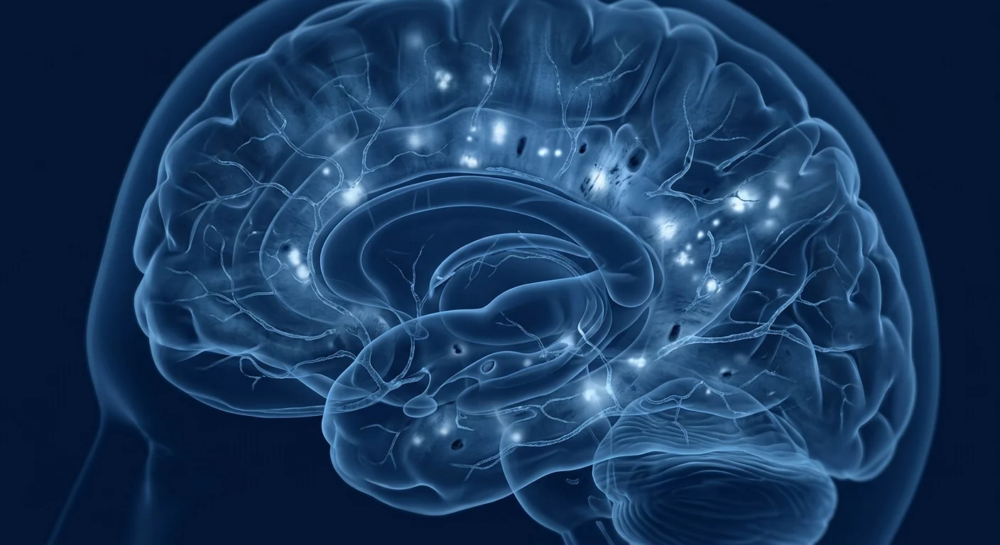
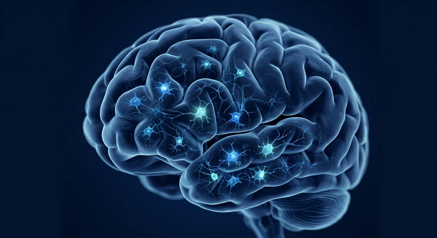
FTD is defined by progressive atrophy of the frontal and temporal lobes. Unlike Alzheimer's, where hippocampal memory networks are the first casualties, FTD targets the prefrontal cortex, anterior temporal lobes, and insula — regions that underpin social cognition, emotional regulation, language production, and executive planning. On MRI, the pattern is unmistakable: knife-edge frontal gyri, widened sulci, and ventricular enlargement disproportionate to age.[3]
The molecular pathology converges on two proteins: tau and TDP-43. Approximately 40–50% of FTD cases involve abnormal accumulations of hyperphosphorylated tau, the same protein that forms tangles in Alzheimer's but in a different distribution — concentrated in frontal and temporal neurons and glia. The remaining cases, and the majority of those associated with ALS, are driven by TDP-43 (TAR DNA-binding protein 43), a nuclear protein that in disease mislocalizes to the cytoplasm, forms toxic aggregates, and loses its normal RNA-processing functions. Both pathways converge on synaptic failure, neuronal death, and progressive brain atrophy.[4][5]
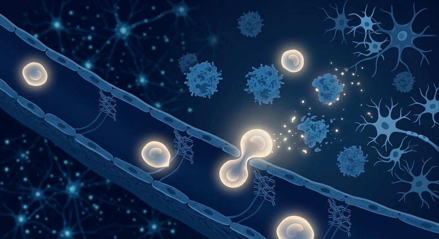
Neuroinflammation is not a bystander — it is a driver. Activated microglia and reactive astrocytes surround the degenerating neurons, releasing pro-inflammatory cytokines (IL-1β, TNF-α, IL-6) that create a self-reinforcing cycle of tissue damage. This neuroinflammatory milieu is what makes MSC therapy a rational target: the cells' primary therapeutic mechanism — paracrine immunomodulation — speaks directly to the inflammatory loop driving FTD progression.[6][7]
FTD subtypes — why one-size treatment does not work
FTD is clinically heterogeneous. The three major subtypes reflect different anatomical targets within the frontal-temporal network:
- Behavioural variant FTD (bvFTD). The most common subtype. Patients develop disinhibition, apathy, loss of empathy, compulsive or ritualistic behaviours, and dietary changes (hyperphagia, carbohydrate craving). Memory is relatively preserved early on, which is why bvFTD is often misdiagnosed as a psychiatric condition for years. Atrophy is most pronounced in the medial and orbitofrontal cortices, anterior cingulate, and anterior insula.[8]
- Semantic variant primary progressive aphasia (svPPA). Progressive loss of word and object meaning. Patients can speak fluently but use vague, empty language ("the thing," "that place"). The anterior temporal lobes — particularly the left — are the primary site of atrophy. Most cases are TDP-43 pathology (Type C).[9]
- Nonfluent/agrammatic variant PPA (nfvPPA). Effortful, halting speech with grammatical errors. Patients know what they want to say but cannot produce it. Atrophy targets the left inferior frontal gyrus (Broca's area) and insula. Most cases are tau pathology (4R tau).[10]
Each subtype affects different networks, but all three share a common thread: neuroinflammation. The microglial and astrocytic response in FTD is regionally specific but mechanistically similar regardless of whether tau or TDP-43 is the primary proteinopathy. This shared inflammatory biology is why a broad immunomodulatory strategy — such as MSC therapy — is conceptually appealing across subtypes.
Why researchers study mesenchymal stem cells for FTD
The rationale for MSC therapy in FTD rests on four mechanisms, each targeting a different dimension of the disease:
1. Immunomodulation and microglial reprogramming. MSCs are potent regulators of innate immunity. When they encounter an inflamed environment, they secrete a cocktail of anti-inflammatory mediators — IL-10, TGF-β, PGE2, TSG-6, IDO — that shift microglia from a pro-inflammatory (M1-like) to a reparative (M2-like) phenotype. In the FTD brain, where chronically activated microglia drive ongoing neuronal damage, this phenotypic switch is one of the most direct ways a cell therapy could alter disease trajectory.[11][12]
2. Progranulin enhancement. Progranulin (PGRN) is a neurotrophic and anti-inflammatory protein, and GRN loss-of-function mutations are the second most common genetic cause of FTD (after C9orf72). Haploinsufficiency — having only one functional copy — reduces PGRN levels by ~50%, impairing lysosomal function and neuronal survival. MSCs naturally secrete PGRN, and in preclinical models, MSC-derived PGRN has been shown to reduce microgliosis, restore lysosomal function, and slow neurodegeneration. This is one of the most compelling disease-specific rationales for MSC therapy in FTD: the cells directly supplement the very protein that haploinsufficient patients lack.[13][14]
3. Neurotrophic support. MSCs secrete brain-derived neurotrophic factor (BDNF), glial cell line-derived neurotrophic factor (GDNF), nerve growth factor (NGF), and insulin-like growth factor-1 (IGF-1) — proteins that promote neuronal survival, synaptic plasticity, and axonal sprouting. In FTD, where synaptic loss precedes neuronal death, neurotrophic support may extend the functional lifespan of vulnerable frontal and temporal neurons.[15]
4. TDP-43 clearance and autophagy enhancement. Emerging evidence suggests MSC-derived factors, including extracellular vesicles, can enhance autophagic clearance of aggregated proteins. In TDP-43 proteinopathies — which account for roughly half of all FTD cases — restoring autophagic flux could reduce the cytoplasmic TDP-43 burden that drives neuronal toxicity. This mechanism is early-stage and experimental, but it aligns with a growing recognition that protein clearance, not just inflammation control, is a viable therapeutic axis.[16]
Preclinical evidence — what animal models show
Preclinical FTD research faces a fundamental challenge: no single mouse model recapitulates the full clinical and pathological spectrum of human FTD. Researchers use a patchwork of transgenic models — P301L tau mice for tau-driven pathology, GRN-knockout mice for progranulin deficiency, TDP-43 overexpression models for TDP-43 proteinopathy — each capturing one dimension of the disease.
In P301L tau transgenic mice, intravenous or intracerebroventricular MSC administration has been reported to reduce tau hyperphosphorylation, decrease microglial activation, and modestly improve cognitive performance on object-recognition and Morris water maze tasks. In GRN-deficient mice, MSC infusion elevated brain PGRN levels, reduced lipofuscin accumulation (a marker of lysosomal dysfunction), and attenuated microgliosis in frontal cortical regions. In TDP-43 models, MSC-derived extracellular vesicles reduced cytoplasmic TDP-43 aggregation in cultured neurons and, in one study, extended survival in a TDP-43 overexpression model.[13][17][18]
Caveats are essential. Mouse models overexpress mutant proteins at supraphysiological levels; they do not capture the decades-long, slowly progressive nature of human FTD. Positive results in a transgenic mouse are a promising signal, not a prediction of clinical success. Many therapies that worked in mice have failed in human trials — and neurodegeneration trials have a particularly high attrition rate.
Clinical evidence — what human data exist
There are no completed Phase II or III MSC trials specifically for FTD. The clinical evidence base is indirect — drawn from related neurodegenerative conditions where MSC safety has been tested — and from small, early-phase studies that included FTD patients within broader dementia cohorts.
A small open-label safety study from South Korea (2015) delivered umbilical cord blood-derived MSCs via stereotactic injection into the hippocampus and precuneus of patients with various dementia subtypes, including a small number of FTD patients. The primary endpoint was safety: the procedure was tolerated, with no serious adverse events attributed to the cells. Cognitive outcomes were exploratory and underpowered, but some patients showed stable or modestly improved scores on the Alzheimer's Disease Assessment Scale – Cognitive subscale (ADAS-Cog) and the Clinical Dementia Rating Sum of Boxes (CDR-SB).[19]
More broadly, MSC safety has been established across dozens of trials in neurological conditions including ALS, multiple sclerosis, spinal cord injury, and stroke. The safety signal is consistent: when manufactured under GMP conditions from perinatal tissue (umbilical cord), allogeneic MSCs are well-tolerated with low rates of infusion-related reactions and no evidence of tumour formation or ectopic tissue growth.[20]
The honest headline
As of today, no stem cell therapy is a proven or approved treatment for frontotemporal dementia. The rationale — immunomodulation, progranulin delivery, neurotrophic support — is biologically coherent and supported by preclinical data, but human efficacy data in FTD are essentially absent. Any clinic offering a "stem cell cure" for FTD is making a claim the evidence does not support. The credible work remains investigational and should be conducted within registered clinical trials.
How FTD outcomes are measured — and why it matters
FTD is harder to measure than Alzheimer's. Memory tests — the backbone of Alzheimer's trials — are relatively preserved in early FTD and poorly capture what the disease actually takes. Instead, FTD trials use instruments that assess the domains FTD attacks:
- FRS (Frontotemporal Dementia Rating Scale). A validated staging tool that quantifies functional decline across behaviour, language, and motor domains. It correlates with atrophy on MRI and is sensitive to change over 12-month intervals.
- CDR plus NACC FTLD (Clinical Dementia Rating plus FTLD module). An adaptation of the standard CDR that incorporates behavioural and language domains relevant to FTD. Increasingly used as a primary endpoint in FTD clinical trials.
- NPI (Neuropsychiatric Inventory). Quantifies the frequency and severity of behavioural symptoms — apathy, disinhibition, agitation, aberrant motor behaviour — that are the primary burden for caregivers.
- Brain volumetry (MRI). Serial MRI with automated segmentation tracks frontal and temporal lobe atrophy rates. This is arguably the most objective biomarker — atrophy rates of 3–5% per year in target regions are typical in FTD, and slowing that rate would be a meaningful signal of disease modification.
- CSF and blood biomarkers. Neurofilament light chain (NfL) is elevated in FTD and correlates with disease severity and rate of progression. PGRN levels in CSF are a pharmacodynamic marker for therapies targeting progranulin deficiency.
A therapy that genuinely modifies FTD should slow atrophy on MRI, reduce NfL rise, and preserve functional ability on the FRS or CDR-FTLD — not just produce anecdotes of improvement. The disease's own fluctuations, particularly in behaviour, can easily be mistaken for treatment effect without a controlled comparison.
How a responsible MSC protocol for neurodegeneration works
At centres that offer MSC therapy within an evidence-guided framework (not a commercial "cure" programme), the process is conservative and transparent:
- Pre-treatment neurological assessment. MRI, cognitive testing (FRS, CDR-FTLD, NPI), CSF or blood NfL levels, and comprehensive neurological examination to establish a baseline against which any change can be measured.
- Informed consent with honest counselling. The physician explains that MSC therapy for FTD is investigational, not proven; that the goal is immunomodulation and disease modulation, not cure; and that the expected trajectory remains progressive. Families are told plainly that no guarantees can be made.
- Cell administration. Most neurodegeneration protocols use intravenous (IV) infusion of allogeneic, GMP-manufactured, umbilical cord-derived MSCs. The IV route relies on the cells' homing to inflammatory signals and their systemic immunomodulatory effects — the cells do not need to enter the brain parenchyma in large numbers to exert paracrine effects. Some experimental protocols use intrathecal administration to bypass the blood-brain barrier, but this is more invasive and its superiority over IV is unproven.
- Serial monitoring. Follow-up MRI at 6 and 12 months, repeat cognitive testing, and NfL monitoring. Objective change — or its absence — is documented transparently.
What the evidence supports — and what it does not
The fair summary is carefully bounded:
- MSC safety is well-established in neurological conditions when cells are GMP-manufactured from perinatal tissue.
- The mechanistic rationale for FTD is strong — immunomodulation, progranulin delivery, and neurotrophic support each target a validated disease pathway. Among neurodegenerative conditions, FTD is unusually well-matched to MSC biology because of the progranulin axis.
- Preclinical data are encouraging across tau, TDP-43, and progranulin-deficient models, but animal models are imperfect.
- Human efficacy data in FTD are lacking. No MSC trial has demonstrated a reproducible, meaningful benefit in slowing FTD progression or improving symptoms. The evidence base is preclinical plus safety data from related conditions — it has not reached the threshold of clinical proof.
- Delivery remains an open question. It is unknown whether IV infusion, intrathecal injection, or a combination yields the best risk-benefit profile for FTD specifically.
FTD is one of the cruelest diagnoses in medicine — it takes the person before the body. That very cruelty makes families vulnerable to anyone who promises to stop it. The most respectful thing we can offer is the truth about what the science can and cannot do today, and the discipline to demand evidence, not testimonials.
— VELAR Clinical Team
Frequently Asked Questions
What is the difference between FTD and Alzheimer's disease?
FTD primarily affects behaviour, personality, and language — memory is relatively preserved early on — while Alzheimer's typically begins with memory loss. FTD also strikes younger (45–65 vs. 65+), progresses faster on average, and has a distinct protein pathology (tau or TDP-43 vs. amyloid-beta and tau in Alzheimer's). The two conditions affect different brain regions and require different assessment tools.
Is there a genetic test for FTD?
Yes. Approximately 30–40% of FTD cases have a family history, and three genes account for most genetic FTD: C9orf72 (the most common), GRN (progranulin), and MAPT (tau). Genetic testing is available and recommended when there is a strong family history of FTD, ALS, or related conditions. Genetic counselling should accompany testing because a positive result has implications for family members.
How much does stem cell therapy cost for FTD in Thailand?
At legitimate, GMP-compliant centres in Bangkok, a single IV MSC infusion for neurodegenerative conditions typically ranges from USD 8,000 to 15,000, depending on cell dose, manufacturing standards, and the extent of pre-treatment assessment and follow-up monitoring. Prices above this range warrant scrutiny. Legitimate centres provide transparent, itemized pricing, honest counselling about the investigational nature of the treatment, and objective outcome tracking — not promises or testimonials.
Can MSC therapy reverse FTD symptoms?
No. There is no evidence — preclinical or clinical — that MSC therapy reverses FTD. The realistic goal is disease modulation: reducing neuroinflammation, supplementing deficient proteins like progranulin, and potentially slowing the rate of atrophy and functional decline. Any clinic claiming reversal, recovery, or cure is making claims unsupported by evidence.
How do I evaluate whether a stem cell clinic is legitimate?
Ask five questions: (1) Is the treatment part of a registered clinical trial? (2) What published, peer-reviewed evidence supports the specific claim for FTD? (3) How will outcomes be measured objectively (MRI volumetry, validated rating scales, biomarkers)? (4) What are the cell source, manufacturing standards, and quality-control data (viability, sterility, identity)? (5) Does the clinic provide honest, written informed consent that explicitly states the treatment is investigational and unproven? A legitimate centre answers all five clearly and provides documentation. Commercial clinics that avoid these questions or charge large sums without objective outcome tracking should be approached with caution.
The VELAR perspective
At VELAR Center, we follow neurodegenerative research closely. FTD is among the conditions where the preclinical rationale for MSC therapy is genuinely compelling — the progranulin axis, the immunomodulatory match to microglial-driven pathology, and the neurotrophic support for vulnerable frontal and temporal neurons each represent a coherent therapeutic strategy. But "compelling rationale" is not the same as "proven treatment." The gap between preclinical promise and clinical proof is wide, and no responsible clinician bridges it with marketing.
We offer MSC therapy within an evidence-guided framework for conditions where the evidence base is more mature, and we monitor the FTD research landscape with interest. If a family wants an honest, unhurried conversation about what regenerative medicine can and cannot offer for a neurodegenerative condition — including FTD — we are here for exactly that conversation. No promises, no testimonials, no pressure. Just the evidence, plainly presented.
Limitations and honest caveats
- No proven disease-modifying treatment exists for FTD. MSC therapy is investigational, and no human trial has demonstrated it slows FTD progression.
- Preclinical data are from animal models that only partially recapitulate human FTD. Translation from mouse to human has a high failure rate in neurodegeneration.
- Progranulin enhancement by MSCs has been demonstrated in vitro and in animal models, but the magnitude and duration of PGRN elevation in humans after MSC infusion is unknown.
- FTD is clinically heterogeneous. Whether bvFTD, svPPA, and nfvPPA respond differently to MSC therapy — and whether tau-driven vs. TDP-43-driven pathology matters for treatment response — is entirely unknown.
- Cost is significant and not reimbursed by insurance for an investigational indication. Families must weigh this honestly against the uncertainty of benefit.
- This article reflects the evidence available as of August 2026 and is not a substitute for neurological consultation. Treatment decisions should be made with a neurologist who understands both FTD and the MSC evidence base.
References
- Bang J, Spina S, Miller BL. Frontotemporal dementia. The Lancet. 2015;386(10004):1672-1682. doi:10.1016/S0140-6736(15)00461-4 ↩
- Rascovsky K, Hodges JR, Knopman D, et al. Sensitivity of revised diagnostic criteria for the behavioural variant of frontotemporal dementia. Brain. 2011;134(9):2456-2477. doi:10.1093/brain/awr179 ↩
- Seeley WW, Crawford RK, Zhou J, Miller BL, Greicius MD. Neurodegenerative diseases target large-scale human brain networks. Neuron. 2009;62(1):42-52. doi:10.1016/j.neuron.2009.03.024 ↩
- Mackenzie IR, Neumann M, Bigio EH, et al. Nomenclature and nosology for neuropathologic subtypes of frontotemporal lobar degeneration. Acta Neuropathologica. 2010;119(1):1-4. doi:10.1007/s00401-009-0612-2 ↩
- Neumann M, Sampathu DM, Kwong LK, et al. Ubiquitinated TDP-43 in frontotemporal lobar degeneration and amyotrophic lateral sclerosis. Science. 2006;314(5796):130-133. doi:10.1126/science.1134108 ↩
- Bright F, Werry EL, Dobson-Stone C, et al. Neuroinflammation in frontotemporal dementia. Nature Reviews Neurology. 2019;15(9):540-555. doi:10.1038/s41582-019-0231-z ↩
- Lui H, Zhang J, Makinson SR, et al. Progranulin deficiency promotes circuit-specific synaptic pruning by microglia via complement activation. Cell. 2016;165(4):921-935. doi:10.1016/j.cell.2016.04.001 ↩
- Piguet O, Hornberger M, Mioshi E, Hodges JR. Behavioural-variant frontotemporal dementia: diagnosis, clinical staging, and management. The Lancet Neurology. 2011;10(2):162-172. doi:10.1016/S1474-4422(10)70299-4 ↩
- Gorno-Tempini ML, Hillis AE, Weintraub S, et al. Classification of primary progressive aphasia and its variants. Neurology. 2011;76(11):1006-1014. doi:10.1212/WNL.0b013e31821103e6 ↩
- Grossman M. The non-fluent/agrammatic variant of primary progressive aphasia. The Lancet Neurology. 2012;11(6):545-555. doi:10.1016/S1474-4422(12)70099-6 ↩
- Uccelli A, Moretta L, Pistoia V. Mesenchymal stem cells in health and disease. Nature Reviews Immunology. 2008;8(9):726-736. doi:10.1038/nri2395 ↩
- Shi Y, Wang Y, Li Q, et al. Immunoregulatory mechanisms of mesenchymal stem and stromal cells in inflammatory diseases. Nature Reviews Nephrology. 2018;14(8):493-507. doi:10.1038/s41581-018-0023-5 ↩
- Arrant AE, Onyilo VC, Unger DE, Roberson ED. Progranulin gene therapy improves lysosomal dysfunction and microglial pathology associated with frontotemporal dementia and neuronal ceroid lipofuscinosis. Journal of Neuroscience. 2018;38(9):2341-2358. doi:10.1523/JNEUROSCI.3081-17.2018 ↩
- Martens LH, Zhang J, Barmada SJ, et al. Progranulin deficiency promotes neuroinflammation and neuron loss following toxin-induced injury. Journal of Clinical Investigation. 2012;122(11):3955-3959. doi:10.1172/JCI63113 ↩
- Teixeira FG, Carvalho MM, Sousa N, Salgado AJ. Mesenchymal stem cells secretome: a new paradigm for central nervous system regeneration? Cellular and Molecular Life Sciences. 2013;70(20):3871-3882. doi:10.1007/s00018-013-1290-8 ↩
- Lee M, Ban JJ, Kim KY, et al. Adipose-derived stem cell exosomes alleviate TDP-43 proteinopathy. Scientific Reports. 2020;10:13356. doi:10.1038/s41598-020-70303-2 ↩
- Yun SP, Kam TI, Panicker N, et al. Block of A1 astrocyte conversion by microglia is neuroprotective in models of Parkinson's disease. Nature Medicine. 2018;24(7):931-938. doi:10.1038/s41591-018-0051-5 ↩
- Kim DH, Lee D, Chang EH, et al. GDF-15 secreted from human umbilical cord blood mesenchymal stem cells delivered through the cerebrospinal fluid promotes hippocampal neurogenesis and synaptic activity in an Alzheimer's disease model. Stem Cells and Development. 2015;24(20):2378-2390. doi:10.1089/scd.2014.0487 ↩
- Kim HJ, Seo SW, Chang JW, et al. Stereotactic brain injection of human umbilical cord blood mesenchymal stem cells in patients with Alzheimer's disease dementia: a phase 1 clinical trial. Alzheimer's & Dementia: Translational Research & Clinical Interventions. 2015;1(2):95-102. doi:10.1016/j.trci.2015.06.007 ↩
- Lalu MM, McIntyre L, Pugliese C, et al. Safety of cell therapy with mesenchymal stromal cells (SafeCell): a systematic review and meta-analysis of clinical trials. PLoS ONE. 2012;7(10):e47559. doi:10.1371/journal.pone.0047559 ↩
额颞叶痴呆(FTD)是一组攻击大脑额叶和颞叶的神经退行性疾病,影响人格、行为、语言和执行功能。与阿尔茨海默病主要侵蚀记忆不同,FTD通常在较早年龄(45–65岁)发病,首先摧毁的是构成一个人本质的特质:同理心、抑制力、判断力和语言能力。目前尚无治愈方法;现有治疗仅能管理症状。间充质干细胞(MSC)疗法正在被研究作为一种缓解FTD进展中神经炎症的方法——不是治愈,而是针对驱动神经退行性变的免疫环境的潜在疾病修饰策略。[1][2]
FTD大脑的病理变化
FTD由额叶和颞叶的进行性萎缩定义。与阿尔茨海默病首先累及海马记忆网络不同,FTD靶向前额叶皮层、前颞叶和岛叶——这些区域主管社会认知、情绪调节、语言产生和执行规划。MRI显示特征性模式:额叶脑回变窄如刀锋、脑沟增宽、脑室扩大。[3]
分子病理学集中于两种蛋白质:tau和TDP-43。约40–50%的FTD病例涉及异常磷酸化tau蛋白积聚——与阿尔茨海默病中的缠结蛋白相同,但分布在额叶和颞叶神经元及胶质细胞中。其余病例以及与ALS相关的多数病例由TDP-43(TAR DNA结合蛋白43)驱动——这种核蛋白在疾病中错位至细胞质,形成毒性聚集体,丧失正常RNA加工功能。两种通路均导致突触衰竭、神经元死亡和进行性脑萎缩。[4][5]
神经炎症并非旁观者——它是驱动因素。活化的小胶质细胞和反应性星形胶质细胞包围退化的神经元,释放促炎细胞因子(IL-1β、TNF-α、IL-6),形成自我强化的组织损伤循环。这种神经炎症环境正是MSC治疗的合理靶点:细胞的主要治疗机制——旁分泌免疫调节——直接针对驱动FTD进展的炎症环路。[6][7]
FTD亚型
- 行为变异型FTD(bvFTD)。最常见亚型。表现为去抑制、冷漠、缺乏同理心、强迫或仪式化行为以及饮食改变。记忆力早期相对保留。萎缩最显著于内侧和眶额叶皮层、前扣带回和前岛叶。[8]
- 语义变异型原发性进行性失语(svPPA)。词汇和物体含义的进行性丧失。患者能流利说话但使用模糊空洞的语言。前颞叶——特别是左侧——是主要萎缩部位。多数为TDP-43病理。[9]
- 非流利/语法缺失变异型PPA(nfvPPA)。费力、停顿的言语伴语法错误。患者知道想说什么但无法表达。萎缩靶向左下额叶(布罗卡区)和岛叶。多数为tau病理。[10]
MSC治疗FTD的机制
1. 免疫调节和小胶质细胞重编程。MSCs分泌抗炎介质(IL-10、TGF-β、PGE2、TSG-6),将小胶质细胞从促炎表型转变为修复性表型。[11][12]
2. 颗粒蛋白前体增强。GRN功能缺失突变是FTD第二常见遗传原因。MSCs天然分泌颗粒蛋白前体(PGRN),在临床前模型中可减少小胶质细胞增生、恢复溶酶体功能、减缓神经退行性变。[13][14]
3. 神经营养支持。MSCs分泌BDNF、GDNF、NGF和IGF-1——促进神经元存活和突触可塑性的蛋白质。[15]
4. 自噬增强和TDP-43清除。MSC来源的细胞外囊泡可能增强聚集蛋白的自噬清除。[16]
临床前和临床证据
在P301L tau转基因小鼠中,MSC给药可减少tau过度磷酸化、降低小胶质细胞活化并适度改善认知表现。在GRN缺陷小鼠中,MSC输注提升脑PGRN水平,减少脂褐素积累并减轻额叶皮层的小胶质细胞增生。[17][18]
目前尚无针对FTD的完成Ⅱ期或Ⅲ期MSC试验。一项2015年韩国小型开放标签安全性研究将脐带血MSCs立体定向注射至包括少数FTD患者在内的痴呆患者中——主要终点为安全性,手术可耐受。认知结果为探索性且样本量不足。[19]
更广泛地,MSC安全性已在ALS、多发性硬化、脊髓损伤和中风等数十项神经系统试验中确立:当从围产期组织在GMP条件下制造时,同种异体MSCs耐受性良好,输注相关反应发生率低,无肿瘤形成证据。[20]
诚实声明
截至今日,尚无干细胞疗法被证实或批准用于治疗额颞叶痴呆。其机制——免疫调节、颗粒蛋白前体递送、神经营养支持——在生物学上是合理的并有临床前数据支持,但FTD的人体疗效数据基本缺失。任何声称提供FTD"干细胞治愈"的诊所都在做出证据不支持的声明。
常见问题
FTD和阿尔茨海默病有什么区别?
FTD主要影响行为、人格和语言——记忆力相对保留——而阿尔茨海默病通常始于记忆丧失。FTD发病年龄更早(45–65岁 vs. 65+),进展平均更快,蛋白质病理不同(tau或TDP-43 vs. β淀粉样蛋白和tau)。
泰国FTD干细胞疗法费用多少?
在曼谷符合GMP标准的合法中心,针对神经退行性疾病的单次静脉MSC输注通常为8,000–15,000美元,取决于细胞剂量、制造标准和评估监测范围。合法中心提供透明的逐项定价、诚实的咨询和客观的结局追踪。
MSC疗法能逆转FTD症状吗?
不能。没有临床前或临床证据表明MSC疗法能逆转FTD。现实目标是疾病修饰:减少神经炎症、补充缺陷蛋白(如颗粒蛋白前体)、可能减缓萎缩和功能衰退的速度。
参考文献
- Bang J, Spina S, Miller BL. Frontotemporal dementia. The Lancet. 2015;386(10004):1672-1682. doi:10.1016/S0140-6736(15)00461-4 ↩
- Rascovsky K, Hodges JR, Knopman D, et al. Sensitivity of revised diagnostic criteria for the behavioural variant of frontotemporal dementia. Brain. 2011;134(9):2456-2477. doi:10.1093/brain/awr179 ↩
- Seeley WW, et al. Neurodegenerative diseases target large-scale human brain networks. Neuron. 2009;62(1):42-52. doi:10.1016/j.neuron.2009.03.024 ↩
- Mackenzie IR, et al. Nomenclature for neuropathologic subtypes of FTLD. Acta Neuropathologica. 2010;119(1):1-4. doi:10.1007/s00401-009-0612-2 ↩
- Neumann M, et al. Ubiquitinated TDP-43 in FTLD and ALS. Science. 2006;314(5796):130-133. doi:10.1126/science.1134108 ↩
- Bright F, et al. Neuroinflammation in frontotemporal dementia. Nature Reviews Neurology. 2019;15(9):540-555. doi:10.1038/s41582-019-0231-z ↩
- Lui H, et al. Progranulin deficiency promotes synaptic pruning by microglia. Cell. 2016;165(4):921-935. doi:10.1016/j.cell.2016.04.001 ↩
- Piguet O, et al. Behavioural-variant FTD: diagnosis, staging, management. The Lancet Neurology. 2011;10(2):162-172. doi:10.1016/S1474-4422(10)70299-4 ↩
- Gorno-Tempini ML, et al. Classification of primary progressive aphasia. Neurology. 2011;76(11):1006-1014. doi:10.1212/WNL.0b013e31821103e6 ↩
- Grossman M. Non-fluent/agrammatic variant of PPA. The Lancet Neurology. 2012;11(6):545-555. doi:10.1016/S1474-4422(12)70099-6 ↩
- Uccelli A, Moretta L, Pistoia V. MSCs in health and disease. Nature Reviews Immunology. 2008;8(9):726-736. doi:10.1038/nri2395 ↩
- Shi Y, et al. Immunoregulatory mechanisms of MSCs. Nature Reviews Nephrology. 2018;14(8):493-507. doi:10.1038/s41581-018-0023-5 ↩
- Arrant AE, et al. Progranulin gene therapy in FTD. Journal of Neuroscience. 2018;38(9):2341-2358. doi:10.1523/JNEUROSCI.3081-17.2018 ↩
- Martens LH, et al. Progranulin deficiency and neuroinflammation. Journal of Clinical Investigation. 2012;122(11):3955-3959. doi:10.1172/JCI63113 ↩
- Teixeira FG, et al. MSCs secretome for CNS regeneration. Cellular and Molecular Life Sciences. 2013;70(20):3871-3882. doi:10.1007/s00018-013-1290-8 ↩
- Lee M, et al. ADSC exosomes alleviate TDP-43 proteinopathy. Scientific Reports. 2020;10:13356. doi:10.1038/s41598-020-70303-2 ↩
- Yun SP, et al. Microglia block A1 astrocyte conversion. Nature Medicine. 2018;24(7):931-938. doi:10.1038/s41591-018-0051-5 ↩
- Kim DH, et al. GDF-15 from UCB-MSCs in Alzheimer's model. Stem Cells and Development. 2015;24(20):2378-2390. doi:10.1089/scd.2014.0487 ↩
- Kim HJ, et al. Brain injection of UCB-MSCs in AD dementia. Alzheimer's & Dementia: TRCI. 2015;1(2):95-102. doi:10.1016/j.trci.2015.06.007 ↩
- Lalu MM, et al. Safety of cell therapy with MSCs. PLoS ONE. 2012;7(10):e47559. doi:10.1371/journal.pone.0047559 ↩
الخرف الجبهي الصدغي (FTD) ليس مرضا واحدا بل عائلة من الاضطرابات التنكسية العصبية التي تهاجم الفصين الجبهي والصدغي في الدماغ — المناطق التي تتحكم في الشخصية والسلوك واللغة والوظائف التنفيذية. على عكس مرض الزهايمر الذي يدمر الذاكرة بشكل أساسي، يصيب FTD عادة في سن أصغر (45–65 عاما) ويدمر أولا الصفات التي تجعل الشخص ما هو عليه: التعاطف والكبح والحكم والقدرة على إيجاد الكلمات. لا يوجد علاج شاف؛ العلاجات الحالية تدير الأعراض فقط. يتم دراسة العلاج بالخلايا الجذعية الوسيطة (MSC) كوسيلة لتهدئة الالتهاب العصبي الذي يسرع تقدم FTD.[1][2]
ما يحدث في دماغ FTD
يتم تعريف FTD بالضمور التدريجي للفصين الجبهي والصدغي. يستهدف القشرة أمام الجبهية والفص الصدغي الأمامي والجزيرة — المناطق المسؤولة عن الإدراك الاجتماعي والتنظيم العاطفي وإنتاج اللغة والتخطيط التنفيذي. يظهر التصوير بالرنين المغناطيسي نمطا مميزا: تلافيف جبهية حادة وتوسع البطينات.[3]
تتقارب الأمراض الجزيئية على بروتينين: tau و TDP-43. حوالي 40–50% من حالات FTD تنطوي على تراكمات غير طبيعية من بروتين tau مفرط الفسفرة. الحالات المتبقية، والأغلبية المرتبطة بـ ALS، مدفوعة بـ TDP-43 — بروتين نووي يتراكم في السيتوبلازم ويشكل تجمعات سامة.[4][5]
الالتهاب العصبي ليس مجرد متفرج — إنه محرك. تطلق الخلايا الدبقية الصغيرة المنشطة والخلايا النجمية التفاعلية سيتوكينات التهابية (IL-1β و TNF-α و IL-6) تخلق دورة ذاتية التعزيز من تلف الأنسجة.[6][7]
الأنواع الفرعية لـ FTD
- النوع السلوكي (bvFTD). الأكثر شيوعا. فقدان التثبيط واللامبالاة وفقدان التعاطف والسلوكيات القهرية. الذاكرة محفوظة نسبيا في البداية.[8]
- الحبسة التقدمية الأولية الدلالية (svPPA). فقدان تدريجي لمعنى الكلمات والأشياء. ضمور الفص الصدغي الأمامي. معظمها أمراض TDP-43.[9]
- الحبسة التقدمية غير الطلقة (nfvPPA). كلام متقطع مع أخطاء نحوية. ضمور منطقة بروكا والجزيرة. معظمها أمراض tau.[10]
آليات MSC لعلاج FTD
١. التعديل المناعي وإعادة برمجة الخلايا الدبقية الصغيرة. تفرز MSCs وسطاء مضادة للالتهابات (IL-10 و TGF-β و PGE2) تحول الخلايا الدبقية الصغيرة إلى نمط تعويضي.[11][12]
٢. تعزيز البروجرانولين. طفرات فقدان الوظيفة في GRN هي ثاني أكثر سبب وراثي شيوعا لـ FTD. تفرز MSCs بروتين البروجرانولين (PGRN) بشكل طبيعي، مما يقلل الالتهاب ويستعيد وظيفة الجسيمات الحالة.[13][14]
٣. الدعم العصبي التغذوي. تفرز MSCs عوامل BDNF و GDNF و NGF و IGF-1 — بروتينات تعزز بقاء الخلايا العصبية والمرونة المشبكية.[15]
٤. تعزيز الالتهام الذاتي وإزالة TDP-43. قد تعزز الحويصلات خارج الخلية المشتقة من MSCs التصفية الذاتية للبروتينات المتجمعة.[16]
الأدلة قبل السريرية والسريرية
في فئران P301L tau المعدلة وراثيا، قلل إعطاء MSCs من فرط فسفرة tau وقلل تنشيط الخلايا الدبقية الصغيرة. في فئران نقص GRN، رفع تسريب MSCs مستويات PGRN الدماغية وخفف من الالتهاب.[17][18]
لا توجد تجارب سريرية مكتملة من المرحلة الثانية أو الثالثة لـ MSC خصيصا لـ FTD. شملت دراسة سلامة كورية صغيرة مفتوحة التسمية (2015) عددا صغيرا من مرضى FTD ضمن مجموعة أوسع من الخرف — كانت نقطة النهاية الأولية هي السلامة وكان الإجراء محتملا. كانت النتائج المعرفية استكشافية.[19]
على نطاق أوسع، تم إثبات سلامة MSCs عبر عشرات التجارب في حالات عصبية تشمل ALS والتصلب المتعدد وإصابة الحبل الشوكي والسكتة الدماغية.[20]
العنوان الصادق
حتى اليوم، لا يوجد علاج بالخلايا الجذعية مثبت أو معتمد لمرض الخرف الجبهي الصدغي. الأساس المنطقي — التعديل المناعي وتوصيل البروجرانولين والدعم العصبي التغذوي — متماسك بيولوجيا، لكن بيانات الفعالية البشرية في FTD غائبة بشكل أساسي.
الأسئلة الشائعة
ما الفرق بين FTD ومرض الزهايمر؟
يؤثر FTD بشكل أساسي على السلوك والشخصية واللغة — الذاكرة محفوظة نسبيا في البداية — بينما يبدأ الزهايمر عادة بفقدان الذاكرة. يصيب FTD في سن أصغر ويتطور بشكل أسرع في المتوسط وله أمراض بروتينية مختلفة (tau أو TDP-43 مقابل بيتا أميلويد و tau في الزهايمر).
كم تكلفة العلاج بالخلايا الجذعية لـ FTD في تايلاند؟
في المراكز المشروعة الملتزمة بمعايير GMP في بانكوك، تتراوح تكلفة تسريب MSC الوريدي الواحد للحالات التنكسية العصبية عادة من 8,000 إلى 15,000 دولار أمريكي. تقدم المراكز المشروعة تسعيرا شفافا واستشارة صادقة وتتبعا موضوعيا للنتائج.
هل يمكن لعلاج MSC عكس أعراض FTD؟
لا. لا يوجد دليل — قبل سريري أو سريري — على أن علاج MSC يعكس FTD. الهدف الواقعي هو تعديل المرض: تقليل الالتهاب العصبي وتعويض البروتينات الناقصة وإبطاء معدل الضمور والتدهور الوظيفي.
المراجع
- Bang J, et al. Frontotemporal dementia. The Lancet. 2015;386(10004):1672-1682. doi:10.1016/S0140-6736(15)00461-4 ↩
- Rascovsky K, et al. Criteria for behavioural variant FTD. Brain. 2011;134(9):2456-2477. doi:10.1093/brain/awr179 ↩
- Seeley WW, et al. Neurodegenerative diseases target brain networks. Neuron. 2009;62(1):42-52. doi:10.1016/j.neuron.2009.03.024 ↩
- Mackenzie IR, et al. Nomenclature for FTLD subtypes. Acta Neuropathologica. 2010;119(1):1-4. doi:10.1007/s00401-009-0612-2 ↩
- Neumann M, et al. TDP-43 in FTLD and ALS. Science. 2006;314(5796):130-133. doi:10.1126/science.1134108 ↩
- Bright F, et al. Neuroinflammation in FTD. Nature Reviews Neurology. 2019;15(9):540-555. doi:10.1038/s41582-019-0231-z ↩
- Lui H, et al. Progranulin deficiency and microglia. Cell. 2016;165(4):921-935. doi:10.1016/j.cell.2016.04.001 ↩
- Piguet O, et al. Behavioural-variant FTD. The Lancet Neurology. 2011;10(2):162-172. doi:10.1016/S1474-4422(10)70299-4 ↩
- Gorno-Tempini ML, et al. Classification of PPA. Neurology. 2011;76(11):1006-1014. doi:10.1212/WNL.0b013e31821103e6 ↩
- Grossman M. Non-fluent/agrammatic PPA. The Lancet Neurology. 2012;11(6):545-555. doi:10.1016/S1474-4422(12)70099-6 ↩
- Uccelli A, et al. MSCs in health and disease. Nature Reviews Immunology. 2008;8(9):726-736. doi:10.1038/nri2395 ↩
- Shi Y, et al. Immunoregulatory mechanisms of MSCs. Nature Reviews Nephrology. 2018;14(8):493-507. doi:10.1038/s41581-018-0023-5 ↩
- Arrant AE, et al. Progranulin gene therapy in FTD. Journal of Neuroscience. 2018;38(9):2341-2358. doi:10.1523/JNEUROSCI.3081-17.2018 ↩
- Martens LH, et al. Progranulin deficiency. Journal of Clinical Investigation. 2012;122(11):3955-3959. doi:10.1172/JCI63113 ↩
- Teixeira FG, et al. MSCs secretome for CNS. Cellular and Molecular Life Sciences. 2013;70(20):3871-3882. doi:10.1007/s00018-013-1290-8 ↩
- Lee M, et al. ADSC exosomes and TDP-43. Scientific Reports. 2020;10:13356. doi:10.1038/s41598-020-70303-2 ↩
- Yun SP, et al. Microglia and A1 astrocytes. Nature Medicine. 2018;24(7):931-938. doi:10.1038/s41591-018-0051-5 ↩
- Kim DH, et al. GDF-15 from UCB-MSCs. Stem Cells and Development. 2015;24(20):2378-2390. doi:10.1089/scd.2014.0487 ↩
- Kim HJ, et al. UCB-MSCs in AD dementia. Alzheimer's & Dementia: TRCI. 2015;1(2):95-102. doi:10.1016/j.trci.2015.06.007 ↩
- Lalu MM, et al. Safety of MSC therapy. PLoS ONE. 2012;7(10):e47559. doi:10.1371/journal.pone.0047559 ↩