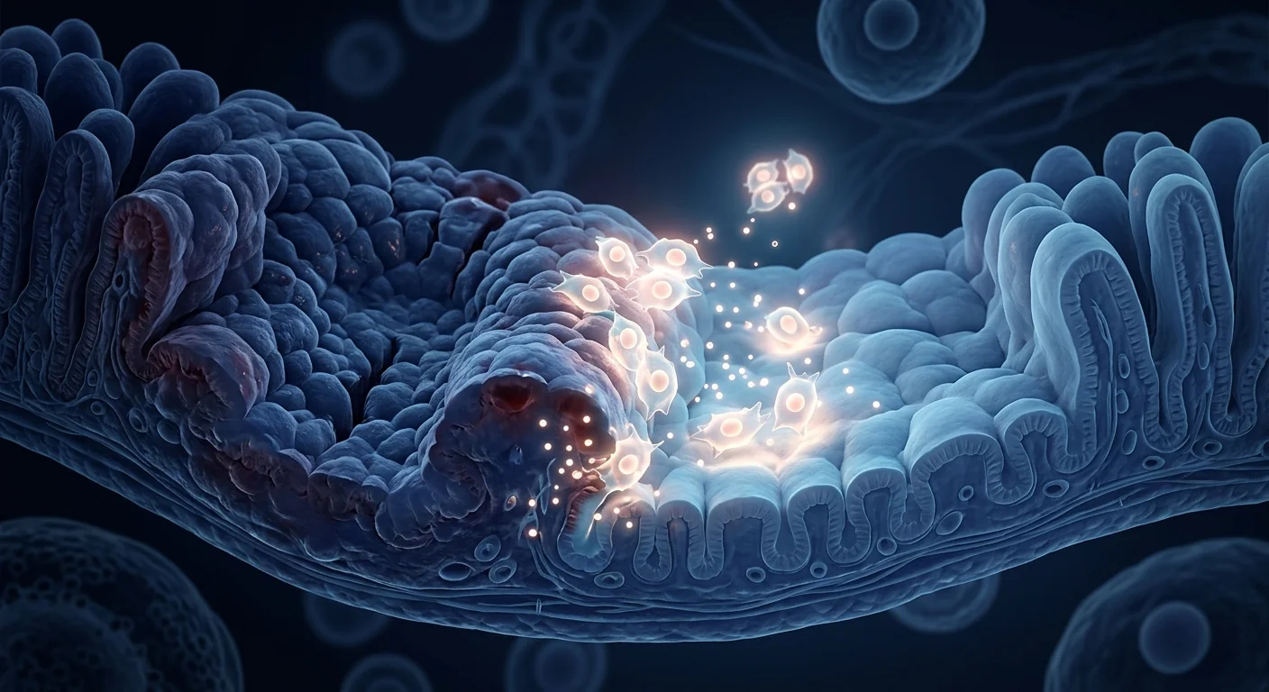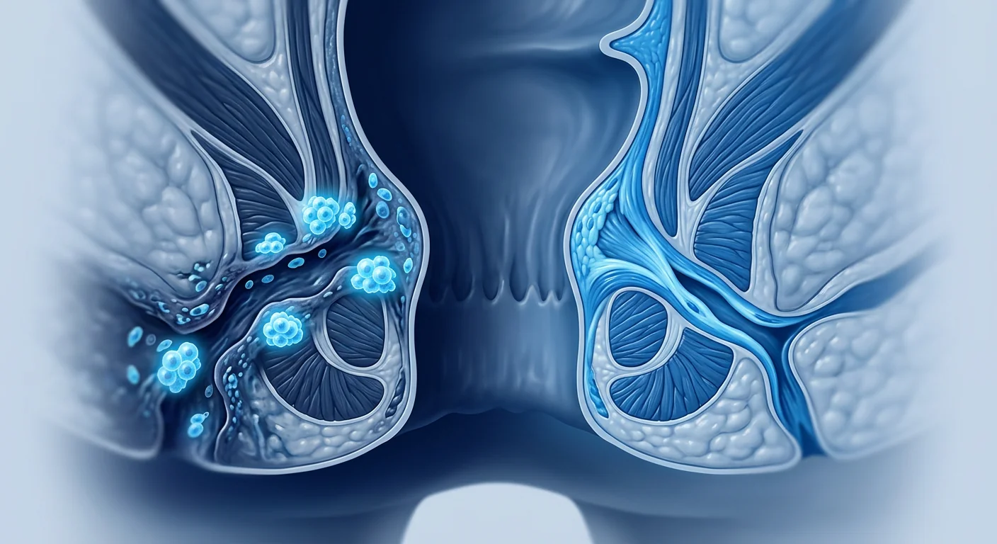
A perianal fistula is an abnormal tunnel that forms between the anal canal and the skin surrounding the anus — a persistent, draining, and often excruciatingly painful connection that refuses to heal. Approximately 30% of Crohn's disease patients will develop a perianal fistula during their lifetime, and for many it becomes the defining feature of their disease: recurrent abscesses, constant drainage, multiple failed surgeries, and a quality of life that collapses under the weight of a condition most people are too embarrassed to discuss [1].
Where conventional treatments fall short. The standard approach — seton drainage, fistulotomy, advancement flaps, and biologic therapy (anti-TNF agents like infliximab) — achieves long-term closure in only 30–50% of complex fistulas. Each failed surgery creates more scar tissue, more sphincter damage, and a progressively higher risk of incontinence. Biologics help control the underlying inflammation but rarely close an established fistula tract on their own [2]. For the patient cycling through their third or fourth surgical repair, the options narrow to a grim choice: live with it or accept a permanent stoma.
The deeper problem is tissue-level. A fistula tract is not simply a hole that needs to be plugged. It is a chronically inflamed, epithelialized tunnel lined with pro-inflammatory macrophages, fibroblasts laying down disorganized collagen, and a local microenvironment that actively resists healing. Systemic immunosuppression quiets the immune drivers but does nothing to remodel the tract itself. Surgery excises the tract but creates a fresh wound in hostile terrain. Neither approach addresses the fundamental pathology: a tissue microenvironment locked in a state of non-healing inflammation [3].
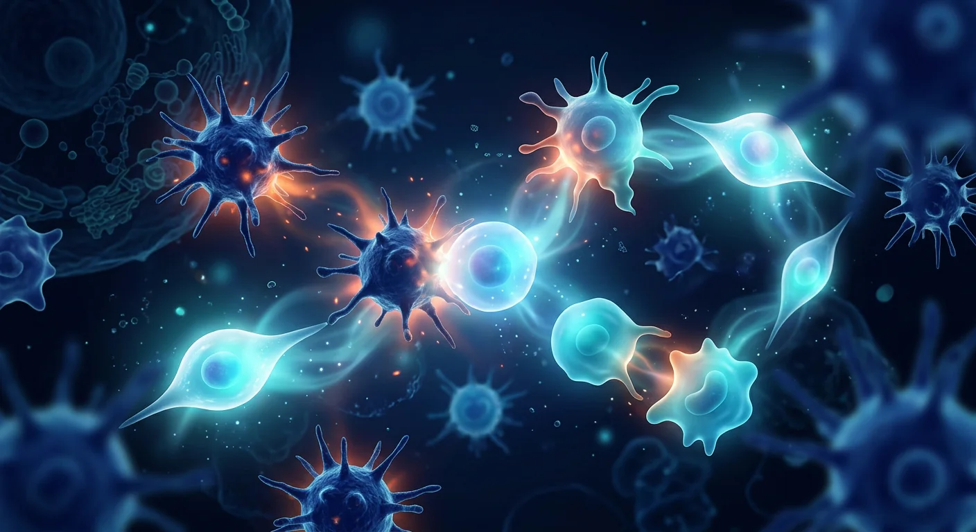
MSC therapy targets both sides of the problem. Rather than just suppressing inflammation or just excising tissue, locally injected mesenchymal stem cells remodel the fistula microenvironment. They suppress the pro-inflammatory cytokines (TNF-α, IL-1β, IL-17) that sustain the tract, polarize macrophages from the tissue-destructive M1 phenotype toward the repair-oriented M2 phenotype, secrete angiogenic factors (VEGF, HGF, FGF-2) that build new blood vessels into the healing zone, and differentiate into fibroblast-like cells that lay down organized collagen to close the tunnel from within [4]. This dual mechanism — anti-inflammatory plus pro-regenerative — is precisely what a fistula tract needs and what neither surgery nor biologics alone can provide.
How MSC Therapy Works for Fistula Closure
Local injection changes the game. Unlike systemic MSC infusion for autoimmune conditions, fistula therapy relies on direct injection of MSCs into the tissue surrounding and lining the fistula tract. This delivers the cells precisely where they are needed — the local injection achieves cell concentrations at the target site that systemic administration cannot match, while minimizing systemic exposure. It is one of the few MSC applications where the mode of delivery is inherently suited to the pathology: a localized, accessible lesion treated with a localized, injectable therapy [5].
The cellular mechanism unfolds in three overlapping phases. First, within hours of injection, MSCs release a burst of paracrine factors — TSG-6, PGE2, IDO, and IL-10 — that rapidly suppress the local inflammatory cascade. Neutrophil infiltration drops, and the destructive feed-forward loop of tissue damage → more inflammation → more tissue damage is interrupted. Second, over days 1–7, macrophage polarization shifts: M1 macrophages (pro-inflammatory, tissue-destructive) are replaced by M2 macrophages (anti-inflammatory, pro-regenerative), which secrete their own growth factors and amplify the repair signal. Third, over weeks 2–8, angiogenesis builds new microvasculature into the healing tract, fibroblasts are recruited and organized, and the epithelialized tunnel is gradually replaced with healthy granulation tissue that eventually remodels into normal connective tissue, closing the fistula from the inside out [6].
Allogeneic MSCs are the clinical standard for fistula therapy. The cells do not need to be the patient's own — umbilical cord-derived or adipose-derived MSCs from healthy donors are immunoprivileged (low MHC class I, no MHC class II), so they survive in the recipient without immunosuppression. This is critical for Crohn's patients who are already immunosuppressed from their disease and medications; adding another layer of immune suppression for an autologous cell procedure would increase risk without benefit. Wharton's jelly-derived MSCs (WJ-MSCs) are particularly well-suited: they secrete higher levels of TSG-6 and PGE2 than bone marrow- or adipose-derived MSCs, show superior immunomodulatory potency in head-to-head comparisons, and are obtained non-invasively from donated umbilical cords after healthy full-term births [7].
The ADMIRE-CD Trial — A Landmark in MSC Clinical Research
Darvadstrocel (Alofisel) is the first and only EMA-approved MSC therapy for fistula. In 2018, the European Medicines Agency granted marketing authorization for darvadstrocel (Cx601, marketed as Alofisel) — expanded allogeneic adipose-derived MSCs — for the treatment of complex perianal fistulas in adults with Crohn's disease who have failed conventional therapy. The approval was based on the ADMIRE-CD phase III randomized, double-blind, placebo-controlled trial (n=212), which remains the gold-standard evidence for MSC fistula therapy [8].
The ADMIRE-CD results were clinically meaningful. At 24 weeks, 50% of patients receiving darvadstrocel achieved combined remission (closure of all treated external openings with absence of collections >2 cm on MRI) compared to 34% in the placebo group — a statistically significant 16-percentage-point advantage. At 52 weeks, the benefit was maintained: 56% vs. 39%. Long-term follow-up at 104 weeks showed durable closure in 56% of treated patients [9]. Importantly, the safety profile was excellent: adverse events were comparable between treatment and placebo arms, with perianal abscess and proctalgia being the most common and occurring at similar rates in both groups. There was no signal of ectopic tissue formation, tumorigenicity, or immune sensitization — safety concerns that had shadowed MSC therapy for years.
Beyond ADMIRE-CD — the broader evidence base. Multiple independent trials confirm the ADMIRE-CD findings. A 2015 phase II trial by Molendijk et al. using allogeneic bone marrow-derived MSCs reported fistula healing in 75% of patients receiving 30 million cells vs. 57% for 10 million cells, suggesting a dose-response relationship. A Korean phase II trial (Cho et al., 2015) using autologous adipose-derived MSCs reported complete fistula closure in 82% at 8 weeks. A 2017 study by Wainstein et al. using bone marrow MSC injection into fistula tracts showed a 73% closure rate at 12 months. Meta-analyses consistently estimate overall fistula healing rates of 55–70% with MSC therapy — substantially higher than the ~30% rate for placebo or conventional care alone in refractory patients [10] [11] [12].
VELAR Treatment Protocol for Fistula
1. Pre-Treatment Assessment
Complete evaluation including pelvic MRI with fistula protocol to characterize tract anatomy (number of tracts, branching, abscess, sphincter involvement), colonoscopy to assess luminal disease activity, and baseline labs (CRP, fecal calprotectin, complete blood count). Patients with undrained abscesses undergo drainage and seton placement first — MSC injection into an active abscess is contraindicated. All patients are evaluated jointly by a gastroenterologist and colorectal surgeon.
2. Cell Product & Dose
Allogeneic Wharton's jelly-derived MSCs (WJ-MSCs), 30–120 million cells depending on fistula complexity and number of tracts. For a single simple tract: 30 million cells injected circumferentially around the internal opening and along the tract wall. For branching or multiple tracts: 60–120 million cells distributed proportionally. WJ-MSCs are selected for their superior TSG-6/PGE2 secretion profile — the same paracrine factors that drive fistula closure in the clinical literature.
3. Injection Procedure
Performed under local anesthesia with ultrasound or MRI guidance as needed. The internal opening is identified and closed with a dissolvable suture. MSCs are injected into the tissue surrounding the fistula tract — not into the lumen — using a fine-gauge needle to distribute cells evenly along the tract wall and at the internal opening. The procedure takes 20–40 minutes. Patients go home the same day. A single injection session is standard; a second session at 4–6 weeks may be offered for incomplete responders.
4. Monitoring & Follow-up
Clinical assessment at 4, 12, and 24 weeks post-injection evaluating: closure of external openings, cessation of drainage, pain scores (VAS), and perianal disease activity index (PDAI). Pelvic MRI at 24 weeks to confirm deep remission (absence of collections >2 cm). Fecal calprotectin and CRP monitored in parallel. Target endpoints: ≥1-point reduction in PDAI at 12 weeks, MRI-confirmed fistula closure at 24 weeks. Any adverse events documented per CTCAE criteria.
Benefits and Realistic Expectations
What MSC therapy can realistically achieve. Based on the clinical trial data, patients with complex perianal fistulas who have failed at least one conventional therapy can expect: closure of 50–70% of treated fistula tracts within 12–24 weeks, significant reduction in drainage and pain (often within 4 weeks of injection), and the ability to reduce or eliminate corticosteroid use in a substantial proportion of patients. Importantly, fistula closure with MSC therapy is durable — the 2-year follow-up data from ADMIRE-CD shows that patients who achieve closure at 24 weeks overwhelmingly maintain it at 104 weeks [13].
What it cannot do. MSC therapy does not cure Crohn's disease. It treats the fistula — a localized manifestation — not the systemic autoimmune process. Patients should continue their prescribed Crohn's disease management (biologics, immunomodulators) unless their gastroenterologist specifically recommends otherwise. MSC therapy also does not guarantee closure: approximately 30–50% of patients will not achieve complete fistula closure, and those with multiple branching tracts, active proctitis, or longstanding fibrosis are at the lower end of the response curve. The therapy is not a replacement for good surgical judgment — undrained sepsis, high transsphincteric tracts with significant sphincter involvement, and rectovaginal fistulas require individualized surgical assessment before any cell-based intervention [14].
Why Bangkok for Fistula MSC Therapy?
Thailand occupies a unique position in the global regenerative medicine landscape: a GMP-compliant cell manufacturing infrastructure, internationally trained colorectal surgeons and gastroenterologists, accreditation to ISO 9001:2015 and ISO/IEC 17025:2017 standards, and treatment costs typically 40–60% lower than equivalent care in Western Europe or North America. The VELAR clinical team includes gastroenterologists with specific experience in perianal fistula management and regenerative protocols, supported by an in-house quality-control laboratory that independently verifies every cell product before release. For the patient who has exhausted surgical options at home and is facing the prospect of a permanent stoma, MSC therapy in Bangkok represents not a cure guarantee but a rationally grounded, evidence-supported alternative worth serious consideration.
Frequently Asked Questions
Is MSC therapy for fistula proven to work?
For complex perianal fistulas in Crohn's disease, the evidence is stronger than for virtually any other MSC application. The phase III ADMIRE-CD trial demonstrated a statistically significant 16% absolute improvement in combined remission at 24 weeks (50% vs. 34% placebo). The EMA granted marketing authorization for darvadstrocel (Alofisel) in 2018 based on these data. Multiple independent phase II trials have replicated fistula closure rates of 55–82%. However, approximately 30–50% of patients do not achieve closure, and the evidence applies specifically to Crohn's-related perianal fistulas — not all fistula types have the same level of evidence.
How much does stem cell therapy for fistula cost in Thailand?
At VELAR Center, a single-session local MSC injection for perianal fistula typically ranges from $5,000–$9,000 USD, depending on the number of tracts treated and the total cell dose required. This includes the pre-treatment MRI, the cell product, the injection procedure, and 6 months of follow-up assessments. By comparison, darvadstrocel (Alofisel) in Europe costs approximately €40,000–60,000 per treatment course. A detailed, personalized quote is provided after the initial clinical review.
Do I need to stop my Crohn's medications?
No. MSC therapy for fistula is complementary to, not a replacement for, standard Crohn's disease management. Patients should continue their prescribed biologic therapy (infliximab, adalimumab, ustekinumab, etc.) and immunomodulators unless specifically instructed otherwise by their treating gastroenterologist. The MSC injection is local — it does not interfere with systemic immunosuppression, and there is no evidence that concomitant biologic therapy reduces MSC efficacy.
What is the recovery like after MSC fistula injection?
The procedure is performed under local anesthesia and takes 20–40 minutes. Most patients go home the same day. There is typically mild perianal discomfort for 24–48 hours, managed with simple analgesics. Drainage from the external opening may persist for 1–2 weeks as the tract remodels — this is normal and not a sign of treatment failure. Patients can return to desk work within 24 hours and resume normal activities within 3–5 days. Strenuous exercise and heavy lifting should be avoided for 2 weeks.
Can MSC therapy be repeated if the first treatment doesn't work?
Yes. Approximately 20–30% of patients who show partial but incomplete response to a first injection achieve closure after a second injection at 4–6 weeks. The decision to re-treat is based on clinical assessment — if there is evidence of partial response (reduced drainage, smaller tract on exam, improving PDAI score), a second session is reasonable. Patients who show no response at all after the first injection are less likely to benefit from a second, and alternative strategies should be discussed.
Limitations and Cautions
MSC therapy for fistula, while supported by the strongest clinical evidence of any MSC indication, is not a cure and does not work for everyone. The pivotal ADMIRE-CD trial showed a 50% combined remission rate at 24 weeks — meaning half of treated patients did not achieve the primary endpoint. The evidence is specific to Crohn's-related complex perianal fistulas; cryptoglandular fistulas, rectovaginal fistulas, enterocutaneous fistulas, and fistulas arising from other etiologies (radiation, malignancy, trauma) have far less supporting evidence. Patients with undrained perianal sepsis, active proctitis, or rectal stricture are not candidates for local MSC injection until these issues are addressed. As with all MSC therapies, the long-term safety data beyond 5 years remain limited — the theoretical risks of ectopic tissue formation and long-term immunologic effects, while not observed in the available data, cannot be definitively excluded. Patients considering MSC therapy for fistula should do so within a framework of transparent informed consent that explicitly acknowledges both the strength of the evidence base and its limitations, in partnership with a clinical team committed to systematic outcome documentation.
References
- Panes J, Garcia-Olmo D, Van Assche G, et al. Expanded allogeneic adipose-derived mesenchymal stem cells (Cx601) for complex perianal fistulas in Crohn's disease: a phase 3 randomised, double-blind controlled trial. The Lancet. 2016;388(10051):1281-1290. doi:10.1016/S0140-6736(16)31203-X ↩
- Sands BE, Anderson FH, Bernstein CN, et al. Infliximab maintenance therapy for fistulizing Crohn's disease. New England Journal of Medicine. 2004;350(9):876-885. doi:10.1056/NEJMoa030815 ↩
- Scharl M, Rogler G. Pathophysiology of fistula formation in Crohn's disease. World Journal of Gastrointestinal Pathophysiology. 2014;5(3):205-212. doi:10.4291/wjgp.v5.i3.205 ↩
- Shi Y, Wang Y, Li Q, et al. Immunoregulatory mechanisms of mesenchymal stem and stromal cells in inflammatory diseases. Nature Reviews Nephrology. 2018;14(8):493-507. doi:10.1038/s41581-018-0023-5 ↩
- Lightner AL, Wang Z, Zubair AC, Dozois EJ. A systematic review and meta-analysis of mesenchymal stem cell injections for the treatment of perianal Crohn's disease. Diseases of the Colon & Rectum. 2019;62(6):737-747. doi:10.1097/DCR.0000000000001353 ↩
- Bernardo ME, Fibbe WE. Mesenchymal stromal cells: sensors and switchers of inflammation. Cell Stem Cell. 2013;13(4):392-402. doi:10.1016/j.stem.2013.09.006 ↩
- Kim DW, Staples M, Shinozuka K, et al. Wharton's jelly-derived mesenchymal stem cells: phenotypic characterization and optimizing their therapeutic potential for clinical applications. International Journal of Molecular Sciences. 2013;14(6):11692-11712. doi:10.3390/ijms140611692 ↩
- Panes J, Garcia-Olmo D, Van Assche G, et al. Long-term efficacy and safety of darvadstrocel for complex perianal fistulas in Crohn's disease: 2-year follow-up results from the ADMIRE-CD study. Journal of Crohn's and Colitis. 2020;14(Suppl 1):S088. doi:10.1093/ecco-jcc/jjz203.082 ↩
- Garcia-Olmo D, Guadalajara H, Rubio-Perez I, et al. Recurrent anal fistulae: limited surgery supported by stem cells. World Journal of Gastroenterology. 2015;21(11):3330-3336. doi:10.3748/wjg.v21.i11.3330 ↩
- Molendijk I, Bonsing BA, Roelofs H, et al. Allogeneic bone marrow-derived mesenchymal stromal cells promote healing of refractory perianal fistulas in patients with Crohn's disease. Gastroenterology. 2015;149(4):918-927.e6. doi:10.1053/j.gastro.2015.06.014 ↩
- Cho YB, Lee WY, Park KJ, et al. Autologous adipose tissue-derived stem cells for the treatment of Crohn's fistula: a phase II clinical study. Cell Transplantation. 2015;24(9):1813-1821. doi:10.3727/096368914X683502 ↩
- Wainstein C, Quera R, Fluxa D, et al. Stem cell therapy in refractory perineal Crohn's disease: long-term follow-up. Colorectal Disease. 2018;20(3):O68-O75. doi:10.1111/codi.14002 ↩
- Ciccocioppo R, Fiorino G, Avanzini MA, et al. Autologous bone marrow-derived mesenchymal stromal cells in the treatment of fistulising Crohn's disease. Gut. 2011;60(6):788-798. doi:10.1136/gut.2010.214841 ↩
- Schwartz DA, Tagarro I, Carmen Diez M, et al. Prevalence of fistulizing Crohn's disease in the United States: a systematic review and claims-based analysis. Inflammatory Bowel Diseases. 2019;25(Suppl 1):S9. doi:10.1093/ibd/izz052 ↩
- Dietz AB, Dozois EJ, Fletcher JG, et al. Autologous mesenchymal stem cells, applied in a bioabsorbable matrix, for treatment of perianal fistulas in patients with Crohn's disease. Gastroenterology. 2017;153(1):59-62.e2. doi:10.1053/j.gastro.2017.04.001 ↩
- Forbes GM, Sturm MJ, Leong RW, et al. A phase 2 study of allogeneic mesenchymal stromal cells for luminal Crohn's disease refractory to biologic therapy. Clinical Gastroenterology and Hepatology. 2014;12(1):64-71. doi:10.1016/j.cgh.2013.06.021 ↩
- De la Portilla F, Alba F, Garcia-Olmo D, et al. Expanded allogeneic adipose-derived stem cells (eASCs) for the treatment of complex perianal fistula in Crohn's disease: results from a multicenter phase I/IIa clinical trial. International Journal of Colorectal Disease. 2013;28(3):313-323. doi:10.1007/s00384-012-1581-9 ↩
- Zhou C, Yang B, Tian Y, et al. Immunomodulatory effect of human umbilical cord Wharton's jelly-derived mesenchymal stem cells on lymphocytes. Cellular Immunology. 2011;272(1):33-38. doi:10.1016/j.cellimm.2011.09.010 ↩
肛瘘是肛管与肛周皮肤之间形成的异常通道——一条持续引流、常常剧痛难忍且拒绝愈合的隧道。约30%的克罗恩病患者一生中会发展出肛瘘,对许多人来说,这成为他们疾病的决定性特征:反复脓肿、持续渗液、多次手术失败,以及在被大多数人羞于讨论的疾病重压下崩溃的生活质量 [1]。
常规治疗的不足之处。标准方案——挂线引流、瘘管切开术、推移瓣修补术和生物制剂治疗(如英夫利昔单抗等抗TNF药物)——在复杂性肛瘘中仅能实现30–50%的长期闭合率。每次失败的手术都会产生更多瘢痕组织、更多括约肌损伤,以及逐渐升高的失禁风险。生物制剂有助于控制基础炎症,但很少能自行闭合已形成的瘘管 [2]。对于经历第三次或第四次手术修复的患者,选择范围缩小到一个严峻的抉择:接受现状或接受永久性造口。
更深层的问题是组织层面的。瘘管不仅仅是一个需要堵塞的洞。它是一条慢性发炎、上皮化的隧道,内衬促炎巨噬细胞、排列紊乱的胶原蛋白的成纤维细胞,以及一个主动抵抗愈合的局部微环境。全身免疫抑制能平息免疫驱动因素,但对重塑瘘管本身毫无作用。手术切除瘘管,但在不利的微环境中制造了新的伤口。两种方法都未能解决根本病理:一个锁定在非愈合性炎症状态的组织微环境 [3]。
间充质干细胞疗法同时针对问题的两个方面。局部注射的间充质干细胞不是简单地抑制炎症,也不是简单地切除组织,而是重塑瘘管微环境。它们抑制维持瘘管的促炎细胞因子(TNF-α、IL-1β、IL-17),将巨噬细胞从组织破坏性M1表型极化为修复导向的M2表型,分泌血管生成因子(VEGF、HGF、FGF-2)在愈合区域建立新血管,并分化为成纤维细胞样细胞,排列有序的胶原蛋白从内部闭合隧道 [4]。这种双重机制——抗炎加促再生——正是瘘管所需要的,也是单纯手术或生物制剂都无法提供的。
间充质干细胞如何实现瘘管闭合
局部注射改变规则。与针对自身免疫性疾病的全身MSC输注不同,瘘管治疗依赖于将MSC直接注射到瘘管周围和衬里组织中。这将细胞精确递送到需要的地方——局部注射在靶点达到的细胞浓度是全身给药无法比拟的,同时最小化全身暴露。这是少数几种给药方式与病理本质相匹配的MSC应用之一:一个局部的、可及的病灶,用局部的、可注射的疗法治疗 [5]。
细胞机制分三个重叠阶段展开。首先,在注射后数小时内,MSC释放大量旁分泌因子——TSG-6、PGE2、IDO和IL-10——迅速抑制局部炎症级联反应。中性粒细胞浸润下降,组织损伤→更多炎症→更多组织损伤的破坏性正反馈循环被打断。其次,在第1–7天,巨噬细胞极化转变:M1型巨噬细胞(促炎、破坏组织)被M2型巨噬细胞(抗炎、促再生)取代,后者分泌自身的生长因子并放大修复信号。第三,在第2–8周,血管生成在愈合的瘘管中建立新的微血管系统,成纤维细胞被招募和组织化,上皮化的隧道逐渐被健康的肉芽组织取代,最终重塑为正常结缔组织,从内向外闭合瘘管 [6]。
异体MSC是瘘管治疗的临床标准。细胞不需要来自患者自身——来自健康供体的脐带源或脂肪源MSC具有免疫豁免特性(低MHC I类,无MHC II类),因此它们能在受体体内存活而无需免疫抑制。这对已经因疾病和药物而免疫抑制的克罗恩病患者至关重要;为自体细胞程序增加另一层免疫抑制会增加风险而无益处。沃顿胶源MSC(WJ-MSC)尤为适合:它们分泌的TSG-6和PGE2水平高于骨髓源或脂肪源MSC,在头对头比较中显示出更强的免疫调节效力,并且是从健康足月分娩后捐赠的脐带中无创获取的 [7]。
ADMIRE-CD试验——MSC临床研究的里程碑
Darvadstrocel(Alofisel)是首个也是唯一获得EMA批准的MSC瘘管疗法。2018年,欧洲药品管理局授予darvadstrocel(Cx601,商品名Alofisel)——扩增的异体脂肪源MSC——上市许可,用于治疗常规治疗失败的成年克罗恩病患者复杂性肛瘘。批准基于ADMIRE-CD III期随机、双盲、安慰剂对照试验(n=212),该试验仍是MSC瘘管治疗的黄金标准证据 [8]。
ADMIRE-CD的结果具有临床意义。在第24周,接受darvadstrocel治疗的患者中有50%达到联合缓解(所有治疗过的外口闭合,MRI证实无>2 cm的积液),而安慰剂组为34%——统计学显著的16个百分点优势。在第52周,获益得以维持:56% vs. 39%。第104周的长期随访显示56%的受治疗患者持久闭合 [9]。重要的是,安全性极佳:不良事件在治疗组和安慰剂组之间相当,肛周脓肿和直肠痛是最常见的,两组发生率相似。无异位组织形成、致瘤性或免疫致敏的信号——这些是多年来困扰MSC治疗的安全性问题。
ADMIRE-CD之外——更广泛的证据基础。多项独立试验证实了ADMIRE-CD的发现。Molendijk等人2015年使用异体骨髓源MSC的II期试验报告,接受3000万细胞的患者中有75%瘘管愈合,而1000万细胞组为57%,提示剂量-反应关系。Cho等人2015年使用自体脂肪源MSC的韩国II期试验报告,第8周完全瘘管闭合率达82%。Wainstein等人2017年使用骨髓MSC注入瘘管的研究显示,12个月闭合率为73%。荟萃分析一致估计MSC治疗的总体瘘管愈合率为55–70%——显著高于难治性患者中约30%的安慰剂或常规治疗率 [10] [11] [12]。
VELAR瘘管治疗方案
1. 治疗前评估
完整评估包括盆腔MRI瘘管协议以明确瘘管解剖(数量、分支、脓肿、括约肌受累)、结肠镜评估肠腔疾病活动度,以及基线实验室检查(CRP、粪钙卫蛋白、全血计数)。未引流的脓肿患者首先接受引流和挂线——禁止向活动性脓肿注射MSC。所有患者由消化内科和结直肠外科医生联合评估。
2. 细胞产品与剂量
异体沃顿胶源MSC(WJ-MSC),根据瘘管复杂性和数量给予3000万–1.2亿细胞。单个简单瘘管:3000万细胞沿内口周围和管壁环形注射。分支或多发瘘管:6000万–1.2亿细胞按比例分配。选择WJ-MSC是因为其优越的TSG-6/PGE2分泌特性——正是临床文献中驱动瘘管闭合的旁分泌因子。
3. 注射程序
在局部麻醉下进行,必要时辅以超声或MRI引导。识别内口并用可吸收缝线闭合。使用细针将MSC注入瘘管周围组织——而非管腔——沿管壁和内口均匀分布细胞。手术时间20–40分钟。患者当天回家。标准为单次注射;不完全应答者可在4–6周进行第二次注射。
4. 监测与随访
注射后第4、12和24周进行临床评估:外口闭合、渗液停止、疼痛评分(VAS)和肛周疾病活动指数(PDAI)。第24周盆腔MRI确认深度缓解(无>2 cm积液)。同步监测粪钙卫蛋白和CRP。目标终点:第12周PDAI降低≥1分,第24周MRI确认瘘管闭合。任何不良事件按CTCAE标准记录。
获益与现实期望
MSC疗法实际能实现什么。基于临床试验数据,至少一种常规治疗失败的复杂性肛瘘患者可以预期:50–70%的瘘管在12–24周内闭合,渗液和疼痛显著减少(通常在注射后4周内),相当比例的患者能够减少或停用皮质类固醇。重要的是,MSC治疗的瘘管闭合是持久的——ADMIRE-CD的2年随访数据显示,在第24周达到闭合的患者绝大多数在第104周保持闭合 [13]。
它不能做什么。MSC疗法不能治愈克罗恩病。它治疗的是瘘管——一种局部表现——而非全身性自身免疫过程。患者应继续其处方的克罗恩病管理(生物制剂、免疫调节剂),除非消化内科医生特别建议其他方案。MSC疗法也不能保证闭合:约30–50%的患者不会达到完全瘘管闭合,多发分支瘘管、活动性直肠炎或长期纤维化的患者处于应答曲线的低端。该疗法不能替代良好的外科判断——未引流的脓毒症、括约肌严重受累的高位经括约肌瘘管和直肠阴道瘘在考虑任何细胞干预前需要个体化外科评估 [14]。
为什么选择曼谷进行瘘管MSC治疗?
泰国在全球再生医学领域占据独特位置:符合GMP标准的细胞生产基础设施、国际培训的结直肠外科和消化内科医生、ISO 9001:2015和ISO/IEC 17025:2017标准认证,治疗费用通常比西欧或北美同等治疗低40–60%。VELAR临床团队包括在肛瘘管理和再生方案方面具有特定经验的消化内科专家,并配有独立质量控制实验室,在每次细胞产品放行前进行独立验证。对于在国内已耗尽手术选择、面临永久造口前景的患者,曼谷的MSC疗法代表了一个有理有据、证据支持且值得认真考虑的替代方案。
常见问题
MSC治疗瘘管被证明有效吗?
对于克罗恩病相关的复杂性肛瘘,证据比几乎任何其他MSC应用都要强。III期ADMIRE-CD试验显示,在第24周联合缓解率有统计学显著的16%绝对改善(50% vs. 34%安慰剂)。EMA基于这些数据于2018年授予darvadstrocel(Alofisel)上市许可。多项独立II期试验重复了55–82%的瘘管闭合率。然而,约30–50%的患者未能达到闭合,证据特别适用于克罗恩病相关肛瘘——并非所有瘘管类型都具有相同水平的证据。
泰国干细胞治疗瘘管的费用是多少?
在VELAR中心,单次局部MSC注射治疗肛瘘的费用通常在5,000–9,000美元之间,取决于治疗的瘘管数量和所需的总细胞剂量。这包括治疗前MRI、细胞产品、注射程序和6个月的随访评估。相比之下,欧洲的darvadstrocel(Alofisel)每个疗程约40,000–60,000欧元。详细的个性化报价在初步临床审查后提供。
我需要停用克罗恩病药物吗?
不需要。MSC治疗瘘管是对标准克罗恩病管理的补充,而非替代。患者应继续其处方的生物制剂治疗(英夫利昔单抗、阿达木单抗、乌司奴单抗等)和免疫调节剂,除非治疗消化内科医生特别指示。MSC注射是局部的——不影响全身免疫抑制,没有证据表明同时使用生物制剂会降低MSC疗效。
MSC瘘管注射后恢复情况如何?
手术在局部麻醉下进行,持续20–40分钟。大多数患者当天回家。通常有24–48小时的轻度肛周不适,用简单止痛药即可控制。随着瘘管重塑,外口渗液可能持续1–2周——这是正常的,不是治疗失败的迹象。患者可在24小时内恢复案头工作,3–5天内恢复正常活动。应避免剧烈运动和重物提举2周。
如果第一次治疗无效,可以重复MSC治疗吗?
可以。约20–30%显示部分但不完全应答的患者在4–6周后第二次注射后达到闭合。再次治疗的决定基于临床评估——如果有部分应答证据(渗液减少、检查示瘘管缩小、PDAI评分改善),第二次注射是合理的。第一次注射后完全无应答的患者不太可能从第二次注射中获益,应讨论替代策略。
局限性与注意事项
MSC治疗瘘管虽然拥有所有MSC适应症中最强的临床证据,但并非治愈方法,也并非对所有人都有效。关键的ADMIRE-CD试验显示第24周联合缓解率为50%——意味着半数受治疗患者未达到主要终点。证据特定于克罗恩病相关复杂性肛瘘;隐窝腺源性瘘管、直肠阴道瘘、肠皮瘘以及由其他病因(放疗、恶性肿瘤、创伤)引起的瘘管支持证据远少于此。未引流肛周脓毒症、活动性直肠炎或直肠狭窄的患者在解决这些问题前不适合局部MSC注射。与所有MSC疗法一样,5年以上的长期安全性数据仍然有限——异位组织形成和长期免疫效应的理论风险在现有数据中未被观察到,但无法明确排除。考虑MSC治疗瘘管的患者应在透明知情同意的框架内进行,明确承认证据基础的强度和局限性,并与致力于系统性结果记录的临床团队合作。
参考文献
- Panes J, Garcia-Olmo D, Van Assche G, et al. Expanded allogeneic adipose-derived mesenchymal stem cells (Cx601) for complex perianal fistulas in Crohn's disease: a phase 3 randomised, double-blind controlled trial. The Lancet. 2016;388(10051):1281-1290. doi:10.1016/S0140-6736(16)31203-X ↩
- Sands BE, Anderson FH, Bernstein CN, et al. Infliximab maintenance therapy for fistulizing Crohn's disease. New England Journal of Medicine. 2004;350(9):876-885. doi:10.1056/NEJMoa030815 ↩
- Scharl M, Rogler G. Pathophysiology of fistula formation in Crohn's disease. World Journal of Gastrointestinal Pathophysiology. 2014;5(3):205-212. doi:10.4291/wjgp.v5.i3.205 ↩
- Shi Y, Wang Y, Li Q, et al. Immunoregulatory mechanisms of mesenchymal stem and stromal cells in inflammatory diseases. Nature Reviews Nephrology. 2018;14(8):493-507. doi:10.1038/s41581-018-0023-5 ↩
- Lightner AL, Wang Z, Zubair AC, Dozois EJ. A systematic review and meta-analysis of mesenchymal stem cell injections for the treatment of perianal Crohn's disease. Diseases of the Colon & Rectum. 2019;62(6):737-747. doi:10.1097/DCR.0000000000001353 ↩
- Bernardo ME, Fibbe WE. Mesenchymal stromal cells: sensors and switchers of inflammation. Cell Stem Cell. 2013;13(4):392-402. doi:10.1016/j.stem.2013.09.006 ↩
- Kim DW, Staples M, Shinozuka K, et al. Wharton's jelly-derived mesenchymal stem cells: phenotypic characterization and optimizing their therapeutic potential for clinical applications. International Journal of Molecular Sciences. 2013;14(6):11692-11712. doi:10.3390/ijms140611692 ↩
- Panes J, Garcia-Olmo D, Van Assche G, et al. Long-term efficacy and safety of darvadstrocel for complex perianal fistulas in Crohn's disease: 2-year follow-up results from the ADMIRE-CD study. Journal of Crohn's and Colitis. 2020;14(Suppl 1):S088. doi:10.1093/ecco-jcc/jjz203.082 ↩
- Garcia-Olmo D, Guadalajara H, Rubio-Perez I, et al. Recurrent anal fistulae: limited surgery supported by stem cells. World Journal of Gastroenterology. 2015;21(11):3330-3336. doi:10.3748/wjg.v21.i11.3330 ↩
- Molendijk I, Bonsing BA, Roelofs H, et al. Allogeneic bone marrow-derived mesenchymal stromal cells promote healing of refractory perianal fistulas in patients with Crohn's disease. Gastroenterology. 2015;149(4):918-927.e6. doi:10.1053/j.gastro.2015.06.014 ↩
- Cho YB, Lee WY, Park KJ, et al. Autologous adipose tissue-derived stem cells for the treatment of Crohn's fistula: a phase II clinical study. Cell Transplantation. 2015;24(9):1813-1821. doi:10.3727/096368914X683502 ↩
- Wainstein C, Quera R, Fluxa D, et al. Stem cell therapy in refractory perineal Crohn's disease: long-term follow-up. Colorectal Disease. 2018;20(3):O68-O75. doi:10.1111/codi.14002 ↩
- Ciccocioppo R, Fiorino G, Avanzini MA, et al. Autologous bone marrow-derived mesenchymal stromal cells in the treatment of fistulising Crohn's disease. Gut. 2011;60(6):788-798. doi:10.1136/gut.2010.214841 ↩
- Schwartz DA, Tagarro I, Carmen Diez M, et al. Prevalence of fistulizing Crohn's disease in the United States: a systematic review and claims-based analysis. Inflammatory Bowel Diseases. 2019;25(Suppl 1):S9. doi:10.1093/ibd/izz052 ↩
- Dietz AB, Dozois EJ, Fletcher JG, et al. Autologous mesenchymal stem cells, applied in a bioabsorbable matrix, for treatment of perianal fistulas in patients with Crohn's disease. Gastroenterology. 2017;153(1):59-62.e2. doi:10.1053/j.gastro.2017.04.001 ↩
- Forbes GM, Sturm MJ, Leong RW, et al. A phase 2 study of allogeneic mesenchymal stromal cells for luminal Crohn's disease refractory to biologic therapy. Clinical Gastroenterology and Hepatology. 2014;12(1):64-71. doi:10.1016/j.cgh.2013.06.021 ↩
- De la Portilla F, Alba F, Garcia-Olmo D, et al. Expanded allogeneic adipose-derived stem cells (eASCs) for the treatment of complex perianal fistula in Crohn's disease: results from a multicenter phase I/IIa clinical trial. International Journal of Colorectal Disease. 2013;28(3):313-323. doi:10.1007/s00384-012-1581-9 ↩
- Zhou C, Yang B, Tian Y, et al. Immunomodulatory effect of human umbilical cord Wharton's jelly-derived mesenchymal stem cells on lymphocytes. Cellular Immunology. 2011;272(1):33-38. doi:10.1016/j.cellimm.2011.09.010 ↩
الناسور الشرجي هو نفق غير طبيعي يتكون بين القناة الشرجية والجلد المحيط بالشرج — اتصال مستمر في التصريف، غالبًا ما يكون مؤلمًا بشدة، ويرفض الالتئام. حوالي 30% من مرضى داء كرون سيصابون بناسور شرجي خلال حياتهم، وبالنسبة للكثيرين يصبح السمة المميزة لمرضهم: خراجات متكررة، تصريف مستمر، عمليات جراحية فاشلة متعددة، وجودة حياة تنهار تحت ثقل حالة يخجل معظم الناس من مناقشتها [1].
حيث تقصر العلاجات التقليدية. النهج القياسي — التصريف بالخيط، فغر الناسور، السدائل المتقدمة، والعلاج البيولوجي (مضادات TNF مثل إنفليكسيماب) — يحقق إغلاقًا طويل الأمد في 30–50% فقط من الناسور المعقد. كل جراحة فاشلة تخلق المزيد من الأنسجة الندبية، المزيد من تلف العضلة العاصرة، وخطرًا متزايدًا تدريجيًا لسلس البراز. تساعد الأدوية البيولوجية في السيطرة على الالتهاب الأساسي لكنها نادرًا ما تغلق مسارًا ناسوريًا قائمًا بمفردها [2]. بالنسبة للمريض الذي يمر بإصلاحه الجراحي الثالث أو الرابع، تضيق الخيارات إلى اختيار قاسٍ: التعايش معه أو قبول فغرة دائمة.
المشكلة الأعمق على مستوى الأنسجة. الناسور ليس مجرد ثقب يحتاج إلى سد. إنه نفق ملتهب بشكل مزمن ومبطن بظهارة، تصطف فيه بلاعم مُحفزة للالتهاب، وخلايا ليفية ترسب الكولاجين بشكل غير منظم، وبيئة دقيقة محلية تقاوم الالتئام بنشاط. يهدئ تثبيط المناعة الجهازي المحفزات المناعية لكنه لا يفعل شيئًا لإعادة تشكيل الناسور نفسه. تستأصل الجراحة الناسور لكنها تخلق جرحًا جديدًا في أرض عدائية. لا يعالج أي من النهجين المرض الأساسي: بيئة دقيقة نسيجية محبوسة في حالة التهاب غير ملتئم [3].
يستهدف علاج MSC جانبي المشكلة. بدلاً من مجرد تثبيط الالتهاب أو مجرد استئصال الأنسجة، تعيد الخلايا الجذعية الوسيطة المحقونة موضعيًا تشكيل البيئة الدقيقة للناسور. تثبط السيتوكينات المُحفزة للالتهاب (TNF-α، IL-1β، IL-17) التي تدعم الناسور، تستقطب البلاعم من النمط M1 المدمر للأنسجة نحو النمط M2 الموجه للإصلاح، تفرز عوامل تولد الأوعية (VEGF، HGF، FGF-2) التي تبني أوعية دموية جديدة في منطقة الالتئام، وتتمايز إلى خلايا شبيهة بالخلايا الليفية ترسب الكولاجين المنظم لإغلاق النفق من الداخل [4]. هذه الآلية المزدوجة — مضادة للالتهاب بالإضافة إلى مُحفزة للتجديد — هي بالضبط ما يحتاجه الناسور وما لا تستطيع الجراحة أو الأدوية البيولوجية وحدها توفيره.
كيف يعمل علاج MSC على إغلاق الناسور
الحقن الموضعي يغير قواعد اللعبة. على عكس التسريب الجهازي للخلايا الجذعية الوسيطة للحالات المناعية الذاتية، يعتمد علاج الناسور على الحقن المباشر للخلايا الجذعية الوسيطة في الأنسجة المحيطة والمبطنة لمسار الناسور. هذا يوصل الخلايا بالضبط حيث تحتاج — يحقق الحقن الموضعي تركيزات خلوية في موقع الهدف لا يمكن للإعطاء الجهازي مضاهاتها، مع تقليل التعرض الجهازي. إنه أحد تطبيقات MSC القليلة التي يتناسب فيها نمط الإعطاء بطبيعته مع المرض: آفة موضعية يمكن الوصول إليها تُعالج بعلاج موضعي قابل للحقن [5].
تتكشف الآلية الخلوية في ثلاث مراحل متداخلة. أولاً، في غضون ساعات من الحقن، تطلق الخلايا الجذعية الوسيطة دفعة من العوامل نظيرة الصماوية — TSG-6، PGE2، IDO، وIL-10 — تثبط بسرعة الشلال الالتهابي الموضعي. ينخفض ارتشاح العدلات، وتنقطع حلقة التغذية الراجعة المدمرة: تلف الأنسجة → المزيد من الالتهاب → المزيد من تلف الأنسجة. ثانيًا، خلال الأيام 1–7، يتحول استقطاب البلاعم: تُستبدل بلاعم M1 (مُحفزة للالتهاب، مدمرة للأنسجة) ببلاعم M2 (مضادة للالتهاب، مُحفزة للتجديد)، التي تفرز عوامل نموها الخاصة وتضخم إشارة الإصلاح. ثالثًا، خلال الأسابيع 2–8، يبني تولد الأوعية أوعية دموية دقيقة جديدة في الناسور الملتئم، تُجند الخلايا الليفية وتُنظم، ويُستبدل النفق المبطن بالظهارة تدريجيًا بنسيج حبيبي سليم يعيد تشكله في النهاية إلى نسيج ضام طبيعي، مغلقًا الناسور من الداخل إلى الخارج [6].
الخلايا الجذعية الوسيطة الخيفية هي المعيار السريري لعلاج الناسور. لا تحتاج الخلايا أن تكون من المريض نفسه — الخلايا الجذعية الوسيطة المشتقة من الحبل السري أو الدهون من متبرعين أصحاء تتمتع بامتياز مناعي (MHC من الفئة الأولى منخفض، ولا MHC من الفئة الثانية)، لذا تبقى في المتلقي دون تثبيط مناعي. هذا أمر بالغ الأهمية لمرضى كرون المثبطين مناعيًا بالفعل بسبب مرضهم وأدويتهم؛ إضافة طبقة أخرى من التثبيط المناعي لإجراء خلوي ذاتي سيزيد المخاطر دون فائدة. الخلايا الجذعية الوسيطة المشتقة من هلام وارتون (WJ-MSCs) مناسبة بشكل خاص: تفرز مستويات أعلى من TSG-6 وPGE2 مقارنة بالخلايا المشتقة من نخاع العظم أو الدهون، وتظهر قوة مناعية فائقة في المقارنات المباشرة، ويتم الحصول عليها بطريقة غير جراحية من الحبال السرية المتبرع بها بعد الولادات الكاملة الصحية [7].
تجربة ADMIRE-CD — علامة فارقة في أبحاث MSC السريرية
دارفادستروسل (ألوفيسيل) هو أول وآخر علاج MSC معتمد من EMA للناسور. في عام 2018، منحت وكالة الأدوية الأوروبية ترخيص التسويق لدارفادستروسل (Cx601، المُسوق باسم ألوفيسيل) — خلايا جذعية وسيطة خيفية موسعة مشتقة من الدهون — لعلاج الناسور الشرجي المعقد لدى البالغين المصابين بداء كرون الذين فشلوا في العلاج التقليدي. استندت الموافقة إلى تجربة ADMIRE-CD، المرحلة الثالثة، العشوائية، مزدوجة التعمية، المضبوطة بالدواء الوهمي (n=212)، التي تظل الدليل المعياري الذهبي لعلاج الناسور بالخلايا الجذعية الوسيطة [8].
كانت نتائج ADMIRE-CD ذات مغزى سريري. في الأسبوع 24، حقق 50% من المرضى المتلقين لدارفادستروسل هدأة مشتركة (إغلاق جميع الفتحات الخارجية المعالجة مع غياب التجمعات >2 سم في الرنين المغناطيسي) مقارنة بـ 34% في مجموعة الدواء الوهمي — فارق 16 نقطة مئوية ذو دلالة إحصائية. في الأسبوع 52، استمرت الفائدة: 56% مقابل 39%. أظهرت المتابعة طويلة الأمد في الأسبوع 104 إغلاقًا دائمًا في 56% من المرضى المعالجين [9]. الأهم من ذلك، كان ملف السلامة ممتازًا: كانت الأحداث الضائرة متقاربة بين مجموعتي العلاج والدواء الوهمي، وكان خراج حول الشرج وألم المستقيم الأكثر شيوعًا وحدثا بمعدلات متشابهة في كلا المجموعتين. لم تكن هناك إشارة على تكوين نسيج منتبذ، أو تولد الأورام، أو التحسس المناعي — مخاوف سلامة طاردت علاج MSC لسنوات.
ما وراء ADMIRE-CD — قاعدة الأدلة الأوسع. تؤكد تجارب مستقلة متعددة نتائج ADMIRE-CD. أبلغت تجربة المرحلة الثانية لعام 2015 بواسطة Molendijk وزملائه باستخدام خلايا جذعية وسيطة خيفية مشتقة من نخاع العظم عن التئام الناسور في 75% من المرضى المتلقين لـ 30 مليون خلية مقابل 57% لـ 10 ملايين خلية، مما يشير إلى علاقة جرعة-استجابة. أبلغت تجربة المرحلة الثانية الكورية (Cho وآخرون، 2015) باستخدام خلايا جذعية وسيطة ذاتية مشتقة من الدهون عن إغلاق كامل للناسور بنسبة 82% في الأسبوع 8. أظهرت دراسة عام 2017 بواسطة Wainstein وزملائه باستخدام حقن MSC من نخاع العظم في مسارات الناسور معدل إغلاق 73% في 12 شهرًا. تقدر التحليلات التلوية باستمرار معدلات التئام الناسور الكلية بـ 55–70% مع علاج MSC — أعلى بكثير من معدل ~30% للدواء الوهمي أو الرعاية التقليدية وحدها في المرضى المقاومين [10] [11] [12].
بروتوكول علاج الناسور في VELAR
١. تقييم ما قبل العلاج
تقييم كامل يشمل الرنين المغناطيسي للحوض مع بروتوكول الناسور لتوصيف تشريح المسار (عدد المسارات، التفرع، الخراج، إصابة العضلة العاصرة)، تنظير القولون لتقييم نشاط المرض اللمعي، والفحوصات المخبرية الأساسية (CRP، كالبروتكتين البرازي، تعداد الدم الكامل). يخضع المرضى الذين لديهم خراجات غير مصرفة للتصريف ووضع الخيط أولاً — حقن MSC في خراج نشط ممنوع. يتم تقييم جميع المرضى بشكل مشترك من قبل أخصائي جهاز هضمي وجراح قولون ومستقيم.
٢. المنتج الخلوي والجرعة
خلايا جذعية وسيطة خيفية مشتقة من هلام وارتون (WJ-MSCs)، 30–120 مليون خلية اعتمادًا على تعقيد الناسور وعدد المسارات. للمسار البسيط الوحيد: 30 مليون خلية محقونة دائريًا حول الفتحة الداخلية وعلى طول جدار المسار. للمسارات المتفرعة أو المتعددة: 60–120 مليون خلية موزعة نسبيًا. تُختار WJ-MSCs لملف إفراز TSG-6/PGE2 المتفوق — نفس العوامل نظيرة الصماوية التي تدفع إغلاق الناسور في الأدبيات السريرية.
٣. إجراء الحقن
يُجرى تحت التخدير الموضعي مع توجيه بالأمواج فوق الصوتية أو الرنين المغناطيسي حسب الحاجة. تُحدد الفتحة الداخلية وتُغلق بخياطة قابلة للذوبان. تُحقن الخلايا الجذعية الوسيطة في الأنسجة المحيطة بمسار الناسور — وليس في اللمعة — باستخدام إبرة دقيقة لتوزيع الخلايا بالتساوي على طول جدار المسار وعند الفتحة الداخلية. يستغرق الإجراء 20–40 دقيقة. يعود المرضى إلى المنزل في نفس اليوم. جلسة حقن واحدة هي المعيار؛ قد تُعرض جلسة ثانية في الأسابيع 4–6 للمستجيبين غير الكاملين.
٤. المراقبة والمتابعة
تقييم سريري في الأسابيع 4 و12 و24 بعد الحقن لتقييم: إغلاق الفتحات الخارجية، توقف التصريف، درجات الألم (VAS)، ومؤشر نشاط المرض حول الشرج (PDAI). رنين مغناطيسي للحوض في الأسبوع 24 لتأكيد الهدأة العميقة (غياب التجمعات >2 سم). مراقبة كالبروتكتين البرازي وCRP بالتوازي. نقاط النهاية المستهدفة: انخفاض ≥1 نقطة في PDAI في الأسبوع 12، إغلاق الناسور المؤكد بالرنين المغناطيسي في الأسبوع 24. توثيق أي أحداث ضائرة وفق معايير CTCAE.
الفوائد والتوقعات الواقعية
ما يمكن أن يحققه علاج MSC بشكل واقعي. بناءً على بيانات التجارب السريرية، يمكن للمرضى الذين يعانون من ناسور شرجي معقد وفشلوا في علاج تقليدي واحد على الأقل أن يتوقعوا: إغلاق 50–70% من مسارات الناسور المعالجة في غضون 12–24 أسبوعًا، انخفاضًا كبيرًا في التصريف والألم (غالبًا في غضون 4 أسابيع من الحقن)، والقدرة على تقليل أو إيقاف استخدام الكورتيكوستيرويد في نسبة كبيرة من المرضى. الأهم من ذلك، إغلاق الناسور بعلاج MSC دائم — تُظهر بيانات المتابعة لمدة سنتين من ADMIRE-CD أن المرضى الذين يحققون الإغلاق في الأسبوع 24 يحافظون عليه بأغلبية ساحقة في الأسبوع 104 [13].
ما لا يمكنه فعله. علاج MSC لا يشفي داء كرون. يعالج الناسور — مظهر موضعي — وليس العملية المناعية الذاتية الجهازية. يجب على المرضى الاستمرار في إدارة داء كرون الموصوفة (الأدوية البيولوجية، معدلات المناعة) ما لم يوصِ أخصائي الجهاز الهضمي المعالج بخلاف ذلك على وجه التحديد. علاج MSC أيضًا لا يضمن الإغلاق: حوالي 30–50% من المرضى لن يحققوا إغلاقًا كاملاً للناسور، وأولئك الذين لديهم مسارات متفرعة متعددة، أو التهاب مستقيم نشط، أو تليف طويل الأمد هم في الطرف الأدنى من منحنى الاستجابة. العلاج ليس بديلاً عن الحكم الجراحي السليم — الإنتان غير المصرف، الناسور العالي العابر للعضلة العاصرة مع إصابة كبيرة للعضلة العاصرة، والناسور المستقيمي المهبلي تتطلب تقييمًا جراحيًا فرديًا قبل أي تدخل قائم على الخلايا [14].
لماذا بانكوك لعلاج الناسور بالخلايا الجذعية الوسيطة؟
تحتل تايلاند موقعًا فريدًا في المشهد العالمي للطب التجديدي: بنية تحتية لتصنيع الخلايا متوافقة مع GMP، جراحو قولون ومستقيم وأطباء جهاز هضمي مدربون دوليًا، اعتماد لمعايير ISO 9001:2015 وISO/IEC 17025:2017، وتكاليف علاج أقل بنسبة 40–60% عادةً من الرعاية المكافئة في أوروبا الغربية أو أمريكا الشمالية. يضم الفريق السريري لـ VELAR أطباء جهاز هضمي لديهم خبرة محددة في إدارة الناسور الشرجي والبروتوكولات التجديدية، مدعومين بمختبر مراقبة جودة داخلي يتحقق بشكل مستقل من كل منتج خلوي قبل الإطلاق. بالنسبة للمريض الذي استنفد الخيارات الجراحية في وطنه ويواجه احتمال فغرة دائمة، يمثل علاج MSC في بانكوك ليس ضمانًا للشفاء ولكنه بديل قائم على أسس عقلانية ومدعوم بالأدلة يستحق النظر الجاد.
أسئلة شائعة
هل علاج MSC للناسور مثبت الفعالية؟
بالنسبة للناسور الشرجي المعقد المرتبط بداء كرون، الأدلة أقوى من أي تطبيق آخر للخلايا الجذعية الوسيطة تقريبًا. أظهرت تجربة ADMIRE-CD من المرحلة الثالثة تحسنًا مطلقًا ذا دلالة إحصائية بنسبة 16% في الهدأة المشتركة في الأسبوع 24 (50% مقابل 34% للدواء الوهمي). منحت EMA ترخيص التسويق لدارفادستروسل (ألوفيسيل) في عام 2018 بناءً على هذه البيانات. كررت تجارب المرحلة الثانية المستقلة المتعددة معدلات إغلاق الناسور بنسبة 55–82%. ومع ذلك، حوالي 30–50% من المرضى لا يحققون الإغلاق، وتنطبق الأدلة بشكل خاص على الناسور الشرجي المرتبط بكرون — ليس لجميع أنواع الناسور نفس مستوى الأدلة.
ما هي تكلفة علاج الناسور بالخلايا الجذعية في تايلاند؟
في مركز VELAR، تتراوح تكلفة جلسة حقن MSC الموضعي الواحدة للناسور الشرجي عادةً من 5,000–9,000 دولار أمريكي، اعتمادًا على عدد المسارات المعالجة والجرعة الخلوية الكلية المطلوبة. يشمل هذا الرنين المغناطيسي قبل العلاج، المنتج الخلوي، إجراء الحقن، و6 أشهر من تقييمات المتابعة. بالمقارنة، تبلغ تكلفة دارفادستروسل (ألوفيسيل) في أوروبا حوالي 40,000–60,000 يورو لكل دورة علاجية. يُقدم عرض سعر مفصل وشخصي بعد المراجعة السريرية الأولية.
هل أحتاج إلى إيقاف أدوية كرون الخاصة بي؟
لا. علاج MSC للناسور مكمل لإدارة داء كرون القياسية، وليس بديلاً عنها. يجب على المرضى الاستمرار في علاجهم البيولوجي الموصوف (إنفليكسيماب، أداليموماب، أوستيكينوماب، إلخ) ومعدلات المناعة ما لم يُطلب منهم خلاف ذلك على وجه التحديد من قبل أخصائي الجهاز الهضمي المعالج. حقن MSC موضعي — لا يتداخل مع التثبيط المناعي الجهازي، ولا يوجد دليل على أن العلاج البيولوجي المصاحب يقلل من فعالية MSC.
كيف يكون التعافي بعد حقن MSC للناسور؟
يُجرى الإجراء تحت التخدير الموضعي ويستغرق 20–40 دقيقة. يعود معظم المرضى إلى المنزل في نفس اليوم. عادة ما يكون هناك انزعاج خفيف حول الشرج لمدة 24–48 ساعة، يُعالج بمسكنات بسيطة. قد يستمر التصريف من الفتحة الخارجية لمدة 1–2 أسبوع مع إعادة تشكيل المسار — هذا طبيعي وليس علامة على فشل العلاج. يمكن للمرضى العودة إلى العمل المكتبي في غضون 24 ساعة واستئناف الأنشطة الطبيعية في غضون 3–5 أيام. يجب تجنب التمارين الشاقة ورفع الأثقال لمدة أسبوعين.
هل يمكن تكرار علاج MSC إذا لم ينجح العلاج الأول؟
نعم. حوالي 20–30% من المرضى الذين يظهرون استجابة جزئية لكن غير كاملة للحقنة الأولى يحققون الإغلاق بعد حقنة ثانية في الأسابيع 4–6. يعتمد قرار إعادة العلاج على التقييم السريري — إذا كان هناك دليل على استجابة جزئية (تصريف أقل، مسار أصغر عند الفحص، تحسن درجة PDAI)، تكون الجلسة الثانية معقولة. المرضى الذين لا يظهرون أي استجابة على الإطلاق بعد الحقنة الأولى هم أقل احتمالاً للاستفادة من حقنة ثانية، ويجب مناقشة استراتيجيات بديلة.
القيود والتحذيرات
علاج MSC للناسور، رغم دعمه بأقوى الأدلة السريرية بين جميع مؤشرات MSC، ليس علاجًا شافيًا ولا ينجح للجميع. أظهرت تجربة ADMIRE-CD المحورية معدل هدأة مشتركة 50% في الأسبوع 24 — مما يعني أن نصف المرضى المعالجين لم يحققوا نقطة النهاية الأولية. الأدلة محددة للناسور الشرجي المعقد المرتبط بداء كرون؛ الناسور خفي المنشأ، الناسور المستقيمي المهبلي، الناسور المعوي الجلدي، والناسور الناشئ عن أسباب أخرى (الإشعاع، الأورام الخبيثة، الرضوض) لديه أدلة داعمة أقل بكثير. المرضى الذين يعانون من إنتان حول الشرج غير مصرف، أو التهاب مستقيم نشط، أو تضيق مستقيمي ليسوا مرشحين لحقن MSC الموضعي حتى تُعالج هذه المشكلات. كما هو الحال مع جميع علاجات MSC، تظل بيانات السلامة طويلة الأمد بعد 5 سنوات محدودة — المخاطر النظرية لتكوين الأنسجة المنتبذة والتأثيرات المناعية طويلة الأمد، رغم عدم ملاحظتها في البيانات المتاحة، لا يمكن استبعادها بشكل قاطع. يجب على المرضى الذين يفكرون في علاج MSC للناسور القيام بذلك في إطار موافقة مستنيرة شفافة تعترف صراحةً بكل من قوة قاعدة الأدلة وقيودها، بالشراكة مع فريق سريري ملتزم بتوثيق منهجي للنتائج.
المراجع
- Panes J, Garcia-Olmo D, Van Assche G, et al. Expanded allogeneic adipose-derived mesenchymal stem cells (Cx601) for complex perianal fistulas in Crohn's disease: a phase 3 randomised, double-blind controlled trial. The Lancet. 2016;388(10051):1281-1290. doi:10.1016/S0140-6736(16)31203-X ↩
- Sands BE, Anderson FH, Bernstein CN, et al. Infliximab maintenance therapy for fistulizing Crohn's disease. New England Journal of Medicine. 2004;350(9):876-885. doi:10.1056/NEJMoa030815 ↩
- Scharl M, Rogler G. Pathophysiology of fistula formation in Crohn's disease. World Journal of Gastrointestinal Pathophysiology. 2014;5(3):205-212. doi:10.4291/wjgp.v5.i3.205 ↩
- Shi Y, Wang Y, Li Q, et al. Immunoregulatory mechanisms of mesenchymal stem and stromal cells in inflammatory diseases. Nature Reviews Nephrology. 2018;14(8):493-507. doi:10.1038/s41581-018-0023-5 ↩
- Lightner AL, Wang Z, Zubair AC, Dozois EJ. A systematic review and meta-analysis of mesenchymal stem cell injections for the treatment of perianal Crohn's disease. Diseases of the Colon & Rectum. 2019;62(6):737-747. doi:10.1097/DCR.0000000000001353 ↩
- Bernardo ME, Fibbe WE. Mesenchymal stromal cells: sensors and switchers of inflammation. Cell Stem Cell. 2013;13(4):392-402. doi:10.1016/j.stem.2013.09.006 ↩
- Kim DW, Staples M, Shinozuka K, et al. Wharton's jelly-derived mesenchymal stem cells: phenotypic characterization and optimizing their therapeutic potential for clinical applications. International Journal of Molecular Sciences. 2013;14(6):11692-11712. doi:10.3390/ijms140611692 ↩
- Panes J, Garcia-Olmo D, Van Assche G, et al. Long-term efficacy and safety of darvadstrocel for complex perianal fistulas in Crohn's disease: 2-year follow-up results from the ADMIRE-CD study. Journal of Crohn's and Colitis. 2020;14(Suppl 1):S088. doi:10.1093/ecco-jcc/jjz203.082 ↩
- Garcia-Olmo D, Guadalajara H, Rubio-Perez I, et al. Recurrent anal fistulae: limited surgery supported by stem cells. World Journal of Gastroenterology. 2015;21(11):3330-3336. doi:10.3748/wjg.v21.i11.3330 ↩
- Molendijk I, Bonsing BA, Roelofs H, et al. Allogeneic bone marrow-derived mesenchymal stromal cells promote healing of refractory perianal fistulas in patients with Crohn's disease. Gastroenterology. 2015;149(4):918-927.e6. doi:10.1053/j.gastro.2015.06.014 ↩
- Cho YB, Lee WY, Park KJ, et al. Autologous adipose tissue-derived stem cells for the treatment of Crohn's fistula: a phase II clinical study. Cell Transplantation. 2015;24(9):1813-1821. doi:10.3727/096368914X683502 ↩
- Wainstein C, Quera R, Fluxa D, et al. Stem cell therapy in refractory perineal Crohn's disease: long-term follow-up. Colorectal Disease. 2018;20(3):O68-O75. doi:10.1111/codi.14002 ↩
- Ciccocioppo R, Fiorino G, Avanzini MA, et al. Autologous bone marrow-derived mesenchymal stromal cells in the treatment of fistulising Crohn's disease. Gut. 2011;60(6):788-798. doi:10.1136/gut.2010.214841 ↩
- Schwartz DA, Tagarro I, Carmen Diez M, et al. Prevalence of fistulizing Crohn's disease in the United States: a systematic review and claims-based analysis. Inflammatory Bowel Diseases. 2019;25(Suppl 1):S9. doi:10.1093/ibd/izz052 ↩
- Dietz AB, Dozois EJ, Fletcher JG, et al. Autologous mesenchymal stem cells, applied in a bioabsorbable matrix, for treatment of perianal fistulas in patients with Crohn's disease. Gastroenterology. 2017;153(1):59-62.e2. doi:10.1053/j.gastro.2017.04.001 ↩
- Forbes GM, Sturm MJ, Leong RW, et al. A phase 2 study of allogeneic mesenchymal stromal cells for luminal Crohn's disease refractory to biologic therapy. Clinical Gastroenterology and Hepatology. 2014;12(1):64-71. doi:10.1016/j.cgh.2013.06.021 ↩
- De la Portilla F, Alba F, Garcia-Olmo D, et al. Expanded allogeneic adipose-derived stem cells (eASCs) for the treatment of complex perianal fistula in Crohn's disease: results from a multicenter phase I/IIa clinical trial. International Journal of Colorectal Disease. 2013;28(3):313-323. doi:10.1007/s00384-012-1581-9 ↩
- Zhou C, Yang B, Tian Y, et al. Immunomodulatory effect of human umbilical cord Wharton's jelly-derived mesenchymal stem cells on lymphocytes. Cellular Immunology. 2011;272(1):33-38. doi:10.1016/j.cellimm.2011.09.010 ↩