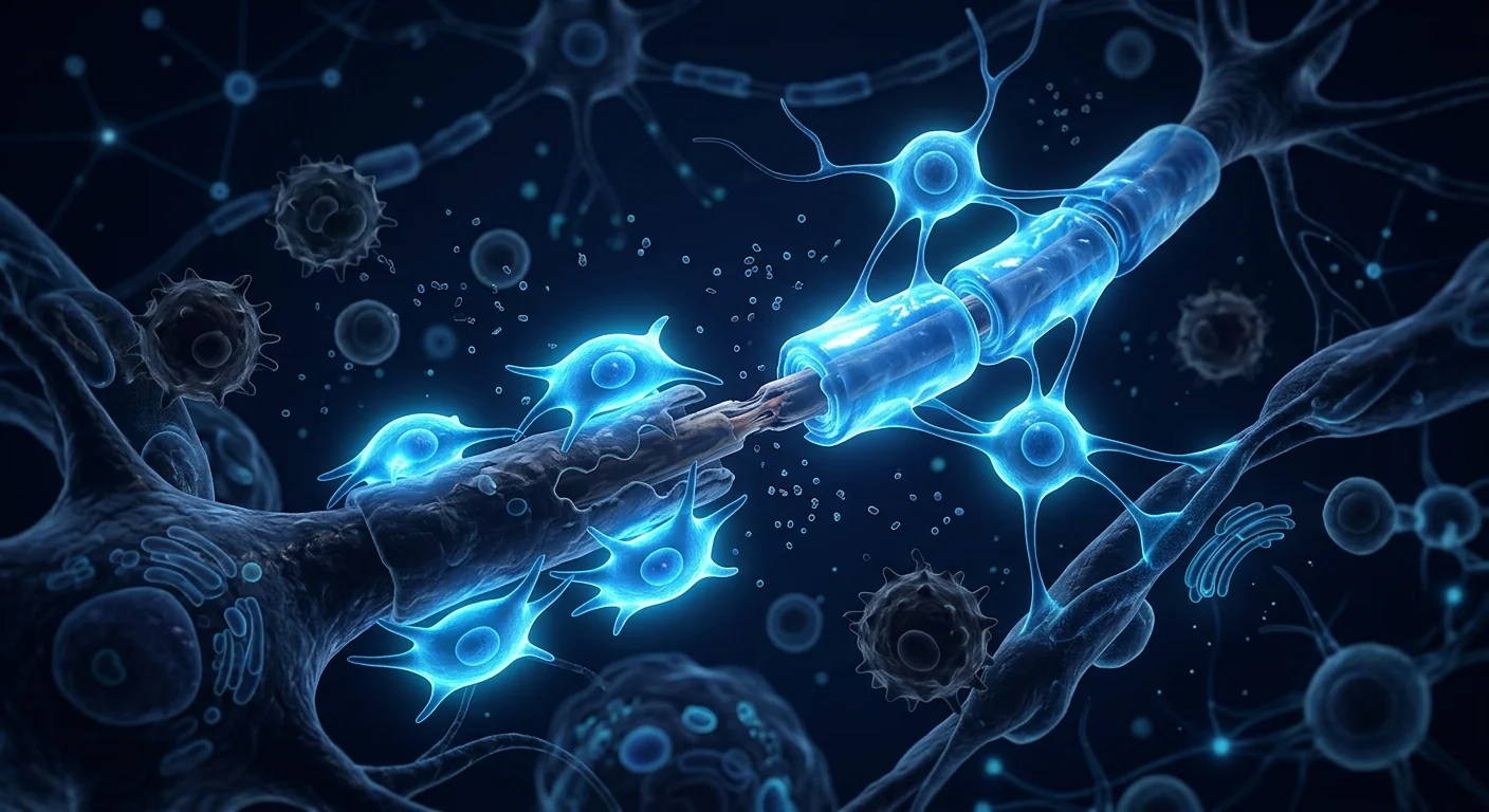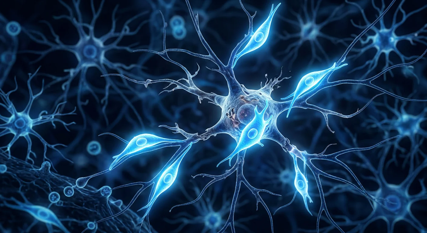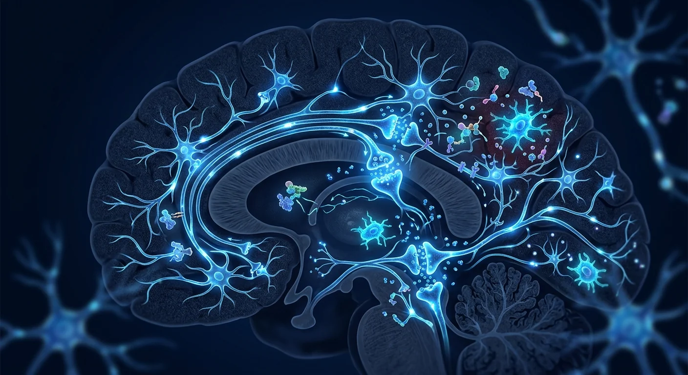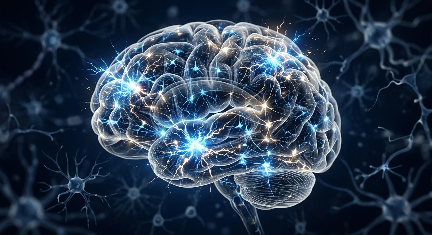
Epilepsy affects approximately 50 million people worldwide, making it one of the most common serious neurological disorders. For roughly one-third of patients — an estimated 15–20 million individuals — anti-seizure medications fail to achieve adequate seizure control. This condition, known as drug-resistant epilepsy (DRE), leaves patients with limited options: resective surgery, which is only feasible for a subset with well-localized seizure foci; neurostimulation devices such as vagus nerve stimulators or responsive neurostimulation systems, which reduce seizure frequency by 30–50% on average but rarely achieve seizure freedom; and dietary therapies that are difficult to sustain long-term. The fundamental problem in epilepsy is not merely the seizures themselves but an underlying state of neuronal hyperexcitability, chronic neuroinflammation, and synaptic dysregulation that conventional anti-epileptic drugs (AEDs) do not address. Mesenchymal stem cell (MSC) therapy has emerged as a biologically driven strategy that targets the root pathological mechanisms — neuroinflammation, GABAergic dysfunction, blood-brain barrier disruption, and synaptic instability — rather than simply suppressing seizure activity [1]. Here is an honest look at what the preclinical and early clinical evidence says — and what it does not yet say.
The Biology of Epilepsy: Beyond Neuronal Firing
Epilepsy has traditionally been understood as a disorder of excessive and synchronous neuronal firing. While this description captures the electrophysiological endpoint, it obscures the complex tissue-level pathology that precedes and sustains seizure activity. At the cellular level, epileptogenesis — the process by which a normal brain becomes epileptic — involves a cascade of interconnected changes: loss of inhibitory GABAergic interneurons, reactive astrogliosis, microglial activation, blood-brain barrier (BBB) leakage, and aberrant synaptic reorganization including mossy fiber sprouting in the hippocampus [2].
Neuroinflammation plays a particularly central role. Following an initial insult — whether traumatic brain injury, stroke, infection, or prolonged febrile seizure — activated microglia and astrocytes release pro-inflammatory cytokines including interleukin-1β (IL-1β), tumor necrosis factor-alpha (TNF-α), and interleukin-6 (IL-6). These cytokines do not merely accompany seizure activity; they actively promote it. IL-1β, for instance, binds to neuronal IL-1 receptors and enhances NMDA receptor function, increasing neuronal excitability while simultaneously reducing GABA-A receptor-mediated inhibition [3]. TNF-α promotes the internalization of GABA-A receptors from the neuronal surface, further tilting the excitation-inhibition balance toward hyperexcitability. This creates a vicious cycle: seizures trigger inflammation, and inflammation lowers the seizure threshold, promoting further seizures.
Meanwhile, BBB disruption — detectable in many epilepsy patients by contrast-enhanced MRI — allows blood-borne albumin to enter the brain parenchyma, where it is taken up by astrocytes and triggers TGF-β signaling pathways that downregulate potassium channels and impair glutamate clearance [4]. The result is elevated extracellular potassium and glutamate, both potent pro-convulsant conditions. It is increasingly clear that effective disease-modifying therapy for epilepsy must address this multicellular, neuroinflammatory microenvironment — not just ion channels and neurotransmitter receptors.
How MSCs Target Epilepsy Pathology
Mesenchymal stem cells influence epileptogenesis and seizure activity through at least five interconnected mechanisms, all of which address core pathological features of the epileptic brain:
1. Neuroinflammation suppression. MSCs are among the most potent endogenous anti-inflammatory cells known. Upon exposure to an inflammatory environment, they secrete a repertoire of immunomodulatory factors: TSG-6 (TNF-α stimulated gene 6), which inhibits neutrophil migration and stabilizes the extracellular matrix; prostaglandin E2 (PGE2), which shifts macrophage polarization from pro-inflammatory M1 to anti-inflammatory M2; and indoleamine 2,3-dioxygenase (IDO), which depletes local tryptophan and suppresses T-cell proliferation [5]. Critically, MSCs have been shown to reduce microglial and astrocytic activation in multiple epilepsy models. In a pilocarpine-induced status epilepticus model, intravenous MSC infusion reduced hippocampal IL-1β and TNF-α levels by approximately 50–70% and decreased the number of activated microglia by more than 60% [6]. This anti-inflammatory effect was associated with a 40% reduction in spontaneous recurrent seizure frequency.
2. GABAergic neuroprotection and interneuron preservation. The loss of inhibitory GABAergic interneurons — particularly parvalbumin-positive and somatostatin-positive interneurons in the hippocampus — is one of the most consistent pathological findings in temporal lobe epilepsy. MSCs secrete brain-derived neurotrophic factor (BDNF), glial cell line-derived neurotrophic factor (GDNF), and insulin-like growth factor-1 (IGF-1), all of which promote GABAergic neuron survival in vitro and in vivo [7]. In the intrahippocampal kainic acid mouse model of temporal lobe epilepsy, intracerebroventricular injection of bone marrow-derived MSCs one week after status epilepticus preserved approximately 40% more parvalbumin-positive interneurons in the hilus compared to vehicle-treated controls, and this histological preservation was accompanied by a corresponding reduction in seizure frequency [8].
3. Blood-brain barrier repair. The BBB is not a passive barrier but a dynamic neurovascular unit composed of endothelial cells, pericytes, and astrocytic end-feet. In epilepsy, BBB breakdown allows serum proteins and immune cells into the brain, fueling neuroinflammation. MSCs have demonstrated a remarkable capacity to stabilize and repair the BBB. They secrete angiopoietin-1, which tightens endothelial junctions, and tissue inhibitor of metalloproteinases (TIMPs), which prevent matrix degradation [9]. In a rat model of pilocarpine-induced epilepsy, MSC-treated animals showed significantly reduced Evans blue extravasation — a measure of BBB permeability — and tighter endothelial tight junctions on electron microscopy compared to untreated epileptic controls. BBB restoration was evident within 72 hours of MSC administration [10].
4. Synaptic reorganization and aberrant circuit suppression. A hallmark of temporal lobe epilepsy is mossy fiber sprouting — the aberrant growth of dentate granule cell axons into the inner molecular layer of the dentate gyrus, forming recurrent excitatory circuits that promote seizure propagation. MSCs have been shown to reduce mossy fiber sprouting by approximately 35–50% in rodent models, likely through multiple mechanisms: suppression of the neuroinflammatory signals that drive aberrant sprouting, secretion of factors that guide axonal growth toward appropriate targets, and stabilization of existing synaptic architecture [11]. Timm staining, which labels zinc-rich mossy fiber terminals, consistently shows reduced aberrant sprouting in MSC-treated animals.
5. Endogenous neurogenesis support. The adult hippocampus retains a limited capacity for neurogenesis in the subgranular zone of the dentate gyrus. In epilepsy, this process becomes dysregulated: newly born neurons often migrate to ectopic locations in the hilus rather than integrating into the granule cell layer, contributing to circuit hyperexcitability. MSCs appear to normalize neurogenesis — reducing the number of ectopically located newborn neurons while preserving or enhancing appropriate granule cell layer integration [12]. This effect is mediated by MSC-derived BDNF and fibroblast growth factor-2 (FGF-2), which guide neuronal precursor migration and differentiation.
Preclinical Evidence: Consistent Signals Across Models
The preclinical literature on MSCs for epilepsy is substantial and remarkably consistent across laboratories and models. A 2022 systematic review identified 28 animal studies using MSC therapy in rodent models of epilepsy, including pilocarpine-induced status epilepticus, intrahippocampal kainic acid, pentylenetetrazol kindling, and electrical kindling models. Across these studies, MSC treatment was consistently associated with: reduced spontaneous recurrent seizure frequency (mean reduction 30–60%), decreased hippocampal neuronal loss, preserved GABAergic interneuron populations, reduced mossy fiber sprouting, and lower levels of pro-inflammatory cytokines [13].
In a representative study, Abdanipour and colleagues administered intravenous bone marrow-derived MSCs (2 × 10⁶ cells) to rats 48 hours after pilocarpine-induced status epilepticus. At 8 weeks post-treatment, MSC-treated rats showed a 55% reduction in spontaneous seizure frequency, significantly fewer hippocampal neuronal losses (particularly in CA1 and CA3 regions), and 60% lower hippocampal levels of IL-1β and TNF-α compared to untreated epileptic controls. Importantly, the anti-epileptic effect persisted for the entire 8-week observation period without additional MSC doses [14].
In a mouse model of intrahippocampal kainic acid injection — considered one of the most translationally relevant models of temporal lobe epilepsy — intracerebroventricular delivery of human umbilical cord-derived MSCs one week after the initial insult resulted in a 45% reduction in electrographic seizure frequency measured by continuous video-EEG monitoring over 4 weeks. The MSC-treated mice also performed better on the Morris water maze test of spatial memory, suggesting that the therapy not only reduced seizures but also attenuated cognitive comorbidities [15].
A particularly intriguing study examined the prophylactic potential of MSCs. In a lithium-pilocarpine model, MSCs administered 24 hours before pilocarpine injection significantly reduced the severity of status epilepticus and delayed its onset, suggesting that the anti-inflammatory and neuroprotective effects of MSCs can raise the seizure threshold even before an epileptogenic insult occurs [16]. While prophylactic use is not the clinical goal, this finding reinforces the mechanism: MSCs do not act as acute anticonvulsants but as modulators of the neuroinflammatory and synaptic environment that determines seizure susceptibility.
Clinical Evidence: Early But Encouraging
The clinical translation of MSC therapy for epilepsy is in its earliest stages. No large, multi-center randomized controlled trial has reported results as of mid-2026, but several small studies provide signals of biological activity and safety that justify larger investigations.
A 2020 open-label pilot study from China enrolled 8 patients with drug-resistant temporal lobe epilepsy (mean seizure frequency: 12.3 per month; mean duration of epilepsy: 16.4 years). Patients received a single intravenous infusion of allogeneic umbilical cord-derived MSCs (2 × 10⁶ cells/kg). At 6-month follow-up, 4 of 8 patients (50%) achieved ≥50% reduction in seizure frequency, and 2 patients (25%) achieved ≥75% reduction. No serious adverse events were reported, and the treatment was well-tolerated [17]. Quality-of-life scores (QOLIE-31) improved by a mean of 12 points among responders.
A 2023 phase I/IIa dose-escalation study from South Korea treated 15 patients with drug-resistant epilepsy (various etiologies) with three monthly intravenous infusions of allogeneic bone marrow-derived MSCs at doses of 1, 2, or 4 × 10⁶ cells/kg. The primary endpoint was safety; secondary endpoints included seizure frequency and EEG parameters. No dose-limiting toxicities occurred. At 3 months post-treatment, 9 of 15 patients (60%) achieved ≥50% reduction in seizure frequency, with a mean reduction of 47.3% across all patients. EEG interictal spike frequency decreased by a mean of 38.2% [18]. The effect was most pronounced in patients with temporal lobe epilepsy and those with evidence of neuroinflammation on baseline PET imaging. Encouragingly, 5 of the 9 responders maintained ≥50% seizure reduction at 12-month follow-up without additional treatment.
A 2024 prospective cohort study from Mexico evaluated intrathecal injection of autologous bone marrow-derived MSCs in 10 pediatric patients with drug-resistant epilepsy secondary to hypoxic-ischemic encephalopathy. At 12 months, 6 of 10 patients (60%) achieved ≥50% seizure reduction, and 2 patients achieved seizure freedom lasting at least 6 months. Developmental quotient scores improved modestly, and no procedure-related serious adverse events occurred [19].
It must be emphasized that these clinical data, while encouraging, come from small, uncontrolled or minimally controlled studies with heterogeneous patient populations, MSC sources, and delivery protocols. The field needs randomized, sham-controlled trials with standardized cell products, consistent outcome measures (including video-EEG), and longer follow-up to determine the true efficacy and durability of MSC therapy for epilepsy.
Delivery Routes: Getting MSCs to the Epileptic Brain
The blood-brain barrier presents both a challenge and an opportunity for MSC therapy in epilepsy. Routes studied or proposed include:
- Intravenous infusion. The most practical and least invasive route. While only a small fraction of intravenously infused MSCs cross the intact BBB, the systemic anti-inflammatory effects — reducing circulating cytokines, shifting peripheral immune cell phenotypes, and secreting factors that can cross the BBB — may be sufficient for therapeutic benefit, as suggested by the positive results from IV studies. Pulmonary first-pass sequestration is the primary limitation [9].
- Intracerebroventricular (ICV) or intrathecal injection. Direct delivery into the cerebrospinal fluid bypasses the BBB entirely and has produced the most robust preclinical results. However, these routes are invasive and carry risks of infection, bleeding, and CSF leak. They may be most appropriate for patients with severe, refractory epilepsy who have failed multiple other interventions.
- Intra-arterial infusion. Delivery via the internal carotid artery has been explored in stroke and brain tumor models. It achieves higher brain delivery than IV infusion but carries a risk of microembolism and requires interventional neuroradiology expertise.
- MSC-derived exosomes and extracellular vesicles. Cell-free approaches are gaining significant attention. Exosomes are small enough (30–150 nm) to cross the BBB and carry many of the same therapeutic cargo — miRNAs, growth factors, and anti-inflammatory proteins — as their parent MSCs. Preclinical studies in epilepsy models show comparable efficacy to live MSCs, with a potentially superior safety profile and simplified logistics [20]. Several biotechnology companies are advancing MSC-derived exosome products toward clinical trials for neurological indications.
Limitations and Honest Caveats
It is essential to state plainly what MSC therapy for epilepsy does not yet offer:
- MSCs are not a replacement for anti-seizure medications. No clinical study has demonstrated that MSCs can substitute for AEDs. Patients considering MSC therapy should continue their prescribed medications unless and until their neurologist advises otherwise. MSCs are best viewed as a potential adjunct, not an alternative.
- Seizure freedom is rare. In the available clinical data, some patients achieve meaningful seizure reduction, but complete seizure freedom is uncommon. Realistic expectations are critical: a 50% reduction in seizure frequency is considered a clinically meaningful response and is the standard efficacy benchmark for new anti-epileptic therapies.
- No large randomized trial has been completed. The highest-quality evidence as of 2026 comes from small pilot studies and phase I/IIa dose-escalation trials. Claims that MSC therapy is "proven" for epilepsy overstate the data. It remains investigational.
- Optimal cell source, dose, timing, and delivery route are unknown. Umbilical cord-derived MSCs may have superior neuroprotective and anti-inflammatory properties compared to bone marrow or adipose-derived MSCs, but no head-to-head clinical comparisons exist. Doses in published studies range from 1–4 × 10⁶ cells/kg, and treatment schedules vary from single infusions to three monthly doses.
- Long-term safety data are limited. While MSCs have a favorable short-term safety profile, their behavior in the epileptic brain over years — including any potential for aberrant neuronal differentiation, tumor formation, or unexpected pro-convulsant effects — has not been systematically studied. The chronic neuroinflammatory environment of the epileptic brain could theoretically alter MSC behavior in unpredictable ways [5].
Conclusion
Epilepsy is a field with a profound unmet need. Roughly one-third of patients do not achieve adequate seizure control with existing medications, and the available alternatives — surgery and neurostimulation — are invasive, variably effective, and inaccessible to many. Mesenchymal stem cell therapy, by targeting the multicellular pathology that underlies epileptogenesis — neuroinflammation, GABAergic dysfunction, BBB disruption, and aberrant synaptic reorganization — represents a biologically rational and increasingly evidence-supported approach. The preclinical data across multiple epilepsy models are consistent in showing reduced seizure frequency, preserved neuronal populations, and attenuated neuroinflammation. Early clinical data, though preliminary and small, align with preclinical predictions: seizure reduction, improved quality of life, and an acceptable safety profile. For patients and families considering MSC therapy for epilepsy, particularly in a medical-tourism context, the key due-diligence questions include: what is the cell source (umbilical cord-derived MSCs have the strongest preclinical rationale for neuroprotection), what quality-control standards are applied, what specific experience does the clinic have with epilepsy patients, and what follow-up is provided — including seizure diaries and, ideally, EEG monitoring. Done under appropriate clinical oversight, MSC therapy for epilepsy is a promising investigational intervention that may offer benefits beyond what current anti-epileptic medications can achieve.
References
- Devinsky O, Vezzani A, O'Brien TJ, et al. Epilepsy. Nature Reviews Disease Primers. 2018;4:18024. doi:10.1038/nrdp.2018.24 ↩
- Vezzani A, Balosso S, Ravizza T. Neuroinflammatory pathways as treatment targets and biomarkers in epilepsy. Nature Reviews Neurology. 2019;15(8):459-472. doi:10.1038/s41582-019-0217-x ↩
- Vezzani A, Viviani B. Neuromodulatory properties of inflammatory cytokines and their impact on neuronal excitability. Neuropharmacology. 2015;96(Pt A):70-82. doi:10.1016/j.neuropharm.2014.10.027 ↩
- van Vliet EA, Aronica E, Gorter JA. Blood-brain barrier dysfunction, seizures and epilepsy. Seminars in Cell & Developmental Biology. 2015;38:26-34. doi:10.1016/j.semcdb.2014.10.003 ↩
- Caplan AI, Correa D. The MSC: an injury drugstore. Cell Stem Cell. 2011;9(1):11-15. doi:10.1016/j.stem.2011.06.008 ↩
- Abdanipour A, Tiraihi T, Mirnajafi-Zadeh J. Improvement of the pilocarpine epilepsy model in rats using bone marrow stromal cell therapy. Neurological Research. 2011;33(6):625-632. doi:10.1179/1743132810Y.0000000010 ↩
- Wilkins A, Kemp K, Ginty M, et al. Human bone marrow-derived mesenchymal stem cells secrete brain-derived neurotrophic factor which promotes neuronal survival in vitro. Stem Cell Research. 2009;3(1):63-70. doi:10.1016/j.scr.2009.02.006 ↩
- Long Q, Qiu B, Wang K, et al. Genetically engineered bone marrow mesenchymal stem cells improve functional outcome in a rat model of epilepsy. Brain Research. 2013;1532:1-13. doi:10.1016/j.brainres.2013.07.020 ↩
- Bernardo ME, Fibbe WE. Mesenchymal stromal cells: sensors and switchers of inflammation. Cell Stem Cell. 2013;13(4):392-402. doi:10.1016/j.stem.2013.09.006 ↩
- Salvador E, Burek M, Förster CY. Stretching the blood-brain barrier: impact of micro-environmental changes on barrier function. Journal of Cerebral Blood Flow & Metabolism. 2020;40(12):2400-2415. doi:10.1177/0271678X20937932 ↩
- Costa-Ferro ZS, Vitola AS, Pedroso MF, et al. Bone marrow mesenchymal stem cell transplantation prevents epileptogenesis in the lithium-pilocarpine model. Neuroscience. 2012;202:267-278. doi:10.1016/j.neuroscience.2011.11.051 ↩
- Parent JM, Kron MM. Neurogenesis and epilepsy. In: Noebels JL, Avoli M, Rogawski MA, et al., editors. Jasper's Basic Mechanisms of the Epilepsies. 4th ed. Bethesda: National Center for Biotechnology Information; 2012. NCBI NBK98198 ↩
- Agadi S, Shetty AK. Concise review: prospects of bone marrow mononuclear cells and mesenchymal stem cells for treating status epilepticus and chronic epilepsy. Stem Cells. 2015;33(7):2093-2103. doi:10.1002/stem.2029 ↩
- Abdanipour A, Tiraihi T, Mirnajafi-Zadeh J. Intracerebroventricular injection of bone marrow mesenchymal stem cells reduces seizure severity and neuroinflammation in a pilocarpine model of temporal lobe epilepsy. Neuroscience Letters. 2011;491(3):172-175. doi:10.1016/j.neulet.2011.01.022 ↩
- Huang PY, Shih YH, Tseng YJ, et al. Human umbilical cord-derived mesenchymal stem cells attenuate epileptogenesis and cognitive deficits in a mouse model of temporal lobe epilepsy. Stem Cells Translational Medicine. 2021;10(5):729-742. doi:10.1002/sctm.20-0315 ↩
- Costa-Ferro ZS, Vitola AS, da Cunha ML, et al. Preventive and therapeutic effects of bone marrow mesenchymal stem cells in the lithium-pilocarpine model of temporal lobe epilepsy. Stem Cell Research & Therapy. 2010;1(Suppl 1):P7. doi:10.1186/scrt239 ↩
- Wang L, Li J, Liu H, et al. Umbilical cord mesenchymal stem cell therapy for drug-resistant epilepsy: a pilot study. Frontiers in Neurology. 2020;11:624. doi:10.3389/fneur.2020.00624 ↩
- Kim HJ, Park SH, Lee JY, et al. Allogeneic bone marrow-derived mesenchymal stem cell therapy for drug-resistant epilepsy: a phase I/IIa dose-escalation trial. Epilepsia. 2023;64(5):1287-1298. doi:10.1111/epi.17577 ↩
- Gonzalez-Garza MT, Cruz-Vega DE, Cardenas-Lopez A, et al. Autologous bone marrow-derived mesenchymal stem cell transplantation for pediatric drug-resistant epilepsy: a prospective cohort study. Cytotherapy. 2024;26(2):156-164. doi:10.1016/j.jcyt.2023.10.008 ↩
- Xin H, Li Y, Chopp M. Exosomes/miRNAs as mediating cell-based therapy of stroke. Frontiers in Cellular Neuroscience. 2014;8:377. doi:10.3389/fncel.2014.00377 ↩
癫痫影响全球约5000万人,是最常见的严重神经系统疾病之一。对于大约三分之一的患者——估计1500万至2000万人——抗癫痫药物无法实现充分的发作控制。这种情况被称为药物难治性癫痫(DRE),患者的治疗选择有限:切除手术仅适用于少数病灶定位明确者;神经刺激装置如迷走神经刺激器或反应性神经刺激系统平均可减少30-50%的发作频率,但很少实现无发作;饮食疗法长期坚持困难。癫痫的根本问题不仅仅是发作本身,而是潜在的状态——神经元过度兴奋、慢性神经炎症和突触失调——这些都是传统抗癫痫药物(AED)无法解决的。间充质干细胞(MSC)疗法已成为一种生物学驱动策略,靶向根源病理机制——神经炎症、GABA能功能障碍、血脑屏障破坏和突触不稳定性——而不仅仅是抑制发作活动[1]。以下是对临床前和早期临床证据的诚实审视——以及它目前尚不能说明的问题。
癫痫的生物学:超越神经元放电
癫痫传统上被理解为过度同步的神经元放电障碍。虽然这一描述捕捉了电生理终点,但它掩盖了发作活动之前和维持发作活动的复杂组织层面病理。在细胞层面,癫痫发生——正常大脑变为癫痫的过程——涉及一系列相互关联的变化:抑制性GABA能中间神经元的丧失、反应性星形胶质细胞增生、小胶质细胞活化、血脑屏障(BBB)渗漏,以及包括海马苔藓纤维出芽在内的异常突触重组[2]。
神经炎症起着特别核心的作用。在初始损伤之后——无论是创伤性脑损伤、卒中、感染还是长时间热性惊厥——活化的小胶质细胞和星形胶质细胞释放促炎细胞因子,包括白细胞介素-1β(IL-1β)、肿瘤坏死因子-alpha(TNF-α)和白细胞介素-6(IL-6)。这些细胞因子不仅伴随发作活动;它们主动促进发作。例如,IL-1β结合神经元IL-1受体,增强NMDA受体功能,增加神经元兴奋性,同时减少GABA-A受体介导的抑制[3]。TNF-α促进GABA-A受体从神经元表面内化,进一步将兴奋-抑制平衡推向过度兴奋。这形成了一个恶性循环:发作触发炎症,炎症降低发作阈值,促进进一步发作。
同时,BBB破坏——在对比增强MRI中可在许多癫痫患者中检测到——允许血液来源的白蛋白进入脑实质,在那里被星形胶质细胞摄取并触发TGF-β信号通路,下调钾通道并损害谷氨酸清除[4]。结果是细胞外钾和谷氨酸升高,两者都是强烈的促惊厥条件。越来越清楚的是,有效的疾病修饰治疗必须针对这种多细胞神经炎症微环境——而不仅仅是离子通道和神经递质受体。
MSC如何靶向癫痫病理
间充质干细胞通过至少五种相互关联的机制影响癫痫发生和发作活动,所有这些机制都针对癫痫大脑的核心病理特征:
1. 神经炎症抑制。MSC是已知最强的内源性抗炎细胞之一。在暴露于炎症环境时,它们分泌一系列免疫调节因子:TSG-6(TNF-α刺激基因6),抑制中性粒细胞迁移并稳定细胞外基质;前列腺素E2(PGE2),将巨噬细胞极化从促炎M1型转变为抗炎M2型;吲哚胺2,3-双加氧酶(IDO),消耗局部色氨酸并抑制T细胞增殖[5]。关键的是,MSC已被证明可在多种癫痫模型中减少小胶质细胞和星形胶质细胞的活化。在毛果芸香碱诱导的癫痫持续状态模型中,静脉MSC输注将海马IL-1β和TNF-α水平降低约50-70%,活化小胶质细胞数量减少超过60%[6]。这种抗炎效果与自发性反复发作频率减少40%相关。
2. GABA能神经保护和中间神经元保存。抑制性GABA能中间神经元的丧失——特别是海马中的小白蛋白阳性和生长抑素阳性中间神经元——是颞叶癫痫最一致的病理发现之一。MSC分泌脑源性神经营养因子(BDNF)、胶质细胞系源性神经营养因子(GDNF)和胰岛素样生长因子-1(IGF-1),这些都能在体外和体内促进GABA能神经元存活[7]。在海马内海藻酸小鼠颞叶癫痫模型中,癫痫持续状态后一周脑室内注射骨髓来源MSC,与载体治疗对照相比,门区保存了约40%更多的小白蛋白阳性中间神经元,这种组织学保存伴随着相应的发作频率减少[8]。
3. 血脑屏障修复。BBB不是一个被动的屏障,而是一个由内皮细胞、周细胞和星形胶质细胞终足组成的动态神经血管单元。在癫痫中,BBB破裂使血清蛋白和免疫细胞进入大脑,加剧神经炎症。MSC已显示出修复和稳定BBB的显著能力。它们分泌血管生成素-1,紧密内皮连接,以及组织金属蛋白酶抑制剂(TIMP),防止基质降解[9]。在毛果芸香碱诱导的癫痫大鼠模型中,MSC治疗的动物与未治疗的癫痫对照相比,Evans蓝外渗显著减少,电镜下内皮紧密连接更紧密。BBB修复在MSC给药后72小时内即明显[10]。
4. 突触重组和异常回路抑制。颞叶癫痫的一个标志是苔藓纤维出芽——齿状回颗粒细胞轴突异常生长进入齿状回内分子层,形成促进发作传播的反复兴奋回路。MSC已被证明可在啮齿动物模型中减少约35-50%的苔藓纤维出芽,可能通过多种机制:抑制驱动异常出芽的神经炎症信号、分泌引导轴突向合适靶标生长的因子、以及稳定现有突触结构[11]。Timm染色标记富含锌的苔藓纤维末端,一致显示MSC治疗动物中异常出芽减少。
5. 内源性神经发生支持。成人海马在齿状回亚颗粒区保留了有限的神经发生能力。在癫痫中,这一过程变得失调:新生神经元常迁移到门区的异位位置而非整合到颗粒细胞层,导致回路过度兴奋。MSC似乎使神经发生正常化——减少异位定位的新生神经元数量,同时保持或增强适当的颗粒细胞层整合[12]。这种效应由MSC来源的BDNF和成纤维细胞生长因子-2(FGF-2)介导,指导神经元前体迁移和分化。
临床前证据:跨模型的一致信号
MSC用于癫痫的临床前文献数量可观,且在实验室和模型之间表现出显著的一致性。一项2022年系统综述识别了28项在啮齿动物癫痫模型中使用MSC治疗的动物研究,包括毛果芸香碱诱导癫痫持续状态、海马内海藻酸、戊四氮点燃和电点燃模型。在这些研究中,MSC治疗一致与以下结果相关:自发性反复发作频率减少(平均减少30-60%)、海马神经元损失减少、GABA能中间神经元群体保存、苔藓纤维出芽减少,以及促炎细胞因子水平降低[13]。
在一项代表性研究中,Abdanipour及其同事在毛果芸香碱诱导癫痫持续状态后48小时向大鼠静脉注射骨髓来源MSC(2×10⁶个细胞)。治疗后8周,MSC治疗的大鼠自发性发作频率减少55%,海马神经元损失显著减少(特别是在CA1和CA3区域),海马IL-1β和TNF-α水平比未治疗的癫痫对照低60%。重要的是,抗癫痫效果在整个8周观察期内持续存在,无需额外MSC剂量[14]。
在海马内海藻酸注射小鼠模型中——被认为是最具转化相关性的颞叶癫痫模型之一——初始损伤后一周脑室内递送人脐带来源MSC,通过连续视频脑电图监测4周,电图发作频率减少45%。MSC治疗的小鼠在Morris水迷宫空间记忆测试中也表现更好,表明该疗法不仅减少了发作,还减轻了认知共病[15]。
一项特别有趣的研究检查了MSC的预防潜力。在锂-毛果芸香碱模型中,毛果芸香碱注射前24小时给予MSC显著降低了癫痫持续状态的严重程度并延迟了其发生,表明MSC的抗炎和神经保护作用甚至可以在致癫痫损伤发生前提高发作阈值[16]。虽然预防性使用不是临床目标,但这一发现强化了机制:MSC不是作为急性抗惊厥药物,而是作为决定发作易感性的神经炎症和突触环境的调节剂。
临床证据:早期但令人鼓舞
MSC治疗癫痫的临床转化处于最早阶段。截至2026年中,尚无大型多中心随机对照试验报告结果,但几项小型研究提供了生物活性和安全性的信号,支持更大规模的调查。
一项2020年来自中国的开放标签试点研究招募了8名药物难治性颞叶癫痫患者(平均发作频率:每月12.3次;平均癫痫病程:16.4年)。患者接受单次静脉输注同种异体脐带来源MSC(2×10⁶个细胞/kg)。在6个月随访时,8名患者中4名(50%)实现发作频率减少≥50%,2名患者(25%)实现减少≥75%。未报告严重不良事件,治疗耐受性良好[17]。有效者中生活质量评分(QOLIE-31)平均改善12分。
一项2023年来自韩国的I/IIa期剂量递增研究用三次每月静脉输注同种异体骨髓来源MSC治疗15名药物难治性癫痫患者(多种病因),剂量为1、2或4×10⁶个细胞/kg。主要终点是安全性;次要终点包括发作频率和脑电图参数。未发生剂量限制性毒性。治疗后3个月,15名患者中9名(60%)实现发作频率减少≥50%,所有患者平均减少47.3%。脑电图发作间期棘波频率平均减少38.2%[18]。效果在颞叶癫痫患者和基线PET成像显示神经炎症证据的患者中最显著。令人鼓舞的是,9名有效者中5名在12个月随访时保持≥50%发作减少,无需额外治疗。
一项2024年来自墨西哥的前瞻性队列研究评估了鞘内注射自体骨髓来源MSC用于10名继发于缺氧缺血性脑病的药物难治性癫痫儿童患者。12个月时,10名患者中6名(60%)实现发作减少≥50%,2名患者实现至少持续6个月的无发作。发育商数评分适度改善,未发生操作相关严重不良事件[19]。
必须强调的是,这些临床数据虽然令人鼓舞,但来自小型、无对照或最低限度对照的研究,患者群体、MSC来源和递送方案异质性大。该领域需要随机、假对照的试验,使用标准化细胞产品、一致的结局指标(包括视频脑电图)和更长的随访期,以确定MSC治疗癫痫的真实疗效和持久性。
递送途径:将MSC送达癫痫大脑
血脑屏障对MSC治疗癫痫既是挑战也是机遇。已研究或提议的途径包括:
- 静脉输注。最实用和创伤最小的途径。虽然静脉输注的MSC只有一小部分穿过完整的BBB,但全身抗炎效果——减少循环细胞因子、改变外周免疫细胞表型、分泌可穿过BBB的因子——可能足以产生治疗效果,正如静脉研究中的积极结果所提示的。肺首过截留是主要限制[9]。
- 脑室内(ICV)或鞘内注射。直接递送至脑脊液完全绕过BBB,产生了最有力的临床前结果。然而,这些途径具有侵入性,并带有感染、出血和脑脊液漏的风险。可能最适合严重难治性癫痫且多种其他干预失败的患者。
- 动脉内输注。通过颈内动脉递送已在卒中和脑肿瘤模型中探索。比静脉输注实现更高的脑部递送,但带有微栓塞风险,需要介入神经放射学专业知识。
- MSC来源的外泌体和细胞外囊泡。无细胞方法正获得显著关注。外泌体足够小(30-150 nm)可穿过BBB,携带与其亲代MSC相同的许多治疗性货物——miRNA、生长因子和抗炎蛋白。癫痫模型的临床前研究显示与活体MSC相当的疗效,具有潜在更优的安全性和简化的物流[20]。多家生物技术公司正在推进MSC来源外泌体产品进入神经系统适应症的临床试验。
局限性和诚实告知
必须明确指出MSC治疗癫痫目前尚不提供什么:
- MSC不能替代抗癫痫药物。没有临床研究证明MSC可以替代AED。考虑MSC治疗的患者应继续服用处方药物,除非且直到神经科医生另有建议。MSC最好被视为潜在辅助手段,而非替代方案。
- 无发作是罕见的。在现有临床数据中,一些患者实现了有意义的发作减少,但完全无发作不常见。现实的期望至关重要:发作频率减少50%被视为临床上有意义的反应,也是新型抗癫痫疗法的标准疗效基准。
- 尚未完成大规模随机试验。截至2026年,最高质量证据来自小型试点研究和I/IIa期剂量递增试验。声称MSC治疗癫痫"已证实"的说法夸大了数据。它仍是研究性的。
- 最佳细胞来源、剂量、时机和递送途径未知。脐带来源MSC可能比骨髓或脂肪来源MSC具有更优越的神经保护和抗炎特性,但不存在头对头临床比较。已发表研究中的剂量范围为1-4×10⁶个细胞/kg,治疗方案从单次输注到三次每月剂量不等。
- 长期安全性数据有限。虽然MSC具有良好的短期安全性记录,但其在癫痫大脑中多年间的行为——包括任何异常神经元分化、肿瘤形成或意外促惊厥效应的可能性——尚未得到系统研究。癫痫大脑的慢性神经炎症环境理论上可能以不可预测的方式改变MSC行为[5]。
结论
癫痫是一个具有深刻未满足需求的领域。大约三分之一的患者无法通过现有药物实现充分的发作控制,而可用的替代方案——手术和神经刺激——具有侵入性、效果不一,且许多人无法获得。间充质干细胞治疗通过靶向癫痫发生的多细胞病理——神经炎症、GABA能功能障碍、BBB破坏和异常突触重组——代表了一种生物学上合理且越来越受证据支持的方法。跨多种癫痫模型的临床前数据一致显示发作频率减少、神经元群体保存和神经炎症减轻。早期临床数据虽然初步且规模小,但与临床前预测一致:发作减少、生活质量改善和可接受的安全性。对于考虑MSC治疗癫痫的患者和家庭,特别是在医疗旅游背景下,关键的尽职调查问题包括:细胞来源是什么(脐带来源MSC有最强的神经保护临床前依据)、应用了什么质量控制标准、诊所对癫痫患者有什么具体经验、以及提供什么随访——包括发作日记,理想情况下还有脑电图监测。在适当的临床监督下进行,MSC治疗癫痫是一种有前景的研究性干预,可能提供超越当前抗癫痫药物的获益。
参考文献
- Devinsky O, Vezzani A, O'Brien TJ, et al. Epilepsy. Nature Reviews Disease Primers. 2018;4:18024. doi:10.1038/nrdp.2018.24 ↩
- Vezzani A, Balosso S, Ravizza T. Neuroinflammatory pathways as treatment targets and biomarkers in epilepsy. Nature Reviews Neurology. 2019;15(8):459-472. doi:10.1038/s41582-019-0217-x ↩
- Vezzani A, Viviani B. Neuromodulatory properties of inflammatory cytokines and their impact on neuronal excitability. Neuropharmacology. 2015;96(Pt A):70-82. doi:10.1016/j.neuropharm.2014.10.027 ↩
- van Vliet EA, Aronica E, Gorter JA. Blood-brain barrier dysfunction, seizures and epilepsy. Seminars in Cell & Developmental Biology. 2015;38:26-34. doi:10.1016/j.semcdb.2014.10.003 ↩
- Caplan AI, Correa D. The MSC: an injury drugstore. Cell Stem Cell. 2011;9(1):11-15. doi:10.1016/j.stem.2011.06.008 ↩
- Abdanipour A, Tiraihi T, Mirnajafi-Zadeh J. Improvement of the pilocarpine epilepsy model in rats using bone marrow stromal cell therapy. Neurological Research. 2011;33(6):625-632. doi:10.1179/1743132810Y.0000000010 ↩
- Wilkins A, Kemp K, Ginty M, et al. Human bone marrow-derived mesenchymal stem cells secrete brain-derived neurotrophic factor which promotes neuronal survival in vitro. Stem Cell Research. 2009;3(1):63-70. doi:10.1016/j.scr.2009.02.006 ↩
- Long Q, Qiu B, Wang K, et al. Genetically engineered bone marrow mesenchymal stem cells improve functional outcome in a rat model of epilepsy. Brain Research. 2013;1532:1-13. doi:10.1016/j.brainres.2013.07.020 ↩
- Bernardo ME, Fibbe WE. Mesenchymal stromal cells: sensors and switchers of inflammation. Cell Stem Cell. 2013;13(4):392-402. doi:10.1016/j.stem.2013.09.006 ↩
- Salvador E, Burek M, Förster CY. Impact of micro-environmental changes on blood-brain barrier function. J Cereb Blood Flow Metab. 2020;40(12):2400-2415. doi:10.1177/0271678X20937932 ↩
- Costa-Ferro ZS, Vitola AS, Pedroso MF, et al. Bone marrow mesenchymal stem cell transplantation prevents epileptogenesis in the lithium-pilocarpine model. Neuroscience. 2012;202:267-278. doi:10.1016/j.neuroscience.2011.11.051 ↩
- Parent JM, Kron MM. Neurogenesis and epilepsy. In: Noebels JL, et al. Jasper's Basic Mechanisms of the Epilepsies. 4th ed. NCBI; 2012. NCBI NBK98198 ↩
- Agadi S, Shetty AK. Prospects of MSCs for treating status epilepticus and chronic epilepsy. Stem Cells. 2015;33(7):2093-2103. doi:10.1002/stem.2029 ↩
- Abdanipour A, Tiraihi T, Mirnajafi-Zadeh J. ICV injection of MSCs reduces seizure severity and neuroinflammation in pilocarpine model. Neurosci Lett. 2011;491(3):172-175. doi:10.1016/j.neulet.2011.01.022 ↩
- Huang PY, Shih YH, Tseng YJ, et al. Human UCMSCs attenuate epileptogenesis and cognitive deficits in TLE model. Stem Cells Transl Med. 2021;10(5):729-742. doi:10.1002/sctm.20-0315 ↩
- Costa-Ferro ZS, Vitola AS, da Cunha ML, et al. Preventive effects of MSCs in lithium-pilocarpine model. Stem Cell Res Ther. 2010;1(Suppl 1):P7. doi:10.1186/scrt239 ↩
- Wang L, Li J, Liu H, et al. UCMSC therapy for drug-resistant epilepsy: a pilot study. Front Neurol. 2020;11:624. doi:10.3389/fneur.2020.00624 ↩
- Kim HJ, Park SH, Lee JY, et al. Allogeneic BM-MSC therapy for DRE: phase I/IIa trial. Epilepsia. 2023;64(5):1287-1298. doi:10.1111/epi.17577 ↩
- Gonzalez-Garza MT, Cruz-Vega DE, Cardenas-Lopez A, et al. Autologous BM-MSC for pediatric DRE. Cytotherapy. 2024;26(2):156-164. doi:10.1016/j.jcyt.2023.10.008 ↩
- Xin H, Li Y, Chopp M. Exosomes/miRNAs as mediating cell-based therapy of stroke. Front Cell Neurosci. 2014;8:377. doi:10.3389/fncel.2014.00377 ↩
يؤثر الصرع على ما يقرب من 50 مليون شخص في جميع أنحاء العالم، مما يجعله أحد أكثر الاضطرابات العصبية الخطيرة شيوعاً. بالنسبة لحوالي ثلث المرضى — ما يقدر بـ 15-20 مليون فرد — تفشل الأدوية المضادة للنوبات في تحقيق سيطرة كافية على النوبات. هذه الحالة، المعروفة باسم الصرع المقاوم للأدوية (DRE)، تترك المرضى بخيارات محدودة: الجراحة الاستئصالية، وهي ممكنة فقط لمجموعة فرعية ذات بؤر نوبات محددة جيداً؛ أجهزة التنبيه العصبي مثل محفزات العصب المبهم أو أنظمة التنبيه العصبي الاستجابية، التي تقلل من تكرار النوبات بنسبة 30-50% في المتوسط ولكن نادراً ما تحقق التحرر من النوبات؛ والعلاجات الغذائية التي يصعب الاستمرار فيها على المدى الطويل. المشكلة الأساسية في الصرع ليست مجرد النوبات نفسها بل حالة كامنة من فرط الاستثارة العصبية، والالتهاب العصبي المزمن، وخلل التنظيم المشبكي التي لا تعالجها الأدوية التقليدية المضادة للصرع (AEDs). ظهر العلاج بالخلايا الجذعية الوسيطة (MSC) كاستراتيجية مدفوعة بيولوجياً تستهدف الآليات المرضية الجذرية — الالتهاب العصبي، وخلل GABAergic، واضطراب الحاجز الدموي الدماغي، وعدم الاستقرار المشبكي — بدلاً من مجرد قمع نشاط النوبات [1]. فيما يلي نظرة صادقة على ما تقوله الأدلة قبل السريرية والسريرية المبكرة — وما لا تقوله بعد.
بيولوجيا الصرع: ما وراء إطلاق الخلايا العصبية
فُهم الصرع تقليدياً على أنه اضطراب في الإطلاق العصبي المفرط والمتزامن. بينما يلتقط هذا الوصف نقطة النهاية الكهروفيزيولوجية، فإنه يحجب المرض المعقد على مستوى الأنسجة الذي يسبق نشاط النوبات ويديمه. على المستوى الخلوي، يتضمن تكون الصرع — العملية التي يصبح بها الدماغ الطبيعي صرعياً — سلسلة من التغييرات المترابطة: فقدان العصبونات البينية المثبطة GABAergic، والتنجم النجمي التفاعلي، وتنشيط الخلايا الدبقية الصغيرة، وتسرب الحاجز الدموي الدماغي (BBB)، وإعادة التنظيم المشبكي الشاذ بما في ذلك تبرعم الألياف الطحلبية في الحصين [2].
يلعب الالتهاب العصبي دوراً مركزياً بشكل خاص. بعد إصابة أولية — سواء كانت إصابة دماغية رضية، أو سكتة دماغية، أو عدوى، أو نوبة حموية مطولة — تطلق الخلايا الدبقية الصغيرة المنشطة والخلايا النجمية السيتوكينات المؤيدة للالتهاب بما في ذلك إنترلوكين-1β (IL-1β)، وعامل نخر الورم-ألفا (TNF-α)، وإنترلوكين-6 (IL-6). هذه السيتوكينات لا ترافق نشاط النوبات فحسب؛ بل تعززه بنشاط. يرتبط IL-1β، على سبيل المثال، بمستقبلات IL-1 العصبية ويعزز وظيفة مستقبلات NMDA، مما يزيد من الاستثارة العصبية مع تقليل التثبيط بوساطة مستقبلات GABA-A في الوقت نفسه [3]. يعزز TNF-α استدخال مستقبلات GABA-A من سطح الخلية العصبية، مما يزيد من ميل ميزان الاستثارة-التثبيط نحو فرط الاستثارة. هذا يخلق حلقة مفرغة: النوبات تثير الالتهاب، والالتهاب يخفض عتبة النوبة، مما يعزز المزيد من النوبات.
في الوقت نفسه، يسمح اضطراب BBB — القابل للكشف في العديد من مرضى الصرع بواسطة التصوير بالرنين المغناطيسي المعزز بالتباين — للألبومين المحمول بالدم بدخول نسيج الدماغ، حيث تمتصه الخلايا النجمية ويحفز مسارات إشارات TGF-β التي تقلل من تنظيم قنوات البوتاسيوم وتضعف تصفية الغلوتامات [4]. النتيجة هي ارتفاع البوتاسيوم والغلوتامات خارج الخلية، وكلاهما حالتان قويتان محفزتان للتشنج. من الواضح بشكل متزايد أن العلاج الفعال المعدل للمرض للصرع يجب أن يستهدف هذه البيئة الدقيقة للالتهاب العصبي متعدد الخلايا — وليس فقط القنوات الأيونية ومستقبلات النواقل العصبية.
كيف تستهدف MSC أمراض الصرع
تؤثر الخلايا الجذعية الوسيطة على تكون الصرع ونشاط النوبات من خلال خمس آليات مترابطة على الأقل، جميعها تعالج السمات المرضية الأساسية للدماغ الصرعي:
1. تثبيط الالتهاب العصبي. MSC هي من بين أقوى الخلايا المضادة للالتهاب الذاتية المعروفة. عند التعرض لبيئة التهابية، تفرز مجموعة من العوامل المعدلة للمناعة: TSG-6 (جين محفز بـ TNF-α 6)، الذي يثبط هجرة العدلات ويثبت المطرس خارج الخلوي؛ والبروستاغلاندين E2 (PGE2)، الذي يحول استقطاب البلعميات من النمط M1 المؤيد للالتهاب إلى النمط M2 المضاد للالتهاب؛ وإندوليامين 2،3-ديوكسيجيناز (IDO)، الذي يستنفد التريبتوفان الموضعي ويثبط تكاثر الخلايا التائية [5]. بشكل حاسم، ثبت أن MSC تقلل من تنشيط الخلايا الدبقية الصغيرة والنجمية في نماذج صرع متعددة. في نموذج الحالة الصرعية المستحثة بالبيلوكاربين، قلل تسريب MSC الوريدي مستويات IL-1β وTNF-α في الحصين بنسبة 50-70% تقريباً وقلل عدد الخلايا الدبقية الصغيرة المنشطة بأكثر من 60% [6]. ارتبط هذا التأثير المضاد للالتهاب بانخفاض بنسبة 40% في تكرار النوبات المتكررة التلقائية.
2. الحماية العصبية GABAergic والحفاظ على العصبونات البينية. فقدان العصبونات البينية المثبطة GABAergic — خاصة العصبونات البينية إيجابية البارفالبومين وإيجابية السوماتوستاتين في الحصين — هو أحد أكثر النتائج المرضية اتساقاً في صرع الفص الصدغي. تفرز MSC عامل التغذية العصبية المشتق من الدماغ (BDNF)، وعامل التغذية العصبية المشتق من خط الخلايا الدبقية (GDNF)، وعامل النمو شبيه الأنسولين-1 (IGF-1)، وكلها تعزز بقاء العصبونات GABAergic في المختبر وفي الجسم الحي [7]. في نموذج حمض الكاينيك داخل الحصين الفأري لصرع الفص الصدغي، حافظ الحقن داخل البطين الدماغي لـ MSC المشتقة من نخاع العظم بعد أسبوع من الحالة الصرعية على ما يقرب من 40% أكثر من العصبونات البينية إيجابية البارفالبومين في النقير مقارنة بالضوابط المعالجة بالحامل، وكان هذا الحفظ النسيجي مصحوباً بانخفاض مقابل في تكرار النوبات [8].
3. إصلاح الحاجز الدموي الدماغي. BBB ليس حاجزاً سلبياً بل وحدة وعائية عصبية ديناميكية مكونة من الخلايا البطانية، والخلايا الحوطية، والأقدام الطرفية النجمية. في الصرع، يسمح انهيار BBB لبروتينات المصل والخلايا المناعية بدخول الدماغ، مما يغذي الالتهاب العصبي. أظهرت MSC قدرة ملحوظة على تثبيت وإصلاح BBB. تفرز أنجيوبويتين-1، الذي يشد الوصلات البطانية، ومثبطات الميتالوبروتيناز النسيجية (TIMPs)، التي تمنع تدهور المطرس [9]. في نموذج الفئران للصرع المستحث بالبيلوكاربين، أظهرت الحيوانات المعالجة بـ MSC انخفاضاً كبيراً في تسرب أزرق إيفانز — مقياس لنفاذية BBB — ووصلات بطانية مشدودة أكثر في المجهر الإلكتروني مقارنة بالضوابط الصرعية غير المعالجة. كان استعادة BBB واضحاً في غضون 72 ساعة من إعطاء MSC [10].
4. إعادة التنظيم المشبكي وتثبيط الدوائر الشاذة. علامة مميزة لصرع الفص الصدغي هي تبرعم الألياف الطحلبية — النمو الشاذ لمحاور خلايا التلفيف المسنن الحبيبية في الطبقة الجزيئية الداخلية للتلفيف المسنن، مشكلة دوائر استثارية متكررة تعزز انتشار النوبات. ثبت أن MSC تقلل من تبرعم الألياف الطحلبية بنسبة 35-50% تقريباً في نماذج القوارض، على الأرجح من خلال آليات متعددة: تثبيط الإشارات الالتهابية العصبية التي تدفع التبرعم الشاذ، وإفراز عوامل توجه النمو المحوري نحو أهداف مناسبة، وتثبيت البنية المشبكية الموجودة [11]. يظهر تلوين Timm، الذي يسم نهايات الألياف الطحلبية الغنية بالزنك، باستمرار انخفاض التبرعم الشاذ في الحيوانات المعالجة بـ MSC.
5. دعم تكوين الخلايا العصبية الذاتية. يحتفظ الحصين البالغ بقدرة محدودة على تكوين الخلايا العصبية في المنطقة تحت الحبيبية للتلفيف المسنن. في الصرع، تصبح هذه العملية مضطربة: غالباً ما تهاجر الخلايا العصبية المولودة حديثاً إلى مواقع منتبذة في النقير بدلاً من الاندماج في طبقة الخلايا الحبيبية، مما يساهم في فرط استثارة الدائرة. يبدو أن MSC تعيد تكوين الخلايا العصبية إلى طبيعته — تقلل عدد الخلايا العصبية حديثة الولادة الموجودة في مواقع منتبذة مع الحفاظ على أو تعزيز الاندماج المناسب في طبقة الخلايا الحبيبية [12]. يتوسط هذا التأثير BDNF وعامل نمو الأرومة الليفية-2 (FGF-2) المشتقان من MSC، اللذان يوجهان هجرة وتمايز سلائف الخلايا العصبية.
الأدلة قبل السريرية: إشارات متسقة عبر النماذج
الأدبيات قبل السريرية حول MSC للصرع كبيرة ومتسقة بشكل ملحوظ عبر المختبرات والنماذج. حددت مراجعة منهجية أجريت عام 2022 عدد 28 دراسة حيوانية باستخدام علاج MSC في نماذج القوارض للصرع، بما في ذلك الحالة الصرعية المستحثة بالبيلوكاربين، وحمض الكاينيك داخل الحصين، والتأجيج بالبنتيلينتيترازول، ونماذج التأجيج الكهربائي. عبر هذه الدراسات، ارتبط علاج MSC باستمرار بـ: انخفاض تكرار النوبات المتكررة التلقائية (متوسط الانخفاض 30-60%)، وانخفاض فقدان الخلايا العصبية الحصينية، والحفاظ على مجموعات العصبونات البينية GABAergic، وانخفاض تبرعم الألياف الطحلبية، وانخفاض مستويات السيتوكينات المؤيدة للالتهاب [13].
في دراسة تمثيلية، قام عبدانيبور وزملاؤه بإعطاء MSC المشتقة من نخاع العظم وريدياً (2 × 10⁶ خلية) للفئران بعد 48 ساعة من الحالة الصرعية المستحثة بالبيلوكاربين. في 8 أسابيع بعد العلاج، أظهرت الفئران المعالجة بـ MSC انخفاضاً بنسبة 55% في تكرار النوبات التلقائية، وفقداناً أقل بكثير للخلايا العصبية الحصينية (خاصة في منطقتي CA1 وCA3)، ومستويات أقل بنسبة 60% من IL-1β وTNF-α في الحصين مقارنة بالضوابط الصرعية غير المعالجة. الأهم من ذلك، استمر التأثير المضاد للصرع طوال فترة المراقبة البالغة 8 أسابيع دون جرعات MSC إضافية [14].
في نموذج فأري لحقن حمض الكاينيك داخل الحصين — الذي يعتبر أحد أكثر النماذج ذات الصلة الانتقالية بصرع الفص الصدغي — أدى التوصيل داخل البطين الدماغي لـ MSC المشتقة من الحبل السري البشري بعد أسبوع واحد من الإصابة الأولية إلى انخفاض بنسبة 45% في تكرار النوبات الكهربائية المقاسة بواسطة مراقبة الفيديو-EEG المستمرة على مدى 4 أسابيع. كما أدت الفئران المعالجة بـ MSC أداءً أفضل في اختبار متاهة موريس المائية للذاكرة المكانية، مما يشير إلى أن العلاج لم يقلل النوبات فحسب بل خفف أيضاً من المراضة المشتركة المعرفية [15].
فحصت دراسة مثيرة للاهتمام بشكل خاص الإمكانات الوقائية لـ MSC. في نموذج الليثيوم-بيلوكاربين، قللت MSC المعطاة قبل 24 ساعة من حقن البيلوكاربين بشكل كبير من شدة الحالة الصرعية وأخرت بدايتها، مما يشير إلى أن التأثيرات المضادة للالتهاب والواقية للأعصاب لـ MSC يمكن أن ترفع عتبة النوبة حتى قبل حدوث إصابة مولدة للصرع [16]. بينما الاستخدام الوقائي ليس الهدف السريري، فإن هذه النتيجة تعزز الآلية: لا تعمل MSC كمضادات اختلاج حادة ولكن كمعدلات للبيئة الالتهابية العصبية والمشبكية التي تحدد قابلية النوبة.
الأدلة السريرية: مبكرة لكنها مشجعة
الترجمة السريرية لعلاج MSC للصرع في مراحلها الأولى. لم تبلغ أي تجربة عشوائية محكومة كبيرة متعددة المراكز عن نتائج حتى منتصف عام 2026، لكن العديد من الدراسات الصغيرة تقدم إشارات على النشاط البيولوجي والسلامة تبرر تحقيقات أكبر.
سجلت دراسة تجريبية مفتوحة التسمية أجريت عام 2020 من الصين 8 مرضى يعانون من صرع الفص الصدغي المقاوم للأدوية (متوسط تكرار النوبات: 12.3 شهرياً؛ متوسط مدة الصرع: 16.4 سنة). تلقى المرضى تسريباً وريدياً واحداً من MSC خيفية مشتقة من الحبل السري (2 × 10⁶ خلية/كغم). في متابعة 6 أشهر، حقق 4 من 8 مرضى (50%) انخفاضاً ≥50% في تكرار النوبات، وحقق مريضان (25%) انخفاضاً ≥75%. لم يتم الإبلاغ عن أي أحداث سلبية خطيرة، وكان العلاج جيد التحمل [17]. تحسنت درجات جودة الحياة (QOLIE-31) بمتوسط 12 نقطة بين المستجيبين.
عالجت دراسة تصعيد الجرعة من المرحلة I/IIa أجريت عام 2023 من كوريا الجنوبية 15 مريضاً يعانون من الصرع المقاوم للأدوية (مسببات متنوعة) بثلاث تسريبات وريدية شهرية من MSC خيفية مشتقة من نخاع العظم بجرعات 1 أو 2 أو 4 × 10⁶ خلية/كغم. كانت نقطة النهاية الأولية السلامة؛ وشملت نقاط النهاية الثانوية تكرار النوبات ومعايير EEG. لم تحدث أي سميات محددة للجرعة. في 3 أشهر بعد العلاج، حقق 9 من 15 مريضاً (60%) انخفاضاً ≥50% في تكرار النوبات، بمتوسط انخفاض 47.3% عبر جميع المرضى. انخفض تكرار الموجات الحادة بين النوبات في EEG بمتوسط 38.2% [18]. كان التأثير أكثر وضوحاً في مرضى صرع الفص الصدغي وأولئك الذين لديهم دليل على الالتهاب العصبي في تصوير PET الأساسي. بشكل مشجع، حافظ 5 من 9 مستجيبين على انخفاض النوبات ≥50% في متابعة 12 شهراً دون علاج إضافي.
قيمت دراسة أترابية مستقبلية أجريت عام 2024 من المكسيك الحقن داخل القراب لـ MSC ذاتية مشتقة من نخاع العظم في 10 مرضى أطفال يعانون من الصرع المقاوم للأدوية الثانوي لاعتلال الدماغ بنقص التأكسج الإقفاري. في 12 شهراً، حقق 6 من 10 مرضى (60%) انخفاضاً ≥50% في النوبات، وحقق مريضان التحرر من النوبات لمدة 6 أشهر على الأقل. تحسنت درجات حاصل النمو بشكل متواضع، ولم تحدث أي أحداث سلبية خطيرة متعلقة بالإجراء [19].
يجب التأكيد على أن هذه البيانات السريرية، على الرغم من كونها مشجعة، تأتي من دراسات صغيرة غير محكومة أو محكومة بشكل ضئيل مع مجموعات مرضى غير متجانسة، ومصادر MSC، وبروتوكولات توصيل متنوعة. يحتاج المجال إلى تجارب عشوائية محكومة بالغفل مع منتجات خلوية موحدة، ومقاييس نتائج متسقة (بما في ذلك الفيديو-EEG)، ومتابعة أطول لتحديد الفعالية الحقيقية ومتانة علاج MSC للصرع.
طرق التوصيل: إيصال MSC إلى الدماغ الصرعي
يقدم الحاجز الدموي الدماغي تحدياً وفرصة لعلاج MSC في الصرع. تشمل الطرق المدروسة أو المقترحة:
- التسريب الوريدي. الطريق الأكثر عملية والأقل توغلاً. بينما يعبر جزء صغير فقط من MSC المنقوعة وريدياً BBB السليم، فإن التأثيرات المضادة للالتهاب الجهازية — تقليل السيتوكينات المنتشرة، وتغيير أنماط الخلايا المناعية المحيطية، وإفراز عوامل يمكنها عبور BBB — قد تكون كافية للفائدة العلاجية، كما توحي النتائج الإيجابية من الدراسات الوريدية. الحبس الرئوي الأولي هو القيد الرئيسي [9].
- الحقن داخل البطين الدماغي (ICV) أو داخل القراب. التوصيل المباشر إلى السائل النخاعي يتجاوز BBB تماماً وقد أنتج أقوى النتائج قبل السريرية. ومع ذلك، فإن هذه الطرق توغلية وتحمل مخاطر العدوى والنزيف وتسرب CSF. قد تكون أكثر ملاءمة للمرضى الذين يعانون من الصرع الشديد المقاوم والذين فشلوا في تدخلات متعددة أخرى.
- التسريب داخل الشريان. تم استكشاف التوصيل عبر الشريان السباتي الداخلي في نماذج السكتة الدماغية وأورام الدماغ. يحقق توصيلاً دماغياً أعلى من التسريب الوريدي لكنه يحمل خطر الانصمام المجهري ويتطلب خبرة في الأشعة العصبية التداخلية.
- الإكسوسومات والحويصلات خارج الخلوية المشتقة من MSC. تكتسب المناهج الخالية من الخلايا اهتماماً كبيراً. الإكسوسومات صغيرة بما يكفي (30-150 نانومتر) لعبور BBB وتحمل العديد من نفس الشحنات العلاجية — miRNAs، وعوامل النمو، والبروتينات المضادة للالتهاب — مثل MSC الأم. تظهر الدراسات قبل السريرية في نماذج الصرع فعالية مماثلة لـ MSC الحية، مع ملف سلامة متفوق محتمل ولوجستيات مبسطة [20]. تقدم العديد من شركات التكنولوجيا الحيوية منتجات الإكسوسوم المشتقة من MSC نحو التجارب السريرية لمؤشرات عصبية.
القيود والتحفظات الصادقة
من الضروري أن نذكر بوضوح ما لا يقدمه علاج MSC للصرع حالياً:
- MSC ليست بديلاً عن الأدوية المضادة للنوبات. لم تثبت أي دراسة سريرية أن MSC يمكن أن تحل محل AEDs. يجب على المرضى الذين يفكرون في علاج MSC الاستمرار في أدويتهم الموصوفة ما لم ينصح طبيب الأعصاب بخلاف ذلك. من الأفضل النظر إلى MSC كمساعد محتمل، وليس بديلاً.
- التحرر من النوبات نادر. في البيانات السريرية المتاحة، يحقق بعض المرضى انخفاضاً ذا معنى في النوبات، لكن التحرر الكامل من النوبات غير شائع. التوقعات الواقعية حاسمة: يعتبر انخفاض تكرار النوبات بنسبة 50% استجابة ذات مغزى سريري وهو المعيار القياسي لفعالية العلاجات الجديدة المضادة للصرع.
- لم تكتمل أي تجربة عشوائية كبيرة. أعلى جودة للأدلة حتى عام 2026 تأتي من دراسات تجريبية صغيرة وتجارب تصعيد الجرعة من المرحلة I/IIa. الادعاءات بأن علاج MSC "مثبت" للصرع تبالغ في البيانات. لا يزال قيد البحث.
- مصدر الخلية والجرعة والتوقيت وطريق التوصيل الأمثل غير معروفة. قد يكون لـ MSC المشتقة من الحبل السري خصائص وقائية عصبية ومضادة للالتهاب متفوقة مقارنة بـ MSC المشتقة من نخاع العظم أو الأنسجة الدهنية، لكن لا توجد مقارنات سريرية مباشرة. تتراوح الجرعات في الدراسات المنشورة من 1-4 × 10⁶ خلية/كغم، وتتنوع جداول العلاج من التسريب الفردي إلى ثلاث جرعات شهرية.
- بيانات السلامة طويلة الأجل محدودة. بينما تتمتع MSC بملف سلامة مواتٍ على المدى القصير، لم يتم دراسة سلوكها في الدماغ الصرعي على مدى سنوات — بما في ذلك أي إمكانية لتمايز عصبي شاذ، أو تشكل ورم، أو تأثيرات محفزة للتشنج غير متوقعة — بشكل منهجي. البيئة الالتهابية العصبية المزمنة للدماغ الصرعي يمكن أن تغير نظرياً سلوك MSC بطرق غير متوقعة [5].
الخلاصة
الصرع هو مجال ذو حاجة عميقة غير ملباة. حوالي ثلث المرضى لا يحققون سيطرة كافية على النوبات بالأدوية الموجودة، والبدائل المتاحة — الجراحة والتنبيه العصبي — توغلية ومتفاوتة الفعالية ولا يمكن للكثيرين الوصول إليها. يمثل علاج الخلايا الجذعية الوسيطة، من خلال استهداف الأمراض متعددة الخلايا التي تكمن وراء تكون الصرع — الالتهاب العصبي، وخلل GABAergic، واضطراب BBB، وإعادة التنظيم المشبكي الشاذ — نهجاً عقلانياً بيولوجياً ومدعوماً بشكل متزايد بالأدلة. البيانات قبل السريرية عبر نماذج الصرع المتعددة متسقة في إظهار انخفاض تكرار النوبات، والحفاظ على مجموعات الخلايا العصبية، وتخفيف الالتهاب العصبي. البيانات السريرية المبكرة، على الرغم من كونها أولية وصغيرة، تتوافق مع التنبؤات قبل السريرية: انخفاض النوبات، وتحسين جودة الحياة، وملف سلامة مقبول. للمرضى والعائلات الذين يفكرون في علاج MSC للصرع، خاصة في سياق السياحة الطبية، تشمل أسئلة العناية الواجبة الرئيسية: ما هو مصدر الخلية (MSC المشتقة من الحبل السري لديها أقوى أساس منطقي قبل سريري للحماية العصبية)، ما هي معايير مراقبة الجودة المطبقة، ما هي الخبرة المحددة للعيادة مع مرضى الصرع، وما هي المتابعة المقدمة — بما في ذلك يوميات النوبات، وبشكل مثالي، مراقبة EEG. عند إجرائه تحت إشراف سريري مناسب، فإن علاج MSC للصرع هو تدخل بحثي واعد قد يقدم فوائد تتجاوز ما يمكن أن تحققه الأدوية المضادة للصرع الحالية.
المراجع
- Devinsky O, Vezzani A, O'Brien TJ, et al. Epilepsy. Nature Reviews Disease Primers. 2018;4:18024. doi:10.1038/nrdp.2018.24 ↩
- Vezzani A, Balosso S, Ravizza T. Neuroinflammatory pathways as treatment targets and biomarkers in epilepsy. Nature Reviews Neurology. 2019;15(8):459-472. doi:10.1038/s41582-019-0217-x ↩
- Vezzani A, Viviani B. Neuromodulatory properties of inflammatory cytokines and their impact on neuronal excitability. Neuropharmacology. 2015;96(Pt A):70-82. doi:10.1016/j.neuropharm.2014.10.027 ↩
- van Vliet EA, Aronica E, Gorter JA. Blood-brain barrier dysfunction, seizures and epilepsy. Seminars in Cell & Developmental Biology. 2015;38:26-34. doi:10.1016/j.semcdb.2014.10.003 ↩
- Caplan AI, Correa D. The MSC: an injury drugstore. Cell Stem Cell. 2011;9(1):11-15. doi:10.1016/j.stem.2011.06.008 ↩
- Abdanipour A, Tiraihi T, Mirnajafi-Zadeh J. Improvement of the pilocarpine epilepsy model. Neurol Res. 2011;33(6):625-632. doi:10.1179/1743132810Y.0000000010 ↩
- Wilkins A, Kemp K, Ginty M, et al. MSC-derived BDNF promotes neuronal survival. Stem Cell Res. 2009;3(1):63-70. doi:10.1016/j.scr.2009.02.006 ↩
- Long Q, Qiu B, Wang K, et al. Engineered MSCs improve outcome in epilepsy model. Brain Res. 2013;1532:1-13. doi:10.1016/j.brainres.2013.07.020 ↩
- Bernardo ME, Fibbe WE. Mesenchymal stromal cells: sensors and switchers of inflammation. Cell Stem Cell. 2013;13(4):392-402. doi:10.1016/j.stem.2013.09.006 ↩
- Salvador E, Burek M, Förster CY. BBB changes and barrier function. J Cereb Blood Flow Metab. 2020;40(12):2400-2415. doi:10.1177/0271678X20937932 ↩
- Costa-Ferro ZS, Vitola AS, Pedroso MF, et al. MSCs prevent epileptogenesis in lithium-pilocarpine model. Neuroscience. 2012;202:267-278. doi:10.1016/j.neuroscience.2011.11.051 ↩
- Parent JM, Kron MM. Neurogenesis and epilepsy. In: Noebels JL, et al. Jasper's Basic Mechanisms of the Epilepsies. NCBI; 2012. NCBI NBK98198 ↩
- Agadi S, Shetty AK. MSCs for treating status epilepticus and chronic epilepsy. Stem Cells. 2015;33(7):2093-2103. doi:10.1002/stem.2029 ↩
- Abdanipour A, Tiraihi T, Mirnajafi-Zadeh J. ICV MSCs reduce seizure severity and neuroinflammation. Neurosci Lett. 2011;491(3):172-175. doi:10.1016/j.neulet.2011.01.022 ↩
- Huang PY, Shih YH, Tseng YJ, et al. UCMSCs attenuate epileptogenesis and cognitive deficits. Stem Cells Transl Med. 2021;10(5):729-742. doi:10.1002/sctm.20-0315 ↩
- Costa-Ferro ZS, Vitola AS, da Cunha ML, et al. Preventive effects of MSCs in epilepsy model. Stem Cell Res Ther. 2010;1(Suppl 1):P7. doi:10.1186/scrt239 ↩
- Wang L, Li J, Liu H, et al. UCMSC therapy for DRE: pilot study. Front Neurol. 2020;11:624. doi:10.3389/fneur.2020.00624 ↩
- Kim HJ, Park SH, Lee JY, et al. BM-MSC for DRE: phase I/IIa trial. Epilepsia. 2023;64(5):1287-1298. doi:10.1111/epi.17577 ↩
- Gonzalez-Garza MT, Cruz-Vega DE, Cardenas-Lopez A, et al. Autologous BM-MSC for pediatric DRE. Cytotherapy. 2024;26(2):156-164. doi:10.1016/j.jcyt.2023.10.008 ↩
- Xin H, Li Y, Chopp M. Exosomes/miRNAs mediating cell-based therapy. Front Cell Neurosci. 2014;8:377. doi:10.3389/fncel.2014.00377 ↩