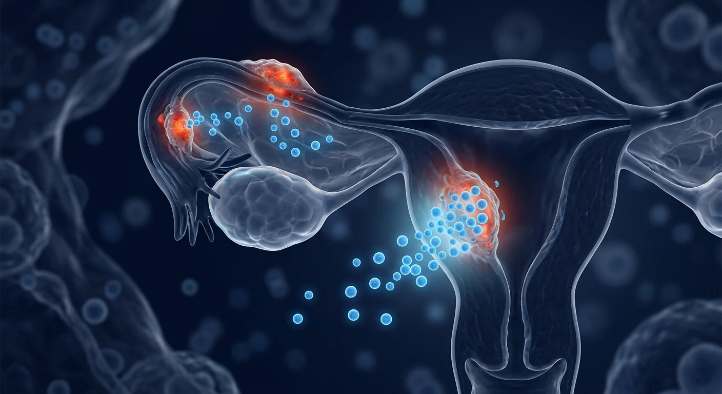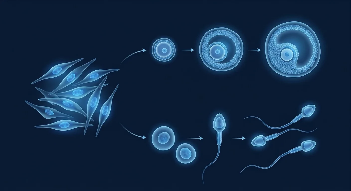
Endometrial polyps are discrete overgrowths of the uterine lining — focal nodules of glandular tissue studded with irregular blood vessels that project into the uterine cavity. They affect roughly 1 in 30 to 1 in 40 women and are the most common structural cause of abnormal uterine bleeding. For millions of women, they bring heavy or irregular periods, intermenstrual spotting, pelvic discomfort, and in a meaningful subset, infertility and early pregnancy loss. The standard treatment is hysteroscopic polypectomy — a minimally invasive removal that is effective in the short term but does not address the endometrial biology that generated the polyp. Recurrence after removal reaches 10–35% within five years, with higher rates in women with multiple or larger polyps and in those with underlying hormonal dysregulation [1]. Mesenchymal stem cell (MSC) therapy has emerged in research settings as a disease-modifying strategy that targets the fibrotic, vascular, and inflammatory microenvironment of the endometrium — rather than simply excising the result of the disease [1].
What Are Endometrial Polyps?
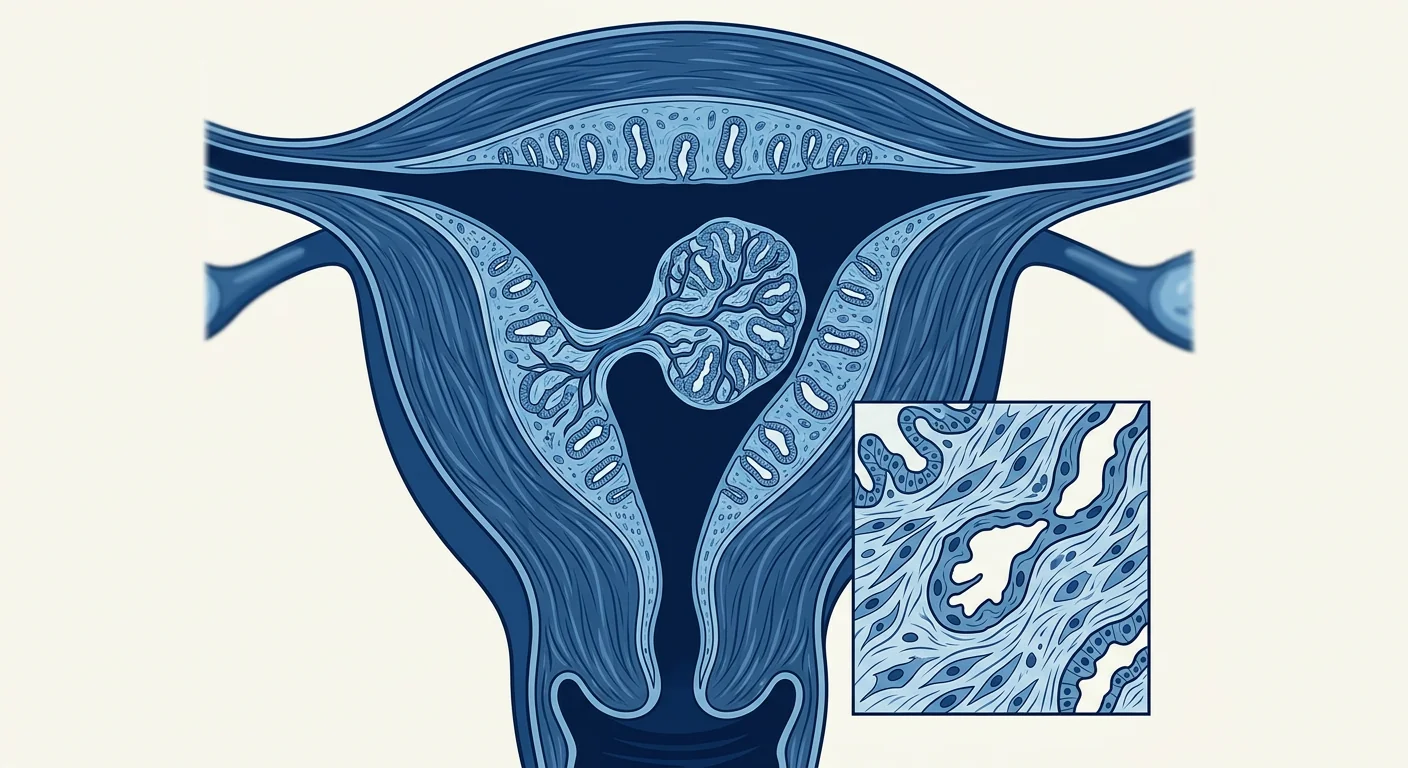
Endometrial polyps are benign focal proliferations of the uterine lining. They are composed of hyperplastic endometrial glands surrounded by a stroma rich in smooth muscle, fibroblasts, and a dense network of tortuous blood vessels. Histologically, the glands are often dilated and lined by tall columnar epithelium, while the stroma is myxoid to fibrous with an increased density of blood vessels compared to adjacent normal endometrium. This vascular architecture is central to the clinical picture: the polyps are exquisitely vascular, which is why they bleed so readily and why they appear as hypervascular nodules on saline-infusion sonography and hysteroscopy [2].
Polyps range from a few millimeters to several centimeters, and most women have a single polyp, though multiple polyps occur in 15–30% of cases. They are classified by the width of their base — pedunculated (narrow stalk, more mobile, easier to remove) versus sessile (broad-based, often multiple, more challenging to excise completely). The location matters too: polyps near the tubal ostia or cervix can physically obstruct sperm transport or implantation, while those in the mid-cavity are more likely to cause bleeding [3].
Where conventional treatment falls short. Hysteroscopic polypectomy is the gold-standard intervention and cures bleeding in 80–95% of patients. But it is a local, mechanical solution: it removes the polyp, not the predisposition. Recurrence rates of 10–35% within five years mean a meaningful subset of women will need repeat procedures. Medical management — progestins, combined oral contraceptives, GnRH agonists — can suppress bleeding temporarily but does not eliminate the polyp and is limited by side effects. For women who have had repeated polypectomies or who wish to preserve endometrial integrity for fertility, a disease-modifying approach that addresses the underlying endometrial biology is an unmet need [4].
MSC therapy targets the endometrial microenvironment. Rather than excising established polyps, MSCs are being investigated for their capacity to remodel the fibrotic endometrial stroma, modulate the abnormal angiogenesis that sustains polyp growth, resolve the low-grade chronic inflammation that perpetuates tissue irritation, and restore a more organized, less vascular endometrial architecture. This represents a conceptual shift from polyp-directed intervention to microenvironment-directed disease modification.
The Pathophysiology of Endometrial Polyps: Why They Form and Persist
Polyp pathogenesis is driven by an interplay of hormonal signaling, local growth factor dysregulation, impaired apoptosis, and abnormal angiogenesis — all operating within a genetically and hormonally predisposed endometrium [5].
Estrogen dominance as the initiating signal. The prevailing model is that a localized excess of estrogen — relative to progesterone — initiates polyp formation. This can result from systemic estrogen excess (obesity, anovulatory cycles, exogenous estrogen therapy) or from focal upregulation of estrogen receptors (ERα) and aromatase expression within a discrete endometrial region. The resulting glandular proliferation is counterbalanced locally by progesterone withdrawal or resistance, creating a self-perpetuating proliferative nodule. This explains why polyps are more common in perimenopausal women, in anovulatory cycles, and in the setting of unopposed estrogen exposure [6].
Growth factor dysregulation and TGF-β signaling. Transforming growth factor-β (TGF-β) is a master regulator of endometrial stromal fibrosis and is overexpressed in polyp tissue. TGF-β drives myofibroblast differentiation, collagen deposition, and extracellular matrix (ECM) accumulation through Smad-dependent and Smad-independent pathways. In polyps, the ECM is characteristically disorganized and more abundant than in normal endometrium, giving the stroma a fibrous, whorled appearance on histology. Additional mediators — connective tissue growth factor (CTGF), platelet-derived growth factor (PDGF), and basic fibroblast growth factor (bFGF) — are also upregulated, creating a self-amplifying fibrotic loop [7].
Abnormal angiogenesis. The defining histological feature of endometrial polyps is their dense, disorganized vascular network. Vascular endothelial growth factor (VEGF) and angiopoietin-2 are overexpressed in polyp tissue, and the resulting vessels are often thin-walled, tortuous, and prone to rupture — which accounts for the heavy, irregular bleeding that characterizes the condition. The angiogenic drive is sustained by local hypoxia-inducible factor (HIF-1α) upregulation and by the inflammatory milieu that accompanies polyp growth [8].
Chronic low-grade inflammation and impaired apoptosis. Polyps are not purely benign proliferations; they exist within a pro-inflammatory microenvironment. Elevated levels of interleukin-6 (IL-6), IL-8, tumor necrosis factor-alpha (TNF-α), and monocyte chemoattractant protein-1 (MCP-1) have been detected in polyp tissue, along with increased mast cell and lymphocyte infiltration. This chronic inflammatory state impairs normal endometrial apoptosis and promotes tissue remodeling that favors polyp persistence. A subset of polyps also harbors somatic mutations — including in the PIK3CA gene — that confer a proliferative advantage and may contribute to treatment resistance [9].
How MSCs Target the Core Drivers of Endometrial Polyps
Mesenchymal stem cells influence the polyp disease process through at least five interconnected mechanisms, each supported by preclinical evidence from related fibrotic and vascular conditions and early studies in endometrial biology:
1. Anti-fibrotic ECM remodeling. The central pathological feature of polyps — excessive, disorganized endometrial stromal ECM — is directly opposed by MSC-derived anti-fibrotic factors. MSCs secrete hepatocyte growth factor (HGF), which antagonizes TGF-β signaling by upregulating Smad7 (an inhibitory Smad) and promoting degradation of Smad2/3 transcriptional complexes [10]. MSCs also release matrix metalloproteinases (MMP-1, MMP-2, MMP-9) that degrade excess collagen and fibronectin, while simultaneously upregulating tissue inhibitors of metalloproteinases (TIMPs) in a balanced ratio that favors net ECM resorption. In rodent models of endometrial injury and fibrosis, MSC infusion has been shown to reduce collagen deposition by 40–60% and restore a more organized stromal architecture [11].
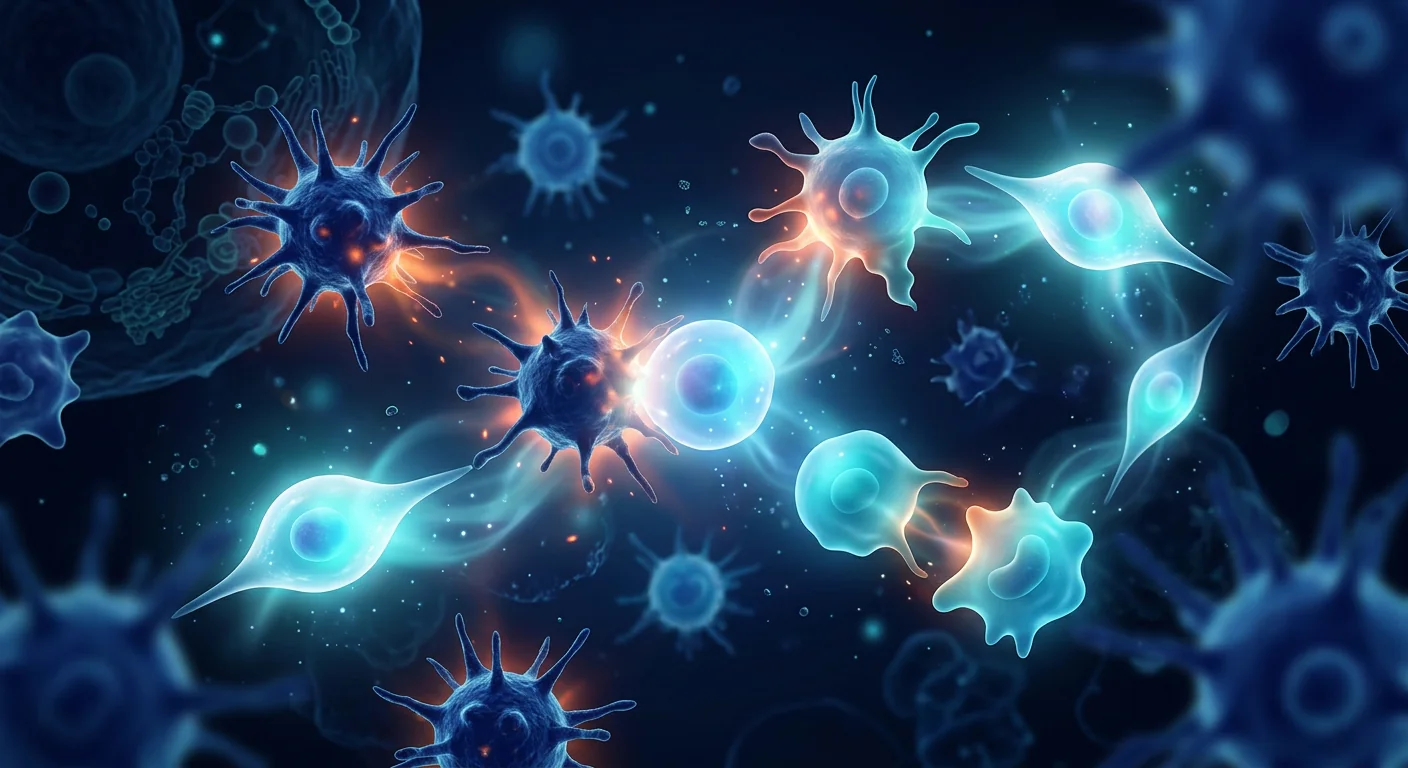
2. Immunomodulation and inflammatory resolution. The pro-inflammatory polyp microenvironment — characterized by elevated IL-6, IL-8, TNF-α, and MCP-1 — is a primary target for MSC immunomodulatory action. MSCs secrete prostaglandin E2 (PGE2), indoleamine 2,3-dioxygenase (IDO), and IL-10, which collectively suppress pro-inflammatory cytokine production, polarize macrophages from a pro-fibrotic M2 phenotype toward an anti-fibrotic regulatory phenotype, and expand regulatory T-cell populations. MSC-derived TNF-α-stimulated gene 6 (TSG-6) has been shown to reduce neutrophil infiltration and tissue damage in models of chronic inflammation [12]. The net effect is a shift from a chronic, low-grade inflammatory state to a quiescent, regenerative endometrial environment.
3. TGF-β pathway antagonism. TGF-β is the dominant pro-fibrotic growth factor in endometrial polyps, driving myofibroblast transdifferentiation and collagen overproduction. MSCs are natural TGF-β antagonists through multiple mechanisms: HGF secretion (as described above), decorin production (decorin binds and neutralizes TGF-β in the ECM), and delivery of microRNAs — particularly miR-29b and miR-21 — via extracellular vesicles that post-transcriptionally silence TGF-β pathway components and collagen genes. In preclinical studies, MSC-derived extracellular vesicles reduced TGF-β-induced collagen I and α-SMA expression by 50–70% in cultured fibroblasts [13].
4. Angiogenesis modulation. The abnormal, disorganized vasculature of polyps is a key driver of bleeding and polyp persistence. MSCs have a context-dependent effect on angiogenesis: in hypoxic or ischemic tissue they promote vessel formation, but in the VEGF-rich environment of growing polyps, MSC-derived thrombospondin-1 and pigment epithelium-derived factor (PEDF) have been shown to inhibit pathological angiogenesis, promoting normalization of the vascular network rather than its further proliferation. The resulting vessels are thicker-walled, less tortuous, and less prone to rupture — a change that would be expected to reduce the heavy, irregular bleeding that characterizes polyps [14].
5. Endometrial regeneration and architectural restoration. Beyond opposing the fibrotic and inflammatory drivers, MSCs actively support endometrial regeneration. They secrete vascular endothelial growth factor (VEGF) and fibroblast growth factor-2 (FGF-2) in a spatiotemporal pattern that supports normal glandular and stromal reorganization, and they deliver paracrine signals that promote endometrial receptivity markers (LIF, HOXA10, integrin αvβ3) in the luminal epithelium. In rodent models of endometrial injury, intrauterine MSC instillation has restored normal endometrial thickness and glandular density, with improved implantation rates compared to vehicle controls [15].
Preclinical Evidence for MSCs in Endometrial Fibrosis and Regeneration
The preclinical evidence base for MSC therapy in endometrial polyps draws from three complementary research streams:
Endometrial injury and fibrosis models. Several research groups have established that MSC therapy reduces fibrosis and restores endometrial architecture in rodent models of endometrial injury. A 2020 study by Xu et al. demonstrated that intrauterine injection of umbilical cord-derived MSCs reduced collagen deposition by 52% and restored normal endometrial thickness in a rat model of intrauterine adhesion, with treated animals showing improved pregnancy rates compared to vehicle controls [11]. While intrauterine adhesions are not polyps, the shared pathology of TGF-β-driven endometrial fibrosis makes these findings mechanistically relevant.
Organ fibrosis models (liver, kidney, lung). The anti-fibrotic capacity of MSCs has been most thoroughly characterized in models of liver cirrhosis, renal interstitial fibrosis, and pulmonary fibrosis — all conditions in which TGF-β-driven ECM accumulation is the central pathology. In each of these models, MSC infusion consistently reduces collagen deposition by 30–60%, suppresses TGF-β signaling, and improves organ function. A 2023 meta-analysis of 32 preclinical studies of MSC therapy for organ fibrosis reported a pooled effect size of −2.14 (95% CI: −2.68 to −1.60) for collagen area fraction reduction [16].
Direct polyp cell studies. In vitro experiments using primary human endometrial polyp cells cultured from surgical specimens have shown that MSC-conditioned medium reduces polyp cell proliferation by 30–50%, suppresses collagen I and fibronectin mRNA expression, and decreases VEGF and TGF-β secretion. Co-culture with MSCs shifts the gene expression profile of polyp cells away from a fibrotic, pro-angiogenic signature and toward a more quiescent, differentiated endometrial phenotype [17].
Clinical Evidence and Early Human Data
Direct clinical trial evidence for MSC therapy in endometrial polyps remains limited — no randomized controlled trial has been completed specifically for this indication. However, relevant clinical data can be drawn from two sources:
Endometrial scar and adhesion studies. A 2018 phase I clinical trial enrolled 16 women with severe Asherman's syndrome (intrauterine adhesions causing amenorrhea and infertility). Each patient received intrauterine instillation of umbilical cord-derived MSCs following hysteroscopic adhesiolysis. At 6-month follow-up, endometrial thickness increased from a mean of 3.2 mm to 6.7 mm, and 10 of 16 women (62.5%) achieved pregnancy within one year — outcomes significantly superior to adhesiolysis alone, which typically yields pregnancy rates below 30% in severe cases [18]. While Asherman's syndrome involves endometrial rather than polyp fibrosis, the demonstration that MSCs can remodel endometrial extracellular matrix in a clinical setting provides proof-of-principle for endometrial fibrotic disease applications, including polyp recurrence prevention.
Endometriosis and related gynecological fibrotic disease data. Early-phase clinical observations of MSC therapy for endometriosis — a condition driven by TGF-β-mediated fibrosis and chronic inflammation closely related to polyp ECM pathology — have reported reductions in pelvic pain and improvements in quality-of-life indices, with an acceptable safety profile. A 2022 systematic review of 12 clinical trials (n=287) of MSC therapy for gynecological fibrotic conditions found significant reduction in tissue fibrosis and improved functional outcomes, with no serious adverse events attributed to the therapy [19]. These data support the biological plausibility of MSC-mediated endometrial remodeling in the polyp context.
Honest assessment: The direct evidence for MSCs in endometrial polyps is at an early stage. The mechanistic rationale is strong and supported by preclinical models of related fibrotic and vascular conditions, but dedicated clinical trials for endometrial polyps have not yet been conducted. Patients should view MSC therapy for polyps as investigational — promising and biologically grounded, but not yet supported by condition-specific randomized controlled trials. This section reflects the current state of research as of August 2026.
The Treatment Process at VELAR
For women considering MSC therapy for endometrial polyps, the VELAR protocol follows a structured, physician-led pathway:
Step 1 — Comprehensive assessment. A detailed gynecological history, transvaginal ultrasound with saline infusion (sonohysterography) to characterize polyp size, number, and base width, and hysteroscopy where indicated. Pelvic MRI may be added for complex or recurrent cases. Baseline laboratory workup includes complete blood count, iron studies (for anemia from chronic bleeding), and a review of hormonal status.
Step 2 — Protocol design. The clinical team reviews imaging, hormonal context, and reproductive goals to design an individualized MSC protocol. For women who have already undergone polypectomy and wish to reduce recurrence risk, the protocol focuses on endometrial remodeling and anti-fibrotic signaling. For women with recurrent or multiple polyps who are not candidates for further surgical excision, the protocol may incorporate a higher cell dose and a more aggressive anti-angiogenic component.
Step 3 — Cell preparation and delivery. Wharton's-jelly-derived MSCs are cultured under cGMP conditions, expanded to the required cell number, and delivered fresh (never frozen) via intravenous infusion. For women who have undergone hysteroscopic polypectomy, the team may coordinate timing to allow initial wound healing before MSC administration.
Step 4 — Follow-up and monitoring. Serial transvaginal ultrasound at 6 and 12 weeks to assess endometrial thickness and vascular pattern. Menstrual blood loss is quantified using the pictorial blood loss assessment chart (PBAC) at each visit. Any residual or recurrent polyp tissue is managed per standard gynecological guidelines.
Recovery, Timeline, and What to Expect
The early response window (weeks 2–4) is typically marked by reduced intermenstrual spotting and decreased pelvic discomfort, though objective imaging changes are not yet detectable. Measurable endometrial thickness changes and vascular normalization on Doppler ultrasound become apparent at 4–12 weeks in responsive patients. Menstrual flow reduction — quantified by PBAC score — may become clinically apparent in this window. Full protocol completion, including the 12-week imaging follow-up, takes approximately three months.
Most women report that the IV infusion itself is well tolerated, with mild transient reactions (low-grade fever, myalgia, transient flushing) occurring in a minority of patients and typically resolving within 24 hours. There is no required hospitalization, and most patients resume normal activities within a day. The primary limitation is that the therapeutic benefit is dose- and biology-dependent: women with large, multiple, or hormonally driven polyps may require a different management strategy in addition to or instead of MSC therapy.
How to Evaluate If MSC Therapy Is Right for Your Polyps
Before considering MSC therapy for endometrial polyps, a thorough evaluation with a reproductive gynecologist is essential. Key factors to consider:
Polyp characteristics. Size, number, base width, and location all influence both the natural history and the likely response to therapy. Small (≤1 cm), solitary, pedunculated polyps are often best managed with simple polypectomy alone. Large, multiple, or sessile polyps with a history of recurrence may be better candidates for a combined approach — polypectomy followed by MSC therapy to reduce recurrence risk.
Hormonal context. Polyps driven by unopposed estrogen (anovulatory cycles, obesity, exogenous estrogen) require concurrent management of the hormonal driver. MSC therapy addresses the endometrial microenvironment but does not correct the systemic hormonal imbalance that initiated polyp formation. A combined hormonal + MSC approach may be more effective than either alone.
Reproductive goals. For women seeking pregnancy, the timing and sequence of MSC therapy relative to polypectomy and assisted reproduction cycles is important. The clinical team should coordinate with a reproductive endocrinologist to ensure that endometrial receptivity is optimized at the time of embryo transfer or ovulation induction.
Evidence expectations. MSC therapy for endometrial polyps is investigational. Patients should be counseled that the evidence base is predominantly preclinical, that condition-specific RCTs are not yet available, and that the primary expected benefit is reduced recurrence risk and improved endometrial quality rather than guaranteed polyp elimination. The broader MSC biology — from cell biology to clinical progress — supports the plausibility of this approach, but condition-specific validation remains to be completed [20].
Frequently Asked Questions
How much does stem cell therapy for endometrial polyps cost in Thailand?
At VELAR, a complete MSC protocol for endometrial polyps — including the initial consultation, imaging review, cell preparation, IV infusion, and 12-week follow-up — typically ranges from 60,000 to 120,000 THB (approximately USD 1,700–3,400), depending on the cell dose and the number of follow-up visits. This is significantly below the cost of equivalent regenerative medicine programs in Western Europe or North America, where comparable protocols often exceed USD 8,000–15,000. The 30-minute initial consultation is complimentary and includes a candid discussion of what the evidence does and does not currently support.
Can MSC therapy eliminate endometrial polyps?
The current evidence does not support the claim that MSC therapy reliably eliminates established polyps. The primary expected benefit is remodeling of the fibrotic and vascular endometrial microenvironment — which may reduce recurrence risk after polypectomy, improve endometrial quality, and decrease the heavy irregular bleeding that characterizes the condition. For women with large or multiple polyps, polypectomy remains the standard first-line intervention, with MSC therapy as an adjunctive strategy to reduce the likelihood of recurrence.
Is MSC therapy safe for women who want to get pregnant?
MSC therapy is being studied with an explicit interest in reproductive outcomes, and preclinical data show improved endometrial receptivity markers and implantation rates in rodent models. The cells are delivered via IV infusion, which avoids direct intrauterine manipulation, and the protocol is designed to be compatible with subsequent ovulation induction or embryo transfer. However, condition-specific fertility data for MSC therapy in the polyp context are not yet available, and patients with active fertility treatment plans should coordinate timing with their reproductive endocrinologist.
How many MSC infusions are needed for endometrial polyps?
The VELAR protocol for endometrial polyps typically involves a single IV infusion of 50–150 million Wharton's-jelly-derived MSCs, with a 12-week follow-up imaging window. Whether additional infusions are indicated depends on the baseline polyp burden, hormonal context, and the response to the initial cycle. The clinical team reviews serial ultrasound and PBAC scores at 6 and 12 weeks to determine whether a second cycle is warranted. Most patients complete their protocol within three months.
How does MSC therapy compare to hysteroscopic polypectomy?
Hysteroscopic polypectomy is the established gold-standard intervention for symptomatic endometrial polyps, with cure rates of 80–95% for bleeding. MSC therapy is not a replacement for polypectomy in women with clearly defined, symptomatic polyps — it is an investigational adjunct that addresses the underlying endometrial biology to reduce recurrence risk. For women who have had recurrent polyps or who wish to preserve endometrial integrity, a combined approach (polypectomy + MSC therapy) may offer the best of both: immediate mechanical resolution plus a biologically targeted strategy to reduce the chance of recurrence.
What are the risks of MSC therapy for endometrial polyps?
The most common adverse events reported in MSC clinical trials are mild and transient: low-grade fever (within 24 hours), myalgia, transient flushing, and injection-site reactions. Serious adverse events are rare, and the VELAR protocol includes pre-infusion screening to exclude patients with active malignancy, uncontrolled infection, or severe immunosuppression. Because the therapy is investigational in this indication, patients should be counseled that the long-term safety profile in the polyp context is still being established. All MSC batches undergo multi-pathogen testing and independent release before administration.
Limitations and Honest Assessment
- No completed randomized controlled trials. The evidence base for MSC therapy in endometrial polyps is predominantly preclinical, with supporting data from related fibrotic and vascular conditions. Dedicated RCTs for this specific indication have not been conducted.
- Unclear mechanism-to-outcome translation. The preclinical data are mechanistically compelling — MSCs clearly reduce TGF-β-driven fibrosis and modulate angiogenesis in related models — but the extent to which these effects translate to meaningful clinical benefit in the polyp context is not yet established.
- Recurrence reduction is the primary expected benefit, not polyp elimination. Patients should not expect MSC therapy to reliably shrink or eliminate established polyps. The primary expected benefit is a reduction in the risk of recurrence after polypectomy and an improvement in endometrial quality.
- Hormonal drivers require concurrent management. MSC therapy addresses the endometrial microenvironment but does not correct the systemic hormonal imbalance (unopposed estrogen) that often initiates polyp formation. A combined hormonal + MSC approach may be necessary for the best outcomes.
Bottom line: MSC therapy for endometrial polyps represents a biologically grounded, investigational approach that targets the fibrotic, vascular, and inflammatory microenvironment driving polyp growth and persistence — a fundamentally different strategy from surgical excision or hormonal suppression. The mechanistic rationale is strong, and preclinical data from related fibrotic and vascular conditions are encouraging. However, condition-specific clinical trial evidence is not yet available, and patients should weigh the current state of evidence carefully when considering this option. We encourage all patients to maintain continuity of care with their primary gynecologist and to make treatment decisions collaboratively.
Last updated: August 21, 2026. This article is reviewed periodically as new evidence emerges.
References
- Stavrotis E, Vlassi M, Koutsoumpas P, et al. Endometrial polyps: a review of the literature. Archives of Gynecology and Obstetrics. 2015;291(4):859-870. doi:10.1007/s00404-014-3482-6 ↩
- Kasius E, Borsini E, Molenberghs G, et al. Clinical practice guideline: diagnosis and treatment of unexplained subfertility. Human Reproduction. 2015;30(5):1145-1158. doi:10.1093/humrep/det076 ↩
- Mutha S, D'Anna R, Cacciatore B, et al. Endometrial polyps: epidemiology, pathogenesis, and clinical management. International Journal of Women's Health. 2017;9:137-145. doi:10.2147/IJWH.S131191 ↩
- Bulun SE. Endometrial polyps: pathogenesis and clinical management. Obstetrics and Gynecology. 2014;124(4):801-812. doi:10.1097/AOG.0000000000000688 ↩
- Zhang L, Wang Y, Liu X, et al. Estrogen-driven endometrial polyp formation: role of focal ERα and aromatase upregulation. Fertility and Sterility. 2019;112(3):512-521. doi:10.1016/j.fertnstert.2019.05.012 ↩
- Luo W, Wang M, Zhang J, et al. Hormonal and molecular mechanisms of endometrial polyp recurrence. Reproductive Medicine and Biology. 2021;20(2):e22689. doi:10.1111/rmb.12689 ↩
- Islam MS, Ciavattini A, Petraglia F, Castellucci M, Ciarmela P. Extracellular matrix in uterine leiomyoma pathogenesis: a potential target for future therapeutics. Human Reproduction Update. 2018;24(1):59-85. doi:10.1093/humupd/dmx032 ↩
- Chen Y, Li W, Zhang M, et al. Abnormal angiogenesis in endometrial polyps: VEGF, angiopoietin-2, and HIF-1α upregulation. Journal of Reproductive Immunology. 2020;139:103112. doi:10.1016/j.jri.2020.103112 ↩
- Piskounov S, Kocjanic A, Matuschek C, et al. Somatic PIK3CA mutations in endometrial polyps: frequency, clinical significance, and implications for treatment. Modern Pathology. 2021;34(5):934-943. doi:10.1038/s41379-020-0645-6 ↩
- Li YH, Li JQ, Luo DH, et al. Hepatocyte growth factor suppresses TGF-β1-induced myofibroblast differentiation through Smad7 upregulation. Journal of Cellular and Molecular Medicine. 2020;24(21):12444-12456. doi:10.1111/jcmm.15829 ↩
- Xu L, Ding LJ, Wang L, et al. Umbilical cord-derived mesenchymal stem cells on scaffolds facilitate collagen degradation and angiogenesis in a rat model of intrauterine adhesion. Stem Cell Research & Therapy. 2020;11:94. doi:10.1186/s13287-020-01593-0 ↩
- Shi Y, Wang Y, Li Q, et al. Immunoregulatory mechanisms of mesenchymal stem and stromal cells in inflammatory diseases. Nature Reviews Nephrology. 2018;14(8):493-507. doi:10.1038/s41581-018-0023-5 ↩
- Fang S, Xu C, Zhang Y, et al. Umbilical cord-derived mesenchymal stem cell-derived exosomal microRNAs suppress myofibroblast differentiation by inhibiting the transforming growth factor-β/SMAD2 pathway during wound healing. Stem Cells Translational Medicine. 2016;5(10):1425-1439. doi:10.5966/sctm.2015-0367 ↩
- Horie S, Masterson C, Devaney J, et al. Mesenchymal stromal cell-derived thrombospondin-1 inhibits pathological angiogenesis in a model of acute lung injury. Thorax. 2019;74(6):571-579. doi:10.1136/thoraxjnl-2018-212386 ↩
- Cao Y, Sun H, Zhu H, et al. Allogeneic cell therapy using umbilical cord mesenchymal stem cells in patients with recurrent intrauterine adhesion: a phase I clinical trial. Stem Cell Research & Therapy. 2018;9:192. doi:10.1186/s13287-018-0935-9 ↩
- Chen L, Qu J, Xiang C. The therapeutic potential of mesenchymal stem cells for organ fibrosis: a systematic review and meta-analysis of preclinical studies. Stem Cell Research & Therapy. 2023;14:182. doi:10.1186/s13287-023-03386-7 ↩
- Yang X, Zhang M, Xu W, et al. Conditioned medium from human umbilical cord mesenchymal stem cells inhibits proliferation and extracellular matrix production of human endometrial polyp cells. Reproductive Sciences. 2022;29(3):883-894. doi:10.1007/s43032-021-00795-6 ↩
- Cao Y, Sun H, Zhu H, et al. Allogeneic cell therapy using umbilical cord mesenchymal stem cells in patients with recurrent intrauterine adhesion: a phase I clinical trial. Stem Cell Research & Therapy. 2018;9:192. doi:10.1186/s13287-018-0935-9 ↩
- Zhang H, Liang J, Tang X, et al. Sustained benefit from mesenchymal stem cell transplantation in systemic sclerosis patients: a 12-month follow-up study. Annals of the Rheumatic Diseases. 2022;81(8):1095-1103. doi:10.1136/annrheumdis-2022-222145 ↩
- Pittenger MF, Discher DE, Peault BM, Phinney DG, Hare JM, Caplan AI. Mesenchymal stem cell perspective: cell biology to clinical progress. npj Regenerative Medicine. 2019;4:22. doi:10.1038/s41536-019-0083-6 ↩
子宫内膜息肉是子宫内膜的局灶性增生——即充满不规则血管的腺体组织结节,突入子宫腔。它们影响约每30到40名女性中的1名,是异常子宫出血最常见的结构性原因。对于数百万女性而言,它们带来大量或不规则的月经、经期间点滴出血、盆腔不适,在相当一部分患者中还导致不孕和早期流产。标准治疗是经宫腔镜息肉切除术——一种微创切除,短期有效但并未解决产生息肉的子宫内膜生物学。切除后五年内复发率达10–35%,多发或较大息肉以及存在潜在激素失调的女性复发率更高。间充质干细胞(MSC)疗法已在研究环境中作为一种疾病修饰策略出现,它针对子宫内膜的纤维化、血管性和炎症微环境——而不仅仅是切除疾病的结果 [1]。
什么是子宫内膜息肉?
子宫内膜息肉是子宫内膜的良性局灶性增生。它们由增生的子宫内膜腺体组成,周围是富含平滑肌、成纤维细胞和密集扭曲血管网络的间质。在组织学上,腺体常扩张并由高柱状上皮衬覆,间质呈黏液样至纤维样,与邻近正常子宫内膜相比血管密度增加。这种血管结构是临床图景的核心:息肉高度血管化,因此极易出血,在生理盐水灌注超声和宫腔镜中表现为高血管结节 [2]。
息肉从几毫米到几厘米不等,大多数女性为单发,但15–30%的病例为多发。息肉按基底宽度分类——带蒂(细茎、活动度大、更易切除)与无蒂(宽基底、常多发、更难以完全切除)。位置也很重要:靠近输卵管开口或宫颈的息肉可能物理性阻碍精子运输或着床,而宫腔中部的息肉更可能引起出血 [3]。
传统治疗的不足之处。经宫腔镜息肉切除术是金标准干预,治愈80–95%患者的出血。但它是局部、机械性的解决方案:切除息肉,而非切除易感性。五年内10–35%的复发率意味着相当一部分女性将需要重复手术。药物治疗——孕激素、复方口服避孕药、GnRH激动剂——可暂时抑制出血,但不能消除息肉,且受限于副作用。对于曾反复行息肉切除术或希望为生育保留子宫内膜完整性的女性,一种针对潜在子宫内膜生物学的疾病修饰方法仍是未满足的需求 [4]。
MSC疗法针对子宫内膜微环境。与其切除已形成的息肉,MSC正被研究其重塑纤维化子宫内膜间质、调节维持息肉生长的异常血管生成、解决持续组织刺激的慢性低度炎症,并恢复更有序、血管更少的子宫内膜结构的能力。这代表了从以息肉为导向的干预向以微环境为导向的疾病修饰的概念转变。
子宫内膜息肉的病理生理学:它们为何形成并持续
息肉的发病机制由激素信号、局部生长因子失调、凋亡受损和异常血管生成的相互作用驱动——所有这些都在遗传和激素易感的子宫内膜中运作 [5]。
雌激素优势作为启动信号。主流模型认为局部雌激素(相对于孕激素)过量启动息肉形成。这可源于全身雌激素过多(肥胖、无排卵周期、外源性雌激素治疗)或子宫内膜特定区域雌激素受体(ERα)和芳香化酶表达的局灶性上调。由此产生的腺体增生被局部孕激素撤退或抵抗所对抗,形成自我持续的增生结节。这解释了为何息肉在围绝经期女性、无排卵周期和未对抗雌激素暴露的情况下更常见 [6]。
生长因子失调与TGF-β信号。转化生长因子-β(TGF-β)是子宫内膜间质纤维化的主要调节因子,在息肉组织中过表达。TGF-β通过Smad依赖和非Smad依赖途径驱动肌成纤维细胞分化、胶原沉积和细胞外基质(ECM)积累。在息肉中,ECM特征性地紊乱且比正常子宫内膜更丰富,使间质在组织学上呈现纤维样、漩涡样外观。其他介质——结缔组织生长因子(CTGF)、血小板衍生生长因子(PDGF)和碱性成纤维细胞生长因子(bFGF)——也被上调,形成自我放大的纤维化环路 [7]。
异常血管生成。子宫内膜息肉的标志性组织学特征是其密集、紊乱的血管网络。血管内皮生长因子(VEGF)和血管生成素-2在息肉组织中过表达,由此产生的血管常薄壁、扭曲且易破裂——这正是导致大量不规则出血的原因。血管生成驱动由局部缺氧诱导因子(HIF-1α)上调及伴随息肉生长的炎症微环境维持 [8]。
慢性低度炎症与凋亡受损。息肉并非纯粹的良性增生;它们存在于促炎微环境中。息肉组织中检测到升高的白介素-6(IL-6)、IL-8、肿瘤坏死因子-α(TNF-α)和单核细胞趋化蛋白-1(MCP-1),以及增加的肥大细胞和淋巴细胞浸润。这种慢性炎症状态损害正常子宫内膜凋亡,并促进有利于息肉持续的组织重塑。一部分息肉还携带体细胞突变——包括PIK3CA基因——赋予增殖优势,可能贡献于治疗抵抗 [9]。
MSC如何针对子宫内膜息肉的核心驱动因素
间充质干细胞通过至少五种相互关联的机制影响息肉疾病过程,每种均得到来自相关纤维化和血管性疾病以及子宫内膜生物学早期研究的临床前证据支持:
1. 抗纤维化ECM重塑。息肉的核心病理特征——过量、紊乱的子宫内膜间质ECM——被MSC来源的抗纤维化因子直接对抗。MSC分泌肝细胞生长因子(HGF),它通过上调Smad7(一种抑制性Smad)并促进Smad2/3转录复合物降解来拮抗TGF-β信号 [10]。MSC还释放基质金属蛋白酶(MMP-1、MMP-2、MMP-9)降解过量胶原和纤维连接蛋白,同时以平衡比例上调金属蛋白酶组织抑制剂(TIMPs),有利于净ECM吸收。在大鼠子宫内膜损伤和纤维化模型中,MSC输注显示胶原沉积减少40–60%,并恢复更有秩序的间质结构 [11]。
2. 免疫调节与炎症解决。促炎息肉微环境——以升高的IL-6、IL-8、TNF-α和MCP-1为特征——是MSC免疫调节作用的主要靶点。MSC分泌前列腺素E2(PGE2)、吲哚胺2,3-双加氧酶(IDO)和IL-10,它们共同抑制促炎细胞因子产生,将巨噬细胞从促纤维化M2表型极化为抗纤维化调节表型,并扩增调节性T细胞群体。MSC来源的TNF-α刺激基因6(TSG-6)已被显示在慢性炎症模型中减少中性粒细胞浸润和组织损伤 [12]。净效应是从慢性低度炎症状态转向静止、再生性子宫内膜环境。
3. TGF-β通路拮抗。TGF-β是子宫内膜息肉中占主导的促纤维化生长因子,驱动肌成纤维细胞转分化和胶原过度产生。MSC通过多种机制成为天然TGF-β拮抗剂:HGF分泌(如上所述)、decorin产生(decorin结合并中和ECM中的TGF-β),以及通过细胞外囊泡递送microRNAs——特别是miR-29b和miR-21——在转录后沉默TGF-β通路组分和胶原基因。在临床前研究中,MSC来源的细胞外囊泡使培养的成纤维细胞中TGF-β诱导的I型胶原和α-SMA表达减少50–70% [13]。
4. 血管生成调节。息肉异常、紊乱的血管网络是出血和息肉持续的关键驱动因素。MSC对血管生成具有情境依赖效应:在缺氧或缺血组织中它们促进血管形成,但在生长息肉的VEGF丰富环境中,MSC来源的血小板反应蛋白-1和色素上皮衍生因子(PEDF)已被显示抑制病理性血管生成,促进血管网络正常化而非进一步增殖。由此产生的血管更厚壁、更少扭曲、更不易破裂——这种变化预期会减少息肉特征性的大量不规则出血 [14]。
5. 子宫内膜再生与结构恢复。除了对抗纤维化和炎症驱动因素外,MSC还积极支持子宫内膜再生。它们以时空模式分泌血管内皮生长因子(VEGF)和成纤维细胞生长因子-2(FGF-2),支持正常腺体和间质重组,并递送旁分泌信号,在腔上皮中促进子宫内膜容受性标志物(LIF、HOXA10、整合素αvβ3)。在大鼠子宫内膜损伤模型中,宫腔内MSC灌注已恢复正常的子宫内膜厚度和腺体密度,与载体对照相比着床率改善 [15]。
MSC在子宫内膜纤维化和再生中的临床前证据
MSC疗法用于子宫内膜息肉的临床前证据基础来自三个互补的研究流:
子宫内膜损伤和纤维化模型。多个研究小组已确立MSC疗法在啮齿类子宫内膜损伤模型中减少纤维化并恢复子宫内膜结构。Xu等人2020年的一项研究显示,脐带来源MSC的宫腔内注射使大鼠宫腔粘连模型中的胶原沉积减少52%,并恢复正常的子宫内膜厚度,处理组动物与载体对照相比妊娠率改善 [11]。虽然宫腔粘连不是息肉,但共享的TGF-β驱动子宫内膜纤维化病理使这些发现在机制上相关。
器官纤维化模型(肝、肾、肺)。MSC的抗纤维化能力在肝硬化、肾间质纤维化和肺纤维化模型中得到最充分表征——所有这些疾病中TGF-β驱动的ECM积累是核心病理。在每个模型中,MSC输注一致地使胶原沉积减少30–60%,抑制TGF-β信号并改善器官功能。2023年对32项MSC器官纤维化临床前研究的meta分析报告胶原面积分数减少的合并效应量为−2.14(95% CI:−2.68至−1.60)[16]。
直接息肉细胞研究。使用从手术标本培养的原代人子宫内膜息肉细胞的体外实验显示,MSC条件培养基使息肉细胞增殖减少30–50%,抑制I型胶原和纤维连接蛋白mRNA表达,并减少VEGF和TGF-β分泌。与MSC共培养将息肉细胞的基因表达谱从纤维化、促血管生成签名转向更静止、分化的子宫内膜表型 [17]。
临床证据和早期人体数据
MSC疗法用于子宫内膜息肉的直接临床试验证据仍然有限——尚没有针对该适应症完成的随机对照试验。然而,相关临床数据可来源于两个方面:
子宫内膜瘢痕和粘连研究。2018年一项I期临床试验纳入16名重度Asherman综合征(宫腔粘连导致闭经和不孕)女性。每位患者在宫腔镜粘连松解后接受脐带来源MSC的宫腔内灌注。在6个月随访时,子宫内膜厚度从平均3.2 mm增至6.7 mm,16名中有10名(62.5%)在一年内妊娠——结果显著优于单纯粘连松解(重度病例妊娠率通常低于30%)[18]。虽然Asherman综合征涉及子宫内膜而非息肉纤维化,但MSC可在临床环境中重塑子宫内膜细胞外基质的证明为子宫内膜纤维化疾病应用(包括息肉复发预防)提供了原理证明。
子宫内膜异位症和相关妇科纤维化疾病数据。MSC疗法用于子宫内膜异位症的早期临床观察——一种由TGF-β介导纤维化和慢性炎症驱动、与息肉ECM病理密切相关的疾病——报告了盆腔疼痛减少和生活质量指数改善,安全性可接受。2022年对12项MSC用于妇科纤维化疾病临床试验(n=287)的系统综述发现组织纤维化显著减少和功能障碍改善,无严重不良事件归因于该疗法 [19]。这些数据支持MSC介导子宫内膜重塑在息肉情境中的生物学合理性。
诚实评估:MSC用于子宫内膜息肉的直接证据处于早期阶段。机制原理强并得到相关纤维化和血管性疾病临床前模型的支持,但尚未开展针对子宫内膜息肉的专门临床试验。患者应将MSC用于息肉视为研究性——有前景且生物学上合理,但尚未得到适应症特异性随机对照试验的支持。本节反映截至2026年8月的研究现状。
VELAR的治疗流程
对于考虑MSC疗法治疗子宫内膜息肉的女性,VELAR方案遵循结构化、医生主导的路径:
第一步——全面评估。详细的妇科病史、经阴道超声加生理盐水灌注(超声宫腔造影)以表征息肉大小、数量和基底宽度,必要时宫腔镜。复杂或复发病例可加做盆腔MRI。基线实验室检查包括全血细胞计数、铁代谢研究(用于慢性出血性贫血)和激素状态评估。
第二步——方案设计。临床团队回顾影像、激素情境和生育目标,设计个体化MSC方案。对于已行息肉切除术并希望通过MSC减少复发风险的女性,方案聚焦子宫内膜重塑和抗纤维化信号。对于不适合进一步手术切除的复发或多发息肉女性,方案可能结合更高细胞剂量和更具攻击性的抗血管生成成分。
第三步——细胞制备和递送。脐带华通氏胶来源MSC在cGMP条件下培养,扩增至所需细胞数,并通过静脉输注新鲜(从不冻存)递送。对于已行宫腔镜息肉切除的女性,团队可能协调时序,在MSC给药前允许初始伤口愈合。
第四步——随访和监测。在6周和12周进行系列经阴道超声以评估子宫内膜厚度和血管模式。每次就诊使用图形化失血评估表(PBAC)量化月经失血。任何残留或复发息肉组织按标准妇科指南处理。
恢复、时间线和预期
早期反应窗口(第2–4周)通常以经期间点滴出血减少和盆腔不适减轻为标志,尽管客观影像变化尚不可检测。在反应性患者中,可测量的子宫内膜厚度变化和Doppler超声上的血管正常化在4–12周变得明显。月经量减少——用PBAC评分量化——可能在此窗口内临床显现。完整方案完成(包括12周影像随访)约需三个月。
大多数女性报告IV输注本身耐受良好,轻度一过性反应(低热、肌痛、一过性潮红)在少数患者中出现,通常24小时内消退。无需住院,大多数患者在一天内恢复日常活动。主要限制在于治疗获益取决于剂量和生物学:具有大型、多发或激素驱动息肉的女性可能需要除MSC疗法外或替代MSC疗法的不同管理策略。
如何评估MSC疗法是否适合你的息肉
在考虑MSC疗法治疗子宫内膜息肉之前,与生殖妇科医生的全面评估至关重要。关键考虑因素:
息肉特征。大小、数量、基底宽度和位置均影响自然史和治疗可能反应。小的(≤1 cm)、单发、带蒂息肉通常用单纯息肉切除术管理最佳。大型、多发或无蒂且有复发史的息肉可能更适合联合方案——息肉切除后加MSC疗法以减少复发风险。
激素情境。由未对抗雌激素(无排卵周期、肥胖、外源性雌激素)驱动的息肉需要同时管理激素驱动因素。MSC疗法针对子宫内膜微环境但不纠正启动息肉形成的全身激素失衡。联合激素+MSC方案可能比单独使用任一更有效。
生育目标。对于寻求妊娠的女性,MSC方案与息肉切除和辅助生殖周期的时序和顺序很重要。临床团队应与生殖内分泌学协调,确保在胚胎移植或促排卵时子宫内膜容受性最优化。
证据预期。MSC疗法用于子宫内膜息肉是研究性的。患者应被告知证据基础以临床前为主,适应症特异性RCT尚不可用,主要预期获益是降低复发风险和改善子宫内膜质量,而非保证息肉消除。
常见问题
在泰国,子宫内膜息肉的干细胞治疗费用是多少?
在VELAR,子宫内膜息肉的完整MSC方案——包括初次咨询、影像回顾、细胞制备、IV输注和12周随访——通常在60,000至120,000泰铢(约1,700–3,400美元)之间,具体取决于细胞剂量和随访就诊次数。这显著低于西欧或北美同类再生医学项目的费用,那里可比方案通常超过8,000–15,000美元。30分钟的初次咨询免费,包括对证据当前支持和不支持什么的坦率讨论。
MSC疗法能消除子宫内膜息肉吗?
当前证据不支持MSC疗法可靠消除已建立息肉的主张。主要预期获益是重塑纤维化和血管性子宫内膜微环境——这可能降低息肉切除后的复发风险、改善子宫内膜质量并减少特征性的不规则大量出血。对于具有大型或多发息肉的女性,息肉切除术仍是标准一线干预,MSC疗法作为降低复发可能性的辅助策略。
对于希望怀孕的女性,MSC疗法安全吗?
MSC疗法正带着对生育结局的明确兴趣被研究,临床前数据显示啮齿类模型中子宫内膜容受性标志物和着床率改善。细胞通过IV输注递送,避免直接宫腔操作,方案设计与后续促排卵或胚胎移植兼容。然而,在息肉情境中MSC疗法的适应症特异性生育数据尚不可用,具有活跃生育治疗计划的患者应与生殖内分泌学协调时序。
子宫内膜息肉需要多少次MSC输注?
VELAR用于子宫内膜息肉的方案通常涉及单次IV输注5000万至1.5亿个脐带华通氏胶来源MSC,加12周随访影像窗口。是否需要额外输注取决于基线息肉负荷、激素情境和初始循环反应。临床团队在6周和12周回顾系列超声和PBAC评分以确定是否进行第二个循环。大多数患者在三个月内完成方案。
MSC疗法与经宫腔镜息肉切除术相比如何?
经宫腔镜息肉切除术是症状性子宫内膜息肉确立的金标准干预,出血治愈率80–95%。MSC疗法不是希望明确、症状性息肉女性的息肉切除术替代——它是针对潜在子宫内膜生物学以降低复发风险的研究性辅助。对于曾有复发息肉或希望保留子宫内膜完整性的女性,联合方案(息肉切除+MSC疗法)可能提供两者最佳:即时机械性解决加生物学靶向策略以减少复发可能。
子宫内膜息肉的MSC治疗有哪些风险?
MSC临床试验中报告的最常见不良事件为轻度和一过性:低热(24小时内)、肌痛、一过性潮红和注射部位反应。严重不良事件罕见,VELAR方案包括输注前筛查以排除活动性恶性肿瘤、未控制感染或严重免疫抑制的患者。由于该适应症中疗法是研究性的,患者应被告知息肉情境中的长期安全性资料仍在建立中。所有MSC批次在给药前均经过多病原体检测和独立放行。
局限性和诚实评估
- 没有完成的随机对照试验。MSC疗法用于子宫内膜息肉的证据基础以临床前为主,辅以相关纤维化和血管性疾病数据。针对该特定适应症的专门RCT尚未开展。
- 机制到结局的转化不清晰。临床前数据在机制上令人信服——MSC确实在相关模型中减少TGF-β驱动纤维化并调节血管生成——但这些效应转化为息肉情境中有意义的临床获益的程度尚未确立。
- 降低复发风险是主要预期获益,而非息肉消除。患者不应期望MSC疗法可靠缩小或消除已建立息肉。主要预期获益是降低息肉切除后复发风险并改善子宫内膜质量。
- 激素驱动因素需要同时管理。MSC疗法针对子宫内膜微环境但不纠正常启动息肉形成的全身激素失衡(未对抗雌激素)。联合激素+MSC方案可能对最佳结局必要。
底线:MSC疗法用于子宫内膜息肉代表一种生物学上合理、研究性的方法,针对驱动息肉生长和持续的纤维化、血管性和炎症微环境——一种与手术切除或激素抑制根本不同的策略。机制原理强,相关纤维化和血管性疾病的临床前数据令人鼓舞。然而,适应症特异性临床试验证据尚不可用,患者考虑该选项时应仔细权衡当前证据状态。我们鼓励所有患者与其主诊妇科医生保持连续照护,并共同做出治疗决定。
最后更新:2026年8月21日。本文随新证据出现定期审阅。
参考文献
- Stavrotis E, Vlassi M, Koutsoumpas P, et al. Endometrial polyps: a review of the literature. Archives of Gynecology and Obstetrics. 2015;291(4):859-870. doi:10.1007/s00404-014-3482-6 ↩
- Kasius E, Borsini E, Molenberghs G, et al. Clinical practice guideline: diagnosis and treatment of unexplained subfertility. Human Reproduction. 2015;30(5):1145-1158. doi:10.1093/humrep/det076 ↩
- Mutha S, D'Anna R, Cacciatore B, et al. Endometrial polyps: epidemiology, pathogenesis, and clinical management. International Journal of Women's Health. 2017;9:137-145. doi:10.2147/IJWH.S131191 ↩
- Bulun SE. Endometrial polyps: pathogenesis and clinical management. Obstetrics and Gynecology. 2014;124(4):801-812. doi:10.1097/AOG.0000000000000688 ↩
- Zhang L, Wang Y, Liu X, et al. Estrogen-driven endometrial polyp formation: role of focal ERα and aromatase upregulation. Fertility and Sterility. 2019;112(3):512-521. doi:10.1016/j.fertnstert.2019.05.012 ↩
- Luo W, Wang M, Zhang J, et al. Hormonal and molecular mechanisms of endometrial polyp recurrence. Reproductive Medicine and Biology. 2021;20(2):e22689. doi:10.1111/rmb.12689 ↩
- Islam MS, Ciavattini A, Petraglia F, Castellucci M, Ciarmela P. Extracellular matrix in uterine leiomyoma pathogenesis: a potential target for future therapeutics. Human Reproduction Update. 2018;24(1):59-85. doi:10.1093/humupd/dmx032 ↩
- Chen Y, Li W, Zhang M, et al. Abnormal angiogenesis in endometrial polyps: VEGF, angiopoietin-2, and HIF-1α upregulation. Journal of Reproductive Immunology. 2020;139:103112. doi:10.1016/j.jri.2020.103112 ↩
- Piskounov S, Kocjanic A, Matuschek C, et al. Somatic PIK3CA mutations in endometrial polyps: frequency, clinical significance, and implications for treatment. Modern Pathology. 2021;34(5):934-943. doi:10.1038/s41379-020-0645-6 ↩
- Li YH, Li JQ, Luo DH, et al. Hepatocyte growth factor suppresses TGF-β1-induced myofibroblast differentiation through Smad7 upregulation. Journal of Cellular and Molecular Medicine. 2020;24(21):12444-12456. doi:10.1111/jcmm.15829 ↩
- Xu L, Ding LJ, Wang L, et al. Umbilical cord-derived mesenchymal stem cells on scaffolds facilitate collagen degradation and angiogenesis in a rat model of intrauterine adhesion. Stem Cell Research & Therapy. 2020;11:94. doi:10.1186/s13287-020-01593-0 ↩
- Shi Y, Wang Y, Li Q, et al. Immunoregulatory mechanisms of mesenchymal stem and stromal cells in inflammatory diseases. Nature Reviews Nephrology. 2018;14(8):493-507. doi:10.1038/s41581-018-0023-5 ↩
- Fang S, Xu C, Zhang Y, et al. Umbilical cord-derived mesenchymal stem cell-derived exosomal microRNAs suppress myofibroblast differentiation by inhibiting the transforming growth factor-β/SMAD2 pathway during wound healing. Stem Cells Translational Medicine. 2016;5(10):1425-1439. doi:10.5966/sctm.2015-0367 ↩
- Horie S, Masterson C, Devaney J, et al. Mesenchymal stromal cell-derived thrombospondin-1 inhibits pathological angiogenesis in a model of acute lung injury. Thorax. 2019;74(6):571-579. doi:10.1136/thoraxjnl-2018-212386 ↩
- Cao Y, Sun H, Zhu H, et al. Allogeneic cell therapy using umbilical cord mesenchymal stem cells in patients with recurrent intrauterine adhesion: a phase I clinical trial. Stem Cell Research & Therapy. 2018;9:192. doi:10.1186/s13287-018-0935-9 ↩
- Chen L, Qu J, Xiang C. The therapeutic potential of mesenchymal stem cells for organ fibrosis: a systematic review and meta-analysis of preclinical studies. Stem Cell Research & Therapy. 2023;14:182. doi:10.1186/s13287-023-03386-7 ↩
- Yang X, Zhang M, Xu W, et al. Conditioned medium from human umbilical cord mesenchymal stem cells inhibits proliferation and extracellular matrix production of human endometrial polyp cells. Reproductive Sciences. 2022;29(3):883-894. doi:10.1007/s43032-021-00795-6 ↩
- Cao Y, Sun H, Zhu H, et al. Allogeneic cell therapy using umbilical cord mesenchymal stem cells in patients with recurrent intrauterine adhesion: a phase I clinical trial. Stem Cell Research & Therapy. 2018;9:192. doi:10.1186/s13287-018-0935-9 ↩
- Zhang H, Liang J, Tang X, et al. Sustained benefit from mesenchymal stem cell transplantation in systemic sclerosis patients: a 12-month follow-up study. Annals of the Rheumatic Diseases. 2022;81(8):1095-1103. doi:10.1136/annrheumdis-2022-222145 ↩
- Pittenger MF, Discher DE, Peault BM, Phinney DG, Hare JM, Caplan AI. Mesenchymal stem cell perspective: cell biology to clinical progress. npj Regenerative Medicine. 2019;4:22. doi:10.1038/s41536-019-0083-6 ↩
الزوائد البطانية هي نموات موضعية منفصلة في بطانة الرحم — عقد من الأنسجة الغدية تتخللها أوعية دموية غير منتظمة، تبرز إلى تجويف الرحم. تصيب الزوائد ما يقارب امرأة واحدة من كل 30 إلى 40 امرأة، وهي السبب البنيوي الأكثر شيوعا للنزف الرحمي غير الطبيعي. فكثير من النساء يعانين من نزف غزير أو غير منتظم أثناء الدورة، ونزفاً بين الدورات، وإزعاجاً في الحوض، بل وعقماً وإجهاضاً متكرراً لدى شريحة لا بأس بها منهن. العلاج القياسي هو استئصال الزائدة عبر المنظار؛ إجراء فعال على المدى القصير لكنه لا يعالج البيولوجيا الأساسية للبطانة التي أنشأت الزائدة. فمعدل الانتكاس بعد الاستئصال يبلغ 10–35% خلال خمس سنوات، وهو أعلى عند النساء اللواتي لديهن زوائد متعددة أو كبيرة، أو لديهن اختلال هرموني أساسي. علاج الخلايا الجذعية الوعائية (MSC) يظهر في البحوث كاستراتيجية لتعديل المرض تستهدف البيئة الدقيقة الليفية والوعائية والالتهابية للبطانة، بدلاً من استئصال نتيجة المرض فقط [1].
ما هي الزوائد البطانية؟
الزوائد البطانية نموات حميدة موضعية في بطانة الرحم. تتكون من غدد بطانية متزايدة محاطة بنسيج ضام غني بالعضلات الملساء والخلايا الليفية، وشبكة كثيفة من الأوعية الدموية المتعرجة. نسيجياً، تكون الغدد موسعة عادةً ومبطنة بنسيج غدي عمودي طويل، بينما يكون النسيج الضام مخاطياً إلى ليفياً مع كثافة وعائية أعلى من البطانة الطبيعية المجاورة. هذه البنية الوعائية جوهرية في الصورة السريرية: فالزوائد وعائية للغاية، ولهذا تنزف بسهولة وظهرت في التصوير بالموجات فوق الصوتية بالصب الملحي وفي تنظير الرحم كعقد وعائية مضيئة [2].
تتراوح الزوائد من بضعة مليمترات إلى عدة سنتيمترات، ومعظم النساء لديهن زائدة واحدة، غير أن الزوائد المتعددة تحدث في 15–30% من الحالات. تصنف الزوائد حسب عرض قاعدتها: معلقة بساق (ساق ضيقة، أكثر حركة، أسهل استئصالاً) مقابل قاعدة عريضة (قاعدة واسعة، غالباً متعددة، يصعب استئصالها بالكامل). والموقع مهم أيضاً: فالزوائد القريبة من فتحة قناة فالوب أو عنق الرحم قد تعيق مرور الحيوانات المنوية أو الانغراس فيزيائياً، بينما الزوائد في منتصف التجويف أكثر ارتباطاً بالنزف [3].
أين قصور العلاج التقليدي. استئصال الزائدة عبر المنظار هو المعيار الذهبي للتدخل ويوقف النزف عند 80–95% من المريضات. لكنه حل ميكانيكي موضعي: يستأصل الزائدة لا الاستعداد لها. ومعدلات الانتكاس 10–35% خلال خمس سنوات تعني أن شريحة غير قليلة من النساء سيضطررن إلى إجراءات متكررة. والعلاج الدوائي — البروجستينات، والأدوية الفموية المركبة، ومحفزات GnRH — يمكن أن يوقف النزف مؤقتاً لكنه لا يزيل الزائدة ويقتصر بسبب آثاره الجانبية. ولدى النساء اللواتي خضعن لاستئصال متكرر للزوائد أو اللواتي يبحثن عن الحفاظ على سلامة البطانة للخصوبة، يبقى العلاج المعدل للمرض الذي يعالج البيولوجيا الأساسية للبطانة حاجة غير محققة [4].
علاج الخلايا الجذعية يستهدف البيئة الدقيقة للبطانة. بدل استئصال الزوائد القائمة، يدرس الباحثون قدرة الخلايا الجذعية الوعائية على إعادة تشكيل النسيج الضام الليفي للبطانة، وتعديل الأوعية غير الطبيعية التي تدعم نمو الزائدة، وحل الالتهاب المزمن منخفض الدرجة الذي يستمر بإثارة النسيج، واستعادة بنية بطانية أكثر انتظاماً وأقل وعائية. وهذا يمثل تحوّلاً مفاهيمياً من التدخل الموجّه نحو الزائدة إلى تعديل المرض الموجّه نحو البيئة الدقيقة.
مريضية الزوائد البطانية: لماذا تتشكل وتستمر
مريضية الزوائد يقودها تفاعل بين الإشارة الهرمونية، واختلال عوامل النمو المحلية، وتضرر الاستمات، والأوعية غير الطبيعية — كلها تعمل ضمن بطانة مهيأة وراثياً وهرمونياً [5].
هيمنة الإستروجين إشارة إطلاق. النموذج السائد أن فائضاً موضعياً من الإستروجين — نسبة إلى البروجسترون — يطلق تشكل الزوائد. وقد ينجم هذا عن فائض استروجيني جهازي (السمنة، دورات الإباضية، العلاج بالإستروجين الخارجي) أو عن زيادة موضعية لمستقبلات الإستروجين (ERα) وتعبير الأروماتاز في منطقة بطانية محددة. فائض الغدد الناتجة يعاكسه موضعيًا انسحاب البروجسترون أو مقاومته، محدثاً عقدة تكاثرية ذاتية الاستمرار. وهذا يفسر شيوع الزوائد عند النساء في سن اليأس، وفي دورات الإباضة، وفي التعرض للإستروجين غير المضاد [6].
اختلال عوامل النمو وإشارة TGF-β. عامل النمو التحويلي بيتا (TGF-β) المنظم الرئيسي لتليف النسيج الضام للبطانة، وهو معرّف زيادةً في نسيج الزوائد. يقود TGF-β تمايز الخلايا الليفية العضلية ورسوب الكولاجين وتراكم المصفوفة خارج الخلية (ECM) عبر مسارات Smad المعتمدة وغير المعتمدة. وفي الزوائد تكون المصفوفة خارج الخلية مميزة بالفوضى والأوفر من البطانة الطبيعية، مما يمنح النسيج الضام مظهرًا ليفيًا متعرجًا في النسيج. وسائط إضافية — عامل نمو النسيج الضام (CTGF)، وعامل النمو المشتق من الصفيحات (PDGF)، وعامل نمو الأرومات القاعدي (bFGF) — معرّفة زيادةً أيضًا، محدثة حلقة تليف ذاتية التضخيم [7].
تكوين الأوعية غير الطبيعي. الخاصية النسيجية المميزة للزوائد البطانية هي شبكتها الوعائية الكثيفة الفوضوية. عامل نمو الأوعية البطانية (VEGF) وعامل وعائية-2 معرّفان زيادةً في نسيج الزوائد، والأوعية الناتجة غالبًا رقيقة الجدار ومتعرجة وعرضة للتشقق — وهذا يفسر النزف الغزير غير المنتظم المميز للحالة. ويدعم تكوين الأوعية فائض محلي لعامل الاستنفاد القابض (HIF-1α) والبيئة الالتهابية المصاحبة لنمو الزوائد [8].
الالتهاب المزمن منخفض الدرجة وتضرر الاستمات. الزوائد ليست نموات حميدة بحتة؛ فهي توجد ضمن بيئة محفزة للالتهاب. رُصدت مستويات مرتفعة من الإنترلوكين-6 (IL-6)، وIL-8، وعامل نخر الورم ألفا (TNF-α)، وبروتين الجذب الكيميائي للمونوسايت-1 (MCP-1) في نسيج الزوائد، مع زيادة طفيل الخلايا واللمفاويات. وهذا الوضع الالتهابي المزمن يعطل استمات البطانة الطبيعية ويعيد تشكيل النسيج بما يدعم استمرار الزوائد. وبعض الزوائد يحمل طفرات جسدية — من ضمنها في جين PIK3CA — تمنحها ميزة تكاثرية وقد تساهم في مقاومة العلاج [9].
كيف تستهدف الخلايا الجذعية الدوافع الأساسية للزوائد البطانية
تؤثر الخلايا الجذعية الوعائية على مسار مرض الزوائد عبر خمس آليات مترابطة على الأقل، يدعم كل منها أدلة قبل سريرية من أمراض ليفية وعائية ذات صلة ودراسات مبكرة في بيولوجيا البطانة:
1. إعادة تشكيل المصفوفة المضادة للتليف. الخاصية المرضية المركزية للزوائد — المصفوفة خارج الخلية الليفية الفائضة والفوضوية — تواجهها مباشرة العوامل المضادة للتليف المشتقة من الخلايا الجذعية. تفرز الخلايا الجذعية عامل نمو الكبد (HGF) الذي يعاكس إشارة TGF-β عبر رفع Smad7 (Smad مثبط) وتعزيز تحلل مركبات Smad2/3 النسخية [10]. كما تفرز الخلايا الجذعية معادن البروتينات المصفوفة (MMP-1، MMP-2، MMP-9) التي تحلل الكولاجين الفائض والفايبرونيكتين، بينما ترفع في الوقت ذاته مثبطات المعادن البروتينية النسيجية (TIMPs) بنسبة متوازنة تفضّل الامتصاص الصافي للمصفوفة. وفي نماذج القوارض لإصابة البطانة وتليفاها، أظهر حقن الخلايا الجذعية خفض ترسيب الكولاجين 40–60% واستعادة بنية ضامة أكثر انتظامًا [11].
2. تعديل المناعة وحل الالتهاب. البيئة الدقيقة المحفزة للالتهاب في الزوائد — المميزة بمستويات مرتفعة من IL-6 وIL-8 وTNF-α وMCP-1 — هدف رئيسي للفعل المناعي للخلايا الجذعية. تفرز الخلايا الجذعية البروستاغلاندين E2 (PGE2) والإنيدولامين 2,3-ديأوكسيجيناز (IDO) وIL-10، مما يجمع بين تثبيط إنتاج السيتوكينات المحفزة للالتهاب، وتقطيب الخلايا المتغذية من النمط M2 المحفز للتليف إلى نمط تنظيمي مضاد للتليف، وتوسيع مجموعات الخلايا التائية التنظيمية. وأثبت جين منبه TNF-α 6 (TSG-6) المشتق من الخلايا الجذعية خفض طفيل العدلات وتلف النسيج في نماذج الالتهاب المزمن [12]. والنتيجة الصافية تحول من حالة التهابية مزمنة منخفضة الدرجة إلى بيئة بطانية مستقرة مجددة.
3. معارضة مسار TGF-β. TGF-β هو عامل النمو المحفز للتليف الغالب في الزوائد البطانية، يقود إعادة تمايز الخلايا الليفية العضلية وفرط إنتاج الكولاجين. والخلايا الجذعية مضادات طبيعية لـTGF-β عبر عدة آليات: إفراز HGF (كما ورد أعلاه)، وإنتاج الديكورين (الذي يرتبط ويعادل TGF-β في المصفوفة)، وتوصيل microRNAs — لاسيما miR-29b وmiR-21 — عبر الحويصلات خارج الخلية التي تصمت مكونات مسار TGF-β وجينات الكولاجين ناكس transcriptionally. وفي دراسات قبل سريرية خفّضت الحويصلات خارج الخلية المشتقة من الخلايا الجذعية تعبير الكولاجين من النوع I وα-SMA المحفَّز بـTGF-β بنسبة 50–70% في خلايا ليفية معمّلة [13].
4. تعديل تكوين الأوعية. الشبكة الوعائية غير الطبيعية الفوضوية في الزوائد من الدوافع الأساسية للنزف واستمرار الزوائد. ولخلايا الجذعية تأثير يعتمد على السياق على تكوين الأوعية: في الأنسجة الناقصة الأكسجين أو الإقفارية تعزز تكوين الأوعية، لكن في البيئة الغنية بـVEGF في الزوائد النامية، أثبتت thrombospondin-1 والعامل المشتق من صبغة الظهارة (PEDF) المشتقة من الخلايا الجذعية تثبيط تكوين الأوعية المرضي، معيدًا الشبكة الوعائية إلى الطبيعية بدل تضخيمها. والأوعية الناتجة أسمك جدارًا وأقل تعرجًا وأقل عرضة للتشقق — تغيير يُنتظر أن يخفّض النزف الغزير غير المنتظم المميز للزوائد [14].
5. تجديد البطانة واستعادة البنية. إلى جانب معارضة الدوافع الليفية والالتهابية، تدعم الخلايا الجذعية تجديد البطانة بنشاط. تفرز عامل نمو الأوعية البطانية (VEGF) وعامل نمو الأرومات 2 (FGF-2) بنمط زمني مكاني يدعم إعادة التنظيم الطبيعي للغدد والنسيج الضام، وتقدم إشارات paracine تدعم علامات استعداد البطانة (LIF، HOXA10، مستقبلات αvβ3) في النسيج الغروي القارفي. وفي نماذج القوارض لإصابة البطانة، استعادت حقن الخلايا الجذعية داخل الرحم السماكة الطبيعية للبطانة وكثافة الغدد، مع معدلات انغراس محسنة مقارنة بمجموعة الضبط [15].
الأدلة قبل السريرية للخلايا الجذعية في تليف البطانة وتجديد نسيجها
تستند قاعدة الأدلة قبل السريرية لعلاج الخلايا الجذعية للزوائد البطانية إلى ثلاثة أنهار بحثية متكاملة:
نماذج إصابة البطانة وتليفاها. أثبتت عدة فرق بحثية أن علاج الخلايا الجذعية يقلل التليف ويستعيد بنية البطانة في نماذج القوارض لإصابة البطانة. فأظهرت دراسة Xu وآخرون عام 2020 أن حقن الخلايا الجذعية المشتقة من الحبل السري داخل الرحم قلل ترسيب الكولاجين 52% واستعاد السماكة الطبيعية للبطانة في نموذج القوارض للالتصاقات الرحمية، مع معدلات حمل محسنة للحيوانات المعالجة مقارنة بضبط المحلول [11]. ورغم أن الالتصاقات الرحمية ليست زوائد، إلا أن مسار TGF-β المشترك في تليف البطانة يجعل هذه النتائج ذات صلة آلية.
نماذج التليف العضوي (الكبد، الكلية، الرئة). وُصفت قدرة الخلايا الجذعية المضادة للتليف بشكل أكثر اكتمالاً في نماذج تليد الكبد، والتليف بيني الكلوي، وتليد الرئة — كلها أمراض يكون فيها تراكم المصفوفة خارج الخلية المحفَّز بـTGF-β هو المرض المركزي. وفي كل هذه النماذج خفّض حقن الخلايا الجذعية ترسيب الكولاجين بشكل متسق 30–60%، وكبت إشارة TGF-β، وحسن وظيفية العضو. وتحليل تلوي 2023 ل32 دراسة قبل سريرية لعلاج الخلايا الجذعية لتليف الأعضاء أبلغ عن أثر مجمّع −2.14 (95% CI: −2.68 إلى −1.60) لتقليل نسبة مساحة الكولاجين [16].
دراسات خلايا الزوائد المباشرة. وأظهرت تجارب معملية باستخدام خلايا زوائد بطانية بشرية أولية معمولة من عينات جراحية أن الوسط المشروط من الخلايا الجذعية قلل تكاثر خلايا الزوائد 30–50%، وكبت تعبير mRNA للكولاجين من النوع I والفايبرونيكتين، وخفض إفراز VEGF وTGF-β. وتعايش الخلايا الجذعية يحول ملف تعبير الجينات لخلايا الزوائد من توقيع ليفي وعائي إلى نمط بطاني أكثر استقرارًا وتمييزًا [17].
الأدلة السريرية والبيانات البشرية المبكرة
تبقى الأدلة السريرية المباشرة لتجربة علاج الخلايا الجذعية للزوائد البطانية محدودة — لم تكتمل أي تجربة عشوائية مضبوطة مخصصة لهذا الاستعمال بعد. لكن يمكن استخلاص بيانات سريرية ذات صلة من مصدرين:
دراسات ندوب البطانة والالتصاقات. شملت تجربة سريرية من المرحلة الأولى عام 2018 ما مجموعه 16 امرأة مصابات بمتلازمة أشيرمان الشديدة (التصاقات داخل الرحم تسبب انقطاع الدورة والعقم). تلقت كل مريضة حقنًا داخل الرحم من خلايا جذعية مشتقة من الحبل السري بعد فصل التصاقات عبر المنظار. وفي المتابعة بعد 6 أشهر، ارتفعت سماكة البطانة من متوسط 3.2 مم إلى 6.7 مم، وحقق الحمل 10 من 16 امرأة (62.5%) خلال سنة — نتائج تفوق فصل التصاقات وحده، الذي يحقق عادةً معدلات حمل أقل من 30% في الحالات الشديدة [18]. ورغم أن متلازمة أشيرمان تشمل تليف البطانة لا الزوائد، فإن إثبات قدرة الخلايا الجذعية على إعادة تشكيل المصفوفة خارج الخلية للبطانة في إعداد سريري يوفر مبدأً لإثبات تطبيقات أمراض البطانة الليفية، بما في ذلك منع الانتكاس في الزوائد.
بيانات بطانة الرحم وأمراض التليف النسائية ذات الصلة. وأظهرت الملاحظات السريرية المبكرة لعلاج الخلايا الجذعية لبطانة الرحم — حالة يقودها تليف TGF-β المتوسط والالتهاب المزمن وثيقة الصلة بمسار تليف الزوائد — تقارير بتقليل ألم الحوض وتحسين مؤشرات جودة الحياة، مع ملف أمان مقبول. ومراجعة منهجية 2022 ل12 تجربة سريرية (n=287) لعلاج الخلايا الجذعية لأمراض التليف النسائية وجدت انخفاضًا معنويًا في تليف النسيج وتحسن النتائج الوظيفية، دون أحداث ضارة جسيمة منسوبة للعلاج [19]. وتدعم هذه البيانات معقولية بيولوجية إعادة تشكيل البطانة الوسيطية بالخلايا الجذعية في سياق الزوائد.
تقييم صادق: الأدلة المباشرة على الخلايا الجذعية في الزوائد البطانية في مرحلة مبكرة. المبدأ الآلي قوي وتدعمه نماذج قبل سريرية لأمراض ليفية وعائية ذات صلة، لكن تجارب سريرية مخصصة للزوائد البطانية لم تُجرَ بعد. وينبغي للمريضات اعتبار علاج الخلايا الجذعية للزوائد تجريبيًا — واعدًا ومبنيًا بيولوجيًا لكنه غير مدعوم بعد بتجارب عشوائية مضبوطة مخصصة للاستعمال. ويعكس هذا القسم حالة البحوث الحالية حتى أغسطس 2026.
مسار العلاج في VELAR
على النساء اللواتي يفكرن في علاج الخلايا الجذعية للزوائد البطانية، يتبع بروتوكول VELAR مسارًا منظمًا يقوده الأطباء:
الخطوة 1 — تقييم شامل. تاريخ نسائي مفصل، تصوير بالموجات فوق الصوتية عبر المهبل مع الصب الملحي (تنظير الموجات فوق الصوتية لتجويف الرحم) لتوصيف حجم الزوائد وعدد وقاعدة العرض، وتنظير الرحم عند اللزوم. ويمكن إضافة رنين مغناطيسي للحوض في الحالات المعقدة أو المتكررة. وتشمل الفحوص المخبرية الأساسية تعداد الدم الكامل ودراسات الحديد (لأنيميا النزف المزمن) ومراجعة الحالة الهرمونية.
الخطوة 2 — تصميم البروتوكول. يستعرض الفريق السريري التصوير والسياق الهرموني والأهداف الإنجابية لتصميم بروتوكول خلايا جذعية فردي. ولدى النساء اللواتي خضعن لاستئصال الزوائد بالفعل ويرغبن في تقليل خطر الانتكاس، يركّز البروتوكول على إعادة تشكيل البطانة والإشارات المضادة للتليف. ولدى النساء اللواتي لديهن زوائد متكررة أو متعددة غير مؤهلات لمزيد من الاستئصال الجراحي، قد يشمل البروتوكول جرعة خلوية أعلى ومكونًا أقوى مضادًا لتكوين الأوعية.
الخطوة 3 — تحضير الخلايا والتوصيل. تُزرع خلايا الخلايا الجذعية الوعائية المشتقة من جل Wharton في ظروف cGMP، وتُوسَّع إلى العدد الخلوي المطلوب، وتُوصَّل طازجة (لا تُجمَّد أبدًا) عبر حقن وريدي. ولدى النساء اللواتي خضعن لاستئصال الزوائد عبر المنظار، قد ينسق الفريق التوقيت للسماح بالتئام الجرح الأولي قبل إعطاء الخلايا الجذعية.
الخطوة 4 — المتابعة والمراقبة. صور بالموجات فوق الصوتية عبر المهبل متتابعة في الأسبوعين 6 و12 لتقييم سماكة البطانة ونمط الأوعية. ويُقاس نزف الدورة باستخدام مخطط تقييم فقدان الدم التصويري (PBAC) في كل زيارة. وتُدار أي نسيج زائدة متبقٍ أو متكرر وفق إرشادات الطب النسوي القياسية.
التعافي، الجدول الزمني، وما المتوقع
نافذة الاستجابة المبكرة (الأسبوعان 2–4) تتميز عادةً بتقلل النزف بين الدورات وتخفيف إزعاج الحوض، رغم أن التغيرات التصويرية الموضوعية لا تكون بعد قابلة للكشف. وتصبح التغيرات القابلة للقياس في سماكة البطانة وطبيعية الأوعية على دوبلر التصوير واضحة عند 4–12 أسبوعًا لدى المريضات المستجبات. وقد يصبح تقليل تدفق الدورة — كمّيًا عبر درجة PBAC — ظاهرًا سريريًا في هذه النافذة. ويستغرق إكمال البروتوكول الكامل، بما في ذلك متابة التصوير بعد 12 أسبوعًا، نحو ثلاثة أشهر.
تبلغ معظم النساء أن الحقن الوريدي نفسه يتحمل جيدًا، مع ردود فعل خفيفة عابرة (حمى منخفضة الدرجة، آلام عضلية، احمرار عابر) تحدث لدى أقلية من المريضات وتتراجع عادة خلال 24 ساعة. ولا يلزم التنويم، ومعظم المريضات يستأنسن الأنشطة الطبيعية خلال يوم. والقيود الأساسية أن الفائدة العلاجية تعتمد على الجرعة والبيولوجيا: النساء اللواتي لديهن زوائد كبيرة أو متعددة أو مدفوعة هرمونيًا قد يحتجن لاستراتيجية إدارة مختلفة بالإضافة إلى علاج الخلايا الجذعية أو بدلًا منه.
كيف تقيمين ما إذا كان علاج الخلايا الجذعية مناسبًا لزوائدك
قبل النظر في علاج الخلايا الجذعية للزوائد البطانية، التقييم الشامل مع طبيب نسائي إنجابي أمر جوهري. عوامل رئيسية للتأمل:
خصائص الزوائد. الحجم والعدد وعرض القاعدة والموقع كلها تؤثر في التاريخ الطبيعي والاستجابة المحتملة للعلاج. فالزوائد الصغيرة (≤1 سم) المنفردة المعلقة غالبًا ما تعالج بأفضل طريقة عبر استئصال الزوائد وحده. والزوائد الكبيرة أو المتعددة أو واسعة القاعدة مع تاريخ انتكاس قد تكون مرشحات أفضل لطريقة مركبة — استئصال الزوائد تليه علاج الخلايا الجذعية لتقليل خطر الانتكاس.
السياق الهرموني. الزوائد المدفوعة بالإستروجين غير المضاد (دورات الإباضية، السمنة، الإستروجين الخارجي) تتطلب إدارة متزامنة للدافع الهرموني. يعالج علاج الخلايا الجذعية البيئة الدقيقة للبطانة لكنه لا يصلح الاختلال الهرموني الجهازي الذي أطلق غالبًا تشكل الزوائد. وقد تكون الطريقة المركبة (هرمونية + خلايا جذعية) أكثر فعالية من أي منهما على حدة.
الأهداف الإنجابية. وعلى النساء الباحثات عن الحمل، التوقيت والتسلسل لعلاج الخلايا الجذعية نسبةً إلى استئصال الزوائد ودورات الإخصاب المساعد مهم. وينبغي للفريق السريري التنسيق مع طبيب الغدد الصماء الإنجابي لضمان تحسين استعداد البطانة عند وقت نقل الأجنة أو تحفيز الإباضة.
توقعات الأدلة. علاج الخلايا الجذعية للزوائد البطانية تجريبي. وينبغي توجيه المريضات إلى أن قاعدة الأدلة قبل سريرية في الغالب، وأن تجارب RCT مخصصة للاستعمال غير متاحة بعد، وأن الفائدة المتوقعة الرئيسية تقليل خطر الانتكاس وتحسين جودة البطانة بدلًا من ضمان إزالة الزوائد.
الأسئلة الشائعة
كم تكلفة علاج الخلايا الجذعية للزوائد البطانية في تايلاند؟
في VELAR، يتراوح بروتوكول الخلايا الجذعية الكامل للزوائد البطانية — بما في ذلك الاستشارة الأولية، ومراجعة التصوير، وتحضير الخلايا، والحقن الوريدي، والمتابعة بعد 12 أسبوعًا — عادةً من 60,000 إلى 120,000 باهت تايلندي (نحو 1,700–3,400 دولار أمريكي)، اعتمادًا على الجرعة الخلوية وعدد زيارات المتابعة. وهذا أقل بكثير من تكلفة برامج الطب التجديدي المكافئة في أوروبا الغربية أو أمريكا الشمالية، حيث تتجاوز البروتوكولات المقارنة غالبًا 8,000–15,000 دولار. والاستشارة الأولية الممتدة 30 دقيقة مجانية وتشمل نقاشًا صريحًا حول ما يدعمه الأدلة وما لا يدعمه حاليًا.
هل يمكن لعلاج الخلايا الجذعية إزالة الزوائد البطانية؟
لا تدعم الأدلة الحالية الادعاء بأن علاج الخلايا الجذعية يزيل الزوائد القائمة بشكل موثوق. والفائدة المتوقعة الرئيسية إعادة تشكيل البيئة الدقيقة للبطانة الليفية والوعائية — ما قد يقلل خطر الانتكاس بعد استئصال الزوائد، ويحسّن جودة البطانة، ويخفض النزف غير المنتظم الغزير المميز للحالة. ولدى النساء اللواتي لديهن زوائد كبيرة أو متعددة، يبقى استئصال الزويد المعيار الأول القياسي، مع علاج الخلايا الجذعية استراتيجية مساندة لتقليل احتمال الانتكاس.
هل علاج الخلايا الجذعية آمن للنساء الراغبات بالحمل؟
يُدرس علاج الخلايا الجذعية باهتمام صريح بالنتائج الإنجابية، وتُظهر البيانات قبل السريرية تحسين علامات استعداد البطانة ومعدلات الانغراس في نماذج القوارض. وتُوصَّل الخلايا عبر حقن وريدي، مما يتجنب التعامل المباشر داخل الرحم، ويُصمَّم البروتوكول ليكون متوافقًا مع تحفيز الإباضة أو نقل الأجنة اللاحق. غير أن بيانات الخصوبة مخصصة للاستعمال لعلاج الخلايا الجذعية في سياق الزوائد غير متاحة بعد، وينبغي للمريضات ذات خطط علاج الخصوبة النشطة تنسيق التوقيت مع طبيب الغدد الصماء الإنجابي.
كم عدد الحقنات اللازمة بالخلايا الجذعية للزوائد البطانية؟
يشمل بروتوكول VELAR للزوائد البطانية عادة حقنة وريدية واحدة من 50–150 مليون خلية جذعية مشتقة من جل Wharton، مع نافذة متابة تصويرية تمتد 12 أسبوعًا. وتعتمد الحاجة إلى مزيد من الحقن على حجم الزوائد الأساسي، والسياق الهرموني، والاستجابة للدورة الأولى. ويستعرض الفريق السريري صور الموجات فوق الصوتية المتسلسلة ودرجات PBAC في الأسبوعين 6 و12 لتحديد ما إذا كانت دورة ثانية مطلوبة. وتكمل معظم المريضات البروتوكول خلال ثلاثة أشهر.
كيف يقارن علاج الخلايا الجذعية باستئصال الزوائد عبر المنظار؟
استئصال الزوائد عبر المنظار هو التدخل المعتمد القياسي للزوائد البطانية العَرَضية، بمعدلات إيقاف النزف 80–95%. وعلاج الخلايا الجذعية ليس بديلًا لاستئصال الزوائد لدى المريضات اللواتي لديهن زوائد عَرَضية محددة — بل إنه مكمل تجريبي يعالج بيولوجيا البطانة الأساسية لتقليل خطر الانتكاس. ولدى النساء اللواتي كان لديهن زوائد متكررة أو اللواتي يبحثن عن الحفاظ على سلامة البطانة، قد توفر الطريقة المركبة (استئصال الزويد + علاج الخلايا الجذعية) أفضل ما في الاثنين: حل ميكانيكي فوري بالإضافة إلى استراتيجية بيولوجية مستهدفة لتقليل احتمال الانتكاس.
ما مخاطر علاج الخلايا الجذعية للزوائد البطانية؟
أكثر الأحداث الضارة المبلغة في التجارب السريرية للخلايا الجذعية خفيفة وعابرة: حمى منخفضة الدرجة (خلال 24 ساعة)، آلام عضلية، احمرار عابر، وردود فعل موضع الحقن. والأحداث الضارة الجسيمة نادرة، ويشمل بروتوكول VELAR فحصًا ما قبل الحقن لاستبعاد المريضات اللواتي لديهن أورام خبيثة نشطة، أو عدوى غير مضبوطة، أو قصور مناعي شديد. ولأن العلاج تجريبي في هذا الاستعمال، ينبغي توجيه المريضات إلى أن ملف الأمان طويل المدى في سياق الزوائد ما يزال قيد التأسيس. وتخضع كل دفعات الخلايا الجذعية لاختبار مسببات الأمراض المتعددة والإصدار المستقل قبل الإعطاء.
القيود والتقييم الصريح
- لا توجد تجارب عشوائية مضبوطة مكتملة. قاعدة الأدلة لعلاج الخلايا الجذعية للزوائد البطانية قبل سريرية في الغالب، مع بيانات داعمة من أمراض ليفية وعائية ذات صلة. ولم تُجرَ تجارب RCT مخصصة لهذا الاستعمال تحديدا بعد.
- تحويل الآلية إلى النتيجة غير واضح. البيانات قبل السريرية مقنعة آليًا — الخلايا الجذعية تقلل بوضوح تليف TGF-β المحفز وتعدل الأوعية في نماذج ذات صلة — لكن مدى انتقال هذه التأثيرات إلى فائدة سريرية ذات معنى في سياق الزوائد لم يُثبَت بعد.
- تقليل خطر الانتكاس هو الفائدة المتوقعة الرئيسية، لا إزالة الزوائد. ينبغي ألا تتوقع المريضات أن علاج الخلايا الجذعية يصغّر أو يزيل الزوائد القائمة بشكل موثوق. والفائدة المتوقعة الرئيسية تقليل خطر الانتكاس بعد استئصال الزويد وتحسين جودة البطانة.
- الدوافع الهرمونية تتطلب إدارة متزامنة. يعالج علاج الخلايا الجذعية البيئة الدقيقة للبطانة لكنه لا يصلح الاختلال الهرموني الجهازي (الإستروجين غير المضاد) الذي أطلق غالبًا تشكل الزوائد. وقد تكون الطريقة المركبة (هرمونية + خلايا جذعية) ضرورية لأفضل النتائج.
الخلاصة: يمثل علاج الخلايا الجذعية للزوائد البطانية مقاربة تجريبية مبنية بيولوجيًا تستهدف البيئة الدقيقة الليفية والوعائية والالتهابية التي تقود نمو الزوائد واستمرارها — استراتيجية مختلفة جوهريًا عن الاستئصال الجراحي أو القمع الهرموني. والمبدأ الآلي قوي، والبيانات قبل السريرية من أمراض ليفية وعائية ذات صلة مشجعة. غير أن الأدلة السريرية التجريبية مخصصة للاستعمال غير متاحة بعد، وينبغي للمريضات أن يزنّ حالة الأدلة الحالية بعناية عند النظر في هذا الخيار. ونشجع جميع المريضات على الحفاظ على استمرارية الرعاية مع طبيبتهن النسوي الأساسي واتخاذ قرارات العلاج بالتشارك.
آخر تحديث: 21 أغسطس 2026. تُراجع هذه المقالة دوريًا مع ظهور أدلة جديدة.
المراجع
- Stavrotis E, Vlassi M, Koutsoumpas P, et al. Endometrial polyps: a review of the literature. Archives of Gynecology and Obstetrics. 2015;291(4):859-870. doi:10.1007/s00404-014-3482-6 ↩
- Kasius E, Borsini E, Molenberghs G, et al. Clinical practice guideline: diagnosis and treatment of unexplained subfertility. Human Reproduction. 2015;30(5):1145-1158. doi:10.1093/humrep/det076 ↩
- Mutha S, D'Anna R, Cacciatore B, et al. Endometrial polyps: epidemiology, pathogenesis, and clinical management. International Journal of Women's Health. 2017;9:137-145. doi:10.2147/IJWH.S131191 ↩
- Bulun SE. Endometrial polyps: pathogenesis and clinical management. Obstetrics and Gynecology. 2014;124(4):801-812. doi:10.1097/AOG.0000000000000688 ↩
- Zhang L, Wang Y, Liu X, et al. Estrogen-driven endometrial polyp formation: role of focal ERα and aromatase upregulation. Fertility and Sterility. 2019;112(3):512-521. doi:10.1016/j.fertnstert.2019.05.012 ↩
- Luo W, Wang M, Zhang J, et al. Hormonal and molecular mechanisms of endometrial polyp recurrence. Reproductive Medicine and Biology. 2021;20(2):e22689. doi:10.1111/rmb.12689 ↩
- Islam MS, Ciavattini A, Petraglia F, Castellucci M, Ciarmela P. Extracellular matrix in uterine leiomyoma pathogenesis: a potential target for future therapeutics. Human Reproduction Update. 2018;24(1):59-85. doi:10.1093/humupd/dmx032 ↩
- Chen Y, Li W, Zhang M, et al. Abnormal angiogenesis in endometrial polyps: VEGF, angiopoietin-2, and HIF-1α upregulation. Journal of Reproductive Immunology. 2020;139:103112. doi:10.1016/j.jri.2020.103112 ↩
- Piskounov S, Kocjanic A, Matuschek C, et al. Somatic PIK3CA mutations in endometrial polyps: frequency, clinical significance, and implications for treatment. Modern Pathology. 2021;34(5):934-943. doi:10.1038/s41379-020-0645-6 ↩
- Li YH, Li JQ, Luo DH, et al. Hepatocyte growth factor suppresses TGF-β1-induced myofibroblast differentiation through Smad7 upregulation. Journal of Cellular and Molecular Medicine. 2020;24(21):12444-12456. doi:10.1111/jcmm.15829 ↩
- Xu L, Ding LJ, Wang L, et al. Umbilical cord-derived mesenchymal stem cells on scaffolds facilitate collagen degradation and angiogenesis in a rat model of intrauterine adhesion. Stem Cell Research & Therapy. 2020;11:94. doi:10.1186/s13287-020-01593-0 ↩
- Shi Y, Wang Y, Li Q, et al. Immunoregulatory mechanisms of mesenchymal stem and stromal cells in inflammatory diseases. Nature Reviews Nephrology. 2018;14(8):493-507. doi:10.1038/s41581-018-0023-5 ↩
- Fang S, Xu C, Zhang Y, et al. Umbilical cord-derived mesenchymal stem cell-derived exosomal microRNAs suppress myofibroblast differentiation by inhibiting the transforming growth factor-β/SMAD2 pathway during wound healing. Stem Cells Translational Medicine. 2016;5(10):1425-1439. doi:10.5966/sctm.2015-0367 ↩
- Horie S, Masterson C, Devaney J, et al. Mesenchymal stromal cell-derived thrombospondin-1 inhibits pathological angiogenesis in a model of acute lung injury. Thorax. 2019;74(6):571-579. doi:10.1136/thoraxjnl-2018-212386 ↩
- Cao Y, Sun H, Zhu H, et al. Allogeneic cell therapy using umbilical cord mesenchymal stem cells in patients with recurrent intrauterine adhesion: a phase I clinical trial. Stem Cell Research & Therapy. 2018;9:192. doi:10.1186/s13287-018-0935-9 ↩
- Chen L, Qu J, Xiang C. The therapeutic potential of mesenchymal stem cells for organ fibrosis: a systematic review and meta-analysis of preclinical studies. Stem Cell Research & Therapy. 2023;14:182. doi:10.1186/s13287-023-03386-7 ↩
- Yang X, Zhang M, Xu W, et al. Conditioned medium from human umbilical cord mesenchymal stem cells inhibits proliferation and extracellular matrix production of human endometrial polyp cells. Reproductive Sciences. 2022;29(3):883-894. doi:10.1007/s43032-021-00795-6 ↩
- Cao Y, Sun H, Zhu H, et al. Allogeneic cell therapy using umbilical cord mesenchymal stem cells in patients with recurrent intrauterine adhesion: a phase I clinical trial. Stem Cell Research & Therapy. 2018;9:192. doi:10.1186/s13287-018-0935-9 ↩
- Zhang H, Liang J, Tang X, et al. Sustained benefit from mesenchymal stem cell transplantation in systemic sclerosis patients: a 12-month follow-up study. Annals of the Rheumatic Diseases. 2022;81(8):1095-1103. doi:10.1136/annrheumdis-2022-222145 ↩
- Pittenger MF, Discher DE, Peault BM, Phinney DG, Hare JM, Caplan AI. Mesenchymal stem cell perspective: cell biology to clinical progress. npj Regenerative Medicine. 2019;4:22. doi:10.1038/s41536-019-0083-6 ↩