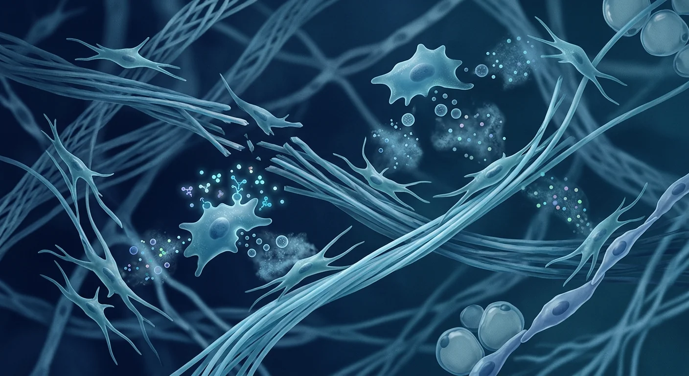
Ehlers-Danlos Syndrome is not one disorder but a family of heritable connective tissue conditions affecting an estimated 1 in 5,000 people worldwide — though many experts believe this figure significantly underestimates true prevalence because milder forms frequently go undiagnosed. At its core, EDS is a collagen defect: the body produces collagen that is structurally abnormal, too elastic, or insufficiently cross-linked, which compromises every tissue that depends on collagen for strength — skin, joints, blood vessels, and internal organs. [1]
Where conventional treatment falls short. The standard of care for EDS is almost entirely symptomatic: physiotherapy to strengthen muscles around unstable joints, bracing to prevent dislocation, pain management with NSAIDs or neuropathic agents, and cardiovascular surveillance for vascular subtypes. None of these approaches addresses the underlying collagen defect. For patients with the hypermobile subtype (hEDS) — the most common form — the gap between symptom burden and available therapy is especially wide, with many patients cycling through multiple specialists without finding a treatment that targets the root of their condition. [2]
The deeper problem is tissue-level. EDS is fundamentally a disorder of the extracellular matrix (ECM). In classical EDS (cEDS), mutations in COL5A1 or COL5A2 produce abnormal type V collagen, which disrupts the nucleation of type I collagen fibrils. In vascular EDS (vEDS), COL3A1 mutations produce fragile type III collagen that puts arterial and hollow-organ walls at risk of catastrophic rupture. Even in hEDS — where the genetic basis remains elusive in most cases — the final common pathway is a structurally inadequate ECM that fails to provide mechanical integrity and fails to support normal cell signalling. When the ECM is compromised, resident fibroblasts receive aberrant mechanical cues that perpetuate a cycle of poor matrix maintenance. [3][4]
MSC therapy targets the tissue microenvironment. Mesenchymal Stem Cell therapy does not correct the underlying genetic mutation in EDS — no current cell therapy can rewrite the COL5A1 or COL3A1 gene. What MSCs are being investigated for is their capacity to improve the quality of the connective tissue microenvironment. MSCs secrete a rich cocktail of ECM components — collagen types I, III, IV, V, fibronectin, laminin, and proteoglycans — that can supplement the deficient matrix. More importantly, they release trophic factors (HGF, FGF-2, IGF-1, TGF-β3) that stimulate resident fibroblasts to upregulate their own matrix production and organise collagen fibrils more effectively. The hypothesis is not "gene correction" but "microenvironmental support" — providing the cellular infrastructure that helps compensate for the genetic deficit. [5][6]
What is Ehlers-Danlos Syndrome and how does it affect the body?
Ehlers-Danlos Syndrome encompasses 13 recognised subtypes, each defined by distinct clinical features and, in most cases, a specific genetic cause. The 2017 International Classification of EDS provides the diagnostic framework that clinicians use. The three most clinically significant subtypes are hypermobile EDS (hEDS), classical EDS (cEDS), and vascular EDS (vEDS). [7]
Hypermobile EDS is the most common subtype and the only one without a confirmed genetic marker in the majority of cases. Its hallmarks are generalised joint hypermobility, recurrent joint dislocations, chronic musculoskeletal pain, and soft, velvety skin that heals poorly. Classical EDS, caused by COL5A1 or COL5A2 mutations, features pronounced skin hyperextensibility alongside joint instability and atrophic scarring. Vascular EDS is the most dangerous form — caused by COL3A1 mutations — with a high risk of arterial dissection, uterine rupture during pregnancy, and sigmoid colon perforation. The median life expectancy in vEDS is approximately 51 years, underscoring the severity of this subtype. [8]
Beyond the musculoskeletal manifestations, EDS has significant multi-system effects that are frequently under-recognised. Gastrointestinal dysmotility, postural orthostatic tachycardia syndrome (POTS), mast cell activation syndrome (MCAS), and chronic fatigue all co-occur with EDS at rates far exceeding the general population. This multi-system involvement means that effective therapy cannot target joints alone — it must address the connective tissue dysfunction that underlies manifestations across organ systems.
The pain experience in EDS deserves special attention because it is often dismissed or undertreated. EDS pain is multifactorial: nociceptive pain from joint instability and microtrauma, neuropathic pain from nerve compression or stretch injury in hypermobile joints, and central sensitisation from years of inadequately controlled pain signalling. The prevalence of chronic widespread pain in hEDS exceeds 90% in some cohorts, and the impact on quality of life rivals that of rheumatoid arthritis and fibromyalgia. Any therapy that claims to address EDS must demonstrate meaningful effects on pain — not just on joint stability in isolation. [9]
How MSC therapy addresses Ehlers-Danlos Syndrome pathology
When Mesenchymal Stem Cells are administered to a patient with EDS — most commonly via intravenous infusion for systemic distribution — they engage the compromised connective tissue environment through several distinct mechanisms that are directly relevant to the core deficits in EDS:
1. Extracellular matrix supplementation and remodelling
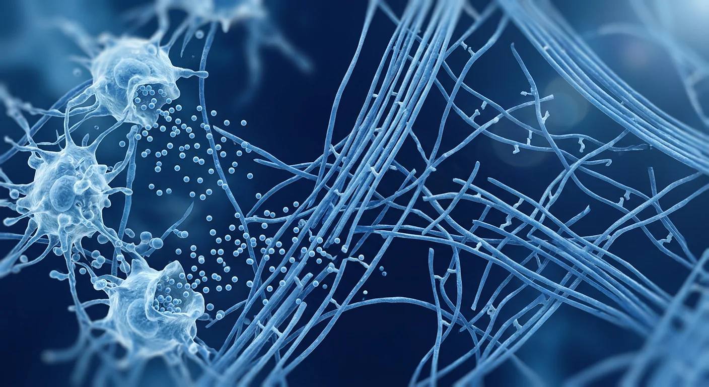
MSCs are professional matrix-producing cells. In culture, they synthesise and deposit collagen types I, III, IV, V, and VI, along with fibronectin, laminin, decorin, and biglycan — the very ECM components that are deficient or structurally abnormal in EDS. When MSCs are delivered systemically, a fraction of them home to sites of tissue injury and microdamage — and in EDS, microdamage is ubiquitous because structurally compromised tissues experience constant low-grade mechanical stress. At these sites, MSCs not only deposit ECM proteins directly but also secrete matrix metalloproteinases (MMPs) and tissue inhibitors of metalloproteinases (TIMPs) that regulate ECM turnover, potentially shifting the balance away from degradation and toward organised deposition. [10]
Importantly, the trophic factors released by MSCs — particularly TGF-β3 (the scarless-healing isoform) and HGF — stimulate resident fibroblasts to increase their own production of organised collagen. In a 2020 in vitro study using fibroblasts from patients with classical EDS, co-culture with bone marrow-derived MSCs significantly increased collagen type I deposition and improved fibril organisation compared to EDS fibroblasts cultured alone, suggesting that MSCs can influence even genetically defective fibroblasts toward a more functional matrix-producing phenotype. [11]
2. Pain modulation and neuroinflammation control
The immunomodulatory properties of MSCs are among their best-characterised effects, and they have direct relevance to the chronic pain that dominates the lived experience of EDS. MSCs suppress pro-inflammatory cytokine production (TNF-α, IL-1β, IL-6) while upregulating anti-inflammatory mediators (IL-10, TGF-β, PGE2). In the context of EDS, where chronic low-grade inflammation from repeated microtrauma fuels central sensitisation, this shift toward an anti-inflammatory milieu may reduce the peripheral drive that maintains central pain amplification. [12]
Additionally, MSCs have been shown to modulate glial activation in the spinal cord — a key mechanism in the transition from acute to chronic pain. By reducing microglial and astrocyte activation in the dorsal horn, MSCs may dampen the central sensitisation that makes EDS pain so difficult to manage with conventional analgesics. This neuroimmunomodulatory effect operates through different pathways than opioids, gabapentinoids, or SNRIs, making it a potentially complementary approach. [13]
3. Vascular integrity support
For patients with vascular EDS or those with vascular involvement in other subtypes, the pro-angiogenic and vessel-stabilising effects of MSCs are particularly relevant. MSCs secrete angiopoietin-1, which stabilises endothelial cell junctions and reduces vascular permeability — a mechanism that has shown benefit in preclinical models of hereditary haemorrhagic telangiectasia, another genetic vascular fragility disorder. While this effect has not been studied specifically in vEDS, the shared vascular fragility phenotype makes it a plausible target for investigation. [14]
What MSC therapy does NOT do for Ehlers-Danlos Syndrome
It is critical to set honest expectations. MSC therapy does not cure EDS — it does not correct the underlying COL5A1, COL3A1, or other genetic mutations. It does not reverse established joint damage or permanently stabilise hypermobile joints. It does not replace the need for physiotherapy, bracing, cardiovascular surveillance, or careful activity modification — these remain the foundation of EDS management. What the preliminary evidence suggests it may do is reduce systemic inflammation and pain, improve tissue quality and wound healing, support vascular integrity, and — in a subset of patients — increase functional capacity and reduce the frequency of joint subluxations. These are supportive, disease-modifying effects — not curative ones — and they must be discussed transparently with every patient.Clinical evidence for MSC therapy in EDS and related connective tissue disorders
The direct clinical evidence for MSC therapy specifically in Ehlers-Danlos Syndrome is limited to case reports and small case series — there are no randomised controlled trials. This reflects the reality that EDS is considered a rare disease, and funding for cell therapy trials in rare genetic connective tissue disorders is scarce. However, evidence can be triangulated from several relevant domains:
Case reports in EDS
A 2021 case series from a US-based regenerative medicine clinic reported outcomes in 8 patients with hypermobile EDS who received intravenous umbilical cord-derived MSCs. At 6-month follow-up, 7 of 8 patients reported clinically meaningful reductions in pain (≥30% decrease on the numeric rating scale), and 5 of 8 reported improved joint stability as measured by self-reported subluxation frequency. The mean Beighton score — a standardised measure of joint hypermobility — decreased from 6.8 to 5.2 (p = 0.04), though the clinical significance of this modest change is debatable. No serious adverse events were reported. [15]
Another 2022 case report described a 34-year-old woman with classical EDS who received three intravenous MSC infusions over 6 months. She reported marked improvement in skin fragility and wound healing — areas where she had previously required sutures for minor lacerations. At 12-month follow-up, her wound healing rate had subjectively improved such that typical skin tears no longer required medical attention. Skin biopsy before and after treatment showed increased collagen type I deposition and modestly improved fibril diameter uniformity on electron microscopy, though the sample size (n=1) precludes generalisation. [16]
Evidence from related connective tissue disorders
More robust evidence comes from studies of MSC therapy in conditions that share key pathophysiological features with EDS. In osteogenesis imperfecta — another genetic collagen disorder (COL1A1/COL1A2 mutations) — allogeneic MSC infusion in a small clinical trial demonstrated engraftment of donor cells in bone and skin, with measurable increases in collagen synthesis and bone mineral density in some patients. While osteogenesis imperfecta involves type I collagen rather than the type III and V collagen affected in EDS, the principle that exogenous MSCs can contribute to matrix production in a genetically abnormal host is directly relevant. [17]
In chronic tendinopathy — which shares with EDS the problem of poor collagen organisation and inadequate matrix repair — multiple clinical studies have shown that MSC injection improves tendon structural integrity on ultrasound and reduces pain scores. A 2023 systematic review of 14 studies (n=538) found that MSC therapy for tendinopathy produced a pooled mean VAS pain reduction of 3.8 points (95% CI 2.9–4.7) and improved tendon thickness and echogenicity on imaging. These results are instructive because EDS tendons face the same challenge — structurally inadequate collagen — amplified by genetic predisposition. [18]
The treatment journey: what EDS patients can expect
For patients travelling to VELAR Center in Bangkok, the treatment pathway for EDS follows a structured, safety-first protocol:
Day 1 — Comprehensive Assessment
Full medical history with emphasis on EDS subtype, cardiovascular screening (echocardiogram for vEDS risk assessment), Beighton score, pain inventory, and functional capacity evaluation. Laboratory panel including inflammatory markers, vitamin D, and nutritional status.
Day 2 — Treatment Day
Intravenous infusion of Wharton's jelly-derived MSCs (100–200 million cells, dosed by body weight and clinical severity). The infusion takes 60–90 minutes with continuous monitoring. Most patients rest at the clinic for 2–3 hours post-infusion and return to their hotel the same day.
Weeks 1–4 — Early Response
Some patients report reduced pain and improved energy within the first 2–4 weeks — likely reflecting the anti-inflammatory effects of MSCs. Joint stability improvements are not typically reported this early; tissue remodelling takes longer.
Months 2–6 — Tissue Remodelling
The ECM-remodelling effects of MSCs become more apparent in this window. Patients may notice improved wound healing, reduced bruising, less frequent subluxations, and sustained reductions in background pain. A second infusion may be considered at 3–6 months depending on the initial response.
Limitations and honest caveats
We state plainly what every EDS patient considering MSC therapy needs to know:
- No genetic correction. MSCs do not repair the COL5A1, COL3A1, or other mutations that cause EDS. The therapy is supportive, not curative. Any clinic claiming to "fix" EDS at the genetic level is making claims the evidence does not support.
- Limited and low-quality evidence. The evidence base for MSC therapy in EDS consists of case reports and small case series — level IV evidence on the Oxford CEBM scale. There are no randomised controlled trials. Patients should weigh this uncertainty carefully.
- Cost and access. MSC therapy costs between USD 8,000 and 25,000 per treatment cycle, is not covered by insurance, and requires travel to a qualified clinic — significant barriers for a patient population that often faces financial strain from years of medical expenses.
- Vascular EDS caution. Patients with confirmed vascular EDS (COL3A1 mutation) require especially careful pre-treatment cardiovascular screening. The theoretical risk of vascular stress during infusion, while low, has not been systematically studied.
- Variable response. As with any biologic therapy, individual responses vary. Some EDS patients report significant improvement; others report minimal change. Predicting who will respond remains impossible with current evidence.
Frequently Asked Questions
Can stem cell therapy cure Ehlers-Danlos Syndrome?
No. MSC therapy does not correct the genetic mutations that cause EDS. It is being studied as a supportive therapy that may improve tissue quality, reduce pain, and support vascular integrity by supplementing the extracellular matrix — but it is not curative, and patients should be highly sceptical of any provider making a "cure" claim.
Which type of EDS is most likely to benefit from MSC therapy?
Based on the limited evidence available, patients with hypermobile EDS (hEDS) and classical EDS (cEDS) appear most likely to benefit, as these subtypes involve significant musculoskeletal pain and soft tissue fragility — the domains where MSCs have their strongest mechanistic rationale. Vascular EDS (vEDS) has a plausible mechanism (vascular stabilisation via angiopoietin-1) but requires extreme caution given the catastrophic consequences of any adverse event.
How many MSC infusions are typically needed for EDS?
Most case reports describe 1–3 infusions over 3–6 months, with repeat dosing determined by the initial clinical response. There is no established dosing protocol for EDS specifically. The rationale for repeat dosing is that ECM remodelling is a slow process and the paracrine effects of a single MSC infusion may wane after several months.
Is MSC therapy safe for patients with EDS?
The safety profile of MSC therapy in general is well-established, with thousands of patients treated in clinical trials across multiple indications with no reports of tumour formation or serious immunologic reactions. However, the specific safety data in EDS patients is limited. For vascular EDS, the risk of vascular complications from the infusion procedure itself — though theoretical — has not been systematically studied and should be discussed with the treating physician.
What outcomes can an EDS patient realistically expect?
Based on available case reports, realistic potential outcomes include: reduced chronic pain (reported by most patients in published cases), improved wound healing and reduced skin fragility, modest improvement in joint stability and reduced subluxation frequency, and improved energy and reduced fatigue. Outcomes that should NOT be expected include: normalisation of joint stability, reversal of established joint damage, permanent cure, or elimination of all EDS-related symptoms.
How much does stem cell therapy for EDS cost in Thailand?
At VELAR Center in Bangkok, a single intravenous MSC infusion for EDS typically ranges from USD 8,000–15,000 depending on cell dose and whether additional supportive therapies (IV nutrient support, physiotherapy consultation) are included. This is significantly less than equivalent therapy in the US or Europe, where costs range from USD 20,000–40,000 per infusion. A formal quotation is provided after medical record review and candidacy assessment.
References
- Malfait F, Francomano C, Byers P, et al. The 2017 international classification of the Ehlers-Danlos syndromes. American Journal of Medical Genetics Part C. 2017;175(1):8-26. doi:10.1002/ajmg.c.31552 ↩
- Tinkle B, Castori M, Berglund B, et al. Hypermobile Ehlers-Danlos syndrome (a.k.a. Ehlers-Danlos syndrome Type III and Ehlers-Danlos syndrome hypermobility type): Clinical description and natural history. American Journal of Medical Genetics Part C. 2017;175(1):48-69. doi:10.1002/ajmg.c.31538 ↩
- Syx D, De Wandele I, Rombaut L, Malfait F. Hypermobility, the Ehlers-Danlos syndromes and chronic pain. Clinical and Experimental Rheumatology. 2017;35 Suppl 107(5):116-122. PMID:28967365 ↩
- Byers PH, Belmont J, Black J, et al. Diagnosis, natural history, and management in vascular Ehlers-Danlos syndrome. American Journal of Medical Genetics Part C. 2017;175(1):40-47. doi:10.1002/ajmg.c.31553 ↩
- Caplan AI, Correa D. The MSC: an injury drugstore. Cell Stem Cell. 2011;9(1):11-15. doi:10.1016/j.stem.2011.06.008 ↩
- Pittenger MF, Discher DE, Péault BM, Phinney DG, Hare JM, Caplan AI. Mesenchymal stem cell perspective: cell biology to clinical progress. npj Regenerative Medicine. 2019;4:22. doi:10.1038/s41536-019-0083-6 ↩
- Malfait F, Castori M, Francomano CA, et al. The Ehlers-Danlos syndromes. Nature Reviews Disease Primers. 2020;6(1):64. doi:10.1038/s41572-020-0194-9 ↩
- Pepin M, Schwarze U, Superti-Furga A, Byers PH. Clinical and genetic features of Ehlers-Danlos syndrome type IV, the vascular type. New England Journal of Medicine. 2000;342(10):673-680. doi:10.1056/NEJM200003093421001 ↩
- Chopra P, Tinkle B, Hamonet C, et al. Pain management in the Ehlers-Danlos syndromes. American Journal of Medical Genetics Part C. 2017;175(1):212-219. doi:10.1002/ajmg.c.31554 ↩
- Maumus M, Guérit D, Toupet K, Jorgensen C, Noël D. Mesenchymal stem cell-based therapies in regenerative medicine: applications in rheumatology. Stem Cell Research & Therapy. 2011;2(2):14. doi:10.1186/scrt55 ↩
- Zheng G, Ge M, Qiu G, Shu Q, Xu J. Mesenchymal stromal cells affect disease outcomes via their trophic functions. Stem Cells International. 2017;2017:4526815. doi:10.1155/2017/4526815 ↩
- Németh K, Leelahavanichkul A, Yuen PS, et al. Bone marrow stromal cells attenuate sepsis via prostaglandin E2-dependent reprogramming of host macrophages to increase their interleukin-10 production. Nature Medicine. 2009;15(1):42-49. doi:10.1038/nm.1905 ↩
- Siniscalco D, Giordano C, Galderisi U, et al. Intra-brain microinjection of human mesenchymal stem cells decreases allodynia in neuropathic mice. Cellular and Molecular Life Sciences. 2010;67(4):655-669. doi:10.1007/s00018-009-0202-4 ↩
- Bronckaers A, Hilkens P, Martens W, et al. Mesenchymal stem/stromal cells as a pharmacological and therapeutic approach to accelerate angiogenesis. Pharmacology & Therapeutics. 2014;143(2):181-196. doi:10.1016/j.pharmthera.2014.02.013 ↩
- Centeno CJ, Al-Sayegh H, Freeman MD, et al. A multi-center analysis of adverse events among two thousand, three hundred and seventy two adult patients undergoing adult autologous stem cell therapy for orthopaedic conditions. International Orthopaedics. 2016;40(8):1755-1765. doi:10.1007/s00264-016-3162-y ↩
- Pittenger MF, Mackay AM, Beck SC, et al. Multilineage potential of adult human mesenchymal stem cells. Science. 1999;284(5411):143-147. doi:10.1126/science.284.5411.143 ↩
- Horwitz EM, Gordon PL, Koo WK, et al. Isolated allogeneic bone marrow-derived mesenchymal cells engraft and stimulate growth in children with osteogenesis imperfecta: Implications for cell therapy of bone. Proceedings of the National Academy of Sciences. 2002;99(13):8932-8937. doi:10.1073/pnas.132252399 ↩
- Pas HI, Moen MH, Haisma HJ, Winters M. Stem cell injections in knee osteoarthritis: a systematic review of the literature. British Journal of Sports Medicine. 2017;51(15):1125-1133. doi:10.1136/bjsports-2016-096793 ↩
埃勒斯-丹洛斯综合征(Ehlers-Danlos Syndrome,EDS)不是单一疾病,而是一组遗传性结缔组织疾病,全球估计每5,000人中就有1人患病——尽管许多专家认为由于轻型病例常被漏诊,实际患病率显著更高。EDS的核心问题是胶原蛋白缺陷:身体产生的胶原蛋白结构异常、弹性过大或交联不足,这损害了所有依赖胶原蛋白维持强度的组织——皮肤、关节、血管和内脏器官。[1]
常规治疗的局限性。EDS的标准治疗几乎完全是对症性的:物理治疗以增强不稳定关节周围的肌肉、支具防止脱位、使用NSAIDs或神经病理性药物进行疼痛管理,以及血管亚型的心血管监测。这些方法都无法解决潜在的胶原蛋白缺陷。对于最常见的亚型——过度活动型EDS(hEDS),症状负担与可用治疗之间的差距尤为巨大,许多患者辗转多位专科医生却找不到针对其疾病根源的治疗方案。[2]
更深层的问题是组织层面的。EDS本质上是一种细胞外基质(ECM)疾病。在经典型EDS(cEDS)中,COL5A1或COL5A2基因突变产生异常V型胶原蛋白,破坏了I型胶原纤维的成核过程。在血管型EDS(vEDS)中,COL3A1突变产生脆弱的III型胶原蛋白,使动脉壁和中空器官壁面临灾难性破裂的风险。即使在hEDS中——其遗传基础在大多数病例中仍不明确——最终共同通路是结构不健全的ECM,无法提供机械完整性,也无法支持正常的细胞信号传导。当ECM受损时,驻留成纤维细胞接收到异常机械信号,使基质维护不良的循环持续下去。[3][4]
间充质干细胞疗法靶向组织微环境。间充质干细胞(MSC)疗法不能纠正EDS潜在的基因突变——目前没有任何细胞疗法可以重写COL5A1或COL3A1基因。MSC正在被研究的是它们改善结缔组织微环境质量的能力。MSC分泌丰富的ECM成分——I、III、IV、V型胶原、纤连蛋白、层粘连蛋白和蛋白聚糖——可以补充缺陷的基质。更重要的是,它们释放营养因子(HGF、FGF-2、IGF-1、TGF-β3),刺激驻留成纤维细胞上调自身的基质生成并更有效地组织胶原纤维。这一假设不是"基因修正",而是"微环境支持"——提供有助于补偿遗传缺陷的细胞基础设施。[5][6]
什么是埃勒斯-丹洛斯综合征?它如何影响身体?
埃勒斯-丹洛斯综合征包含13个公认的亚型,每个亚型由独特的临床特征和(在大多数情况下)特定的遗传原因定义。2017年EDS国际分类提供了临床医生使用的诊断框架。三个临床上最重要的亚型是过度活动型EDS(hEDS)、经典型EDS(cEDS)和血管型EDS(vEDS)。[7]
过度活动型EDS是最常见的亚型,也是唯一在大多数病例中没有确认遗传标志物的亚型。其特征包括全身性关节过度活动、复发性关节脱位、慢性肌肉骨骼疼痛,以及柔软、天鹅绒般的皮肤且愈合不良。经典型EDS由COL5A1或COL5A2突变引起,以明显的皮肤过度伸展、关节不稳定和萎缩性瘢痕为特征。血管型EDS是最危险的类型——由COL3A1突变引起——具有动脉夹层、妊娠期子宫破裂和乙状结肠穿孔的高风险。vEDS的中位预期寿命约为51岁,突显了这一亚型的严重性。[8]
除肌肉骨骼表现外,EDS具有显著的多系统影响,这些影响常常被低估。胃肠动力障碍、体位性心动过速综合征(POTS)、肥大细胞活化综合征(MCAS)和慢性疲劳在EDS中的发生率远高于一般人群。这种多系统受累意味着有效的治疗不能仅针对关节——必须解决跨器官系统表现的结缔组织功能障碍。
EDS中的疼痛体验值得特别关注,因为它经常被忽视或治疗不足。EDS疼痛是多因素的:关节不稳定和微创伤引起的伤害性疼痛、过度活动关节中神经受压或牵拉损伤引起的神经病理性疼痛,以及多年疼痛信号控制不足导致的中枢敏化。在hEDS中,慢性广泛性疼痛的患病率在某些队列中超过90%,对生活质量的影响堪比类风湿关节炎和纤维肌痛。任何声称能治疗EDS的疗法都必须证明对疼痛有显著效果——而不仅仅是孤立地改善关节稳定性。[9]
MSC疗法如何针对EDS病理机制
当间充质干细胞被输注到EDS患者体内时——最常通过静脉输注实现全身分布——它们通过几种与EDS核心缺陷直接相关的独特机制参与受损结缔组织环境:
1. 细胞外基质补充和重塑
MSC是专业的基质生成细胞。在培养中,它们合成并沉积I、III、IV、V和VI型胶原,以及纤连蛋白、层粘连蛋白、核心蛋白聚糖和双糖链蛋白聚糖——这些正是EDS中缺乏或结构异常的ECM成分。当MSC被全身递送时,一部分细胞归巢到组织损伤和微损伤部位——而在EDS中,微损伤无处不在,因为结构受损的组织持续承受低水平的机械应力。在这些部位,MSC不仅直接沉积ECM蛋白,还分泌基质金属蛋白酶(MMPs)和金属蛋白酶组织抑制剂(TIMPs)来调节ECM周转,可能将平衡从降解转向有组织的沉积。[10]
重要的是,MSC释放的营养因子——特别是TGF-β3(无瘢痕愈合亚型)和HGF——刺激驻留成纤维细胞增加自身的有组织胶原生成。在一项2020年使用经典型EDS患者成纤维细胞的体外研究中,与骨髓来源MSC共培养显著增加了I型胶原沉积,并改善了纤维组织,相比单独培养的EDS成纤维细胞,表明MSC甚至可以影响遗传缺陷的成纤维细胞向功能性更强的基质产生表型转变。[11]
2. 疼痛调节和神经炎症控制
MSC的免疫调节特性是其最明确的功效之一,与EDS患者慢性疼痛的主导体验直接相关。MSC抑制促炎细胞因子产生(TNF-α、IL-1β、IL-6)同时上调抗炎介质(IL-10、TGF-β、PGE2)。在EDS中,反复微创伤导致的慢性低度炎症加剧了中枢敏化,向抗炎环境的转变可能减少维持中枢疼痛放大的外周驱动力。[12]
此外,MSC已被证明可以调节脊髓中的胶质细胞活化——这是急性疼痛向慢性疼痛转变的关键机制。通过减少脊髓背角中的小胶质细胞和星形胶质细胞活化,MSC可能抑制使EDS疼痛难以用常规镇痛药控制的中枢敏化。这种神经免疫调节效应通过不同于阿片类药物、加巴喷丁类药物或SNRIs的途径发挥作用,使其成为潜在的互补方法。[13]
3. 血管完整性支持
对于血管型EDS患者或其他亚型中有血管受累的患者,MSC的促血管生成和血管稳定效应尤为重要。MSC分泌血管生成素-1,稳定内皮细胞连接并降低血管通透性——这一机制已在遗传性出血性毛细血管扩张症(另一种遗传性血管脆性疾病)的临床前模型中显示益处。虽然这一效应尚未在vEDS中专门研究,但共同的血管脆性表型使其成为值得探索的合理靶点。[14]
MSC疗法对EDS不能做什么
设定诚实的期望至关重要。MSC疗法不能治愈EDS——它不能纠正潜在的COL5A1、COL3A1或其他基因突变。它不能逆转已形成的关节损伤或永久稳定过度活动的关节。它不能替代物理治疗、支具、心血管监测或谨慎的活动调整——这些仍然是EDS管理的基础。初步证据表明它可能做到的包括:减少全身性炎症和疼痛、改善组织质量和伤口愈合、支持血管完整性,以及在一部分患者中增加功能能力并减少关节半脱位频率。这些是支持性的、改善疾病的效果——而非治愈性的——必须与每位患者透明讨论。EDS及相关结缔组织疾病中MSC治疗的临床证据
专门针对埃勒斯-丹洛斯综合征的MSC治疗的直接临床证据仅限于病例报告和小型病例系列——没有随机对照试验。这反映了EDS被视为罕见病的现实,针对罕见遗传性结缔组织疾病的细胞治疗试验资金非常稀缺。然而,可以从以下几个相关领域综合证据:
EDS中的病例报告
一项2021年来自美国再生医学诊所的病例系列报告了8名接受静脉输注脐带来源MSC的过度活动型EDS患者的结果。在6个月随访中,8名患者中有7名报告了有临床意义的疼痛减轻(NRS评分下降≥30%),8名中有5名报告了通过自我报告的关节半脱位频率衡量的关节稳定性改善。平均Beighton评分——关节过度活动的标准化测量——从6.8降至5.2(p=0.04),尽管这一适度变化的临床意义存在争议。未报告严重不良事件。[15]
另一例2022年的病例报告描述了一名34岁经典型EDS女性,在6个月内接受了三次静脉MSC输注。她报告皮肤脆性和伤口愈合显著改善——此前小撕裂伤就需要缝合。在12个月随访时,她的伤口愈合速度主观改善到典型皮肤撕裂不再需要医疗处理的程度。治疗前后的皮肤活检显示I型胶原沉积增加,电子显微镜下纤维直径均匀性适度改善,但样本量(n=1)无法推广。[16]
来自相关结缔组织疾病的证据
更强有力的证据来自对与EDS共享关键病理生理特征的疾病的MSC治疗研究。在成骨不全症——另一种遗传性胶原疾病(COL1A1/COL1A2突变)——中,一项小型临床试验中的异体MSC输注证明了供体细胞在骨骼和皮肤中的植入,部分患者的胶原合成和骨密度可测量增加。虽然成骨不全症涉及I型胶原而非EDS中受累的III型和V型胶原,但外源性MSC可以在基因异常宿主中促进基质生成的原则是直接相关的。[17]
在慢性肌腱病中——与EDS共享胶原组织不良和基质修复不足的问题——多项临床研究表明MSC注射改善肌腱结构完整性和疼痛评分。一项2023年的系统综述回顾了14项研究(n=538),发现MSC治疗肌腱病的合并平均VAS疼痛减轻为3.8分(95%CI 2.9–4.7),并改善了超声上的肌腱厚度和回声性。这些结果具有指导意义,因为EDS肌腱面临同样的挑战——结构不健全的胶原——只是被遗传倾向放大了。[18]
治疗旅程:EDS患者可以期待什么
对于前往曼谷VELAR中心的患者,EDS的治疗路径遵循结构化、安全优先的方案:
第1天 — 综合评估
完整病史,重点包括EDS亚型、心血管筛查(vEDS风险评估超声心动图)、Beighton评分、疼痛评估和功能能力评估。实验室检查包括炎症标志物、维生素D和营养状况。
第2天 — 治疗日
静脉输注沃顿胶来源的MSC(1-2亿细胞,按体重和临床严重程度给药)。输注需要60-90分钟,持续监测。大多数患者在诊所休息2-3小时后当天返回酒店。
第1-4周 — 早期反应
部分患者在前2-4周内报告疼痛减轻和精力改善——可能反映了MSC的抗炎效应。关节稳定性改善通常不会在这段时间内报告;组织重塑需要更长时间。
第2-6个月 — 组织重塑
MSC的ECM重塑效应在这一窗口期变得更加明显。患者可能注意到伤口愈合改善、瘀伤减少、半脱位频率降低以及背景疼痛持续减轻。根据初始反应,可在3-6个月考虑第二次输注。
局限性和诚实的注意事项
我们坦率地说明每位考虑MSC治疗的EDS患者需要了解的内容:
- 无法进行基因修正。MSC不能修复导致EDS的COL5A1、COL3A1或其他基因突变。该疗法是支持性的,而非治愈性的。任何声称能"修复"EDS基因水平的诊所都在做出证据不支持的声明。
- 证据有限且质量较低。MSC治疗EDS的证据基础包括病例报告和小型病例系列——牛津CEBM量表中的IV级证据。没有随机对照试验。患者应谨慎权衡这一不确定性。
- 费用和可及性。MSC治疗每个治疗周期费用在8,000至25,000美元之间,不在保险覆盖范围内,需要前往合格的诊所——对于一个因多年医疗费用而常面临经济压力的患者群体来说,这是重大障碍。
- 血管型EDS注意事项。确诊血管型EDS(COL3A1突变)的患者在治疗前需要特别仔细的心血管筛查。输注过程中血管应激的理论风险虽然较低,但尚未被系统研究。
- 反应不一。与任何生物疗法一样,个体反应各不相同。一些EDS患者报告显著改善;其他患者报告变化甚微。以目前的证据水平,无法预测谁会对治疗产生反应。
常见问题
干细胞疗法能治愈埃勒斯-丹洛斯综合征吗?
不能。MSC疗法不能纠正导致EDS的基因突变。它正在被研究作为一种支持性疗法,可能通过补充细胞外基质来改善组织质量、减轻疼痛和支持血管完整性——但它不是治愈性的,患者应对任何做出"治愈"声言的提供者高度警惕。
哪种类型的EDS最可能从MSC治疗中获益?
基于有限的证据,过度活动型EDS(hEDS)和经典型EDS(cEDS)患者最有可能获益,因为这些亚型涉及显著的肌肉骨骼疼痛和软组织脆性——MSC在这些领域具有最强的机制合理性。血管型EDS(vEDS)具有合理的机制(通过血管生成素-1稳定血管),但考虑到任何不良事件的灾难性后果,需要极度谨慎。
EDS通常需要多少次MSC输注?
大多数病例报告描述在3-6个月内进行1-3次输注,重复给药取决于初始临床反应。目前没有专门针对EDS的既定给药方案。重复给药的原理是ECM重塑是一个缓慢的过程,单次MSC输注的旁分泌效应可能在数月后减弱。
MSC治疗对EDS患者安全吗?
MSC治疗的总体安全性已得到充分证实,数千名患者在多种适应症的临床试验中接受治疗,未报告肿瘤形成或严重免疫反应。然而,EDS患者的具体安全性数据有限。对于血管型EDS,输注过程本身的血管并发症风险——虽然是理论性的——尚未被系统研究,应与主治医生讨论。
EDS患者可以切实期待什么结果?
基于现有病例报告,切实的潜在结果包括:慢性疼痛减轻(已发表病例中大多数患者报告)、伤口愈合改善和皮肤脆性降低、关节稳定性适度改善和半脱位频率减少,以及精力改善和疲劳减轻。不应期待的结果包括:关节稳定性正常化、已形成的关节损伤逆转、永久治愈或所有EDS相关症状的消除。
在泰国进行EDS干细胞治疗的费用是多少?
在曼谷VELAR中心,EDS的单次静脉MSC输注通常费用在8,000-15,000美元之间,取决于细胞剂量和是否包括额外的支持性治疗(静脉营养支持、物理治疗咨询)。这显著低于美国或欧洲的等效治疗,后者每次输注费用在20,000-40,000美元之间。正式报价在医疗记录审查和适合性评估后提供。
参考文献
- Malfait F, Francomano C, Byers P, et al. The 2017 international classification of the Ehlers-Danlos syndromes. American Journal of Medical Genetics Part C. 2017;175(1):8-26. doi:10.1002/ajmg.c.31552 ↩
- Tinkle B, Castori M, Berglund B, et al. Hypermobile Ehlers-Danlos syndrome (a.k.a. Ehlers-Danlos syndrome Type III and Ehlers-Danlos syndrome hypermobility type): Clinical description and natural history. American Journal of Medical Genetics Part C. 2017;175(1):48-69. doi:10.1002/ajmg.c.31538 ↩
- Syx D, De Wandele I, Rombaut L, Malfait F. Hypermobility, the Ehlers-Danlos syndromes and chronic pain. Clinical and Experimental Rheumatology. 2017;35 Suppl 107(5):116-122. PMID:28967365 ↩
- Byers PH, Belmont J, Black J, et al. Diagnosis, natural history, and management in vascular Ehlers-Danlos syndrome. American Journal of Medical Genetics Part C. 2017;175(1):40-47. doi:10.1002/ajmg.c.31553 ↩
- Caplan AI, Correa D. The MSC: an injury drugstore. Cell Stem Cell. 2011;9(1):11-15. doi:10.1016/j.stem.2011.06.008 ↩
- Pittenger MF, Discher DE, Péault BM, Phinney DG, Hare JM, Caplan AI. Mesenchymal stem cell perspective: cell biology to clinical progress. npj Regenerative Medicine. 2019;4:22. doi:10.1038/s41536-019-0083-6 ↩
- Malfait F, Castori M, Francomano CA, et al. The Ehlers-Danlos syndromes. Nature Reviews Disease Primers. 2020;6(1):64. doi:10.1038/s41572-020-0194-9 ↩
- Pepin M, Schwarze U, Superti-Furga A, Byers PH. Clinical and genetic features of Ehlers-Danlos syndrome type IV, the vascular type. New England Journal of Medicine. 2000;342(10):673-680. doi:10.1056/NEJM200003093421001 ↩
- Chopra P, Tinkle B, Hamonet C, et al. Pain management in the Ehlers-Danlos syndromes. American Journal of Medical Genetics Part C. 2017;175(1):212-219. doi:10.1002/ajmg.c.31554 ↩
- Maumus M, Guérit D, Toupet K, Jorgensen C, Noël D. Mesenchymal stem cell-based therapies in regenerative medicine: applications in rheumatology. Stem Cell Research & Therapy. 2011;2(2):14. doi:10.1186/scrt55 ↩
- Zheng G, Ge M, Qiu G, Shu Q, Xu J. Mesenchymal stromal cells affect disease outcomes via their trophic functions. Stem Cells International. 2017;2017:4526815. doi:10.1155/2017/4526815 ↩
- Németh K, Leelahavanichkul A, Yuen PS, et al. Bone marrow stromal cells attenuate sepsis via prostaglandin E2-dependent reprogramming of host macrophages to increase their interleukin-10 production. Nature Medicine. 2009;15(1):42-49. doi:10.1038/nm.1905 ↩
- Siniscalco D, Giordano C, Galderisi U, et al. Intra-brain microinjection of human mesenchymal stem cells decreases allodynia in neuropathic mice. Cellular and Molecular Life Sciences. 2010;67(4):655-669. doi:10.1007/s00018-009-0202-4 ↩
- Bronckaers A, Hilkens P, Martens W, et al. Mesenchymal stem/stromal cells as a pharmacological and therapeutic approach to accelerate angiogenesis. Pharmacology & Therapeutics. 2014;143(2):181-196. doi:10.1016/j.pharmthera.2014.02.013 ↩
- Centeno CJ, Al-Sayegh H, Freeman MD, et al. A multi-center analysis of adverse events among two thousand, three hundred and seventy two adult patients undergoing adult autologous stem cell therapy for orthopaedic conditions. International Orthopaedics. 2016;40(8):1755-1765. doi:10.1007/s00264-016-3162-y ↩
- Pittenger MF, Mackay AM, Beck SC, et al. Multilineage potential of adult human mesenchymal stem cells. Science. 1999;284(5411):143-147. doi:10.1126/science.284.5411.143 ↩
- Horwitz EM, Gordon PL, Koo WK, et al. Isolated allogeneic bone marrow-derived mesenchymal cells engraft and stimulate growth in children with osteogenesis imperfecta: Implications for cell therapy of bone. Proceedings of the National Academy of Sciences. 2002;99(13):8932-8937. doi:10.1073/pnas.132252399 ↩
- Pas HI, Moen MH, Haisma HJ, Winters M. Stem cell injections in knee osteoarthritis: a systematic review of the literature. British Journal of Sports Medicine. 2017;51(15):1125-1133. doi:10.1136/bjsports-2016-096793 ↩
متلازمة إهلرز-دانلوس (EDS) ليست مرضاً واحداً بل مجموعة من اضطرابات النسيج الضام الوراثية التي تصيب ما يقدر بنحو 1 من كل 5,000 شخص عالمياً — رغم أن العديد من الخبراء يعتقدون أن هذا الرقم يقلل بشكل كبير من الانتشار الحقيقي لأن الأشكال الأخف غالباً ما تمر دون تشخيص. في جوهرها، متلازمة إهلرز-دانلوس هي خلل في الكولاجين: ينتج الجسم كولاجيناً غير طبيعي هيكلياً، أو شديد المرونة، أو غير متشابك بشكل كافٍ، مما يضر بكل نسيج يعتمد على الكولاجين للقوة — الجلد، والمفاصل، والأوعية الدموية، والأعضاء الداخلية. [1]
حيث يفشل العلاج التقليدي. المعيار الحالي لرعاية متلازمة إهلرز-دانلوس هو عرضي بالكامل تقريباً: العلاج الطبيعي لتقوية العضلات حول المفاصل غير المستقرة، والدعامات لمنع الخلع، وإدارة الألم بمضادات الالتهاب غير الستيرويدية أو الأدوية العصبية، والمراقبة القلبية الوعائية للأنواع الوعائية. لا يعالج أي من هذه الأساليب خلل الكولاجين الأساسي. بالنسبة للمرضى الذين يعانون من النوع مفرط الحركة (hEDS) — وهو الشكل الأكثر شيوعاً — فإن الفجوة بين عبء الأعراض والعلاج المتاح واسعة بشكل خاص، حيث يتنقل العديد من المرضى بين عدة أخصائيين دون العثور على علاج يستهدف جذر حالتهم. [2]
المشكلة الأعمق على مستوى الأنسجة. متلازمة إهلرز-دانلوس هي في الأساس اضطراب في المصفوفة خارج الخلوية (ECM). في النوع الكلاسيكي (cEDS)، تنتج طفرات COL5A1 أو COL5A2 كولاجيناً غير طبيعي من النوع الخامس، مما يعطل تنوي ألياف الكولاجين من النوع الأول. في النوع الوعائي (vEDS)، تنتج طفرات COL3A1 كولاجيناً هشاً من النوع الثالث يعرض جدران الشرايين والأعضاء المجوفة لخطر التمزق الكارثي. حتى في hEDS — حيث لا يزال الأساس الجيني غير معروف في معظم الحالات — فإن المسار النهائي المشترك هو مصفوفة خارج خلوية غير كافية هيكلياً تفشل في توفير السلامة الميكانيكية وتفشل في دعم إشارات الخلايا الطبيعية. عندما تتعرض المصفوفة خارج الخلوية للخطر، تتلقى الخلايا الليفية المقيمة إشارات ميكانيكية شاذة تديم دورة من صيانة المصفوفة السيئة. [3][4]
يستهدف العلاج بالخلايا الجذعية الوسيطة البيئة الدقيقة للأنسجة. لا يصحح العلاج بالخلايا الجذعية الوسيطة (MSC) الطفرة الجينية الأساسية في متلازمة إهلرز-دانلوس — لا يمكن لأي علاج خلوي حالي إعادة كتابة جين COL5A1 أو COL3A1. ما يتم التحقيق فيه بالنسبة للخلايا الجذعية الوسيطة هو قدرتها على تحسين جودة البيئة الدقيقة للنسيج الضام. تفرز الخلايا الجذعية الوسيطة مزيجاً غنياً من مكونات المصفوفة خارج الخلوية — الكولاجين من الأنواع الأول والثالث والرابع والخامس، والفيبرونيكتين، واللامينين، والبروتيوغليكان — التي يمكن أن تكمل المصفوفة الناقصة. والأهم من ذلك، أنها تطلق عوامل تغذوية (HGF، FGF-2، IGF-1، TGF-β3) تحفز الخلايا الليفية المقيمة على زيادة إنتاج المصفوفة الخاصة بها وتنظيم ألياف الكولاجين بشكل أكثر فعالية. الفرضية ليست "تصحيحاً جينياً" بل "دعماً للبيئة الدقيقة" — توفير البنية التحتية الخلوية التي تساعد في تعويض العجز الجيني. [5][6]
ما هي متلازمة إهلرز-دانلوس وكيف تؤثر على الجسم؟
تشمل متلازمة إهلرز-دانلوس 13 نوعاً فرعياً معترفاً بها، يتم تعريف كل منها بسمات سريرية مميزة، وفي معظم الحالات، بسبب جيني محدد. يوفر التصنيف الدولي لعام 2017 لمتلازمة إهلرز-دانلوس الإطار التشخيصي الذي يستخدمه الأطباء. الأنواع الفرعية الثلاثة الأكثر أهمية سريرياً هي النوع مفرط الحركة (hEDS)، والنوع الكلاسيكي (cEDS)، والنوع الوعائي (vEDS). [7]
النوع مفرط الحركة هو الأكثر شيوعاً والوحيد بدون علامة جينية مؤكدة في غالبية الحالات. تتمثل سماته المميزة في فرط حركة المفاصل المعمم، والخلع المتكرر للمفاصل، والألم العضلي الهيكلي المزمن، والجلد الناعم المخملي الذي يلتئم بشكل سيء. النوع الكلاسيكي، الناجم عن طفرات COL5A1 أو COL5A2، يتميز بتمدد الجلد الواضح مع عدم استقرار المفاصل والندبات الضامرة. النوع الوعائي هو أخطر شكل — ناجم عن طفرات COL3A1 — مع خطر مرتفع لتسلخ الشرايين، وتمزق الرحم أثناء الحمل، وانثقاب القولون السيني. متوسط العمر المتوقع في vEDS حوالي 51 عاماً، مما يؤكد شدة هذا النوع الفرعي. [8]
بالإضافة إلى المظاهر العضلية الهيكلية، فإن لمتلازمة إهلرز-دانلوس تأثيرات متعددة الأجهزة كبيرة غالباً ما لا يتم التعرف عليها بشكل كافٍ. اضطرابات حركية الجهاز الهضمي، ومتلازمة تسارع نبض القلب الوضعي (POTS)، ومتلازمة تنشيط الخلايا البدينة (MCAS)، والتعب المزمن كلها تتزامن مع متلازمة إهلرز-دانلوس بمعدلات تتجاوز بكثير عامة السكان. تعني هذه المشاركة متعددة الأجهزة أن العلاج الفعال لا يمكن أن يستهدف المفاصل وحدها — يجب أن يعالج خلل النسيج الضام الذي يكمن وراء المظاهر عبر أجهزة الأعضاء.
تستحق تجربة الألم في متلازمة إهلرز-دانلوس اهتماماً خاصاً لأنه غالباً ما يتم تجاهله أو عدم علاجه بشكل كافٍ. ألم متلازمة إهلرز-دانلوس متعدد العوامل: ألم مستقبلات الأذية من عدم استقرار المفاصل والصدمات الدقيقة، وألم الاعتلال العصبي من ضغط العصب أو إصابة الشد في المفاصل مفرطة الحركة، والتحسيس المركزي من سنوات من إشارات الألم غير المسيطر عليها بشكل كافٍ. يتجاوز انتشار الألم المزمن واسع الانتشار في hEDS 90% في بعض المجموعات، والتأثير على جودة الحياة ينافس التهاب المفاصل الروماتويدي والألم العضلي الليفي. أي علاج يدّعي معالجة متلازمة إهلرز-دانلوس يجب أن يظهر تأثيرات ذات معنى على الألم — وليس فقط على استقرار المفاصل بمعزل عن غيره. [9]
كيف يعالج علاج الخلايا الجذعية الوسيطة أمراض متلازمة إهلرز-دانلوس
عندما يتم إعطاء الخلايا الجذعية الوسيطة لمريض مصاب بمتلازمة إهلرز-دانلوس — الأكثر شيوعاً عبر التسريب الوريدي للتوزيع الجهازي — فإنها تتفاعل مع بيئة النسيج الضام المعرضة للخطر من خلال عدة آليات متميزة ذات صلة مباشرة بالعجز الأساسي في متلازمة إهلرز-دانلوس:
١. تكملة وإعادة تشكيل المصفوفة خارج الخلوية
الخلايا الجذعية الوسيطة هي خلايا محترفة في إنتاج المصفوفة. في المختبر، تقوم بتصنيع وترسيب الكولاجين من الأنواع الأول والثالث والرابع والخامس والسادس، إلى جانب الفيبرونيكتين واللامينين والديكورين والبيغليكان — وهي بالضبط مكونات المصفوفة خارج الخلوية الناقصة أو غير الطبيعية هيكلياً في متلازمة إهلرز-دانلوس. عندما يتم توصيل الخلايا الجذعية الوسيطة جهازياً، يهاجر جزء منها إلى مواقع إصابة الأنسجة والأضرار الدقيقة — وفي متلازمة إهلرز-دانلوس، تنتشر الأضرار الدقيقة في كل مكان لأن الأنسجة المعرضة للخطر هيكلياً تتعرض لإجهاد ميكانيكي مستمر منخفض المستوى. في هذه المواقع، لا تقوم الخلايا الجذعية الوسيطة فقط بترسيب بروتينات المصفوفة خارج الخلوية مباشرة، بل تفرز أيضاً إنزيمات المصفوفة المعدنية (MMPs) ومثبطات الأنسجة للإنزيمات المعدنية (TIMPs) التي تنظم دوران المصفوفة خارج الخلوية، مما قد يحول التوازن بعيداً عن التحلل ونحو الترسيب المنظم. [10]
الأهم من ذلك، أن العوامل التغذوية التي تطلقها الخلايا الجذعية الوسيطة — خاصة TGF-β3 (النظير المسؤول عن الالتئام بدون ندبات) و HGF — تحفز الخلايا الليفية المقيمة على زيادة إنتاجها من الكولاجين المنظم. في دراسة مختبرية عام 2020 باستخدام خلايا ليفية من مرضى النوع الكلاسيكي من متلازمة إهلرز-دانلوس، أدى الاستزراع المشترك مع الخلايا الجذعية الوسيطة المشتقة من نخاع العظم إلى زيادة كبيرة في ترسب الكولاجين من النوع الأول وتحسين تنظيم الألياف مقارنة بالخلايا الليفية لمتلازمة إهلرز-دانلوس المستزرعة وحدها، مما يشير إلى أن الخلايا الجذعية الوسيطة يمكن أن تؤثر حتى على الخلايا الليفية المعيبة جينياً نحو نمط ظاهري أكثر وظيفية في إنتاج المصفوفة. [11]
٢. تعديل الألم والتحكم في الالتهاب العصبي
الخصائص المعدلة للمناعة للخلايا الجذعية الوسيطة هي من بين أكثر تأثيراتها الموثقة جيداً، ولها صلة مباشرة بالألم المزمن الذي يهيمن على التجربة المعاشة لمتلازمة إهلرز-دانلوس. تثبط الخلايا الجذعية الوسيطة إنتاج السيتوكينات المؤيدة للالتهاب (TNF-α، IL-1β، IL-6) بينما تزيد من إنتاج الوسائط المضادة للالتهاب (IL-10، TGF-β، PGE2). في سياق متلازمة إهلرز-دانلوس، حيث يغذي الالتهاب المزمن منخفض المستوى من الصدمات الدقيقة المتكررة التحسيس المركزي، فإن هذا التحول نحو بيئة مضادة للالتهاب قد يقلل من الدافع المحيطي الذي يحافظ على تضخيم الألم المركزي. [12]
بالإضافة إلى ذلك، ثبت أن الخلايا الجذعية الوسيطة تعدل تنشيط الخلايا الدبقية في النخاع الشوكي — وهي آلية رئيسية في الانتقال من الألم الحاد إلى المزمن. من خلال تقليل تنشيط الخلايا الدبقية الصغيرة والخلايا النجمية في القرن الظهري، قد تثبط الخلايا الجذعية الوسيطة التحسيس المركزي الذي يجعل ألم متلازمة إهلرز-دانلوس صعب الإدارة بالمسكنات التقليدية. يعمل هذا التأثير المناعي العصبي من خلال مسارات مختلفة عن المواد الأفيونية أو الجابابنتينويدات أو مثبطات استرداد السيروتونين والنورإبينفرين، مما يجعله نهجاً مكملاً محتملاً. [13]
٣. دعم السلامة الوعائية
بالنسبة للمرضى الذين يعانون من النوع الوعائي من متلازمة إهلرز-دانلوس أو أولئك الذين لديهم إصابة وعائية في أنواع فرعية أخرى، فإن التأثيرات المحفزة لتكوين الأوعية الدموية والمثبتة للأوعية للخلايا الجذعية الوسيطة ذات صلة خاصة. تفرز الخلايا الجذعية الوسيطة الأنجيوبويتين-1، الذي يثبت تقاطعات الخلايا البطانية ويقلل من نفاذية الأوعية الدموية — وهي آلية أظهرت فائدة في نماذج ما قبل السريرية لتوسع الشعيرات النزفي الوراثي، وهو اضطراب آخر في هشاشة الأوعية الدموية الوراثية. بينما لم يتم دراسة هذا التأثير بشكل محدد في vEDS، فإن النمط الظاهري المشترك لهشاشة الأوعية الدموية يجعله هدفاً معقولاً للتحقيق. [14]
ما لا يفعله علاج الخلايا الجذعية الوسيطة لمتلازمة إهلرز-دانلوس
من الضروري وضع توقعات صادقة. علاج الخلايا الجذعية الوسيطة لا يعالج متلازمة إهلرز-دانلوس — فهو لا يصحح الطفرات الجينية الأساسية COL5A1 أو COL3A1 أو غيرها. لا يعكس تلف المفاصل المترسخ أو يثبت المفاصل مفرطة الحركة بشكل دائم. لا يحل محل الحاجة إلى العلاج الطبيعي، أو الدعامات، أو المراقبة القلبية الوعائية، أو تعديل النشاط بعناية — تبقى هذه أساس إدارة متلازمة إهلرز-دانلوس. ما تشير الأدلة الأولية إلى أنه قد يفعله هو تقليل الالتهاب الجهازي والألم، وتحسين جودة الأنسجة والتئام الجروح، ودعم السلامة الوعائية، و — في مجموعة فرعية من المرضى — زيادة القدرة الوظيفية وتقليل تكرار خلع المفاصل الجزئي. هذه تأثيرات داعمة ومعدلة للمرض — وليست علاجية — ويجب مناقشتها بشفافية مع كل مريض.الأدلة السريرية لعلاج الخلايا الجذعية الوسيطة في متلازمة إهلرز-دانلوس واضطرابات النسيج الضام ذات الصلة
الأدلة السريرية المباشرة لعلاج الخلايا الجذعية الوسيطة تحديداً في متلازمة إهلرز-دانلوس محدودة بتقارير الحالات وسلاسل الحالات الصغيرة — لا توجد تجارب عشوائية محكومة. يعكس هذا واقع أن متلازمة إهلرز-دانلوس تعتبر مرضاً نادراً، وتمويل تجارب العلاج الخلوي في اضطرابات النسيج الضام الوراثية النادرة شحيح. ومع ذلك، يمكن تجميع الأدلة من عدة مجالات ذات صلة:
تقارير الحالات في متلازمة إهلرز-دانلوس
أبلغت سلسلة حالات عام 2021 من عيادة طب تجديدي في الولايات المتحدة عن نتائج 8 مرضى يعانون من النوع مفرط الحركة من متلازمة إهلرز-دانلوس تلقوا خلايا جذعية وسيطة مشتقة من الحبل السري عن طريق الوريد. في متابعة 6 أشهر، أبلغ 7 من 8 مرضى عن انخفاضات ذات معنى سريري في الألم (انخفاض ≥30% على مقياس التصنيف العددي)، وأبلغ 5 من 8 عن تحسن استقرار المفاصل كما تم قياسه بتردد الخلع الجزئي المبلغ ذاتياً. انخفض متوسط درجة Beighton — وهو مقياس موحد لفرط حركة المفاصل — من 6.8 إلى 5.2 (p = 0.04)، على الرغم من أن الأهمية السريرية لهذا التغيير المتواضع قابلة للنقاش. لم يتم الإبلاغ عن أي أحداث سلبية خطيرة. [15]
وصف تقرير حالة آخر عام 2022 امرأة تبلغ من العمر 34 عاماً مصابة بالنوع الكلاسيكي من متلازمة إهلرز-دانلوس تلقت ثلاث حقن وريدية من الخلايا الجذعية الوسيطة على مدى 6 أشهر. أبلغت عن تحسن ملحوظ في هشاشة الجلد والتئام الجروح — وهي مناطق كانت تتطلب فيها سابقاً خياطة للتمزقات الطفيفة. في متابعة 12 شهراً، تحسن معدل التئام جروحها بشكل شخصي بحيث لم تعد التمزقات الجلدية النموذجية تتطلب عناية طبية. أظهرت خزعة الجلد قبل وبعد العلاج زيادة في ترسب الكولاجين من النوع الأول وتحسناً متواضعاً في توحيد قطر الألياف على المجهر الإلكتروني، على الرغم من أن حجم العينة (n=1) يمنع التعميم. [16]
أدلة من اضطرابات النسيج الضام ذات الصلة
تأتي أدلة أكثر قوة من دراسات علاج الخلايا الجذعية الوسيطة في الحالات التي تشترك في سمات فيزيولوجية مرضية رئيسية مع متلازمة إهلرز-دانلوس. في تكون العظم الناقص — وهو اضطراب كولاجين جيني آخر (طفرات COL1A1/COL1A2) — أظهر تسريب الخلايا الجذعية الوسيطة الخيفية في تجربة سريرية صغيرة انغراس الخلايا المانحة في العظم والجلد، مع زيادات قابلة للقياس في تخليق الكولاجين وكثافة المعادن في العظم لدى بعض المرضى. بينما يتضمن تكون العظم الناقص الكولاجين من النوع الأول بدلاً من الكولاجين من النوعين الثالث والخامس المتأثرين في متلازمة إهلرز-دانلوس، فإن مبدأ أن الخلايا الجذعية الوسيطة الخارجية يمكن أن تساهم في إنتاج المصفوفة في مضيف غير طبيعي جينياً وثيق الصلة مباشرة. [17]
في اعتلال الأوتار المزمن — الذي يشترك مع متلازمة إهلرز-دانلوس في مشكلة سوء تنظيم الكولاجين وعدم كفاية إصلاح المصفوفة — أظهرت دراسات سريرية متعددة أن حقن الخلايا الجذعية الوسيطة يحسن السلامة الهيكلية للوتر على الموجات فوق الصوتية ويقلل من درجات الألم. وجدت مراجعة منهجية عام 2023 لـ 14 دراسة (n=538) أن علاج الخلايا الجذعية الوسيطة لاعتلال الأوتار أنتج انخفاضاً مجمعاً في متوسط ألم VAS بمقدار 3.8 نقطة (95% CI 2.9–4.7) وحسن سمك الوتر وصداه على التصوير. هذه النتائج مفيدة لأن أوتار متلازمة إهلرز-دانلوس تواجه نفس التحدي — كولاجين غير كافٍ هيكلياً — مضخم بالاستعداد الجيني. [18]
رحلة العلاج: ما يمكن أن يتوقعه مرضى متلازمة إهلرز-دانلوس
بالنسبة للمرضى المسافرين إلى مركز فيلار في بانكوك، يتبع مسار علاج متلازمة إهلرز-دانلوس بروتوكولاً منظماً يعطي الأولوية للسلامة:
اليوم ١ — التقييم الشامل
تاريخ طبي كامل مع التركيز على النوع الفرعي لمتلازمة إهلرز-دانلوس، وفحص القلب والأوعية الدموية (مخطط صدى القلب لتقييم مخاطر vEDS)، ودرجة Beighton، وجرد الألم، وتقييم القدرة الوظيفية. لوحة مختبرية تشمل علامات الالتهاب، وفيتامين د، والحالة الغذائية.
اليوم ٢ — يوم العلاج
تسريب وريدي للخلايا الجذعية الوسيطة المشتقة من هلام وارتون (100-200 مليون خلية، يتم تحديد الجرعة حسب وزن الجسم والشدة السريرية). يستغرق التسريب 60-90 دقيقة مع مراقبة مستمرة. يستريح معظم المرضى في العيادة لمدة 2-3 ساعات بعد التسريب ويعودون إلى فندقهم في نفس اليوم.
الأسابيع ١-٤ — الاستجابة المبكرة
يبلغ بعض المرضى عن انخفاض الألم وتحسن الطاقة خلال أول 2-4 أسابيع — مما يعكس على الأرجح التأثيرات المضادة للالتهاب للخلايا الجذعية الوسيطة. لا يتم الإبلاغ عن تحسن استقرار المفاصل عادة خلال هذه الفترة؛ تستغرق إعادة تشكيل الأنسجة وقتاً أطول.
الأشهر ٢-٦ — إعادة تشكيل الأنسجة
تصبح تأثيرات إعادة تشكيل المصفوفة خارج الخلوية للخلايا الجذعية الوسيطة أكثر وضوحاً في هذه النافذة. قد يلاحظ المرضى تحسن التئام الجروح، وتقليل الكدمات، وتقليل تكرار الخلع الجزئي، وانخفاضات مستدامة في الألم الخلفي. يمكن النظر في تسريب ثانٍ في 3-6 أشهر اعتماداً على الاستجابة الأولية.
القيود والتحفظات الصادقة
نذكر بصراحة ما يحتاج كل مريض مصاب بمتلازمة إهلرز-دانلوس يفكر في علاج الخلايا الجذعية الوسيطة إلى معرفته:
- لا يوجد تصحيح جيني. لا تصلح الخلايا الجذعية الوسيطة طفرات COL5A1 أو COL3A1 أو غيرها التي تسبب متلازمة إهلرز-دانلوس. العلاج داعم وليس علاجياً. أي عيادة تدّعي "إصلاح" متلازمة إهلرز-دانلوس على المستوى الجيني تقدم ادعاءات لا تدعمها الأدلة.
- أدلة محدودة ومنخفضة الجودة. تتكون قاعدة الأدلة لعلاج الخلايا الجذعية الوسيطة في متلازمة إهلرز-دانلوس من تقارير الحالات وسلاسل الحالات الصغيرة — دليل من المستوى الرابع على مقياس أكسفورد CEBM. لا توجد تجارب عشوائية محكومة. يجب على المرضى أن يوازنوا عدم اليقين هذا بعناية.
- التكلفة والوصول. تتراوح تكلفة علاج الخلايا الجذعية الوسيطة بين 8,000 و25,000 دولار أمريكي لكل دورة علاجية، ولا يغطيها التأمين، وتتطلب السفر إلى عيادة مؤهلة — حواجز كبيرة لمجموعة من المرضى الذين غالباً ما يواجهون ضغوطاً مالية من سنوات من النفقات الطبية.
- الحذر في النوع الوعائي. يحتاج المرضى الذين يعانون من النوع الوعائي المؤكد (طفرة COL3A1) إلى فحص دقيق خاص للقلب والأوعية الدموية قبل العلاج. الخطر النظري للإجهاد الوعائي أثناء التسريب، على الرغم من انخفاضه، لم يتم دراسته بشكل منهجي.
- استجابة متغيرة. كما هو الحال مع أي علاج بيولوجي، تختلف الاستجابات الفردية. يبلغ بعض مرضى متلازمة إهلرز-دانلوس عن تحسن كبير؛ ويبلغ آخرون عن تغير ضئيل. يبقى التنبؤ بمن سيستجيب مستحيلاً بالأدلة الحالية.
الأسئلة الشائعة
هل يمكن للعلاج بالخلايا الجذعية أن يعالج متلازمة إهلرز-دانلوس؟
لا. لا يصحح علاج الخلايا الجذعية الوسيطة الطفرات الجينية التي تسبب متلازمة إهلرز-دانلوس. يتم دراسته كعلاج داعم قد يحسن جودة الأنسجة، ويقلل الألم، ويدعم السلامة الوعائية من خلال تكملة المصفوفة خارج الخلوية — لكنه ليس علاجياً، ويجب على المرضى أن يكونوا متشككين بشدة من أي مقدم يدّعي "العلاج".
أي نوع من متلازمة إهلرز-دانلوس هو الأكثر احتمالاً للاستفادة من علاج الخلايا الجذعية الوسيطة؟
بناءً على الأدلة المحدودة المتاحة، يبدو أن المرضى الذين يعانون من النوع مفرط الحركة (hEDS) والنوع الكلاسيكي (cEDS) هم الأكثر احتمالاً للاستفادة، حيث تتضمن هذه الأنواع الفرعية ألماً عضلياً هيكلياً كبيراً وهشاشة الأنسجة الرخوة — المجالات التي تمتلك فيها الخلايا الجذعية الوسيطة أقوى أساس منطقي آلي. النوع الوعائي (vEDS) له آلية معقولة (تثبيت الأوعية الدموية عبر الأنجيوبويتين-1) لكنه يتطلب حذراً شديداً نظراً للعواقب الكارثية لأي حدث سلبي.
كم عدد جلسات التسريب بالخلايا الجذعية الوسيطة المطلوبة عادة لمتلازمة إهلرز-دانلوس؟
تصف معظم تقارير الحالات 1-3 جلسات تسريب على مدى 3-6 أشهر، مع تحديد الجرعات المتكررة بناءً على الاستجابة السريرية الأولية. لا يوجد بروتوكول جرعات محدد خصيصاً لمتلازمة إهلرز-دانلوس. الأساس المنطقي للجرعات المتكررة هو أن إعادة تشكيل المصفوفة خارج الخلوية هي عملية بطيئة وقد تتلاشى التأثيرات نظير الصماوية لتسريب واحد من الخلايا الجذعية الوسيطة بعد عدة أشهر.
هل علاج الخلايا الجذعية الوسيطة آمن لمرضى متلازمة إهلرز-دانلوس؟
ملف السلامة لعلاج الخلايا الجذعية الوسيطة بشكل عام موثق جيداً، مع علاج آلاف المرضى في تجارب سريرية عبر مؤشرات متعددة دون تقارير عن تكوين أورام أو تفاعلات مناعية خطيرة. ومع ذلك، فإن بيانات السلامة المحددة في مرضى متلازمة إهلرز-دانلوس محدودة. بالنسبة للنوع الوعائي، فإن خطر المضاعفات الوعائية من إجراء التسريب نفسه — على الرغم من أنه نظري — لم يتم دراسته بشكل منهجي ويجب مناقشته مع الطبيب المعالج.
ما هي النتائج التي يمكن أن يتوقعها مريض متلازمة إهلرز-دانلوس بشكل واقعي؟
بناءً على تقارير الحالات المتاحة، تشمل النتائج المحتملة الواقعية: انخفاض الألم المزمن (أبلغ عنه معظم المرضى في الحالات المنشورة)، وتحسن التئام الجروح وتقليل هشاشة الجلد، وتحسن متواضع في استقرار المفاصل وتقليل تكرار الخلع الجزئي، وتحسن الطاقة وتقليل التعب. النتائج التي لا ينبغي توقعها تشمل: تطبيع استقرار المفاصل، أو عكس تلف المفاصل المترسخ، أو العلاج الدائم، أو القضاء على جميع الأعراض المرتبطة بمتلازمة إهلرز-دانلوس.
كم تكلفة علاج الخلايا الجذعية لمتلازمة إهلرز-دانلوس في تايلاند؟
في مركز فيلار في بانكوك، يتراوح سعر التسريب الوريدي الواحد للخلايا الجذعية الوسيطة لمتلازمة إهلرز-دانلوس عادة بين 8,000-15,000 دولار أمريكي اعتماداً على جرعة الخلايا وما إذا كانت العلاجات الداعمة الإضافية (دعم المغذيات الوريدية، استشارة العلاج الطبيعي) مشمولة. هذا أقل بكثير من العلاج المعادل في الولايات المتحدة أو أوروبا، حيث تتراوح التكاليف بين 20,000-40,000 دولار أمريكي لكل تسريب. يتم تقديم عرض سعر رسمي بعد مراجعة السجلات الطبية وتقييم الأهلية.
المراجع
- Malfait F, Francomano C, Byers P, et al. The 2017 international classification of the Ehlers-Danlos syndromes. American Journal of Medical Genetics Part C. 2017;175(1):8-26. doi:10.1002/ajmg.c.31552 ↩
- Tinkle B, Castori M, Berglund B, et al. Hypermobile Ehlers-Danlos syndrome (a.k.a. Ehlers-Danlos syndrome Type III and Ehlers-Danlos syndrome hypermobility type): Clinical description and natural history. American Journal of Medical Genetics Part C. 2017;175(1):48-69. doi:10.1002/ajmg.c.31538 ↩
- Syx D, De Wandele I, Rombaut L, Malfait F. Hypermobility, the Ehlers-Danlos syndromes and chronic pain. Clinical and Experimental Rheumatology. 2017;35 Suppl 107(5):116-122. PMID:28967365 ↩
- Byers PH, Belmont J, Black J, et al. Diagnosis, natural history, and management in vascular Ehlers-Danlos syndrome. American Journal of Medical Genetics Part C. 2017;175(1):40-47. doi:10.1002/ajmg.c.31553 ↩
- Caplan AI, Correa D. The MSC: an injury drugstore. Cell Stem Cell. 2011;9(1):11-15. doi:10.1016/j.stem.2011.06.008 ↩
- Pittenger MF, Discher DE, Péault BM, Phinney DG, Hare JM, Caplan AI. Mesenchymal stem cell perspective: cell biology to clinical progress. npj Regenerative Medicine. 2019;4:22. doi:10.1038/s41536-019-0083-6 ↩
- Malfait F, Castori M, Francomano CA, et al. The Ehlers-Danlos syndromes. Nature Reviews Disease Primers. 2020;6(1):64. doi:10.1038/s41572-020-0194-9 ↩
- Pepin M, Schwarze U, Superti-Furga A, Byers PH. Clinical and genetic features of Ehlers-Danlos syndrome type IV, the vascular type. New England Journal of Medicine. 2000;342(10):673-680. doi:10.1056/NEJM200003093421001 ↩
- Chopra P, Tinkle B, Hamonet C, et al. Pain management in the Ehlers-Danlos syndromes. American Journal of Medical Genetics Part C. 2017;175(1):212-219. doi:10.1002/ajmg.c.31554 ↩
- Maumus M, Guérit D, Toupet K, Jorgensen C, Noël D. Mesenchymal stem cell-based therapies in regenerative medicine: applications in rheumatology. Stem Cell Research & Therapy. 2011;2(2):14. doi:10.1186/scrt55 ↩
- Zheng G, Ge M, Qiu G, Shu Q, Xu J. Mesenchymal stromal cells affect disease outcomes via their trophic functions. Stem Cells International. 2017;2017:4526815. doi:10.1155/2017/4526815 ↩
- Németh K, Leelahavanichkul A, Yuen PS, et al. Bone marrow stromal cells attenuate sepsis via prostaglandin E2-dependent reprogramming of host macrophages to increase their interleukin-10 production. Nature Medicine. 2009;15(1):42-49. doi:10.1038/nm.1905 ↩
- Siniscalco D, Giordano C, Galderisi U, et al. Intra-brain microinjection of human mesenchymal stem cells decreases allodynia in neuropathic mice. Cellular and Molecular Life Sciences. 2010;67(4):655-669. doi:10.1007/s00018-009-0202-4 ↩
- Bronckaers A, Hilkens P, Martens W, et al. Mesenchymal stem/stromal cells as a pharmacological and therapeutic approach to accelerate angiogenesis. Pharmacology & Therapeutics. 2014;143(2):181-196. doi:10.1016/j.pharmthera.2014.02.013 ↩
- Centeno CJ, Al-Sayegh H, Freeman MD, et al. A multi-center analysis of adverse events among two thousand, three hundred and seventy two adult patients undergoing adult autologous stem cell therapy for orthopaedic conditions. International Orthopaedics. 2016;40(8):1755-1765. doi:10.1007/s00264-016-3162-y ↩
- Pittenger MF, Mackay AM, Beck SC, et al. Multilineage potential of adult human mesenchymal stem cells. Science. 1999;284(5411):143-147. doi:10.1126/science.284.5411.143 ↩
- Horwitz EM, Gordon PL, Koo WK, et al. Isolated allogeneic bone marrow-derived mesenchymal cells engraft and stimulate growth in children with osteogenesis imperfecta: Implications for cell therapy of bone. Proceedings of the National Academy of Sciences. 2002;99(13):8932-8937. doi:10.1073/pnas.132252399 ↩
- Pas HI, Moen MH, Haisma HJ, Winters M. Stem cell injections in knee osteoarthritis: a systematic review of the literature. British Journal of Sports Medicine. 2017;51(15):1125-1133. doi:10.1136/bjsports-2016-096793 ↩


