Dry eye disease (DED) affects an estimated 344 million people globally, making it one of the most prevalent ocular surface disorders in clinical practice. [1] Far more than transient discomfort, chronic DED causes persistent pain, visual disturbance, and a measurable decline in quality of life comparable to angina or dialysis. For millions of patients, artificial tears and anti-inflammatory drops manage symptoms without addressing the underlying loss of ocular surface homeostasis.
Where conventional treatment falls short. The standard therapeutic ladder begins with artificial tears, progresses to topical cyclosporine or lifitegrast, and may include punctal occlusion, scleral contact lenses, or autologous serum tears. These interventions temporarily lubricate or suppress inflammation, but none of them restore the structural integrity of the corneal epithelium, regenerate dysfunctional lacrimal acinar cells, or break the vicious cycle of tear film hyperosmolarity and surface inflammation that defines the disease. [2]
The deeper problem is tissue-level. Dry eye is a multifactorial disease characterized by tear film instability, hyperosmolarity, and chronic inflammation of the ocular surface. The TFOS DEWS II framework describes a self-perpetuating cycle: tear film breakup \u2192 corneal epithelial hyperosmolarity \u2192 activation of stress-activated MAP kinases and NF-\u03baB \u2192 release of pro-inflammatory cytokines (TNF-\u03b1, IL-1\u03b2, IL-6, IFN-\u03b3) \u2192 T-cell recruitment \u2192 further epithelial damage and goblet cell loss. [3] This inflammatory cascade progressively destroys the very cells that maintain a healthy tear film, creating a downward spiral that no single lubricant can arrest.
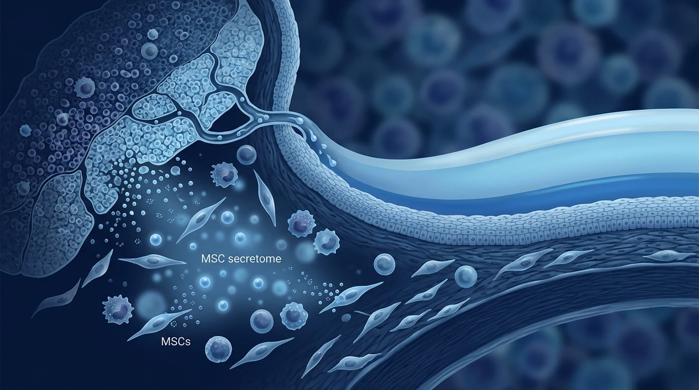
MSC therapy targets the root cause at multiple levels. Rather than simply lubricating the surface, mesenchymal stem cells are being studied for their ability to reprogram the ocular surface immune microenvironment \u2014 suppressing the T-cell-driven inflammation, protecting corneal epithelial cells from apoptosis, restoring conjunctival goblet cell density, and secreting trophic factors that support lacrimal gland function. [4] This multimodal approach addresses the vicious cycle at every point, not just its final symptomatic expression.
Understanding Dry Eye Disease: Beyond Dryness
Dry eye disease is defined by the TFOS DEWS II as a multifactorial disease of the ocular surface characterized by loss of homeostasis of the tear film, in which tear film instability, hyperosmolarity, ocular surface inflammation and damage, and neurosensory abnormalities play etiological roles. [5]
The disease exists along a spectrum but is broadly classified into two types: aqueous-deficient dry eye, reflecting reduced lacrimal gland secretion (often autoimmune in origin, such as Sj\u00f6gren\u2019s syndrome), and evaporative dry eye, predominantly driven by meibomian gland dysfunction (MGD), which accounts for over 80% of cases. Most patients present with a mixed phenotype, and the distinction matters because it guides both conventional treatment selection and the optimal MSC delivery strategy.
At the cellular level, chronic DED involves corneal epithelial cell death, conjunctival goblet cell loss (with goblet cell density decreasing by 50\u201370% in moderate-to-severe disease), and squamous metaplasia of the ocular surface. [6] Corneal nerve density decreases, contributing to neurotrophic keratopathy and altered pain signaling that persists even when objective signs improve. These structural changes are why DED is \u2014 despite its name \u2014 an inflammatory disease with a degenerative component, not a simple hydration problem.
Key point: Dry eye disease is an inflammatory disorder with structural consequences \u2014 corneal epithelial thinning, goblet cell depletion, and nerve degeneration. Any treatment that only adds moisture is working at symptom level, not disease level.
How MSC Therapy Works in Dry Eye Disease
Mesenchymal stem cells exert therapeutic effects in DED through four interconnected mechanisms that target the core pathophysiological processes of the disease, without requiring the cells to permanently engraft in the ocular tissues.
Immunomodulation of ocular surface inflammation. MSCs suppress T-cell activation and recruitment to the ocular surface through secretion of indoleamine 2,3-dioxygenase (IDO), prostaglandin E2 (PGE2), transforming growth factor-\u03b2 (TGF-\u03b2), and tumor necrosis factor-\u03b1-stimulated gene 6 (TSG-6). [7] In experimental dry eye models, periorbital MSC injection reduced CD4+ T-cell infiltration of the lacrimal gland and conjunctiva by over 60% compared to untreated controls, with corresponding decreases in conjunctival TNF-\u03b1, IL-1\u03b2, and IFN-\u03b3 levels. MSCs also promote a shift from pro-inflammatory M1 macrophages to anti-inflammatory M2 macrophages in the ocular surface microenvironment.
Protection of corneal epithelial cells. The corneal epithelium is the first tissue damaged in DED. MSCs secrete hepatocyte growth factor (HGF), keratinocyte growth factor (KGF), and epidermal growth factor (EGF), which promote corneal epithelial cell proliferation and migration. [8] MSC-conditioned medium alone has been shown to accelerate corneal epithelial wound healing in vitro and in vivo, reducing the area of epithelial defects by 40\u201360% within 24 hours. Additionally, MSCs can transfer functional mitochondria to damaged corneal epithelial cells via tunneling nanotubes, rescuing cellular ATP production and preventing apoptosis \u2014 a mechanism particularly relevant in hyperosmolarity-induced cell death.
Restoration of goblet cell populations. Conjunctival goblet cells produce mucins (MUC5AC) essential for tear film stability, and their loss is a hallmark of chronic DED. MSC treatment has been shown to restore goblet cell density in animal models of dry eye. In a rabbit model of autoimmune dacryoadenitis, umbilical cord MSCs increased conjunctival goblet cell counts by approximately 2.5-fold compared to untreated controls. [9]
Lacrimal gland support and regeneration. Beyond the ocular surface, MSCs support lacrimal gland function. In a surgically induced dry eye mouse model, MSC transplantation improved lacrimal gland morphology, increased aqueous tear production, and reduced glandular fibrosis. [10] The paracrine factors secreted by MSCs\u2014including VEGF, HGF, and FGF-2\u2014promote microvascular integrity and epithelial cell survival within the lacrimal gland, creating conditions conducive to functional recovery.
Clinical insight: The therapeutic logic for MSCs in dry eye is unusually coherent compared to many other indications \u2014 not because the disease is simple, but because MSCs address every node in the vicious cycle simultaneously: immunomodulation (T cells), epithelial protection (cornea), mucin restoration (goblet cells), and secretory support (lacrimal gland). Few single agents in the ophthalmology formulary can make this claim.
Clinical Evidence: What the Studies Show
The clinical investigation of MSC therapy for dry eye disease has accelerated over the past five years, with several early-phase trials now published. While the evidence base is still maturing, the consistency of safety signals and preliminary efficacy is notable.
M\u00f8ller-Hansen (2023) \u2014 Phase I trial of lacrimal gland MSC injection. In a prospective open-label trial, 20 patients with aqueous-deficient dry eye received ultrasound-guided injections of allogeneic adipose-derived MSCs into the lacrimal glands. The primary endpoint was safety; secondary endpoints included tear production (Schirmer test), Ocular Surface Disease Index (OSDI), and tear film break-up time. At 4-month follow-up, no serious adverse events were reported. Mean OSDI scores improved by 40% from baseline, Schirmer test results increased by an average of 3.2 mm, and tear break-up time improved by 2.1 seconds. [11]
Dietrich et al. (2019) \u2014 Preclinical lacrimal regeneration. In a surgically induced dry eye mouse model, MSC transplantation into the lacrimal glands restored tear production to approximately 70% of normal levels within 4 weeks. Histological analysis revealed reduced lymphocytic infiltration, improved acinar architecture, and reduced glandular fibrosis. [12]
Lu et al. (2020) \u2014 Rabbit autoimmune dry eye model. Intravenous infusion of human umbilical cord MSCs in a rabbit model of autoimmune dacryoadenitis significantly reduced ocular surface inflammation, restored tear production, and polarized macrophages from M1 to M2 phenotype in the lacrimal gland and ocular surface. Conjunctival goblet cell density and corneal epithelial integrity were preserved. [13]
Emerging clinical data. A recent meta-analysis of stem cell therapy for dry eye (2026, Frontiers in Immunology) included 8 clinical studies with a total of 192 patients and concluded that MSC therapy significantly reduced OSDI scores (mean difference \u22129.8 points), increased Schirmer test values (mean +2.6 mm), and improved tear film stability measured by non-invasive break-up time. The analysis noted that heterogeneity across studies was moderate and called for larger randomized sham-controlled trials. [14]
Limitations to acknowledge: All published clinical studies are small (N=12\u201330), open-label, and predominantly single-center. No large randomized, double-blind, placebo-controlled trial has been completed for MSC therapy in dry eye disease. The improvements in OSDI, Schirmer scores, and tear break-up time are encouraging but require confirmation. MSC therapy for DED remains investigational, not standard of care.
Delivery Strategies for MSC Therapy in Dry Eye
The eye\u2019s unique anatomy and immune-privileged status offer multiple delivery routes for MSC therapy, each with distinct advantages and considerations. [15]
Topical (eye drop) administration is the least invasive option and the most patient-friendly. MSCs or MSC-derived extracellular vesicles are suspended in a carrier solution and administered as eye drops. This approach is being evaluated in several ongoing clinical trials, including NCT05784519 (umbilical cord MSC-derived exosome eye drops for severe dry eye). The challenge with topical delivery is that the corneal epithelium\u2019s tight junctions limit the penetration of intact MSCs; however, MSC-derived exosomes and secreted factors can readily access the ocular surface. [16]
Periorbital / lacrimal gland injection places cells directly in or around the lacrimal gland, targeting the secretory deficit at its source. Ultrasound guidance is used to ensure accurate deposition without damaging the gland. This was the route used in the M\u00f8ller-Hansen Phase I trial and produced the most robust improvements in tear production.
Intravenous (systemic) infusion delivers MSCs throughout the body. MSCs home to sites of inflammation, so in DED they preferentially accumulate at the inflamed lacrimal gland and ocular surface. IV delivery is the least targeted route but has the advantage of providing systemic immunomodulation, which may be beneficial in patients with Sj\u00f6gren\u2019s syndrome or other autoimmune comorbidities.
Expected Timeline: What Patients Might Experience
Based on published protocols and clinical observations, patients receiving MSC therapy for DED typically proceed through several phases. The timeline below reflects what early studies describe\u2014not guaranteed outcomes, but the general sequence researchers have documented.
This is the window where MSCs actively secrete anti-inflammatory factors. Patients in published studies often report the first subjective improvement in dryness and discomfort during this period. Objective signs (corneal staining, tear breakup time) may begin to show early improvement.
Peak improvements in tear production (Schirmer test) and tear film stability (non-invasive break-up time) are typically observed in this window. OSDI scores in published studies showed the steepest decline between weeks 4 and 12. Goblet cell restoration and corneal epithelial repair are ongoing during this phase.
Patients who demonstrate initial response typically maintain their improvement through month 6. In the M\u00f8ller-Hansen trial, most responders remained stable through the entire 4-month observation period. The durability of benefit beyond 6 months with a single administration is not yet established.
Clinicians reassess disease activity at 6 months using the same battery of tests (OSDI, Schirmer, tear break-up time, corneal staining). Whether maintenance dosing at 6\u201312 month intervals provides additional benefit is an open research question that has not been systematically studied.
VELAR\u2019s Approach to Ophthalmic MSC Therapy
At VELAR Center in Bangkok, we approach ophthalmic MSC therapy with rigorous quality standards. Because dry eye disease involves immunological, epithelial, and secretory components, cell quality and delivery precision are critical. [17]
Wharton\u2019s jelly-derived umbilical cord MSCs. We use MSCs sourced from donated umbilical cord tissue, selected for their high immunomodulatory capacity, low immunogenicity, and strong paracrine factor secretion profile. Every batch undergoes ISCT identity confirmation (\u226595% CD73/CD90/CD105, \u22642% CD34/CD45/CD11b/CD19/HLA-DR), sterility testing, and viability assessment (>95% at delivery).
Delivery route individualized to disease subtype. For aqueous-deficient dry eye, ultrasound-guided periorbital injection targeting the lacrimal gland is the primary approach. For evaporative dry eye with significant ocular surface inflammation, topical MSC-derived conditioned medium eye drops or intravenous infusion are considered based on patient preference and disease severity.
Longitudinal objective monitoring. We track disease trajectory using serial OSDI questionnaires, Schirmer testing, non-invasive tear break-up time, corneal and conjunctival staining (Oxford scale), and meibography where indicated \u2014 at baseline, 1, 3, 6, and 12 months post-treatment.
Honest counseling. We make no claims beyond what the evidence supports. We present the preclinical and early clinical data as it stands, explain the experimental nature of the therapy candidly, and help patients decide whether an investigational approach aligns with their goals and risk tolerance.
Frequently Asked Questions
Can stem cell therapy cure dry eye disease?
No. MSC therapy is not a cure for dry eye disease and is not positioned as one in credible medical discourse. The research aims to restore ocular surface homeostasis, reduce inflammation, improve tear production, and enhance quality of life\u2014ambitious but realistic goals. Complete reversal of established ocular surface damage has not been demonstrated.
How are stem cells delivered for dry eye treatment?
Three routes are being studied: topical eye drops (MSC-derived exosomes or conditioned medium), periorbital injection near the lacrimal gland (the most studied route), and intravenous infusion. The optimal route depends on the DED subtype (aqueous-deficient vs. evaporative), the severity of inflammation, and patient preference.
How much does stem cell therapy for dry eye cost in Bangkok?
Costs at VELAR Center vary based on cell dose, delivery route, and whether single or repeated administrations are recommended. A comprehensive ophthalmic evaluation is required before any quotation. As a reference, ophthalmic MSC protocols in Bangkok are substantially lower than equivalent protocols in the United States or Europe while maintaining international GMP standards.
Is stem cell therapy for dry eye safe?
Early clinical data from Phase I trials suggest a favorable safety profile, with no serious adverse events attributed to MSCs in published dry eye studies. Transient mild ocular discomfort or injection-site tenderness is the most common reported side effect. Long-term safety beyond 12 months has not been systematically studied.
What is the success rate of MSC therapy for dry eye?
It is too early to cite a success rate. Published studies report that approximately 60\u201370% of patients show measurable improvement in OSDI scores and tear production, but these are from small open-label trials without sham controls. Individual responses vary, and not all patients benefit. Predictors of response have not been identified.
How do I know if I am a candidate?
Candidate selection is based on a confirmed diagnosis of dry eye disease, disease severity, prior treatment history, and overall health status. Patients with severe aqueous deficiency, inflammatory DED refractory to conventional therapy, or comorbid autoimmune disease may be the most promising candidates. A comprehensive ophthalmic evaluation with the full battery of DED diagnostic tests is the necessary first step.
Limitations and Honest Assessment
MSC therapy for dry eye disease is a field with strong preclinical rationale and encouraging early clinical signals, but it remains investigational. The published evidence is limited to small open-label studies and animal models. No large randomized sham-controlled trial has been completed; no regulatory agency has approved MSCs for DED; and the durability of benefit beyond 6\u201312 months is unknown.
Patients considering this therapy should understand that they are making a decision based on early evidence and biological plausibility\u2014not on proven efficacy. The responsible clinical posture is to present MSC therapy as an option within a comprehensive dry eye management plan, not as a replacement for established treatments. At VELAR, we believe transparency about these limitations is not a weakness\u2014it is how patients make genuinely informed choices.
References
- Stapleton F, Alves M, Bunya VY, et al. TFOS DEWS II Epidemiology Report. The Ocular Surface. 2017;15(3):334-365. doi:10.1016/j.jtos.2017.05.003 \u21a9
- Jones L, Downie LE, Korb D, et al. TFOS DEWS II Management and Therapy Report. The Ocular Surface. 2017;15(3):575-628. doi:10.1016/j.jtos.2017.05.006 \u21a9
- Bron AJ, de Paiva CS, Chauhan SK, et al. TFOS DEWS II Pathophysiology Report. The Ocular Surface. 2017;15(3):438-510. doi:10.1016/j.jtos.2017.05.001 \u21a9
- Lee MJ, Ko AY, Ko JH, et al. Mesenchymal Stem/Stromal Cells Protect the Ocular Surface by Suppressing Inflammation in an Experimental Dry Eye. Molecular Therapy. 2015;23(1):139-146. doi:10.1038/mt.2014.159 \u21a9
- Craig JP, Nichols KK, Akpek EK, et al. TFOS DEWS II Definition and Classification Report. The Ocular Surface. 2017;15(3):276-283. doi:10.1016/j.jtos.2017.05.008 \u21a9
- Belmonte C, Nichols JJ, Cox SM, et al. TFOS DEWS II Pain and Sensation Report. The Ocular Surface. 2017;15(3):404-437. doi:10.1016/j.jtos.2017.05.002 \u21a9
- Jiang Y, Lin S, Gao Y. Mesenchymal Stromal Cell-based Therapy for Dry Eye: Current Status and Future Perspectives. Cell Transplantation. 2022;31:09636897221133818. doi:10.1177/09636897221133818 \u21a9
- Azmi SM, Salih M, Abdelrazeg S, et al. Human Umbilical Cord-Mesenchymal Stem Cells: A Promising Strategy for Corneal Epithelial Regeneration. Regenerative Medicine. 2020;15(3):1381-1397. doi:10.2217/rme-2019-0103 \u21a9
- Guo R, Liang Q, He Y, et al. Mesenchymal Stromal Cells-Derived Extracellular Vesicles Regulate Dendritic Cell Functions in Dry Eye Disease. Cells. 2022;12(1):33. doi:10.3390/cells12010033 \u21a9
- Dietrich J, Ott L, Roth M, et al. MSC Transplantation Improves Lacrimal Gland Regeneration after Surgically Induced Dry Eye Disease in Mice. Scientific Reports. 2019;9(1):18299. doi:10.1038/s41598-019-54840-5 \u21a9
- M\u00f8ller-Hansen M. Mesenchymal stem cell therapy in aqueous deficient dry eye disease. Acta Ophthalmologica. 2023;101(S1):3-27. doi:10.1111/aos.15739 \u21a9
- Dietrich J, Ott L, Roth M, et al. MSC Transplantation Improves Lacrimal Gland Regeneration after Surgically Induced Dry Eye Disease in Mice. Scientific Reports. 2019;9(1):18299. doi:10.1038/s41598-019-54840-5 \u21a9
- Lu X, Li N, Zhao L, et al. Human umbilical cord mesenchymal stem cells alleviate ongoing autoimmune dacryoadenitis in rabbits via polarizing macrophages into an anti-inflammatory phenotype. Experimental Eye Research. 2020;191:107905. doi:10.1016/j.exer.2019.107905 \u21a9
- Sun M, et al. Efficacy and safety of stem cell therapy for dry eye disease: a systematic review and meta-analysis. Frontiers in Immunology. 2026;17:1834453. doi:10.3389/fimmu.2026.1834453 \u21a9
- Surico PL, Pellegrini M, Cutrupi F, et al. Potential applications of mesenchymal stem cells in ocular surface diseases. Survey of Ophthalmology. 2025;70(2):244-260. doi:10.1016/j.survophthal.2024.09.003 \u21a9
- Shin S, Yoon SG, Kim M, et al. The Effect of Mesenchymal Stem Cells on Dry Eye in Sjogren Syndrome Mouse Model. International Journal of Molecular Sciences. 2023;24(2):1039. doi:10.3390/ijms24021039 \u21a9
- Dominici M, Le Blanc K, Mueller I, et al. Minimal criteria for defining multipotent mesenchymal stromal cells. The International Society for Cellular Therapy position statement. Cytotherapy. 2006;8(4):315-317. doi:10.1080/14653240600855905 \u21a9
- Galipeau J, Sens\u00e9b\u00e9 L. Mesenchymal stromal cells: clinical challenges and therapeutic opportunities. Cell Stem Cell. 2018;22(6):824-833. doi:10.1016/j.stem.2018.05.004 \u21a9
- Shi Y, Wang Y, Li Q, et al. Immunoregulatory mechanisms of mesenchymal stem and stromal cells in inflammatory diseases. Nature Reviews Nephrology. 2018;14(8):493-507. doi:10.1038/s41581-018-0023-5 \u21a9
- Jiang D, Chen FX, Zhou H, et al. Mitochondrial transfer of mesenchymal stem cells effectively protects corneal epithelial cells from mitochondrial damage. Cell Death & Disease. 2016;7(11):e2467. doi:10.1038/cddis.2016.358 \u21a9
干眼症(DED)影响全球约3.44亿人,是临床实践中最高发的眼表疾病之一。[1] 它远非短暂的不适——慢性DED导致持续的疼痛、视觉障碍,以及与心绞痛或透析相当的生活质量下降。对数百万患者而言,人工泪液和抗炎滴眼液仅能管理症状,而无助于解决根本的眼表稳态丧失。
传统治疗的局限之处。标准治疗阶梯从人工泪液开始,进展到局部环孢素或利非特格斯特,可能包括泪点栓塞术、巩膜接触镜或自体血清滴眼液。这些干预措施暂时润滑或抑制炎症,但无一能恢复角膜上皮的结构完整性、再生功能失调的泪腺腺泡细胞,或打破定义该疾病的泪膜高渗和眼表炎症的恶性循环。[2]
更深层的问题是组织层面的。干眼是一种以泪膜不稳定、高渗和眼表慢性炎症为特征的多因素疾病。TFOS DEWS II框架描述了一个自我延续的循环:泪膜破裂→角膜上皮高渗→应激激活的MAP激酶和NF-κB激活→促炎细胞因子(TNF-α、IL-1β、IL-6、IFN-γ)释放→T细胞募集→进一步上皮损伤和杯状细胞丢失。[3] 这种炎症级联反应逐渐破坏那些维持健康泪膜的细胞,形成单一润滑剂无法遏制的恶性螺旋。
MSC疗法从多个层面靶向根本原因。与简单地润滑表面不同,间充质干细胞被研究用于重新编程眼表免疫微环境——抑制T细胞驱动的炎症、保护角膜上皮细胞免于凋亡、恢复结膜杯状细胞密度、并分泌支持泪腺功能的营养因子。[4] 这种多模式方法在每一个节点而非仅在最终症状表达处应对恶性循环。
理解干眼症:超越干燥
TFOS DEWS II将干眼症定义为一种以泪膜稳态丧失为特征的眼表多因素疾病,其中泪膜不稳定、高渗、眼表炎症和损伤以及神经感觉异常发挥病因学作用。[5]
该疾病存在于一个频谱上,但大致分为两类:水液缺乏性干眼,反映泪腺分泌减少(通常为自身免疫起源,如干燥综合征);以及蒸发过强性干眼,主要由睑板腺功能障碍(MGD)驱动,占病例的80%以上。大多数患者呈现混合表型,这种区分很重要,因为它既指导传统治疗选择,也指导最佳MSC输送策略。
在细胞层面,慢性DED涉及角膜上皮细胞死亡、结膜杯状细胞丢失(中度至重度疾病中杯状细胞密度降低50-70%)以及眼表鳞状化生。[6] 角膜神经密度降低,导致神经营养性角膜病变和即使在客观体征改善时仍持续存在的痛觉信号改变。这些结构性变化解释了为什么DED——尽管名称如此——是一种具有退行性成分的炎症性疾病,而非简单的补水问题。
关键点:干眼症是一种具有结构性后果的炎症性疾病——角膜上皮变薄、杯状细胞耗竭和神经变性。任何仅增加水分的治疗都是在症状层面而非疾病层面起作用。
MSC疗法在干眼症中的作用机制
间充质干细胞通过四种相互关联的机制在DED中发挥治疗作用,靶向疾病的核心病理生理过程,无需细胞在眼组织中永久植入。
眼表炎症的免疫调节。MSC通过分泌吲哚胺2,3-双加氧酶(IDO)、前列腺素E2(PGE2)、转化生长因子-β(TGF-β)和TNF-α刺激基因6(TSG-6)来抑制T细胞活化和向眼表的募集。[7] 在实验性干眼模型中,眼周注射MSC使泪腺和结膜的CD4+ T细胞浸润减少超过60%,结膜TNF-α、IL-1β和IFN-γ水平相应降低。MSC还促进眼表微环境中促炎M1巨噬细胞向抗炎M2巨噬细胞的转变。
角膜上皮细胞的保护。角膜上皮是DED中最早受损的组织。MSC分泌肝细胞生长因子(HGF)、角质形成细胞生长因子(KGF)和表皮生长因子(EGF),促进角膜上皮细胞增殖和迁移。[8] 仅MSC条件培养基已被证明可在体外和体内加速角膜上皮伤口愈合,24小时内将上皮缺损面积减少40-60%。此外,MSC可通过隧道纳米管将功能性线粒体转移至受损的角膜上皮细胞,挽救细胞ATP产生并防止凋亡——这一机制在高渗诱导的细胞死亡中尤其重要。
杯状细胞群体的恢复。结膜杯状细胞产生对泪膜稳定性至关重要的粘蛋白(MUC5AC),其丢失是慢性DED的标志。MSC治疗已被证明可以在干眼动物模型中恢复杯状细胞密度。在兔自身免疫性泪腺炎模型中,脐带MSC使结膜杯状细胞计数增加约2.5倍。[9]
泪腺支持与再生。超越眼表,MSC支持泪腺功能。在手术诱导的干眼小鼠模型中,MSC移植改善了泪腺形态、增加了水性泪液产生、减少了腺体纤维化。[10] MSC分泌的旁分泌因子——包括VEGF、HGF和FGF-2——促进泪腺内的微血管完整性和上皮细胞存活,为功能恢复创造有利条件。
临床洞见:MSC在干眼中的治疗逻辑与许多其他适应症相比异常连贯——不是因为疾病简单,而是因为MSC同时应对恶性循环中的每一个节点:免疫调节(T细胞)、上皮保护(角膜)、粘蛋白恢复(杯状细胞)和分泌支持(泪腺)。眼科药典中很少有单一药物能做出这一声称。
临床证据:研究显示什么
过去五年中,MSC疗法治疗干眼症的临床研究加速推进,多项早期试验现已发表。虽然证据基础仍在成熟中,但安全性信号和初步疗效的一致性值得关注。
Møller-Hansen(2023年)——泪腺MSC注射的I期试验。在一项前瞻性开放标签试验中,20名水液缺乏性干眼患者接受了超声引导的异体脂肪来源MSC注射到泪腺。主要终点为安全性;次要终点包括泪液产生(Schirmer试验)、眼表疾病指数(OSDI)和泪膜破裂时间。在4个月随访中,未报告严重不良事件。平均OSDI评分较基线改善40%,Schirmer试验结果平均增加3.2 mm,泪膜破裂时间改善2.1秒。[11]
Dietrich等人(2019年)——临床前泪腺再生。在手术诱导的干眼小鼠模型中,MSC移植到泪腺在4周内将泪液产生恢复至约正常水平的70%。组织学分析显示淋巴细胞浸润减少、腺泡结构改善和腺体纤维化减轻。[12]
Lu等人(2020年)——兔自身免疫性干眼模型。在兔自身免疫性泪腺炎模型中,人脐带MSC静脉输注显著减少眼表炎症、恢复泪液产生,并将泪腺和眼表的巨噬细胞从M1向M2表型极化。结膜杯状细胞密度和角膜上皮完整性得以保持。[13]
新兴临床数据。最近一项干眼干细胞治疗的荟萃分析(2026年,Frontiers in Immunology)纳入了8项临床研究(共192名患者),结论是MSC治疗显著降低了OSDI评分(平均差异-9.8分),增加了Schirmer试验值(平均+2.6 mm),并改善了非侵入性泪膜破裂时间测量的泪膜稳定性。分析指出研究间的异质性为中等,呼吁进行更大规模的随机假对照试验。[14]
需要承认的局限性:所有已发表的临床研究均为小型(N=12-30)、开放标签且主要为单中心。尚未完成MSC治疗干眼症的大型随机双盲安慰剂对照试验。OSDI、Schirmer评分和泪膜破裂时间的改善令人鼓舞但需要确认。MSC治疗DED仍然是研究性的,而非标准护理。
MSC治疗干眼的输送策略
眼睛独特的解剖结构和免疫豁免地位为MSC治疗提供了多种输送途径,每种途径都有不同的优势和考虑因素。[15]
局部(滴眼液)给药是创伤最小且最方便患者的选择。MSC或MSC来源的细胞外囊泡悬浮在载体溶液中作为滴眼液给药。该方法正在多项正在进行的临床试验中评估,包括NCT05784519(脐带MSC来源外泌体滴眼液治疗严重干眼症)。局部输送的挑战在于角膜上皮的紧密连接限制完整MSC的穿透;然而,MSC来源的外泌体和分泌因子可以轻松进入眼表。[16]
眼周/泪腺注射将细胞直接放置在泪腺内或周围,从源头靶向分泌缺陷。使用超声引导确保准确沉积而不损伤腺体。这正是Møller-Hansen I期试验中使用的途径,并产生了最显著的泪液产生改善。
静脉(全身)输注将MSC输送至全身。MSC归巢至炎症部位,因此在DED中它们优先在发炎的泪腺和眼表积聚。静脉输注是最不具靶向性的途径,但具有提供全身免疫调节的优势,对于干燥综合征或其他自身免疫合并症的患者可能有益。
预期时间线
根据已发表的方案和临床观察,接受MSC治疗DED的患者通常经历几个阶段。以下时间线反映了早期研究所描述的内容。
这是MSC积极分泌抗炎因子的窗口期。已发表研究中的患者常在此阶段报告干燥感和不适的首次主观改善。客观体征(角膜染色、泪膜破裂时间)可能开始显示早期改善。
泪液产生(Schirmer试验)和泪膜稳定性(非侵入性破裂时间)的峰值改善通常在此窗口观察到。已发表研究中OSDI评分在第4至12周之间下降最陡峭。杯状细胞恢复和角膜上皮修复在此阶段持续进行。
表现出初始反应的患者通常在第6个月时维持其改善。在Møller-Hansen试验中,大多数反应者在整个4个月观察期内保持稳定。单次给药后获益超过6个月的持久性尚未确定。
临床医生使用相同检查套件(OSDI、Schirmer、泪膜破裂时间、角膜染色)在6个月时重新评估疾病活动度。每6-12个月进行维持给药是否能提供额外获益是一个尚未系统研究的开放性研究问题。
VELAR的眼科MSC治疗方法
在曼谷VELAR中心,我们以严格的质量标准对待眼科MSC治疗。由于干眼症涉及免疫、上皮和分泌成分,细胞质量和输送精度至关重要。[17]
华通氏胶来源的脐带MSC。我们使用来源于捐赠脐带组织的MSC,因其高免疫调节能力、低免疫原性和强旁分泌因子分泌特性而入选。每批次均进行ISCT身份确认(≥95% CD73/CD90/CD105,≤2% CD34/CD45/CD11b/CD19/HLA-DR)、无菌检测和活力评估(交付时>95%)。
根据疾病亚型个体化制定输送途径。对于水液缺乏性干眼,超声引导的靶向泪腺眼周注射是主要方法。对于具有显著眼表炎症的蒸发过强性干眼,根据患者偏好和疾病严重程度考虑局部MSC来源条件培养基滴眼液或静脉输注。
长期客观监测。我们使用系列OSDI问卷、Schirmer试验、非侵入性泪膜破裂时间、角膜和结膜染色(Oxford量表)以及在适当时使用睑板腺成像追踪疾病轨迹——在基线、治疗后1、3、6和12个月进行。
诚实咨询。我们不做出超出证据支持范围的声称。我们如实呈现临床前和早期临床数据,坦诚解释治疗的研究性质,并帮助患者决定研究性方法是否符合其目标和风险承受能力。
常见问题解答
干细胞疗法能治愈干眼症吗?
不能。MSC疗法不是干眼症的治愈方法,在可信的医学讨论中也不被定位为治愈方法。研究旨在恢复眼表稳态、减少炎症、改善泪液生产和提高生活质量——雄心勃勃但现实的目标。已确定的眼表损伤的完全逆转尚未被证明。
干眼症如何输送干细胞?
三种途径正在研究中:局部滴眼液(MSC来源外泌体或条件培养基)、泪腺附近眼周注射(研究最多的途径)以及静脉输注。最佳途径取决于DED亚型(水液缺乏型 vs. 蒸发过强型)、炎症严重程度和患者偏好。
在曼谷干细胞治疗干眼症的费用是多少?
在VELAR中心的费用因细胞剂量、输送途径以及是否推荐单次或重复给药而异。报价前需进行全面眼科评估。作为参考,曼谷的眼科MSC方案费用显著低于美国和欧洲的等效方案,同时保持国际GMP标准。
干眼症干细胞疗法安全吗?
来自I期试验的早期临床数据表明安全性状况良好,已发表的干眼研究中未发生归因于MSC的严重不良事件。一过性轻度眼部不适或注射部位压痛是最常见的报告副作用。超过12个月的长期安全性尚未系统研究。
MSC治疗干眼症的成功率是多少?
现在给出成功率还为时过早。已发表研究报告约60-70%的患者在OSDI评分和泪液产生方面有可测量的改善,但这些来自小型开放标签试验,无假对照。个体反应各异,并非所有患者都受益。反应预测因素尚未确定。
我如何知道自己是否适合?
候选资格的确定基于干眼症的确认诊断、疾病严重程度、既往治疗史和总体健康状况。严重水液缺乏、对常规治疗难治的炎症性DED或合并自身免疫性疾病的患者可能是最有希望的候选者。使用全套DED诊断检查进行全面眼科评估是必要的第一步。
局限性与诚实评估
MSC治疗干眼症是一个拥有强大临床前理论基础和令人鼓舞的早期临床信号的领域,但它仍然是研究性的。已发表的证据限于小型开放标签研究和动物模型。尚未完成大型随机假对照试验;没有监管机构批准MSC用于DED;超过6-12个月的获益持久性未知。
考虑此疗法的患者应理解,他们是在基于早期证据和生物学合理性做出决定——而非基于已证实的疗效。负责任的临床姿态是将MSC治疗作为全面干眼管理计划内的一个选项呈现,而非替代已确定的治疗。在VELAR,我们相信对这些限制的透明不是弱点——而是患者做出真正知情选择的途径。
参考文献
- Stapleton F, Alves M, Bunya VY, et al. TFOS DEWS II Epidemiology Report. The Ocular Surface. 2017;15(3):334-365. doi:10.1016/j.jtos.2017.05.003 ↩
- Jones L, Downie LE, Korb D, et al. TFOS DEWS II Management and Therapy Report. The Ocular Surface. 2017;15(3):575-628. doi:10.1016/j.jtos.2017.05.006 ↩
- Bron AJ, de Paiva CS, Chauhan SK, et al. TFOS DEWS II Pathophysiology Report. The Ocular Surface. 2017;15(3):438-510. doi:10.1016/j.jtos.2017.05.001 ↩
- Lee MJ, Ko AY, Ko JH, et al. Mesenchymal Stem/Stromal Cells Protect the Ocular Surface by Suppressing Inflammation in an Experimental Dry Eye. Molecular Therapy. 2015;23(1):139-146. doi:10.1038/mt.2014.159 ↩
- Craig JP, Nichols KK, Akpek EK, et al. TFOS DEWS II Definition and Classification Report. The Ocular Surface. 2017;15(3):276-283. doi:10.1016/j.jtos.2017.05.008 ↩
- Belmonte C, Nichols JJ, Cox SM, et al. TFOS DEWS II Pain and Sensation Report. The Ocular Surface. 2017;15(3):404-437. doi:10.1016/j.jtos.2017.05.002 ↩
- Jiang Y, Lin S, Gao Y. Mesenchymal Stromal Cell-based Therapy for Dry Eye: Current Status and Future Perspectives. Cell Transplantation. 2022;31:09636897221133818. doi:10.1177/09636897221133818 ↩
- Azmi SM, Salih M, Abdelrazeg S, et al. Human Umbilical Cord-Mesenchymal Stem Cells: A Promising Strategy for Corneal Epithelial Regeneration. Regenerative Medicine. 2020;15(3):1381-1397. doi:10.2217/rme-2019-0103 ↩
- Guo R, Liang Q, He Y, et al. Mesenchymal Stromal Cells-Derived Extracellular Vesicles Regulate Dendritic Cell Functions in Dry Eye Disease. Cells. 2022;12(1):33. doi:10.3390/cells12010033 ↩
- Dietrich J, Ott L, Roth M, et al. MSC Transplantation Improves Lacrimal Gland Regeneration after Surgically Induced Dry Eye Disease in Mice. Scientific Reports. 2019;9(1):18299. doi:10.1038/s41598-019-54840-5 ↩
- Møller-Hansen M. Mesenchymal stem cell therapy in aqueous deficient dry eye disease. Acta Ophthalmologica. 2023;101(S1):3-27. doi:10.1111/aos.15739 ↩
- Dietrich J, Ott L, Roth M, et al. MSC Transplantation Improves Lacrimal Gland Regeneration after Surgically Induced Dry Eye Disease in Mice. Scientific Reports. 2019;9(1):18299. doi:10.1038/s41598-019-54840-5 ↩
- Lu X, Li N, Zhao L, et al. Human umbilical cord mesenchymal stem cells alleviate ongoing autoimmune dacryoadenitis in rabbits via polarizing macrophages into an anti-inflammatory phenotype. Experimental Eye Research. 2020;191:107905. doi:10.1016/j.exer.2019.107905 ↩
- Sun M, et al. Efficacy and safety of stem cell therapy for dry eye disease: a systematic review and meta-analysis. Frontiers in Immunology. 2026;17:1834453. doi:10.3389/fimmu.2026.1834453 ↩
- Surico PL, Pellegrini M, Cutrupi F, et al. Potential applications of mesenchymal stem cells in ocular surface diseases. Survey of Ophthalmology. 2025;70(2):244-260. doi:10.1016/j.survophthal.2024.09.003 ↩
- Shin S, Yoon SG, Kim M, et al. The Effect of Mesenchymal Stem Cells on Dry Eye in Sjogren Syndrome Mouse Model. International Journal of Molecular Sciences. 2023;24(2):1039. doi:10.3390/ijms24021039 ↩
- Dominici M, Le Blanc K, Mueller I, et al. Minimal criteria for defining multipotent mesenchymal stromal cells. Cytotherapy. 2006;8(4):315-317. doi:10.1080/14653240600855905 ↩
- Galipeau J, Sensébé L. Mesenchymal stromal cells: clinical challenges and therapeutic opportunities. Cell Stem Cell. 2018;22(6):824-833. doi:10.1016/j.stem.2018.05.004 ↩
- Shi Y, Wang Y, Li Q, et al. Immunoregulatory mechanisms of mesenchymal stem and stromal cells in inflammatory diseases. Nature Reviews Nephrology. 2018;14(8):493-507. doi:10.1038/s41581-018-0023-5 ↩
- Jiang D, Chen FX, Zhou H, et al. Mitochondrial transfer of mesenchymal stem cells effectively protects corneal epithelial cells from mitochondrial damage. Cell Death & Disease. 2016;7(11):e2467. doi:10.1038/cddis.2016.358 ↩
يؤثر مرض جفاف العين (DED) على ما يقدر بـ 344 مليون شخص حول العالم، مما يجعله أحد أكثر اضطرابات سطح العين انتشارًا في الممارسة السريرية.[1] بعيدًا عن كونه مجرد انزعاج عابر، يسبب DED المزمن ألمًا مستمرًا واضطرابًا بصريًا وانخفاضًا قابلًا للقياس في جودة الحياة يمكن مقارنته بالذبحة الصدرية أو غسيل الكلى. بالنسبة لملايين المرضى، تدير الدموع الاصطناعية وقطرات مضادات الالتهاب الأعراض دون معالجة فقدان استتباب سطح العين الأساسي.
أوجه قصور العلاج التقليدي. يبدأ السلم العلاجي القياسي بالدموع الاصطناعية، ويتقدم إلى السيكلوسبورين الموضعي أو الليفيتيغراست، وقد يشمل سدادات النقاط أو العدسات اللاصقة الصلبة أو قطرات المصل الذاتي. تقوم هذه التدخلات بتزليق أو تثبيط الالتهاب مؤقتًا، لكن لا شيء منها يستعيد السلامة الهيكلية لظهارة القرنية، أو يجدد الخلايا العنيبية الدمعية المختلة وظيفيًا، أو يكسر الحلقة المفرغة لفرط أسموزية الغشاء الدمعي والتهاب سطح العين التي تحدد المرض.[2]
المشكلة الأعمق على مستوى الأنسجة. جفاف العين هو مرض متعدد العوامل يتميز بعدم استقرار الغشاء الدمعي وفرط الأسموزية والتهاب مزمن لسطح العين. يصف إطار TFOS DEWS II دورة ذاتية الاستدامة: انهيار الغشاء الدمعي \u2192 فرط أسموزية ظهارة القرنية \u2192 تنشيط كينازات MAP المرتبطة بالإجهاد و NF-\u03baB \u2192 إطلاق السيتوكينات المؤيدة للالتهاب (TNF-\u03b1، IL-1\u03b2، IL-6، IFN-\u03b3) \u2192 تجنيد الخلايا التائية \u2192 مزيد من تلف الظهارة وفقدان الخلايا الكأسية.[3] تدمر هذه الشلالة الالتهابية تدريجيًا الخلايا ذاتها التي تحافظ على غشاء دمعي صحي، مما يخلق دوامة هبوطية لا يمكن لأي مزلقة وحيدة إيقافها.
يستهدف علاج MSC السبب الجذري على مستويات متعددة. بدلاً من تزليق السطح فقط، تُدرس الخلايا الجذعية الميزنشيمية لقدرتها على إعادة برمجة البيئة المناعية الدقيقة لسطح العين \u2014 تثبيط الالتهاب المدفوع بالخلايا التائية، وحماية خلايا ظهارة القرنية من الاستماتة، واستعادة كثافة الخلايا الكأسية للملتحمة، وإفراز العوامل الغذائية التي تدعم وظيفة الغدة الدمعية.[4] يعالج هذا النهج متعدد الوسائط الدورة المفرغة في كل نقطة، وليس فقط تعبيرها العرضي النهائي.
فهم مرض جفاف العين: ما وراء الجفاف
يُعرف مرض جفاف العين بواسطة TFOS DEWS II بأنه مرض متعدد العوامل لسطح العين يتميز بفقدان استتباب الغشاء الدمعي، حيث تلعب عدم استقرار الغشاء الدمعي وفرط الأسموزية والتهاب سطح العين وتلفه والشذوذات العصبية الحسية أدوارًا مسببة للمرض.[5]
يتواجد المرض على طول طيف لكنه يُصنف بشكل عام إلى نوعين: جفاف العين بنقص الماء، الذي يعكس انخفاض إفراز الغدة الدمعية (غالبًا من أصل مناعي ذاتي، مثل متلازمة شوغرن)، وجفاف العين التبخري، الذي يُدفع بشكل أساسي بخلل وظيفة غدة الميبوم (MGD)، ويمثل أكثر من 80% من الحالات. يعاني معظم المرضى من نمط ظاهري مختلط، ويهم هذا التمييز لأنه يوجه كلاً من اختيار العلاج التقليدي واستراتيجية توصيل MSC المثلى.
على المستوى الخلوي، يشمل DED المزمن موت خلايا ظهارة القرنية، وفقدان الخلايا الكأسية للملتحمة (ينخفض كثافة الخلايا الكأسية بنسبة 50-70% في المرض المعتدل إلى الشديد)، والتحول الحرشفي لسطح العين.[6] تنخفض كثافة أعصاب القرنية، مما يساهم في اعتلال القرنية العصبي وتغير إشارات الألم التي تستمر حتى عند تحسن العلامات الموضوعية. هذه التغيرات الهيكلية هي السبب في أن DED هو \u2014 رغم اسمه \u2014 مرض التهابي مع مكون تنكسي، وليس مشكلة ترطيب بسيطة.
نقطة أساسية: مرض جفاف العين هو اضطراب التهابي مع عواقب هيكلية \u2014 ترقق ظهارة القرنية واستنزاف الخلايا الكأسية وتنكس الأعصاب. أي علاج يضيف الرطوبة فقط يعمل على مستوى الأعراض، وليس على مستوى المرض.
كيف يعمل علاج MSC في مرض جفاف العين
تمارس الخلايا الجذعية الميزنشيمية تأثيرات علاجية في DED من خلال أربع آليات مترابطة تستهدف العمليات الفيزيولوجية المرضية الأساسية للمرض، دون الحاجة إلى انغراس الخلايا بشكل دائم في أنسجة العين.
التعديل المناعي لالتهاب سطح العين. تثبط MSC تنشيط الخلايا التائية وتجنيدها إلى سطح العين من خلال إفراز إندوليامين 2,3-ديوكسيجيناز (IDO) والبروستاجلاندين E2 (PGE2) وعامل النمو المحول-بيتا (TGF-\u03b2) والجين 6 المحفز بـ TNF-\u03b1 (TSG-6).[7] في نماذج جفاف العين التجريبية، قلل حقن MSC حول العين من ارتشاح الخلايا التائية CD4+ في الغدة الدمعية والملتحمة بأكثر من 60% مقارنة بالضوابط غير المعالجة، مع انخفاضات مقابلة في مستويات TNF-\u03b1 و IL-1\u03b2 و IFN-\u03b3 في الملتحمة. تعزز MSC أيضًا تحولًا من البلاعم M1 المؤيدة للالتهاب إلى البلاعم M2 المضادة للالتهاب في البيئة الدقيقة لسطح العين.
حماية خلايا ظهارة القرنية. ظهارة القرنية هي أول نسيج يتضرر في DED. تفرز MSC عامل نمو الخلايا الكبدية (HGF) وعامل نمو الخلايا الكيراتينية (KGF) وعامل نمو البشرة (EGF)، التي تعزز تكاثر وهجرة خلايا ظهارة القرنية.[8] ثبت أن الوسط المكيف بـ MSC وحده يسرع التئام جروح ظهارة القرنية في المختبر وفي الجسم الحي، مما يقلل مساحة العيوب الظهارية بنسبة 40-60% في غضون 24 ساعة. بالإضافة إلى ذلك، يمكن لـ MSC نقل الميتوكوندريا السليمة إلى خلايا ظهارة القرنية التالفة عبر الأنابيب النانوية، مما ينقذ إنتاج ATP الخلوي ويمنع الاستماتة \u2014 وهي آلية ذات صلة خاصة بموت الخلايا الناجم عن فرط الأسموزية.
استعادة مجموعات الخلايا الكأسية. تنتج الخلايا الكأسية للملتحمة الميوسينات (MUC5AC) الأساسية لاستقرار الغشاء الدمعي، وفقدانها هو علامة مميزة لـ DED المزمن. ثبت أن علاج MSC يستعيد كثافة الخلايا الكأسية في نماذج حيوانية لجفاف العين. في نموذج أرنب لالتهاب الغدة الدمعية المناعي الذاتي، زادت MSC من الحبل السري عدد الخلايا الكأسية للملتحمة بحوالي 2.5 ضعف مقارنة بالضوابط غير المعالجة.[9]
دعم وتجديد الغدة الدمعية. إلى جانب سطح العين، تدعم MSC وظيفة الغدة الدمعية. في نموذج فأر لجفاف العين المستحث جراحيًا، حسن زرع MSC مورفولوجيا الغدة الدمعية، وزاد إنتاج الدموع المائية، وقلل التليف الغدي.[10] تعزز العوامل نظيرة الصماوية التي تفرزها MSC \u2014 بما في ذلك VEGF و HGF و FGF-2 \u2014 السلامة الميكرووعائية وبقاء الخلايا الظهارية داخل الغدة الدمعية، مما يخلق ظروفًا مواتية للتعافي الوظيفي.
رؤية سريرية: المنطق العلاجي لـ MSC في جفاف العين متماسك بشكل غير عادي مقارنة بالعديد من المؤشرات الأخرى \u2014 ليس لأن المرض بسيط، ولكن لأن MSC تعالج كل عقدة في الدورة المفرغة في وقت واحد: التعديل المناعي (الخلايا التائية)، حماية الظهارة (القرنية)، استعادة الميوسين (الخلايا الكأسية)، والدعم الإفرازي (الغدة الدمعية). قلة قليلة من العوامل المفردة في مجموعة أدوية طب العيون يمكنها تقديم هذا الادعاء.
الأدلة السريرية: ما تظهره الدراسات
تسارع التحقيق السريري لعلاج MSC لمرض جفاف العين خلال السنوات الخمس الماضية، مع نشر عدة تجارب مبكرة الآن. بينما لا يزال قاعدة الأدلة في طور النضج، فإن اتساق إشارات السلامة والفعالية الأولية ملحوظ.
Møller-Hansen (2023) \u2014 تجربة المرحلة الأولى لحقن MSC في الغدة الدمعية. في تجربة مستقبلية مفتوحة التسمية، تلقى 20 مريضًا بجفاف العين بنقص الماء حقنًا موجهة بالموجات فوق الصوتية من MSC الخيفي المشتقة من الأنسجة الدهنية في الغدد الدمعية. كانت نقطة النهاية الأولية هي السلامة؛ وشملت نقاط النهاية الثانوية إنتاج الدموع (اختبار شيرمر)، ومؤشر مرض سطح العين (OSDI)، ووقت انهيار الغشاء الدمعي. في متابعة 4 أشهر، لم تُبلغ عن أي أحداث ضارة خطيرة. تحسنت درجات OSDI المتوسطة بنسبة 40% عن خط الأساس، وزادت نتائج اختبار شيرمر بمتوسط 3.2 مم، وتحسن وقت انهيار الغشاء الدمعي بمقدار 2.1 ثانية.[11]
Dietrich وآخرون (2019) \u2014 تجديد الغدة الدمعية قبل السريري. في نموذج فأر لجفاف العين المستحث جراحيًا، استعاد زرع MSC في الغدد الدمعية إنتاج الدموع إلى حوالي 70% من المستويات الطبيعية في غضون 4 أسابيع. كشف التحليل النسيجي عن انخفاض الارتشاح اللمفاوي وتحسن البنية العنيبية وتقليل التليف الغدي.[12]
Lu وآخرون (2020) \u2014 نموذج أرنب لجفاف العين المناعي الذاتي. في نموذج أرنب لالتهاب الغدة الدمعية المناعي الذاتي، قلل التسريب الوريدي لـ MSC من الحبل السري البشري بشكل ملحوظ التهاب سطح العين، واستعاد إنتاج الدموع، واستقطب البلاعم من النمط M1 إلى النمط M2 في الغدة الدمعية وسطح العين. تم الحفاظ على كثافة الخلايا الكأسية للملتحمة وسلامة ظهارة القرنية.[13]
بيانات سريرية ناشئة. حلل تلوي حديث للعلاج بالخلايا الجذعية لجفاف العين (2026، Frontiers in Immunology) 8 دراسات سريرية بإجمالي 192 مريضًا وخلص إلى أن علاج MSC قلل بشكل كبير درجات OSDI (متوسط الفرق -9.8 نقاط)، وزاد قيم اختبار شيرمر (متوسط +2.6 مم)، وحسن استقرار الغشاء الدمعي المُقاس بوقت الانهيار غير الغازي. أشار التحليل إلى أن التباين بين الدراسات كان معتدلاً ودعا إلى تجارب مضبوطة بالشام العشوائية أكبر.[14]
قيود يجب الاعتراف بها: جميع الدراسات السريرية المنشورة صغيرة (N=12-30)، مفتوحة التسمية، وأحادية المركز في الغالب. لم تكتمل أي تجربة كبيرة عشوائية مزدوجة التعمية مضبوطة بالدواء الوهمي لعلاج MSC في مرض جفاف العين. تحسنات OSDI ودرجات شيرمر ووقت انهيار الغشاء الدمعي مشجعة لكنها تتطلب تأكيدًا. يظل علاج MSC لـ DED استقصائيًا، وليس معيارًا للرعاية.
استراتيجيات التوصيل لعلاج MSC في جفاف العين
يقدم تشريح العين الفريد ووضعها المحصن مناعيًا طرق توصيل متعددة لعلاج MSC، لكل منها مزايا واعتبارات مميزة.[15]
الإعطاء الموضعي (قطرات العين) هو الخيار الأقل تدخلاً والأكثر ملاءمة للمريض. تُعلق MSC أو الحويصلات خارج الخلوية المشتقة من MSC في محلول ناقل وتُعطى كقطرات عين. يتم تقييم هذا النهج في العديد من التجارب السريرية الجارية، بما في ذلك NCT05784519 (قطرات عين من الحويصلات خارج الخلوية المشتقة من MSC الحبل السري لجفاف العين الشديد). يكمن التحدي مع التوصيل الموضعي في أن الوصلات المحكمة لظهارة القرنية تحد من اختراق MSC السليمة؛ ومع ذلك، يمكن للحويصلات خارج الخلوية المشتقة من MSC والعوامل المفرزة الوصول بسهولة إلى سطح العين.[16]
حقن حول العين / الغدة الدمعية يضع الخلايا مباشرة داخل أو حول الغدة الدمعية، مستهدفًا العجز الإفرازي في مصدره. يُستخدم التوجيه بالموجات فوق الصوتية لضمان الترسيب الدقيق دون إتلاف الغدة. كان هذا هو المسار المستخدم في تجربة Møller-Hansen من المرحلة الأولى وأنتج أقوى التحسنات في إنتاج الدموع.
التسريب الوريدي (الجهازي) يوصل MSC في جميع أنحاء الجسم. تنجذب MSC إلى مواقع الالتهاب، لذلك في DED تتراكم بشكل تفضيلي في الغدة الدمعية الملتهبة وسطح العين. التوصيل الوريدي هو أقل المسارات استهدافًا لكن له ميزة توفير تعديل مناعي جهازي، والذي قد يكون مفيدًا في المرضى الذين يعانون من متلازمة شوغرن أو الاعتلالات المشتركة المناعية الذاتية الأخرى.
الجدول الزمني المتوقع: ما قد يختبره المرضى
بناءً على البروتوكولات المنشورة والملاحظات السريرية، يتقدم المرضى الذين يتلقون علاج MSC لـ DED عادة عبر عدة مراحل.
هذه هي النافذة التي تفرز فيها MSC بنشاط العوامل المضادة للالتهاب. أبلغ المرضى في الدراسات المنشورة غالبًا عن أول تحسن ذاتي في الجفاف والانزعاج خلال هذه الفترة. قد تبدأ العلامات الموضوعية (تلطيخ القرنية، وقت انهيار الغشاء الدمعي) في إظهار تحسن مبكر.
عادةً ما تُلاحظ ذروة التحسنات في إنتاج الدموع (اختبار شيرمر) واستقرار الغشاء الدمعي (وقت الانهيار غير الغازي) في هذه النافذة. أظهرت درجات OSDI في الدراسات المنشورة أشد انخفاض بين الأسبوع 4 و 12. يستمر استعادة الخلايا الكأسية وإصلاح ظهارة القرنية خلال هذه المرحلة.
المرضى الذين يظهرون استجابة أولية يحافظون عادةً على تحسنهم خلال الشهر 6. في تجربة Møller-Hansen، بقي معظم المستجيبين مستقرين طوال فترة المراقبة البالغة 4 أشهر. استمرارية الفائدة بعد 6 أشهر بإعطاء واحد لم تُحدد بعد.
يعيد الأطباء تقييم نشاط المرض عند 6 أشهر باستخدام نفس مجموعة الاختبارات (OSDI، شيرمر، وقت انهيار الغشاء الدمعي، تلطيخ القرنية). ما إذا كان إعطاء الصيانة على فترات 6-12 شهرًا يوفر فائدة إضافية هو سؤال بحثي مفتوح لم يُدرس بشكل منهجي.
نهج VELAR لعلاج MSC في طب العيون
في مركز VELAR في بانكوك، نتعامل مع علاج MSC لطب العيون بمعايير جودة صارمة. نظرًا لأن مرض جفاف العين يشمل مكونات مناعية وظهارية وإفرازية، فإن جودة الخلايا ودقة التوصيل أمران حاسمان.
MSC المشتقة من هلام وارتون للحبل السري. نستخدم MSC من أنسجة الحبل السري المتبرع بها، والمختارة لقدرتها العالية على التعديل المناعي وانخفاض مناعيتها وملف إفراز العوامل نظيرة الصماوية القوي. يتم التحقق من كل دفعة وفقًا لمعايير ISCT (≥95% CD73/CD90/CD105، ≤2% CD34/CD45/CD11b/CD19/HLA-DR)، واختبار العقم، وتقييم الحيوية (>95% عند التسليم).
تخصيص مسار التوصيل حسب النوع الفرعي للمرض. لجفاف العين بنقص الماء، يعتبر حقن حول العين الموجه بالموجات فوق الصوتية المستهدف للغدة الدمعية هو النهج الأساسي. لجفاف العين التبخري مع التهاب سطح العين الكبير، يُنظر في قطرات العين بالوسط المكيف المشتق من MSC موضعيًا أو التسريب الوريدي بناءً على تفضيل المريض وشدة المرض.
مراقبة موضوعية طولية. نتتبع مسار المرض باستخدام استبيانات OSDI المتسلسلة، واختبار شيرمر، ووقت انهيار الغشاء الدمعي غير الغازي، وتلطيخ القرنية والملتحمة (مقياس أكسفورد)، وتصوير الغدة الميبومية حيثما هو محدد \u2014 عند خط الأساس، وشهر 1 و 3 و 6 و 12 بعد العلاج.
استشارة صادقة. لا نقدم ادعاءات تتجاوز ما تدعمه الأدلة. نعرض البيانات قبل السريرية والسريرية المبكرة كما هي، ونشرح الطبيعة التجريبية للعلاج بصراحة، ونساعد المرضى في تحديد ما إذا كان النهج الاستقصائي يتوافق مع أهدافهم وتحملهم للمخاطر.
الأسئلة الشائعة
هل يمكن للعلاج بالخلايا الجذعية علاج مرض جفاف العين؟
لا. علاج MSC ليس علاجًا لمرض جفاف العين ولا يتم وضعه على هذا النحو في الخطاب الطبي الموثوق. يهدف البحث إلى استعادة استتباب سطح العين وتقليل الالتهاب وتحسين إنتاج الدموع وجودة الحياة \u2014 أهداف طموحة لكنها واقعية. لم يتم إثبات الانعكاس الكامل لتلف سطح العين المثبت.
كيف تُوصل الخلايا الجذعية لعلاج جفاف العين؟
ثلاث طرق قيد الدراسة: قطرات العين الموضعية (الحويصلات خارج الخلوية المشتقة من MSC أو الوسط المكيف)، وحقن حول العين بالقرب من الغدة الدمعية (الطريقة الأكثر دراسة)، والتسريب الوريدي. تعتمد الطريقة المثلى على النوع الفرعي لـ DED (نقص الماء مقابل تبخري)، وشدة الالتهاب، وتفضيل المريض.
كم تكلفة العلاج بالخلايا الجذعية لجفاف العين في بانكوك؟
تختلف التكاليف في مركز VELAR بناءً على جرعة الخلايا وطريقة التوصيل وما إذا كانت الإعطاءات مفردة أو متكررة. يلزم إجراء تقييم عيون شامل قبل أي عرض سعر. كمرجع، بروتوكولات MSC للعيون في بانكوك أقل بكثير من البروتوكولات المكافئة في الولايات المتحدة أو أوروبا مع الحفاظ على معايير GMP الدولية.
هل العلاج بالخلايا الجذعية لجفاف العين آمن؟
تشير البيانات السريرية المبكرة من تجارب المرحلة الأولى إلى ملف سلامة مواتٍ، مع عدم وجود أحداث ضارة خطيرة منسوبة إلى MSC في دراسات جفاف العين المنشورة. الانزعاج العيني الخفيف العابر أو ألم موقع الحقن هو الأثر الجانبي الأكثر شيوعًا. لم تُدرس السلامة طويلة المدى التي تتجاوز 12 شهرًا بشكل منهجي.
ما هو معدل نجاح علاج MSC لجفاف العين؟
من المبكر جدًا ذكر معدل نجاح. تشير الدراسات المنشورة إلى أن حوالي 60-70% من المرضى يظهرون تحسنًا قابلًا للقياس في درجات OSDI وإنتاج الدموع، لكن هذه من تجارب صغيرة مفتوحة التسمية بدون ضوابط شام. تختلف الاستجابات الفردية، ولا يستفيد جميع المرضى. لم تُحدد منبئات الاستجابة.
كيف أعرف ما إذا كنت مرشحًا؟
يعتمد اختيار المرشح على تشخيص مؤكد لمرض جفاف العين، وشدة المرض، وتاريخ العلاج السابق، والحالة الصحية العامة. المرضى الذين يعانون من نقص الماء الشديد، أو DED الالتهابي المقاوم للعلاج التقليدي، أو مرض مناعي ذاتي مصاحب قد يكونون المرشحين الأكثر وعدًا. تقييم عيون شامل مع مجموعة كاملة من اختبارات تشخيص DED هو الخطوة الأولى الضرورية.
القيود والتقييم الصادق
علاج MSC لمرض جفاف العين هو مجال ذو أساس قبل سريري قوي وإشارات سريرية مبكرة مشجعة، لكنه يظل استقصائيًا. الأدلة المنشورة محدودة بدراسات صغيرة مفتوحة التسمية ونماذج حيوانية. لم تكتمل أي تجربة عشوائية كبيرة مضبوطة بالشام؛ لم توافق أي وكالة تنظيمية على MSC لـ DED؛ واستمرارية الفائدة بعد 6-12 شهرًا غير معروفة.
يجب على المرضى الذين يفكرون في هذا العلاج أن يفهموا أنهم يتخذون قرارًا بناءً على أدلة مبكرة ومعقولية بيولوجية \u2014 وليس على فعالية مثبتة. الموقف السريري المسؤول هو تقديم علاج MSC كخيار ضمن خطة إدارة شاملة لجفاف العين، وليس كبديل عن العلاجات المثبتة. في VELAR، نعتقد أن الشفافية حول هذه القيود ليست نقطة ضعف \u2014 بل هي كيف يتخذ المرضى خيارات مستنيرة حقًا.
المراجع
- Stapleton F, Alves M, Bunya VY, et al. TFOS DEWS II Epidemiology Report. The Ocular Surface. 2017;15(3):334-365. doi:10.1016/j.jtos.2017.05.003 ↩
- Jones L, Downie LE, Korb D, et al. TFOS DEWS II Management and Therapy Report. The Ocular Surface. 2017;15(3):575-628. doi:10.1016/j.jtos.2017.05.006 ↩
- Bron AJ, de Paiva CS, Chauhan SK, et al. TFOS DEWS II Pathophysiology Report. The Ocular Surface. 2017;15(3):438-510. doi:10.1016/j.jtos.2017.05.001 ↩
- Lee MJ, Ko AY, Ko JH, et al. Mesenchymal Stem/Stromal Cells Protect the Ocular Surface by Suppressing Inflammation in an Experimental Dry Eye. Molecular Therapy. 2015;23(1):139-146. doi:10.1038/mt.2014.159 ↩
- Craig JP, Nichols KK, Akpek EK, et al. TFOS DEWS II Definition and Classification Report. The Ocular Surface. 2017;15(3):276-283. doi:10.1016/j.jtos.2017.05.008 ↩
- Belmonte C, Nichols JJ, Cox SM, et al. TFOS DEWS II Pain and Sensation Report. The Ocular Surface. 2017;15(3):404-437. doi:10.1016/j.jtos.2017.05.002 ↩
- Jiang Y, Lin S, Gao Y. Mesenchymal Stromal Cell-based Therapy for Dry Eye. Cell Transplantation. 2022;31:09636897221133818. doi:10.1177/09636897221133818 ↩
- Azmi SM, Salih M, Abdelrazeg S, et al. Human Umbilical Cord-MSCs for Corneal Epithelial Regeneration. Regenerative Medicine. 2020;15(3):1381-1397. doi:10.2217/rme-2019-0103 ↩
- Guo R, et al. MSC-EVs Regulate Dendritic Cell Functions in Dry Eye Disease. Cells. 2022;12(1):33. doi:10.3390/cells12010033 ↩
- Dietrich J, et al. MSC Improves Lacrimal Gland Regeneration. Scientific Reports. 2019;9(1):18299. doi:10.1038/s41598-019-54840-5 ↩
- Møller-Hansen M. MSC therapy in aqueous deficient dry eye disease. Acta Ophthalmologica. 2023;101(S1):3-27. doi:10.1111/aos.15739 ↩
- Dietrich J, et al. MSC Improves Lacrimal Gland Regeneration. Scientific Reports. 2019;9(1):18299. doi:10.1038/s41598-019-54840-5 ↩
- Lu X, et al. hUC-MSCs alleviate autoimmune dacryoadenitis in rabbits. Experimental Eye Research. 2020;191:107905. doi:10.1016/j.exer.2019.107905 ↩
- Sun M, et al. Efficacy and safety of stem cell therapy for DED: meta-analysis. Frontiers in Immunology. 2026;17:1834453. doi:10.3389/fimmu.2026.1834453 ↩
- Surico PL, et al. Potential applications of MSCs in ocular surface diseases. Survey of Ophthalmology. 2025;70(2):244-260. doi:10.1016/j.survophthal.2024.09.003 ↩
- Shin S, et al. Effect of MSCs on Dry Eye in Sjogren Mouse Model. IJMS. 2023;24(2):1039. doi:10.3390/ijms24021039 ↩
- Dominici M, et al. Minimal criteria for defining multipotent MSCs. Cytotherapy. 2006;8(4):315-317. doi:10.1080/14653240600855905 ↩
- Galipeau J, Sens\u00e9b\u00e9 L. Mesenchymal stromal cells: clinical challenges. Cell Stem Cell. 2018;22(6):824-833. doi:10.1016/j.stem.2018.05.004 ↩
- Shi Y, et al. Immunoregulatory mechanisms of MSCs. Nature Reviews Nephrology. 2018;14(8):493-507. doi:10.1038/s41581-018-0023-5 ↩
- Jiang D, et al. Mitochondrial transfer of MSCs protects corneal epithelial cells. Cell Death & Disease. 2016;7(11):e2467. doi:10.1038/cddis.2016.358 ↩