Diverticulitis — acute inflammation of small outpouchings in the colon wall — sends over 200,000 Americans to the hospital each year and is one of the most common gastrointestinal reasons for emergency admission in adults over 40. [1] For most patients, a course of antibiotics and bowel rest resolves the acute episode. But for a significant minority, diverticulitis becomes recurrent, progressive, or complicated by abscess, perforation, or stricture — and that is where treatment options narrow toward surgery, and where MSC therapy is being investigated as a potential disease-modifying intervention.
What is diverticulitis, and why does it recur?
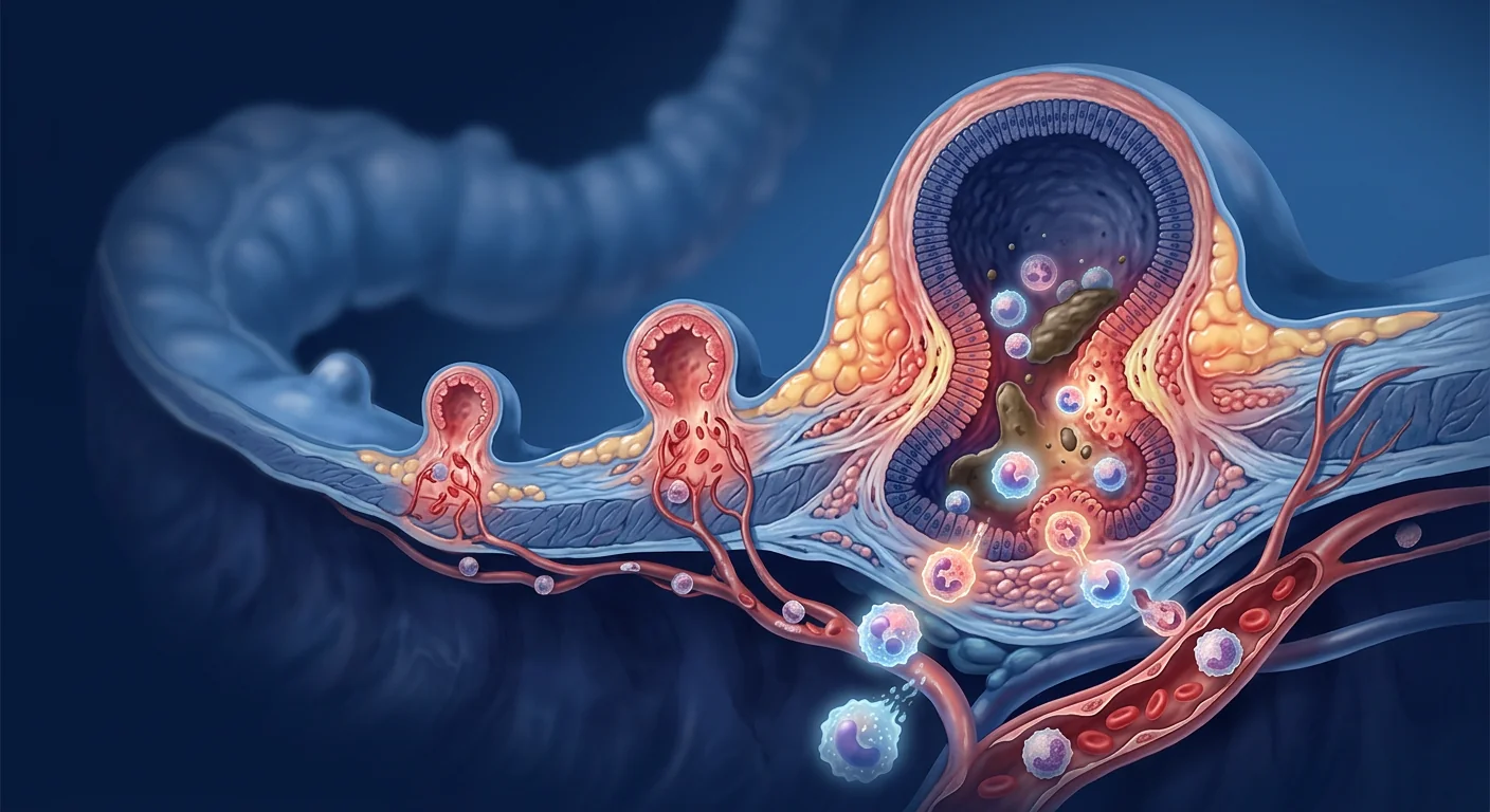
Diverticulosis — the presence of small mucosal outpouchings (diverticula) through weak points in the colonic wall — is present in roughly 60% of people over 60 in Western populations. [2] Most never know they have it. Diverticulitis occurs when one or more of these pouches becomes obstructed, leading to bacterial overgrowth, microperforation, and an acute inflammatory response in the surrounding colonic wall and pericolic fat. [3]
Why recurrence is the real problem. After a first episode of acute diverticulitis, approximately 20–35% of patients will have a second within five years, and recurrence risk climbs with each subsequent episode. [4] The standard pathway — antibiotics for uncomplicated cases, percutaneous drainage for abscesses, and segmental colectomy for complicated or frequently recurrent disease — manages the acute crisis but does not address the underlying colonic vulnerability. The bowel wall remains weakened, the local inflammatory milieu persists, and the microbiome dysbiosis that contributes to diverticular formation is unchanged.
The tissue-level problem. Chronic low-grade inflammation in the colonic mucosa — driven by altered gut permeability, microbial translocation, and innate immune activation — appears to be the common thread linking diverticulosis to diverticulitis and diverticulitis to recurrence. [5] This is fundamentally an inflammatory disease of the colon wall, not simply a mechanical one, which is why anti-inflammatory strategies, including MSC therapy, are being explored.
Why mesenchymal stem cells are studied for diverticulitis
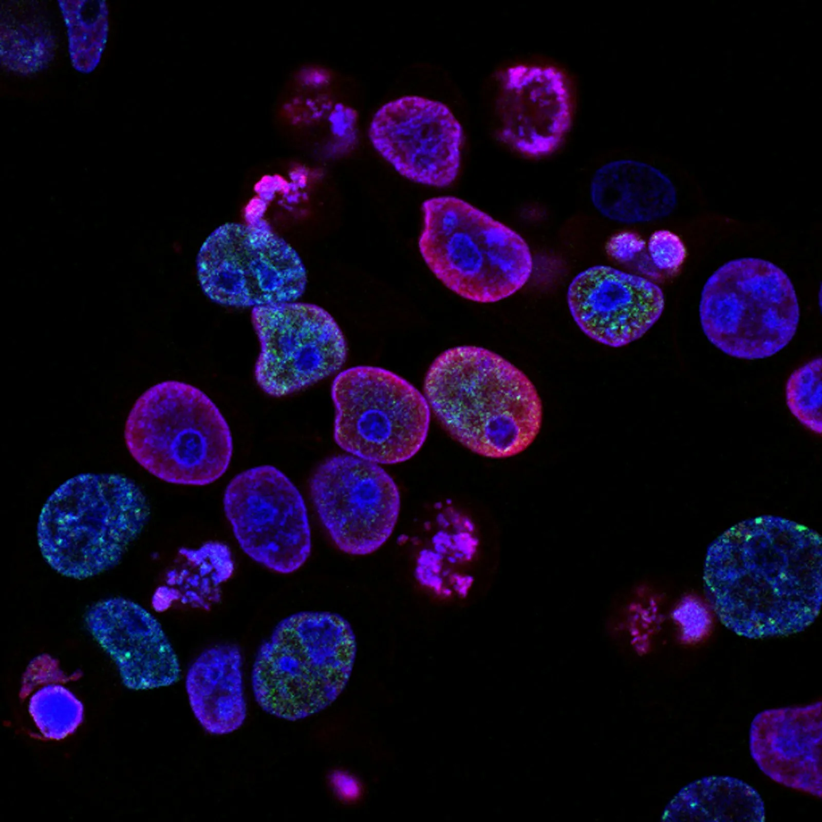
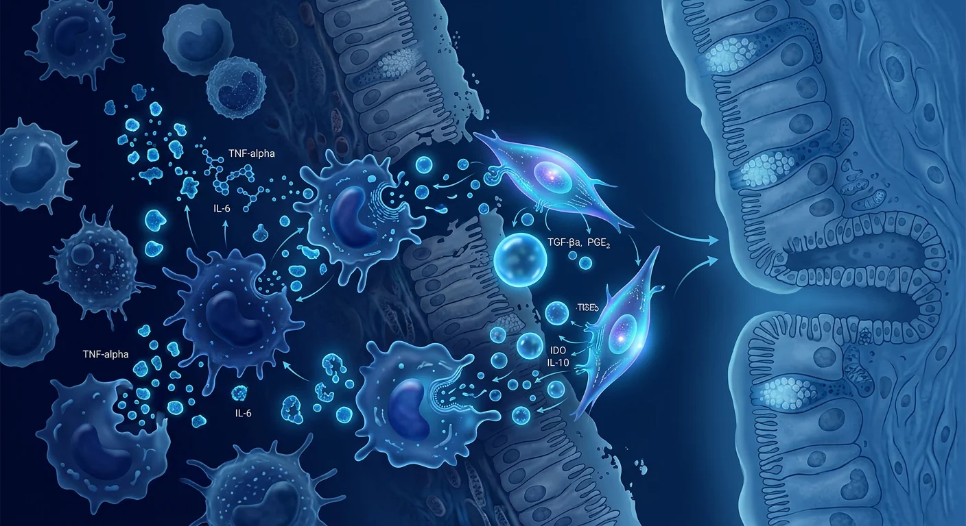
MSCs are potent immunomodulators. When exposed to an inflamed environment, they secrete a cocktail of anti-inflammatory factors — TGF-β, IL-10, PGE2, TSG-6, and IDO — that shift macrophages from a pro-inflammatory M1 phenotype to a reparative M2 phenotype, suppress neutrophil-driven tissue damage, and promote regulatory T-cell activity. [6][7] They also release angiogenic factors (VEGF, HGF, FGF-2) that support microvascular repair and trophic factors that promote epithelial barrier integrity. [8]
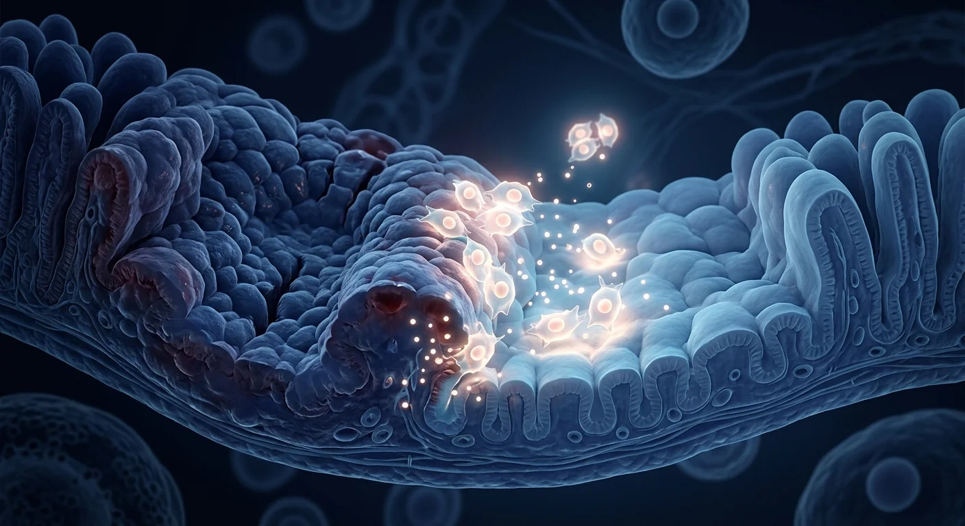
This mechanism set maps directly onto the pathology of recurrent diverticulitis. Reducing the chronic inflammatory tone in the colonic wall could theoretically lower the risk of new acute episodes. Enhancing mucosal barrier function could reduce microbial translocation — the trigger for diverticular inflammation. And promoting vascular health in the pericolic tissue could improve resilience against microperforation. None of this has been proven in a large controlled trial for diverticulitis specifically, but the biological rationale is grounded in well-characterized MSC properties.
Key MSC mechanisms relevant to diverticulitis
- M1→M2 macrophage polarization — shifts colonic immune environment from inflammatory to reparative
- Treg induction + Th17 suppression — rebalances adaptive immunity in the gut wall
- Epithelial barrier repair — tight junction protein upregulation reduces gut permeability
- Anti-fibrotic activity — TGF-β/Smad pathway modulation may reduce post-inflammatory stricturing
- Angiogenesis support — VEGF, HGF, FGF-2 promote microvascular integrity in pericolic tissue
What the clinical evidence says
The direct evidence is early. No large randomized controlled trial has tested MSC therapy specifically for diverticulitis. However, the evidence base from adjacent conditions — and from the broader MSC immunomodulation literature — provides a plausible foundation.
Evidence from IBD and gastrointestinal MSC trials. The use of MSCs in Crohn's disease, particularly for complex perianal fistulas, established that locally delivered MSCs can reduce inflammation, promote tissue closure, and achieve durable healing in the gastrointestinal tract. [9] Systemic MSC infusion for luminal Crohn's and ulcerative colitis has shown safety and some efficacy signals in early-phase trials, suggesting that intravenous MSCs can reach and modulate the gut immune environment. [10]
Preclinical models of colonic inflammation. In animal models of chemically induced colitis (which share features with diverticulitis including mucosal barrier disruption, innate immune activation, and pericolic inflammation), MSC administration consistently reduces histological inflammation scores, lowers pro-inflammatory cytokines (TNF-α, IL-1β, IL-6), upregulates anti-inflammatory IL-10, and accelerates mucosal healing. [11] These are precisely the outcomes one would hope to achieve in diverticulitis.
How MSC therapy for diverticulitis works in practice
For patients with recurrent diverticulitis who are being evaluated for MSC therapy, the treatment approach at VELAR Center follows a structured protocol.
Step 1 — Comprehensive assessment. Before any treatment decision, patients undergo a thorough evaluation including colonoscopy (to rule out alternative diagnoses, assess diverticular burden, and evaluate for stricture or chronic inflammatory changes), CT imaging where indicated, inflammatory biomarker panel (CRP, fecal calprotectin), and nutritional and microbiome assessment. The goal is to confirm that diverticulitis — and not another colonic pathology — is the primary problem and that the patient is an appropriate candidate.
Step 2 — Biomarker baseline and protocol design. Baseline inflammatory markers are established, and a personalized treatment protocol is designed. For diverticulitis, the approach typically involves systemic intravenous MSC infusion — the goal being to deliver immunomodulatory cells throughout the circulatory system so they can home to sites of colonic inflammation. The number of cells, dosing schedule, and whether to combine with local or targeted delivery is determined individually.
Step 3 — MSC infusion. The infusion itself is an outpatient procedure taking 60–90 minutes. VELAR uses fresh, never-frozen Wharton's jelly-derived MSCs — cells are cultured under cGMP conditions in a Class-100 (ISO 5) cleanroom, pass rigorous identity (ISCT criteria), purity (≥95% MSC markers), and viability (>95% at delivery) testing, and are released only after independent quality verification. No cryopreservation, no DMSO.
Step 4 — Follow-up and outcome tracking. Patients are monitored at defined intervals — typically 1 month, 3 months, 6 months, and 12 months post-infusion. Outcomes tracked include: episode frequency (number of diverticulitis flares requiring medical attention), inflammatory biomarkers (CRP, fecal calprotectin), quality-of-life scores (SF-36 or GIQLI), and any need for antibiotics, hospitalization, or surgical intervention.
Honest expectations
MSC therapy for diverticulitis is investigational — it is not a proven cure and is not a substitute for appropriate acute medical or surgical management. The goal is disease modification: reducing the frequency and severity of recurrent episodes by altering the inflammatory environment in the colonic wall. Some patients may see a meaningful reduction in flare frequency; others may not. No responsible clinician can guarantee a specific outcome. Treatment decisions should be made collaboratively, with full understanding of what the evidence does and does not support.
What recovery and follow-up look like
MSC infusion is well tolerated. The most commonly reported side effects are transient — mild fatigue for 24–48 hours, a low-grade temperature elevation, or mild infusion-related symptoms — and resolve without intervention. [12] Patients return to normal activities within 1–2 days. There is no surgical recovery, no bowel prep beyond standard hydration, and no downtime.
Outcome assessment is structured around real clinical endpoints — not just how the patient feels. Inflammatory biomarkers typically begin to trend downward within 4–8 weeks if the treatment is working. Diverticulitis episode frequency should be tracked over 6–12 months to assess whether the treatment has altered the disease trajectory. A patient who previously experienced 3–4 flares per year and drops to 0–1 per year over 12 months of follow-up would represent a clinically meaningful improvement. A patient who sees no change in flare frequency after 6 months should have a frank conversation about whether to continue with additional infusions or pursue alternative strategies.
How to evaluate any offer responsibly
If you are considering MSC therapy for recurrent diverticulitis, bring a critical eye. Ask precisely which cells are used (source, donor screening, culture conditions), what viability and purity testing is performed at release, and how the cells are delivered (fresh vs. frozen, IV vs. local). Request to see the clinic's quality certifications — cGMP, ISO class, and independent release testing documentation. Ask how outcomes are measured: is it based on symptom reports alone, or are inflammatory biomarkers and episode counts tracked objectively over time?
Be deeply skeptical of any clinic that claims to "cure" diverticulitis, promises a specific percentage reduction in flares without citing a published source, or offers MSC therapy without a thorough pre-treatment evaluation to confirm the diagnosis and rule out complications that require surgical intervention. A trustworthy provider will be transparent about the investigational nature of the treatment, will measure outcomes objectively, and will never let hope outrun the data.
Frequently Asked Questions
Can stem cell therapy cure diverticulitis?
No. MSC therapy is investigational for diverticulitis and is not a cure. The goal is disease modification — reducing the frequency and severity of recurrent episodes by dampening the chronic inflammatory environment in the colonic wall. Diverticula themselves do not disappear; the aim is to prevent them from becoming inflamed.
How is MSC therapy different from antibiotics or surgery?
Antibiotics treat the acute infection during a flare but do nothing to prevent the next one. Surgery removes the affected segment of colon but is a major procedure with its own risks and recovery. MSC therapy targets the underlying inflammatory biology — the chronic low-grade immune activation that makes the colonic wall vulnerable to recurrent episodes. It is a disease-modifying approach, not an acute rescue or a structural fix.
How many treatments are needed for diverticulitis?
There is no established protocol for MSC therapy in diverticulitis. In early clinical experience with related gastrointestinal inflammatory conditions, a single intravenous infusion is the most common starting point, with some protocols incorporating a second infusion at 3–6 months. The number and timing of infusions should be individualized based on disease severity, treatment response, and inflammatory biomarker trends.
How much does stem cell therapy for diverticulitis cost in Bangkok?
Costs vary by clinic, cell source, quality standards, and protocol intensity. In Thailand, MSC therapy at cGMP-certified facilities typically ranges from USD 12,000–25,000 per infusion depending on cell dose and ancillary services. A detailed cost breakdown should be provided during consultation, and patients should understand exactly what is included — cell preparation, infusion, follow-up monitoring, and any additional support services.
Is MSC therapy for diverticulitis covered by insurance?
No. MSC therapy for diverticulitis is investigational and not covered by any health insurance plan. It is a self-pay treatment. Some patients use medical savings accounts or health financing options; check with your provider.
Who is a candidate for MSC therapy for diverticulitis?
Patients with documented recurrent diverticulitis (2 or more confirmed episodes) who wish to explore a non-surgical, disease-modifying option may be appropriate candidates. Contraindications include active acute diverticulitis requiring antibiotics or drainage, complicated diverticulitis with free perforation or uncontrolled sepsis, known malignancy, active systemic infection, and pregnancy. Every patient undergoes a comprehensive pre-treatment evaluation to confirm candidacy.
Limitations and honest disclosures
Several important caveats must be stated plainly. First, MSC therapy for diverticulitis is investigational — it has not been validated in a large randomized controlled trial for this specific indication, and the evidence supporting its use is drawn from mechanistic plausibility, preclinical studies, and clinical experience in related gastrointestinal inflammatory conditions, not from diverticulitis-specific trials. Second, not every patient will respond — some may see no reduction in flare frequency despite treatment. Third, MSC therapy does not eliminate diverticula from the colon wall — it targets the inflammatory process, not the anatomical abnormality. Fourth, patients with complicated diverticulitis (abscess, perforation, fistula, or obstruction) require surgical evaluation first — MSC therapy is not a substitute for indicated surgery. Fifth, long-term safety data beyond 5–10 years of follow-up are limited for MSC therapy in general, and essentially nonexistent for diverticulitis specifically. Patients considering this treatment must weigh these uncertainties carefully against the potential for disease modification.
The VELAR perspective
At VELAR Center, we approach diverticulitis with the same framework we apply to every condition: start with the biology, follow the evidence, and let the patient make an informed decision. The immunomodulatory properties of MSCs are real and well-documented. The inflammatory basis of recurrent diverticulitis is well-established. The two fit together logically — but logic is not proof, and we will not pretend otherwise. If a patient with recurrent diverticulitis wants to explore whether MSC therapy can reduce their flare burden, we will provide an honest, evidence-based consultation, rigorous pre-treatment evaluation, transparent outcome tracking, and a commitment to measuring results — not just reporting anecdotes. That is what responsible regenerative medicine looks like.
References
- Strate LL, Morris AM. Epidemiology, pathophysiology, and treatment of diverticulitis. Gastroenterology. 2019;156(5):1282-1298.e1. doi:10.1053/j.gastro.2018.12.033 ↩
- Peery AF, Crockett SD, Murphy CC, et al. Burden and cost of gastrointestinal, liver, and pancreatic diseases in the United States: Update 2021. Gastroenterology. 2022;162(2):621-644. doi:10.1053/j.gastro.2021.10.017 ↩
- Tursi A, Scarpignato C, Strate LL, et al. Colonic diverticular disease. Nature Reviews Disease Primers. 2020;6(1):20. doi:10.1038/s41572-020-0153-5 ↩
- Bharucha AE, Parthasarathy G, Ditah I, et al. Temporal trends in the incidence and natural history of diverticulitis. American Journal of Gastroenterology. 2015;110(11):1589-1596. doi:10.1038/ajg.2015.302 ↩
- Barbara G, Scaioli E, Barbaro MR, et al. Gut microbiota, metabolome and immune signatures in patients with uncomplicated diverticular disease. Gut. 2017;66(7):1252-1261. doi:10.1136/gutjnl-2016-312377 ↩
- Shi Y, Wang Y, Li Q, et al. Immunoregulatory mechanisms of mesenchymal stem and stromal cells in inflammatory diseases. Nature Reviews Nephrology. 2018;14(8):493-507. doi:10.1038/s41581-018-0023-5 ↩
- Galipeau J, Sensébé L. Mesenchymal stromal cells: clinical challenges and therapeutic opportunities. Cell Stem Cell. 2018;22(6):824-833. doi:10.1016/j.stem.2018.05.004 ↩
- Bronckaers A, Hilkens P, Martens W, et al. Mesenchymal stem/stromal cells as a pharmacological approach to accelerate angiogenesis. Pharmacology & Therapeutics. 2014;143(2):181-196. doi:10.1016/j.pharmthera.2014.02.013 ↩
- Panés J, García-Olmo D, Van Assche G, et al. Expanded allogeneic adipose-derived mesenchymal stem cells (Cx601) for complex perianal fistulas in Crohn's disease: a phase 3 randomised, double-blind controlled trial. The Lancet. 2016;388(10051):1281-1290. doi:10.1016/S0140-6736(16)31203-X ↩
- Duijvestein M, Vos ACW, Roelofs H, et al. Autologous bone marrow-derived mesenchymal stromal cell treatment for refractory luminal Crohn's disease. Gut. 2010;59(12):1662-1669. doi:10.1136/gut.2010.215152 ↩
- Gonzalez-Rey E, Anderson P, Gonzalez MA, Rico L, Buscher D, Delgado M. Human adult stem cells derived from adipose tissue protect against experimental colitis and sepsis. Gut. 2009;58(7):929-939. doi:10.1136/gut.2008.168534 ↩
- Lalu MM, McIntyre L, Pugliese C, et al. Safety of cell therapy with mesenchymal stromal cells (SafeCell). PLoS One. 2012;7(10):e47559. doi:10.1371/journal.pone.0047559 ↩
- Pittenger MF, Discher DE, Péault BM, Phinney DG, Hare JM, Caplan AI. Mesenchymal stem cell perspective: cell biology to clinical progress. NPJ Regenerative Medicine. 2019;4:22. doi:10.1038/s41536-019-0083-6 ↩
- Dominici M, Le Blanc K, Mueller I, et al. Minimal criteria for defining multipotent mesenchymal stromal cells. Cytotherapy. 2006;8(4):315-317. doi:10.1080/14653240600855905 ↩
憩室炎——结肠壁小憩室的急性炎症——每年导致超过20万美国人住院,是40岁以上成年人最常见的消化系统急症之一。[1] 对大多数患者而言,一个疗程的抗生素和肠道休息即可解决急性发作。但对相当一部分少数患者来说,憩室炎会变为复发性、进展性,或并发脓肿、穿孔或狭窄——此时治疗选择便趋向手术,而MSC疗法正被研究为一种潜在的疾病修饰性干预手段。
什么是憩室炎,为何会复发?
憩室病——结肠壁薄弱处出现的小黏膜憩室——在西方人群中约60%的60岁以上人群存在。[2] 大多数人并不知道自己患有此病。憩室炎发生在一个或多个憩室被堵塞,导致细菌过度生长、微穿孔,并在周围结肠壁和结肠旁脂肪中引发急性炎症反应。[3]
为何复发才是真正的问题。首次急性憩室炎发作后,约20–35%的患者在五年内会复发第二次,且每次后续发作都会增加复发风险。[4] 标准路径——无并发症病例使用抗生素、脓肿行经皮引流、复杂性或频繁复发病例行节段性结肠切除术——处理了急性危机,但并未解决潜在的结肠脆弱性。肠壁仍然薄弱,局部炎症环境持续存在,导致憩室形成的微生物群失调也未被改变。
组织层面的问题。结肠黏膜中的慢性低度炎症——由肠道通透性改变、微生物移位和先天免疫激活驱动——似乎是连接憩室病与憩室炎、以及憩室炎与复发的共同线索。[5] 这本质上是结肠壁的炎症性疾病,而非单纯的机械问题,正因如此,包含MSC疗法在内的抗炎策略正在被探索。
为何研究间充质干细胞用于憩室炎
MSC是强效的免疫调节剂。暴露于炎症环境中时,它们分泌一系列抗炎因子——TGF-β、IL-10、PGE2、TSG-6和IDO——将巨噬细胞从促炎的M1表型转变为修复性的M2表型,抑制中性粒细胞驱动的组织损伤,并促进调节性T细胞活性。[6][7] 它们还释放支持微血管修复的血管生成因子(VEGF、HGF、FGF-2)和促进上皮屏障完整性的营养因子。[8]
这一机制集合直接映射到复发性憩室炎的病理上。降低结肠壁的慢性炎症基调可从理论上降低新急性发作的风险。增强黏膜屏障功能可减少微生物移位——憩室炎症的触发因素。促进结肠旁组织的血管健康可改善对微穿孔的抵抗力。这些目前尚未在针对憩室炎的大型对照试验中得到证实,但其生物学理论基础建立在充分表征的MSC特性之上。
与憩室炎相关的关键MSC机制
- M1→M2巨噬细胞极化——将结肠免疫环境从炎症转向修复
- Treg诱导+Th17抑制——重新平衡肠壁的适应性免疫
- 上皮屏障修复——紧密连接蛋白上调可降低肠道通透性
- 抗纤维化活性——TGF-β/Smad通路调节可减少炎症后狭窄形成
- 血管生成支持——VEGF、HGF、FGF-2促进结肠旁组织微血管完整性
临床证据说明了什么
直接证据尚属早期。尚无大型随机对照试验专门测试MSC疗法用于憩室炎。然而,来自邻近疾病的证据基础——以及更广泛的MSC免疫调节文献——提供了一个可信的基础。
来自IBD和消化道MSC试验的证据。MSC在克罗恩病中的应用,特别是用于复杂性肛周瘘管,确立了局部递送的MSC可以减少炎症、促进组织闭合并在消化道实现持久愈合。[9] 系统性MSC输注用于肠腔内克罗恩病和溃疡性结肠炎,在早期试验中显示了安全性和一些疗效信号,表明静脉MSC可以抵达并调节肠道免疫环境。[10]
结肠炎症的临床前模型。在化学诱导结肠炎的动物模型中(与憩室炎共享包括黏膜屏障破坏、先天免疫激活和结肠旁炎症在内的特征),MSC给药持续降低组织学炎症评分、降低促炎细胞因子(TNF-α、IL-1β、IL-6)、上调抗炎的IL-10,并加速黏膜愈合。[11] 这些正是在憩室炎中希望达到的结局。
MSC治疗憩室炎的实际操作
对于正在评估MSC治疗的复发性憩室炎患者,VELAR中心的治疗方法遵循结构化方案。
第一步——全面评估。在任何治疗决定之前,患者接受全面评估,包括结肠镜检查(排除替代诊断、评估憩室负荷、评估狭窄或慢性炎症变化)、CT影像(如有指征)、炎症生物标志物组合(CRP、粪便钙卫蛋白)、以及营养和微生物组评估。目标是确认憩室炎——而非其他结肠病变——是主要问题,且患者是合适的候选者。
第二步——生物标志物基线和方案设计。建立基线炎症标志物,设计个性化治疗方案。对于憩室炎,方法通常涉及系统性静脉MSC输注——目标是将免疫调节细胞递送至整个循环系统,使其能够归巢至结肠炎症部位。细胞数量、给药方案以及是否结合局部或靶向递送需个体化确定。
第三步——MSC输注。输注本身是门诊操作,耗时60–90分钟。VELAR使用新鲜、从未冷冻的沃顿胶来源MSC——细胞在cGMP条件下于Class-100(ISO 5)洁净室中培养,通过严格的身份(ISCT标准)、纯度(≥95% MSC标志物)和活性(递送时>95%)测试,并仅在独立质量验证后放行。无冷冻保存,无DMSO。
第四步——随访和结局追踪。患者在确定的时间间隔进行监测——通常为输注后1个月、3个月、6个月和12个月。追踪的结局包括:发作频率(需要医疗处理的憩室炎发作次数)、炎症生物标志物(CRP、粪便钙卫蛋白)、生活质量评分(SF-36或GIQLI),以及任何对抗生素、住院或手术干预的需求。
诚实的预期
MSC治疗憩室炎是研究性的——它不是已被证实的治愈方法,也不能替代适当的急性内科或外科治疗。目标是疾病修饰:通过改变结肠壁的炎症环境来降低复发的频率和严重程度。部分患者可能看到发作频率有意义的减少;另一些则可能不会。没有任何负责任的临床医生能保证特定的结果。治疗决定应在充分理解证据支持与不支持的内容的基础上共同做出。
恢复和随访是什么样的
MSC输注耐受性良好。最常报告的副作用是短暂的——24–48小时轻度疲劳、低度体温升高或轻度输注相关症状——且无需干预即可缓解。[12] 患者在1–2天内恢复正常活动。没有手术恢复期,没有超出标准补水的肠道准备,也没有停机时间。
结局评估围绕真实的临床终点构建——而非仅凭患者的感受。如果治疗有效,炎症生物标志物通常在4–8周内开始呈下降趋势。应在6–12个月内追踪憩室炎发作频率,以评估治疗是否改变了疾病轨迹。一个先前每年经历3–4次发作的患者,在12个月随访中降至每年0–1次,将代表具有临床意义的改善。一个在6个月后发作频率未见变化的患者,应就继续追加输注还是寻求替代策略进行坦诚的讨论。
如何负责任地评估任何提议
如果您正在考虑MSC治疗复发性憩室炎,请带着批判的眼光。精确询问使用的是哪种细胞(来源、供体筛查、培养条件)、放行时进行了哪些活性和纯度检测、以及细胞如何递送(新鲜vs.冷冻、静脉vs.局部)。要求查看诊所的质量认证——cGMP、ISO等级和独立放行检测文件。询问结果如何衡量:仅基于症状报告,还是通过炎症生物标志物和发作次数在时间推移中进行客观追踪?
对任何声称能"治愈"憩室炎、引用未注明来源的具体发作减少百分比、或未进行全面的治疗前评估以确认诊断并排除需要手术干预的并发症就提供MSC疗法的诊所,都要深度怀疑。值得信赖的提供者会对治疗的研究性质保持透明,会客观衡量结果,且决不会让希望超越数据。
常见问题
干细胞疗法能治愈憩室炎吗?
不能。MSC疗法用于憩室炎是研究性的,并非治愈方法。目标是疾病修饰——通过抑制结肠壁的慢性炎症环境来降低复发的频率和严重程度。憩室本身不会消失;目标是防止它们发生炎症。
MSC疗法与抗生素或手术有何不同?
抗生素治疗发作期间的急性感染,但不能预防下一次发作。手术切除受累的结肠段,但是一项有其自身风险和恢复期的大型手术。MSC疗法靶向潜在的炎症生物学——使结肠壁易受复发影响的慢性低度免疫激活。这是一种疾病修饰方法,而非急性救援或结构性修复。
憩室炎需要多少次治疗?
MSC疗法用于憩室炎尚无既定方案。在相关消化道炎症疾病的早期临床经验中,单次静脉输注是最常见的起点,部分方案在3–6个月时加入第二次输注。输注的次数和时机应基于疾病严重程度、治疗反应和炎症生物标志物趋势进行个体化调整。
在曼谷进行干细胞治疗憩室炎的费用是多少?
费用因诊所、细胞来源、质量标准和方案强度而异。在泰国,cGMP认证设施的MSC治疗通常为每次输注USD 12,000–25,000,取决于细胞剂量和辅助服务。应在咨询期间提供详细的费用明细,患者应准确了解所包含的内容——细胞制备、输注、随访监测和任何额外的支持服务。
MSC治疗憩室炎是否有保险覆盖?
没有。MSC治疗憩室炎是研究性的,不在任何健康保险计划的覆盖范围内。这是一项自费治疗。部分患者使用医疗储蓄账户或健康融资方案;请与您的提供商确认。
谁是MSC治疗憩室炎的候选者?
具有记录的复发性憩室炎(2次或更多已确认的发作)并希望探索非手术、疾病修饰选项的患者可能是合适的候选者。禁忌症包括需要抗生素或引流的急性活动性憩室炎、伴游离穿孔或未受控败血症的复杂性憩室炎、已知恶性肿瘤、活动性全身感染和妊娠。每位患者都接受全面的治疗前评估以确认候选资格。
局限性与诚实披露
必须明确指出几个重要的注意事项。第一,MSC治疗憩室炎是研究性的——尚未在此特定适应症的大型随机对照试验中得到验证,支持其使用的证据来自机制合理性、临床前研究以及相关消化道炎症疾病的临床经验,而非憩室炎特异性试验。第二,并非每位患者都会产生应答——部分患者可能尽管治疗但发作频率未见减少。第三,MSC疗法不会消除结肠壁上的憩室——它靶向炎症过程,而非解剖异常。第四,患有复杂性憩室炎(脓肿、穿孔、瘘管或梗阻)的患者需要首先进行外科评估——MSC疗法不能替代有指征的手术。第五,MSC疗法总体上超过5–10年随访的长期安全性数据有限,对憩室炎而言基本不存在。考虑该治疗的患者必须仔细权衡这些不确定性与其潜在的疾病修饰获益。
VELAR的观点
在VELAR中心,我们以适用于每种疾病的同一框架来处理憩室炎:从生物学出发,循证据而行,让患者做出知情决定。MSC的免疫调节特性是真实的且有充分文献记载。复发性憩室炎的炎症基础已被充分确立。二者在逻辑上是契合的——但逻辑并非证据,我们不会假装如此。如果一位复发性憩室炎患者想要探索MSC疗法是否能减少其发作负担,我们将提供诚实的、以证据为基础的咨询、严格的治疗前评估、透明的结局追踪,以及衡量结果——而非仅报告轶事——的承诺。这就是负责任的再生医学应有的样子。
参考文献
- Strate LL, Morris AM. Epidemiology, pathophysiology, and treatment of diverticulitis. Gastroenterology. 2019;156(5):1282-1298.e1. doi:10.1053/j.gastro.2018.12.033 ↩
- Peery AF, Crockett SD, Murphy CC, et al. Burden and cost of gastrointestinal, liver, and pancreatic diseases in the United States: Update 2021. Gastroenterology. 2022;162(2):621-644. doi:10.1053/j.gastro.2021.10.017 ↩
- Tursi A, Scarpignato C, Strate LL, et al. Colonic diverticular disease. Nature Reviews Disease Primers. 2020;6(1):20. doi:10.1038/s41572-020-0153-5 ↩
- Bharucha AE, Parthasarathy G, Ditah I, et al. Temporal trends in the incidence and natural history of diverticulitis. American Journal of Gastroenterology. 2015;110(11):1589-1596. doi:10.1038/ajg.2015.302 ↩
- Barbara G, Scaioli E, Barbaro MR, et al. Gut microbiota, metabolome and immune signatures in patients with uncomplicated diverticular disease. Gut. 2017;66(7):1252-1261. doi:10.1136/gutjnl-2016-312377 ↩
- Shi Y, Wang Y, Li Q, et al. Immunoregulatory mechanisms of mesenchymal stem and stromal cells in inflammatory diseases. Nature Reviews Nephrology. 2018;14(8):493-507. doi:10.1038/s41581-018-0023-5 ↩
- Galipeau J, Sensébé L. Mesenchymal stromal cells: clinical challenges and therapeutic opportunities. Cell Stem Cell. 2018;22(6):824-833. doi:10.1016/j.stem.2018.05.004 ↩
- Bronckaers A, Hilkens P, Martens W, et al. Mesenchymal stem/stromal cells as a pharmacological approach to accelerate angiogenesis. Pharmacology & Therapeutics. 2014;143(2):181-196. doi:10.1016/j.pharmthera.2014.02.013 ↩
- Panés J, García-Olmo D, Van Assche G, et al. Expanded allogeneic adipose-derived mesenchymal stem cells (Cx601) for complex perianal fistulas in Crohn's disease: a phase 3 randomised, double-blind controlled trial. The Lancet. 2016;388(10051):1281-1290. doi:10.1016/S0140-6736(16)31203-X ↩
- Duijvestein M, Vos ACW, Roelofs H, et al. Autologous bone marrow-derived mesenchymal stromal cell treatment for refractory luminal Crohn's disease. Gut. 2010;59(12):1662-1669. doi:10.1136/gut.2010.215152 ↩
- Gonzalez-Rey E, Anderson P, Gonzalez MA, Rico L, Buscher D, Delgado M. Human adult stem cells derived from adipose tissue protect against experimental colitis and sepsis. Gut. 2009;58(7):929-939. doi:10.1136/gut.2008.168534 ↩
- Lalu MM, McIntyre L, Pugliese C, et al. Safety of cell therapy with mesenchymal stromal cells (SafeCell). PLoS One. 2012;7(10):e47559. doi:10.1371/journal.pone.0047559 ↩
- Pittenger MF, Discher DE, Péault BM, Phinney DG, Hare JM, Caplan AI. Mesenchymal stem cell perspective: cell biology to clinical progress. NPJ Regenerative Medicine. 2019;4:22. doi:10.1038/s41536-019-0083-6 ↩
- Dominici M, Le Blanc K, Mueller I, et al. Minimal criteria for defining multipotent mesenchymal stromal cells. Cytotherapy. 2006;8(4):315-317. doi:10.1080/14653240600855905 ↩
التهاب الرتوج — التهاب حاد في الجيوب الصغيرة في جدار القولون — يرسل أكثر من 200,000 أمريكي إلى المستشفى كل عام، وهو أحد أكثر الأسباب الهضمية شيوعاً لدخول الطوارئ لدى البالغين فوق سن الأربعين. [1] بالنسبة لمعظم المرضى، تحل دورة من المضادات الحيوية وراحة الأمعاء النوبة الحادة. لكن لدى أقلية ذات دلالة، يصبح التهاب الرتوج متكرراً أو متقدماً أو معقّداً بالخراج أو الانثقاب أو التضيّق — وهنا تضيق خيارات العلاج نحو الجراحة، وحيث يُبحَث في علاج MSC كتدخّل محتمل معدِّل للمرض.
ما هو التهاب الرتوج، ولماذا يتكرر؟
داء الرتوج — وجود جيوب مخاطية صغيرة (رتوج) عبر نقاط ضعف في جدار القولون — موجود لدى 60% تقريباً ممن تجاوزوا الستين في المجتمعات الغربية. [2] معظمهم لا يعرفون أنهم مصابون به. يحدث التهاب الرتوج حين ينسد واحد أو أكثر من هذه الجيوب، مما يؤدي إلى فرط نمو بكتيري وثقب دقيق واستجابة التهابية حادة في جدار القولون المحيط والدهن حول القولون. [3]
لماذا التكرار هو المشكلة الحقيقية. بعد النوبة الأولى من التهاب الرتوج الحاد، سيصاب 20–35% من المرضى تقريباً بنوبة ثانية خلال خمس سنوات، ويزداد خطر التكرار مع كل نوبة لاحقة. [4] المسار القياسي — المضادات الحيوية للحالات غير المعقّدة، والنزح عبر الجلد للخراجات، واستئصال القولون القطعي للحالات المعقّدة أو المتكررة بكثرة — يدير الأزمة الحادة لكنه لا يعالج الضعف القولوني الكامن. يبقى جدار الأمعاء ضعيفاً، وتستمر البيئة الالتهابية الموضعية، ويبقى اختلال الميكروبيوم الذي يساهم في تشكّل الرتوج دون تغيير.
المشكلة على مستوى النسيج. الالتهاب المزمن منخفض الدرجة في مخاطية القولون — المدفوع بتغيّر نفاذية الأمعاء والانتقال الميكروبي وتفعيل المناعة الفطرية — يبدو أنه الخيط المشترك الذي يربط داء الرتوج بالتهاب الرتوج، والتهاب الرتوج بالتكرار. [5] هذا مرض التهابي لجدار القولون جوهرياً، وليس مجرد مرض ميكانيكي، وهذا هو سبب استكشاف الاستراتيجيات المضادة للالتهاب، بما فيها علاج MSC.
لماذا تُدرَس الخلايا الجذعية الوسيطة لالتهاب الرتوج
MSCs معدِّلات مناعية قوية. حين تتعرض لبيئة ملتهبة، تفرز مزيجاً من العوامل المضادة للالتهاب — TGF-β وIL-10 وPGE2 وTSG-6 وIDO — تحوّل البلاعم من نمط M1 المحرِّض للالتهاب إلى نمط M2 الإصلاحي، وتكبح تلف النسيج المدفوع بالعدلات، وتعزز نشاط الخلايا التائية التنظيمية. [6][7] كما تطلق عوامل مولّدة للأوعية (VEGF وHGF وFGF-2) تدعم إصلاح الأوعية الدقيقة وعوامل مغذّية تعزز سلامة الحاجز الظهاري. [8]
تنطبق مجموعة الآليات هذه مباشرة على أمراضية التهاب الرتوج المتكرر. تقليل النغمة الالتهابية المزمنة في جدار القولون يمكن نظرياً أن يخفض خطر النوبات الحادة الجديدة. تعزيز وظيفة الحاجز المخاطي يمكن أن يقلل الانتقال الميكروبي — محفز التهاب الرتوج. وتعزيز الصحة الوعائية في النسيج حول القولون يمكن أن يحسّن المقاومة ضد الثقب الدقيق. لم يُثبَت أي من هذا في تجربة كبيرة مضبوطة لالتهاب الرتوج تحديداً، لكن الأساس البيولوجي يرتكز على خصائص MSC الموثّقة جيداً.
آليات MSC الرئيسية ذات الصلة بالتهاب الرتوج
- استقطاب البلاعم M1→M2 — ينقل البيئة المناعية القولونية من الالتهاب إلى الإصلاح
- تحريض Treg + تثبيط Th17 — يعيد توازن المناعة التكيّفية في جدار الأمعاء
- إصلاح الحاجز الظهاري — زيادة بروتينات الوصلات المحكمة تقلل نفاذية الأمعاء
- نشاط مضاد للتليف — تعديل مسار TGF-β/Smad قد يقلل التضيّق بعد الالتهاب
- دعم تولّد الأوعية — VEGF وHGF وFGF-2 تعزز سلامة الأوعية الدقيقة في النسيج حول القولون
ماذا تقول الأدلة السريرية
الدليل المباشر مبكر. لم تختبر أي تجربة كبيرة مضبوطة معشّاة علاج MSC تحديداً لالتهاب الرتوج. ومع ذلك، توفر قاعدة الأدلة من الحالات المجاورة — ومن أدبيات تعديل المناعة بـ MSC الأوسع — أساساً معقولاً.
أدلة من تجارب IBD والجهاز الهضمي بـ MSC. استخدام MSCs في داء كرون، خاصةً للنواسير المعقّدة حول الشرج، أثبت أن MSCs المعطاة موضعياً يمكن أن تقلل الالتهاب وتعزز إغلاق النسيج وتحقق التئاماً دائماً في السبيل الهضمي. [9] تسريب MSC الجهازي لداء كرون اللمعي والتهاب القولون التقرحي أظهر سلامة وبعض إشارات الفعالية في تجارب مبكرة الطور، مما يشير إلى أن MSCs الوريدية يمكن أن تصل إلى البيئة المناعية المعوية وتعدّلها. [10]
نماذج ما قبل السريرية لالتهاب القولون. في نماذج حيوانية لالتهاب القولون المستحث كيميائياً (التي تشترك مع التهاب الرتوج في سمات تشمل تمزق الحاجز المخاطي وتفعيل المناعة الفطرية والالتهاب حول القولون)، يقلل إعطاء MSC باستمرار من درجات الالتهاب النسيجي، ويخفض السيتوكينات المحرّضة للالتهاب (TNF-α وIL-1β وIL-6)، ويرفع IL-10 المضاد للالتهاب، ويسرّع التئام المخاطية. [11] هذه بالضبط النتائج التي يأمل المرء تحقيقها في التهاب الرتوج.
كيف يعمل علاج MSC لالتهاب الرتوج عملياً
للمرضى المصابين بالتهاب رتوج متكرر الذين يُقيَّمون لعلاج MSC، يتبع نهج العلاج في مركز VELAR بروتوكولاً منظّماً.
الخطوة 1 — التقييم الشامل. قبل أي قرار علاجي، يخضع المرضى لتقييم شامل يشمل تنظير القولون (لاستبعاد التشخيصات البديلة وتقييم عبء الرتوج وتقييم التضيّق أو التغيرات الالتهابية المزمنة)، وتصوير CT حين يُستدعى، ولوحة المؤشرات الحيوية الالتهابية (CRP وكالبروتكتين البرازي)، وتقييم التغذية والميكروبيوم. الهدف هو تأكيد أن التهاب الرتوج — وليس أمراضاً قولونية أخرى — هو المشكلة الأساسية وأن المريض مرشح مناسب.
الخطوة 2 — خط الأساس للمؤشرات الحيوية وتصميم البروتوكول. تُحدَّد مؤشرات الالتهاب الأساسية، ويُصمَّم بروتوكول علاجي مخصص. بالنسبة لالتهاب الرتوج، يتضمن النهج عادةً تسريب MSC وريدي جهازي — الهدف هو إيصال الخلايا المعدِّلة للمناعة عبر الجهاز الدوري لتتمكن من التوجّه إلى مواقع الالتهاب القولوني. عدد الخلايا وجدول الجرعات وما إذا كان سيُدمَج مع إيصال موضعي أو موجَّه يُحدَّد فردياً.
الخطوة 3 — تسريب MSC. التسريب نفسه إجراء في العيادة الخارجية يستغرق 60–90 دقيقة. تستخدم VELAR خلايا MSC طازجة غير مجمّدة أبداً مشتقة من هلام وارتون — تُزرَع الخلايا تحت ظروف cGMP في غرفة نظيفة من الفئة 100 (ISO 5)، وتجتاز اختبارات هوية صارمة (معايير ISCT) ونقاوة (≥95% واسمات MSC) وحيوية (>95% عند التسليم)، ولا تُصرَف إلا بعد تحقق جودة مستقل. لا حفظ بالتجميد، لا DMSO.
الخطوة 4 — المتابعة وتتبّع النتائج. يُراقَب المرضى على فترات محددة — عادةً بعد شهر و3 أشهر و6 أشهر و12 شهراً من التسريب. النتائج المتتبَّعة تشمل: تواتر النوبات (عدد نوبات التهاب الرتوج التي تتطلب عناية طبية)، والمؤشرات الحيوية الالتهابية (CRP وكالبروتكتين البرازي)، ودرجات جودة الحياة (SF-36 أو GIQLI)، وأي حاجة للمضادات الحيوية أو دخول المستشفى أو التدخل الجراحي.
توقعات صادقة
علاج MSC لالتهاب الرتوج هو بحثي تجريبي — ليس علاجاً مثبتاً وليس بديلاً عن التدبير الطبي أو الجراحي الحاد المناسب. الهدف هو تعديل المرض: تقليل تواتر وشدة النوبات المتكررة عبر تغيير البيئة الالتهابية في جدار القولون. قد يرى بعض المرضى انخفاضاً ذا معنى في تواتر النوبات؛ وقد لا يراه آخرون. لا يمكن لأي طبيب مسؤول أن يضمن نتيجة محددة. يجب أن تُتَّخذ قرارات العلاج بالتعاون، مع فهم كامل لما تدعمه الأدلة وما لا تدعمه.
كيف تبدو فترة التعافي والمتابعة
تسريب MSC جيد التحمل. أكثر التأثيرات الجانبية المبلَّغ عنها عابرة — تعب خفيف لمدة 24–48 ساعة، أو ارتفاع طفيف في درجة الحرارة، أو أعراض خفيفة متعلقة بالتسريب — وتزول دون تدخل. [12] يعود المرضى إلى أنشطتهم الطبيعية خلال 1–2 يوم. لا يوجد تعافٍ جراحي، ولا تحضير معوي يتجاوز الترطيب القياسي، ولا وقت تعطّل.
تقييم النتائج مبني على نقاط نهاية سريرية حقيقية — وليس فقط على شعور المريض. تبدأ المؤشرات الحيوية الالتهابية عادةً بالاتجاه نحو الانخفاض خلال 4–8 أسابيع إذا كان العلاج فعالاً. يجب تتبّع تواتر نوبات التهاب الرتوج على مدى 6–12 شهراً لتقييم ما إذا كان العلاج قد غيّر مسار المرض. مريض كان يعاني سابقاً من 3–4 نوبات سنوياً وينخفض إلى 0–1 نوبة سنوياً خلال 12 شهراً من المتابعة يمثّل تحسناً ذا معنى سريري. مريض لا يرى تغيراً في تواتر النوبات بعد 6 أشهر يجب أن يجري حواراً صريحاً حول متابعة التسريبات الإضافية أو السعي لاستراتيجيات بديلة.
كيف تقيّم أي عرض بمسؤولية
إذا كنت تفكر في علاج MSC لالتهاب الرتوج المتكرر، فاحمل نظرة ناقدة. اسأل تحديداً عن الخلايا المستخدمة (المصدر، فحص المتبرع، ظروف الزرع)، وما هي اختبارات الحيوية والنقاوة التي تُجرى عند الصرف، وكيف تُوصَّل الخلايا (طازجة مقابل مجمّدة، وريدياً مقابل موضعياً). اطلب رؤية شهادات جودة العيادة — cGMP وفئة ISO ووثائق اختبار الصرف المستقل. اسأل كيف تُقاس النتائج: هل تعتمد على تقارير الأعراض وحدها، أم تُتبَّع المؤشرات الحيوية الالتهابية وعدد النوبات بموضوعية عبر الزمن؟
كن شديد الشك تجاه أي عيادة تدّعي "علاج" التهاب الرتوج، أو تعد بنسبة مئوية محددة لانخفاض النوبات دون الاستشهاد بمصدر منشور، أو تقدم علاج MSC دون تقييم شامل قبل العلاج لتأكيد التشخيص واستبعاد المضاعفات التي تتطلب تدخلاً جراحياً. سيكون المزوّد الجدير بالثقة شفافاً بشأن الطبيعة البحثية للعلاج، وسيقيس النتائج بموضوعية، ولن يدع الأمل يتجاوز البيانات أبداً.
الأسئلة الشائعة
هل يمكن للعلاج بالخلايا الجذعية أن يشفي التهاب الرتوج؟
لا. علاج MSC لالتهاب الرتوج بحثي تجريبي وليس شفاءً. الهدف هو تعديل المرض — تقليل تواتر وشدة النوبات المتكررة عبر تثبيط البيئة الالتهابية المزمنة في جدار القولون. الرتوج نفسها لا تختفي؛ الهدف هو منعها من الالتهاب.
كيف يختلف علاج MSC عن المضادات الحيوية أو الجراحة؟
تعالج المضادات الحيوية العدوى الحادة أثناء النوبة لكنها لا تفعل شيئاً لمنع النوبة التالية. تزيل الجراحة الجزء المصاب من القولون لكنها إجراء كبير له مخاطره وفترة تعافيه الخاصة. يستهدف علاج MSC البيولوجيا الالتهابية الكامنة — التنشيط المناعي المزمن منخفض الدرجة الذي يجعل جدار القولون عرضة للنوبات المتكررة. إنه نهج معدِّل للمرض، وليس إنقاذاً حاداً أو إصلاحاً بنيوياً.
كم عدد العلاجات المطلوبة لالتهاب الرتوج؟
لا يوجد بروتوكول معتمد لعلاج MSC في التهاب الرتوج. في الخبرة السريرية المبكرة مع الحالات الالتهابية الهضمية ذات الصلة، التسريب الوريدي الوحيد هو نقطة البداية الأكثر شيوعاً، مع إضافة بعض البروتوكولات تسريباً ثانياً عند 3–6 أشهر. يجب تخصيص عدد وتوقيت التسريبات بناءً على شدة المرض والاستجابة للعلاج واتجاهات المؤشرات الحيوية الالتهابية.
كم تكلفة العلاج بالخلايا الجذعية لالتهاب الرتوج في بانكوك؟
تتفاوت التكاليف حسب العيادة ومصدر الخلايا ومعايير الجودة وشدة البروتوكول. في تايلاند، يتراوح علاج MSC في المنشآت المعتمدة cGMP عادةً بين 12,000–25,000 دولار أمريكي لكل تسريب حسب جرعة الخلايا والخدمات المساعدة. يجب تقديم تفصيل دقيق للتكلفة أثناء الاستشارة، ويجب أن يفهم المرضى تماماً ما هو مشمول — تحضير الخلايا والتسريب ومتابعة المراقبة وأي خدمات دعم إضافية.
هل يغطي التأمين علاج MSC لالتهاب الرتوج؟
لا. علاج MSC لالتهاب الرتوج بحثي تجريبي ولا تغطيه أي خطة تأمين صحي. إنه علاج يُدفع ذاتياً. يستخدم بعض المرضى حسابات الادخار الطبي أو خيارات التمويل الصحي؛ استشر مزوّدك.
من هو المرشح لعلاج MSC لالتهاب الرتوج؟
المرضى الذين لديهم التهاب رتوج متكرر موثّق (نوبتان مؤكدتان أو أكثر) ويرغبون في استكشاف خيار غير جراحي معدِّل للمرض قد يكونون مرشحين مناسبين. موانع الاستعمال تشمل التهاب الرتوج الحاد النشط الذي يتطلب مضادات حيوية أو نزحاً، والتهاب الرتوج المعقّد مع انثقاب حر أو إنتان غير مضبوط، والورم الخبيث المعروف، والعدوى الجهازية النشطة، والحمل. يخضع كل مريض لتقييم شامل قبل العلاج لتأكيد الأهلية.
القيود والإفصاحات الصادقة
يجب ذكر عدة تحفظات مهمة بوضوح. أولاً، علاج MSC لالتهاب الرتوج هو بحثي تجريبي — لم يتم التحقق منه في تجربة كبيرة مضبوطة معشّاة لهذا الاستطباب تحديداً، والأدلة الداعمة لاستخدامه مستمدة من المعقولية الآلياتية والدراسات قبل السريرية والخبرة السريرية في الحالات الالتهابية الهضمية ذات الصلة، وليس من تجارب خاصة بالتهاب الرتوج. ثانياً، ليس كل مريض سيستجيب — قد لا يرى البعض أي انخفاض في تواتر النوبات رغم العلاج. ثالثاً، علاج MSC لا يزيل الرتوج من جدار القولون — إنه يستهدف العملية الالتهابية، وليس الشذوذ التشريحي. رابعاً، المرضى المصابون بالتهاب رتوج معقّد (خراج أو انثقاب أو ناسور أو انسداد) يحتاجون تقييماً جراحياً أولاً — علاج MSC ليس بديلاً عن الجراحة المستطبّة. خامساً، بيانات السلامة طويلة الأمد التي تتجاوز 5–10 سنوات من المتابعة محدودة لعلاج MSC عموماً، ومعدومة أساساً لالتهاب الرتوج تحديداً. يجب على المرضى الذين يفكرون في هذا العلاج أن يوازنوا بدقة بين أوجه عدم اليقين هذه وإمكانية تعديل المرض.
منظور VELAR
في مركز VELAR، نتعامل مع التهاب الرتوج بنفس الإطار الذي نطبقه على كل حالة: نبدأ من البيولوجيا، ونتبع الأدلة، وندع المريض يتخذ قراراً مستنيراً. الخصائص المعدِّلة للمناعة لـ MSCs حقيقية وموثّقة جيداً. الأساس الالتهابي لالتهاب الرتوج المتكرر راسخ. الاثنان يتلاءمان منطقياً — لكن المنطق ليس برهاناً، ولن ندّعي غير ذلك. إذا أراد مريض مصاب بالتهاب رتوج متكرر استكشاف ما إذا كان علاج MSC يمكن أن يقلل عبء نوباته، سنقدم استشارة صادقة قائمة على الأدلة، وتقييماً دقيقاً قبل العلاج، وتتبّعاً شفافاً للنتائج، والتزاماً بقياس النتائج — لا مجرد سرد القصص. هذا ما يبدو عليه الطب التجديدي المسؤول.
المراجع
- Strate LL, Morris AM. Epidemiology, pathophysiology, and treatment of diverticulitis. Gastroenterology. 2019;156(5):1282-1298.e1. doi:10.1053/j.gastro.2018.12.033 ↩
- Peery AF, Crockett SD, Murphy CC, et al. Burden and cost of gastrointestinal, liver, and pancreatic diseases in the United States: Update 2021. Gastroenterology. 2022;162(2):621-644. doi:10.1053/j.gastro.2021.10.017 ↩
- Tursi A, Scarpignato C, Strate LL, et al. Colonic diverticular disease. Nature Reviews Disease Primers. 2020;6(1):20. doi:10.1038/s41572-020-0153-5 ↩
- Bharucha AE, Parthasarathy G, Ditah I, et al. Temporal trends in the incidence and natural history of diverticulitis. American Journal of Gastroenterology. 2015;110(11):1589-1596. doi:10.1038/ajg.2015.302 ↩
- Barbara G, Scaioli E, Barbaro MR, et al. Gut microbiota, metabolome and immune signatures in patients with uncomplicated diverticular disease. Gut. 2017;66(7):1252-1261. doi:10.1136/gutjnl-2016-312377 ↩
- Shi Y, Wang Y, Li Q, et al. Immunoregulatory mechanisms of mesenchymal stem and stromal cells in inflammatory diseases. Nature Reviews Nephrology. 2018;14(8):493-507. doi:10.1038/s41581-018-0023-5 ↩
- Galipeau J, Sensébé L. Mesenchymal stromal cells: clinical challenges and therapeutic opportunities. Cell Stem Cell. 2018;22(6):824-833. doi:10.1016/j.stem.2018.05.004 ↩
- Bronckaers A, Hilkens P, Martens W, et al. Mesenchymal stem/stromal cells as a pharmacological approach to accelerate angiogenesis. Pharmacology & Therapeutics. 2014;143(2):181-196. doi:10.1016/j.pharmthera.2014.02.013 ↩
- Panés J, García-Olmo D, Van Assche G, et al. Expanded allogeneic adipose-derived mesenchymal stem cells (Cx601) for complex perianal fistulas in Crohn's disease: a phase 3 randomised, double-blind controlled trial. The Lancet. 2016;388(10051):1281-1290. doi:10.1016/S0140-6736(16)31203-X ↩
- Duijvestein M, Vos ACW, Roelofs H, et al. Autologous bone marrow-derived mesenchymal stromal cell treatment for refractory luminal Crohn's disease. Gut. 2010;59(12):1662-1669. doi:10.1136/gut.2010.215152 ↩
- Gonzalez-Rey E, Anderson P, Gonzalez MA, Rico L, Buscher D, Delgado M. Human adult stem cells derived from adipose tissue protect against experimental colitis and sepsis. Gut. 2009;58(7):929-939. doi:10.1136/gut.2008.168534 ↩
- Lalu MM, McIntyre L, Pugliese C, et al. Safety of cell therapy with mesenchymal stromal cells (SafeCell). PLoS One. 2012;7(10):e47559. doi:10.1371/journal.pone.0047559 ↩
- Pittenger MF, Discher DE, Péault BM, Phinney DG, Hare JM, Caplan AI. Mesenchymal stem cell perspective: cell biology to clinical progress. NPJ Regenerative Medicine. 2019;4:22. doi:10.1038/s41536-019-0083-6 ↩
- Dominici M, Le Blanc K, Mueller I, et al. Minimal criteria for defining multipotent mesenchymal stromal cells. Cytotherapy. 2006;8(4):315-317. doi:10.1080/14653240600855905 ↩