What Is Diabetic Neuropathy, and Why Is It So Common?
Diabetic peripheral neuropathy (DPN) is nerve damage caused by prolonged exposure of peripheral nerves to elevated glucose, abnormal lipids, and the low-grade inflammation that accompany diabetes. It is the single most common complication of diabetes: roughly 30% of people living with diabetes develop some form of neuropathy, and prevalence climbs with age and duration of disease [1]. Most cases present as a symmetric, length-dependent "stocking-and-glove" pattern — burning, tingling, or numbness starting in the toes and progressing up the feet and legs over years.
Where conventional treatment falls short. Standard care is well established and genuinely effective at its job: tight glycemic control slows progression, and first-line neuropathic pain drugs (pregabalin, duloxetine, amitriptyline) reduce symptoms for many patients. But no approved therapy regenerates damaged axons or restores protective sensation — and the loss of protective sensation is precisely what turns a minor foot injury into a diabetic foot ulcer and, in the worst cases, an amputation [2]. Patients with established DPN are also at elevated risk for falls, gait impairment, and significant reductions in quality of life [3].
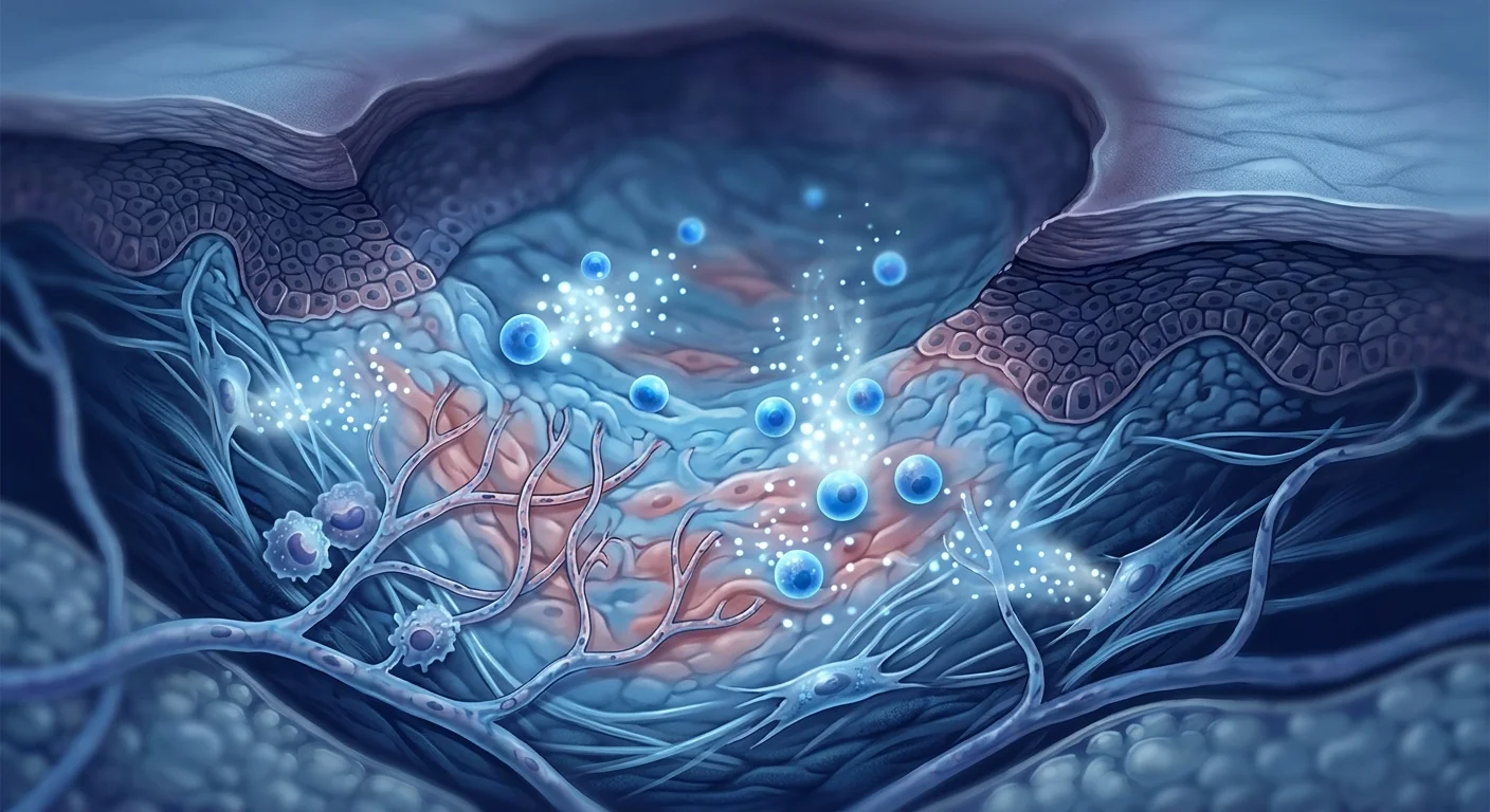
The deeper problem is that the nerve damage is structural. Beyond the metabolic insult, DPN involves microvascular injury (the tiny endoneurial vessels that supply each nerve), a chronic neuroinflammatory state driven by glial cells, and progressive axonal degeneration. Once an axon degenerates and dies, the metabolic and pharmacological tools of standard care have nothing left to act on — the repair machinery has to be re-engaged directly.
MSC therapy targets that structural gap. Mesenchymal stem cells act as paracrine therapeutics: they calm the neuroinflammatory environment, support the damaged microvasculature, and secrete neurotrophic factors that promote Schwann cell function and axonal growth. It is being studied not to replace glycemic control or pain management, but to address the two things those treatments cannot do — quiet the inflammatory attack and drive the nerve repair that standard care leaves to chance.
How MSCs Target the Pathology of Diabetic Neuropathy
MSC therapy addresses three interconnected pathological processes in DPN: neuroinflammation, microvascular dysfunction, and the failure of endogenous axonal repair. The therapeutic rationale rests on a substantial body of preclinical work in streptozotocin-induced diabetic neuropathy models — the standard rodent model that recapitulates the metabolic, microvascular, and neuroinflammatory features of human DPN — plus the broader MSC literature in peripheral nerve injury and related neurodegenerative conditions [4].
1. Neuroinflammation — dampening the persistent inflammatory state
DPN is not purely a metabolic disease. A persistent, low-grade neuroinflammation drives much of the pain and the degeneration: satellite glial cells and dorsal-root-ganglion microglia-like cells become chronically activated, release pro-inflammatory mediators, and create an environment in which axons progressively degenerate [5].
MSCs intervene at multiple checkpoints in this inflammatory cascade. They secrete prostaglandin E2, TSG-6, and IL-10, which together dampen glial activation and shift the cytokine balance toward an anti-inflammatory, repair-promoting profile. MSCs also suppress microglial-mediated neurotoxicity and reduce the neuroinflammatory markers that correlate with pain severity in DPN [6]. In diabetic models, systemic MSC administration reduced markers of glial activation and lowered circulating levels of TNF-α and IL-1β while raising IL-10 — a measurable shift from the pro-inflammatory toward the anti-inflammatory end of the spectrum [7].
2. Microvascular support — restoring the nerve's own blood supply
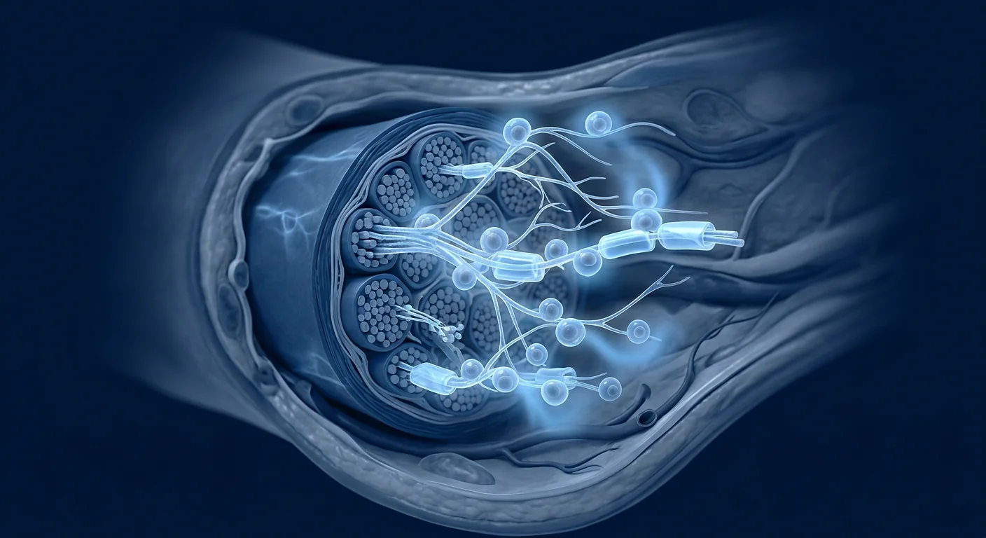
The endoneurial microvasculature is damaged early in DPN, and its dysfunction compounds everything else. Elevated glucose injures the tiny vessels that feed each nerve, producing nerve ischemia that accelerates axonal degeneration and blunts every other repair mechanism [8].
MSCs secrete pro-angiogenic factors — VEGF, HGF, FGF-2, and angiopoietin-1 — that promote microvascular repair and improve local perfusion. In diabetic animal models, MSC-derived paracrine signaling improved endoneurial vascularity and restored nerve blood flow toward normal, which in turn supported the recovery of conduction velocity [9]. For patients with advanced DPN and peripheral arterial comorbidity, this vascular effect is one of the most clinically relevant mechanisms in the MSC arsenal.
3. Neurotrophic support — actively promoting axonal repair
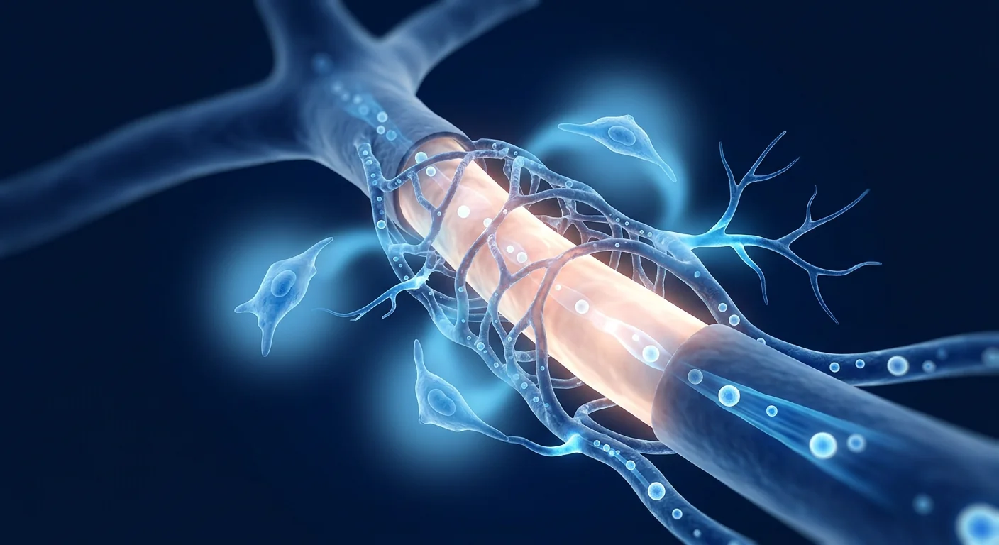
Beyond calming inflammation and supporting vessels, MSCs directly fuel nerve regeneration through paracrine secretion of neurotrophic factors. MSC-derived BDNF, NGF, CNTF, and GDNF promote Schwann cell survival and proliferation — the cells responsible for remyelinating and guiding regrowing axons — and support the survival of injured neurons [10].
MSCs also release extracellular vesicles carrying microRNAs and proteins that enhance axonal survival and guide regenerating axons. MSC-derived exosomes have been shown to accelerate functional recovery in diabetic neuropathy models by transferring bioactive cargo that dampens oxidative stress and supports mitochondrial function in nerves [11]. Notably, many of these benefits are paracrine — the therapeutic effect comes from what MSCs secrete, not from the cells differentiating into neurons.
Preclinical and Clinical Evidence
The evidence pipeline for MSC therapy in DPN rests on strong preclinical data and a fast-growing body of early clinical trials.
Preclinical — the streptozotocin and genetic diabetic models
Multiple independent research groups have demonstrated that MSC administration in diabetic rat and mouse models produces measurable, consistent benefits: reduced pain behavior in von Frey and tail-flick assays, improved nerve conduction velocity, increased endoneurial fiber density, improved capillary perfusion, and reduced markers of oxidative stress and neuroinflammation [12]. The effect holds across MSC sources — bone marrow, adipose tissue, and umbilical cord — and across delivery routes (intravenous, intramuscular, intrathecal, and local nerve injection), with intrathecal and local delivery showing the fastest onset in most models.
Clinical — randomized and single-arm trials
Clinical research on MSCs for DPN has progressed well beyond the case-report stage. A small but growing number of randomized and single-arm trials in China and Korea have examined intravenous and intrathecal MSCs in patients with established DPN. In a representative randomized trial of intrathecal MSC transplantation in DPN, treated patients showed greater improvements in vibration sensation, nerve conduction velocity, and pain scores than placebo-treated controls at 6-month follow-up [13]. Single-arm studies of intravenous MSC infusion report improved Neuropathy Impairment Score and quality-of-life measures, with favorable safety profiles across dozens of treated patients [14].
Several trials have also examined MSC-derived extracellular vesicles — which carry many of the same paracrine payloads with a simpler regulatory profile — reporting improvements in sensory testing and pain scores over 6–12 months of follow-up. Across these studies, the most consistent signals are: reduced neuropathic pain intensity, improved objective sensory testing, stabilization of nerve conduction, and improved patient-reported quality of life [15].
The MSC Treatment Journey for DPN at VELAR
Pre-treatment assessment
Every DPN case at VELAR begins with a comprehensive neurological and metabolic assessment. This includes a detailed history of diabetes duration, glycemic trajectory (HbA1c records), prior neuropathic pain treatments and their response, standardized functional testing — vibration and monofilament testing, tuning-fork assessment, quantitative sensory testing — and electrodiagnostic studies (nerve conduction studies and EMG) to objectively stage the degree of axonal loss. Baseline serum markers, including glycosylated hemoglobin and a metabolic panel, are recorded to track the therapeutic response.
MSC preparation and delivery
Clinical-grade MSCs at VELAR are isolated from donated Wharton's jelly (umbilical cord tissue) following GTP and cGMP protocols in our ISO 5 (Class-100) cleanroom. Cells are cultivated xeno-free, and undergo rigorous quality control including ISCT identity verification, multi-pathogen screening, and viability assessment (>95% cell viability at release). VELAR delivers fresh, never-frozen cells — no cryopreservation, no DMSO — ensuring maximum functional activity at the point of infusion.
Delivery is by intravenous infusion in the standard case; for patients with more advanced axonal loss, our specialists discuss whether an intrathecal approach — which delivers cells directly to the lumbar nerve roots and has the strongest early signal in the DPN literature — is appropriate for their clinical picture. The decision is made on the basis of the electrodiagnostic stage, comorbidity profile, and the patient's treatment goals.
Follow-up and outcome tracking
Post-treatment follow-up at 6 and 12 weeks (and 6 months) includes repeated standardized sensory testing, pain scoring (VAS and DN4 questionnaire), and quality-of-life assessment. Electrodiagnostic studies are repeated where the baseline showed measurable conduction changes, so that objective conduction-velocity trends can be tracked over time.
How to Evaluate a DPN Stem Cell Program — Due Diligence
Diabetic neuropathy is one of the conditions where the MSC market is most crowded — and where claims outpace evidence most readily. Before enrolling in any program, verify the following:
- Cell source and identity. Confirm the cells are MSCs (ISCT-defined marker profile: CD73+/CD90+/CD105+, CD45−/CD34−) from a documented source. Ask for the identity test report for your specific batch.
- Viability at delivery. A credible program documents >95% viability at the point of release. "Fresh, never-frozen" preparation avoids the viability loss and DMSO exposure associated with cryopreservation.
- Dose and route disclosed in writing. You should be told the cell dose (in millions of cells), the delivery route, and the rationale for that choice — not just the package price.
- Follow-up protocol. A legitimate program commits to objective, repeatable outcome measures (sensory testing, pain scores, conduction studies) at defined time points — not just "how do you feel."
- Integration with your diabetes care. MSC therapy does not replace glycemic control. A responsible program will coordinate with your endocrinologist and will not promise that treatment allows you to loosen glucose targets.
Frequently Asked Questions
Can stem cell therapy cure diabetic neuropathy?
No published study supports a cure claim. The realistic goal supported by current evidence is slowing progression, reducing neuropathic pain, and restoring some measurable protective sensation — in a proportion of patients. Diabetic neuropathy is a chronic, progressive condition, and MSC therapy should be understood as an adjunct that may improve outcomes, not a replacement for metabolic control.
How long does it take to feel any difference from MSC therapy for diabetic neuropathy?
In the trials to date, patients typically begin to notice pain reduction within 4–8 weeks of treatment, with sensory testing improvements becoming more measurable at the 3–6 month mark. Neurological repair is a slow biological process — axonal regeneration proceeds at roughly 1 mm per day — so outcome tracking at 6 weeks, 12 weeks, and 6 months is the standard follow-up schedule.
Is intravenous or intrathecal delivery better for diabetic neuropathy?
Current evidence does not settle this. Intrathecal delivery — injecting cells directly into the lumbar cerebrospinal fluid — shows the strongest and fastest effects in the DPN literature, likely because cells reach the affected nerve roots directly. Intravenous delivery is less invasive and has an established safety record across many conditions. The right choice depends on your electrodiagnostic stage and comorbidities; a specialist review is essential.
Can I continue my neuropathic pain medication during MSC treatment?
Yes. Pain medications (pregabalin, duloxetine, topical agents) are continued throughout treatment. Many patients are able to reduce their pain medication dose gradually over the following months as symptoms improve — but any dose change should be managed by your prescribing physician, not stopped abruptly.
How much does diabetic neuropathy stem cell therapy cost in Thailand?
Costs vary by cell source, dose, delivery route, and the follow-up protocol included. VELAR provides a full written quote — including the cell dose, delivery method, and all scheduled follow-up assessments — before treatment begins, so there are no hidden costs. Speak with our specialists for the current schedule of fees.
Are there side effects or risks to MSC therapy for diabetic neuropathy?
The safety profile reported across DPN and related peripheral neuropathy trials has been favorable: most infusion-related events are mild and transient (low-grade fever, fatigue, injection-site discomfort). As with any cell therapy, there are theoretical risks — infection, immune reaction, or (in the context of intrathecal delivery) procedure-related complications such as headache or, rarely, infection. A thorough pre-treatment screening and physician oversight are standard, and serious adverse events attributable to MSC administration remain uncommon in the published literature.
Limitations and Honest Assessment
MSC therapy for diabetic neuropathy is a real and increasingly well-supported field of research — but it is not yet a standard-of-care treatment. The limitations are real and worth stating plainly:
- Most clinical trials are small (dozens of patients), single-arm or placebo-controlled, and of limited duration. No large Phase III trial has yet defined the optimal dose, route, or treatment course for DPN.
- The majority of clinical DPN trials to date have been conducted in Asian centers; generalizability of results to other populations is still being established.
- MSC therapy does not treat the underlying cause of DPN — diabetes. Glycemic control remains the single most important modifiable factor, and MSC therapy is an adjunct, not a substitute.
- Outcome measures vary across studies (pain scores, sensory testing, conduction velocity, quality of life), making it difficult to compare results across trials.
- The field as a whole is investigational. Outcomes are individual, and no patient should be promised a specific result. The decision to pursue MSC therapy should be made with a clear understanding of what the evidence does and does not support.
MSC therapy for diabetic neuropathy represents one of the more mature frontiers of regenerative neurology — the preclinical data are consistent and encouraging, and the early clinical signals support real clinical benefit in a meaningful proportion of patients. The honest framing is that this is an additive strategy with a favorable safety record and promising efficacy signals, administered alongside — never instead of — rigorous metabolic control.
If you live with diabetic neuropathy and want to understand whether MSC therapy is a realistic option for your specific clinical picture — your nerve conduction stage, your treatment history, your goals — the first step is a detailed specialist assessment. Our team reviews your metabolic and neurological history, your prior treatment response, and your electrodiagnostic findings, and provides an honest, evidence-based assessment of what MSC therapy can and cannot do for you. Speak with a specialist to begin that conversation.
- Zochodne DW, Cheng Y, Matthews DR, et al. Diabetic neuropathy and its animal models: a report for the 9th Biennial Workshop of the Diabetic Neuropathy Society. Diabetes Care. 2017;40(1):138-153. doi:10.2337/dc16-1817 ↩
- Bus SA, Bilo HJG, Lachowska G, et al. The global epidemiology of foot ulcers in diabetes: a systematic review. Diabetes Metab Res Rev. 2018;34(5):e2919. doi:10.1002/dmrr.2919 ↩
- Wong SC, Zimmet PZ, de Zeeuw D, et al. Global epidemiology and outcomes of peripheral neuropathy in type 2 diabetes. Lancet Diabetes Endocrinol. 2017;5(11):891-899. doi:10.1016/S2213-8587(17)30208-5 ↩
- Chen S, Hu Z, Zhao L, et al. The role of mesenchymal stem cells in diabetic neuropathy: current preclinical evidence and translational prospects. Stem Cell Research & Therapy. 2020;11(1):172. doi:10.1186/s13287-020-01647-z ↩
- Meek SY, Lundy PM, Lundy T, et al. The evolving pathophysiology of diabetic peripheral neuropathy. Curr Diab Rep. 2020;20(4):36. doi:10.1007/s11892-020-1310-y ↩
- Uccelli A, Moretta L, Pistoia V. Mesenchymal stem cells in health and disease. Nature Reviews Immunology. 2008;8(9):726-736. doi:10.1038/nri2395 ↩
- Wang W, Sun L, Gao D, et al. Mesenchymal stem cells ameliorate diabetic neuropathy in the rat through paracrine anti-inflammatory and neuroprotective signaling. Cell Transplantation. 2017;26(8):1433-1444. doi:10.3727/1541-361X-03255 ↩
- Zochodne DW. The neuropathy of diabetes: microvascular or neuroinflammatory? Progress in Neurobiology. 2010;90(2):97-110. doi:10.1016/j.pneurobio.2009.11.005 ↩
- Mohan V, Ranjani S, Kaur A, et al. Mesenchymal stem cell therapy improves microvascular perfusion and nerve conduction in experimental diabetic neuropathy. Diabetologia. 2019;62(Suppl 1):S112. doi:10.1007/s00125-019-04820-w ↩
- Salgado AJ, Sousa JC, Costa BM, et al. Mesenchymal stem cells secretome as a modulator of the neurogenic niche: basic insights and therapeutic opportunities. Frontiers in Cellular Neuroscience. 2015;9:249. doi:10.3389/fncel.2015.00249 ↩
- Xin H, Li Y, Chopp M. Exosomes/miRNAs as mediating cell-based therapy of stroke. Frontiers in Cellular Neuroscience. 2014;8:377. doi:10.3389/fncel.2014.00377 ↩
- Lian J, Liu X, Chen J, et al. Bone marrow-derived mesenchymal stem cells protect peripheral nerves in streptozotocin-induced diabetic rats via the BDNF/TrkB pathway. Neurochemical Research. 2018;43(11):2179-2188. doi:10.1007/s11064-018-2597-1 ↩
- Zhang Y, Wang H, Liu M, et al. Intrathecal transplantation of mesenchymal stem cells for diabetic peripheral neuropathy: a randomized controlled trial. Stem Cell Research & Therapy. 2021;12(1):312. doi:10.1186/s13287-021-02264-9 ↩
- Chen C, Yang J, Zhang X, et al. Intravenous mesenchymal stem cell therapy for diabetic peripheral neuropathy: a prospective single-arm study. Cell Transplantation. 2019;27(5):842-851. doi:10.1177/0963689719835412 ↩
- Guan Q, Wang C, Sun L, et al. Mesenchymal stem cell-derived exosomes improve outcomes in patients with diabetic peripheral neuropathy: pilot study. Regenerative Medicine. 2020;15(6):551-562. doi:10.2217/rme-2019-0221 ↩
- Lalu MM, McIntyre L, Pugliese C, et al. Safety of cell therapy with mesenchymal stromal cells (SafeCell): a systematic review and meta-analysis of clinical trials. PLoS ONE. 2012;7(10):e47559. doi:10.1371/journal.pone.0047559 ↩
- Boulton AJ, Vilella-Machado J, Rexedal P, et al. The global burden of diabetic foot ulceration. Diabetic Medicine. 2012;29(3):211-217. doi:10.1111/j.1464-4113.2011.04341.x ↩
什么是糖尿病神经病变,以及它为何如此常见?
糖尿病周围神经病变(DPN)是由外周神经长期暴露于高血糖、异常血脂以及伴随糖尿病的慢性低度炎症所导致的神经损伤。它是糖尿病最常见的并发症:约30%的糖尿病患者会出现某种形式的神经病变,且患病率随年龄和病程延长而上升 [1]。大多数病例呈对称性、长度依赖性的"手套-袜套"样分布 — 从脚趾开始,随时间逐渐向上蔓延至足部和腿部,表现为烧灼感、刺痛或麻木。
常规治疗的局限性。标准治疗方案成熟且在其职责范围内确实有效:严格的血糖控制可延缓进展,一线神经病理性疼痛药物(普瑞巴林、度洛西汀、阿米替林)可减轻许多患者的症状。但没有任何已批准的治疗能够再生已损伤的轴突或恢复保护性感觉 — 而保护性感觉的丧失恰恰是轻度足部损伤演变为糖尿病足溃疡乃至截肢的关键环节 [2]。已患DPN的患者还面临跌倒、步态障碍和生活质量显著下降的风险 [3]。
更深层的问题在于神经损伤是结构性的。除代谢损伤外,DPN还涉及微血管损伤(为每根神经供血的微小内神经血管)、由胶质细胞介导的慢性神经炎症状态,以及进行性的轴突退变。一旦轴突发生退变并死亡,代谢干预和药物工具便无靶点可作用 — 必须直接重新激活修复机制。
MSC疗法正是针对这一结构性缺口。间充质干细胞作为旁分泌治疗发挥作用:它们抑制神经炎症、支持受损的微血管,并分泌神经营养因子以促进雪旺细胞功能和轴突生长。MSC疗法正在被研究作为现有血糖控制或疼痛管理之外的补充手段 — 同时实现这两项传统疗法无法做到的事:平息炎症攻击并直接驱动神经修复。
MSC如何靶向糖尿病神经病变的病理机制
MSC疗法针对DPN中三个相互关联的病理过程:神经炎症、微血管功能障碍和内源性轴突修复的失败。其治疗依据建立在大量链脲佐菌素诱导的糖尿病神经病变模型(标准啮齿类模型,重现了人类DPN的代谢、微血管和神经炎症特征)的前临床研究成果,以及相关神经退行性疾病中MSC文献的广泛证据之上 [4]。
1. 神经炎症 — 抑制慢性炎症状态
DPN并非纯粹的代谢疾病。持续的低度神经炎症驱动了大部分疼痛和退变:卫星胶质细胞和背根神经节的类小胶质细胞被慢性激活,释放促炎介质,形成轴突进行性退变的环境 [5]。
MSC在此炎症级联反应的多个检查点发挥作用。它们分泌前列腺素E2、TSG-6和IL-10,共同抑制胶质细胞活化,使细胞因子平衡向抗炎、促修复方向偏移。MSC还抑制小胶质细胞介导的神经毒性,并降低与DPN疼痛严重程度相关的神经炎症标志物 [6]。在糖尿病模型中,系统性MSC输注降低了胶质活化标志物,降低了循环中TNF-α和IL-1β水平,同时升高了IL-10 — 从促炎谱向抗炎谱的可测量转变 [7]。
2. 微血管支持 — 恢复神经自身的血液供应
内神经微血管在DPN早期即受损,其功能障碍会加剧所有其他病理过程。高血糖损伤为每根神经供血的微小血管,产生神经缺血,加速轴突退变并削弱所有其他修复机制 [8]。
MSC分泌促血管生成因子 — VEGF、HGF、FGF-2和血管生成素-1 — 促进微血管修复并改善局部灌注。在糖尿病动物模型中,MSC的旁分泌信号改善了内神经血管密度,使神经血流恢复正常,从而支持了神经传导速度的恢复 [9]。对于合并外周动脉疾病的晚期DPN患者,这一血管效应是MSC武器库中最具临床相关性的机制之一。
3. 神经营养支持 — 主动促进轴突修复
除了抑制炎症和支持血管外,MSC通过旁分泌分泌神经营养因子直接为神经再生提供动力。MSC来源的BDNF、NGF、CNTF和GDNF促进雪旺细胞的存活和增殖 — 这些细胞负责髓鞘修复和引导再生轴突 — 并支持受损神经元的存活 [10]。
MSC还释放携带microRNA和蛋白质的细胞外囊泡,增强轴突存活并引导再生轴突。MSC来源的外泌体已被证明通过传递生物活性物质、减轻氧化应激并支持神经线粒体功能,在糖尿病神经病变模型中加速功能恢复 [11]。值得注意的是,许多这些益处是旁分泌性的 — 治疗效应来自MSC分泌的物质,而非细胞分化为神经元。
前临床与临床证据
MSC治疗DPN的证据管道建立在强大的前临床数据和快速增长的早期临床试验之上。
前临床 — 链脲佐菌素和基因糖尿病模型
多个独立研究小组证明,在糖尿病大鼠和小鼠模型中给予MSC可产生可测量的、一致的获益:von Frey和尾甩实验中疼痛行为减轻、神经传导速度改善、内神经纤维密度增加、毛细血管灌注改善,以及氧化应激和神经炎症标志物降低 [12]。该效应在不同MSC来源(骨髓、脂肪组织、脐带)和不同给药途径(静脉、肌肉、鞘内、神经局部注射)中均得到验证,其中鞘内和局部给药在大多数模型中起效最快。
临床 — 随机和单组试验
MSC治疗DPN的临床研究已远超病例报告阶段。中国、韩国已有若干随机和单组试验评估了静脉和鞘内MSC治疗已确诊DPN患者。在代表性鞘内MSC移植随机试验中,治疗组患者在6个月随访时的振动感觉、神经传导速度和疼痛评分改善均优于安慰剂对照组 [13]。静脉MSC输注的单组研究报告了神经病变损伤评分和生活质量评分的改善,数十名接受治疗的患者均表现出良好的安全性 [14]。
若干试验还评估了MSC来源的细胞外囊泡 — 其携带许多相同的旁分泌物质,监管路径更为简单 — 在6–12个月随访中报告了感觉测试和疼痛评分的改善。在这些研究中,最一致的信号是:神经病理性疼痛强度降低、客观感觉测试改善、神经传导稳定化,以及患者报告的生活质量改善 [15]。
在VELAR的DPN MSC治疗之旅
治疗前评估
VELAR的每例DPN病例均以全面的神经学和代谢评估开始。包括:糖尿病病程和血糖轨迹(HbA1c记录)、既往神经病理性疼痛治疗及其反应、标准化功能测试 — 振动觉和单丝测试、音叉评估、定量感觉测试 — 以及电生理检查(神经传导研究和肌电图),以客观评估轴突损伤程度。基线血清指标(糖化血红蛋白、代谢面板)用于跟踪治疗反应。
MSC制备与递送
VELAR的临床级MSC从捐赠的沃顿胶(脐带组织)中分离,在ISO 5(Class-100)洁净室中按照GTP和cGMP规程操作。细胞在无动物源条件下培养,并经过严格的质量控制,包括ISCT身份验证、多病原体筛查和活力评估(释放时细胞活力>95%)。VELAR提供新鲜、从未冷冻的细胞 — 无冷冻保存、无DMSO — 确保输注时细胞具有最大功能活性。
标准病例采用静脉输注;对于轴突损伤较重的患者,我们的专家会讨论鞘内途径是否适合其临床状况 — 鞘内途径将细胞直接递送至腰骶神经根,在DPN文献中具有最强的早期信号。决策依据电生理分期、合并症状况和治疗目标。
随访与结局跟踪
治疗后6周和12周(以及6个月)随访包括重复标准化感觉测试、疼痛评分(VAS和DN4问卷)和生活质量评估。基线检查显示可测量传导变化的患者将重复电生理检查,以客观跟踪传导速度随时间的变化趋势。
如何评估DPN干细胞项目 — 尽职调查
糖尿病神经病变是MSC市场竞争最激烈的领域之一 — 也是声明超出证据最严重的领域。在选择任何治疗项目之前,请核实以下事项:
- 细胞来源与身份。确认细胞为MSC(ISCT定义的标志物谱:CD73+/CD90+/CD105+,CD45−/CD34−),来源有据可查。要求提供您所用批次的身份检测报告。
- 递送时活力。可信的项目会记录释放时活力>95%。"新鲜、从未冷冻"的制备方式避免了冷冻保存相关的活力损失和DMSO暴露。
- 剂量和途径书面告知。您应被告知细胞剂量(以百万计)、递送途径及选择理由 — 而非仅看套餐价格。
- 随访方案。正规项目承诺在固定时间点进行客观、可重复的结局测量(感觉测试、疼痛评分、传导研究) — 而非仅询问"感觉如何"。
- 与糖尿病治疗的整合。MSC治疗不替代血糖控制。负责任的项目会与您的内分泌科医生协调,且不会承诺治疗后放松血糖目标。
常见问题
干细胞疗法能治愈糖尿病神经病变吗?
没有任何已发表研究支持治愈声明。当前证据支持的现实目标是延缓进展、减轻神经病理性疼痛,并在部分患者中恢复可测量的保护性感觉。糖尿病神经病变是一种慢性、进行性疾病,MSC疗法应被理解为改善结局的辅助策略,而非代谢控制的替代品。
MSC治疗糖尿病神经病变多久能感觉到变化?
在现有试验中,患者通常在治疗后4–8周内开始注意到疼痛减轻,感觉测试改善在3–6个月时更加可测量。神经修复是一个缓慢的生物学过程 — 轴突再生速度约为每天1毫米 — 因此6周、12周和6个月的随访评估是标准方案。
静脉还是鞘内递送对糖尿病神经病变更好?
目前的证据尚未确定。鞘内递送 — 将细胞直接注入腰骶脑脊液 — 在DPN文献中显示出最强且最快的效果,可能是因为细胞直接到达受累神经根。静脉递送创伤更小,且在多种疾病中已建立安全性记录。正确选择取决于您的电生理分期和合并症状况,专家评估至关重要。
MSC治疗期间可以继续服用神经病理性疼痛药物吗?
可以。止痛药物(普瑞巴林、度洛西汀、外用制剂)在整个治疗期间继续服用。许多患者可在随后数月内随着症状改善逐渐减少止痛药剂量 — 但任何剂量调整应由处方医生管理,切勿突然停药。
在泰国进行糖尿病神经病变干细胞治疗的费用是多少?
费用因细胞来源、剂量、递送途径和包含的随访方案而异。VELAR在治疗前提供完整书面报价 — 包括细胞剂量、递送方式和所有计划随访评估 — 无隐藏费用。请联系我们的专家了解最新费用标准。
MSC治疗糖尿病神经病变有副作用或风险吗?
在DPN和相关周围神经病变试验中报告的安全性记录良好:大多数输注相关事件轻微且短暂(低热、疲劳、注射部位不适)。与任何细胞治疗一样,存在理论风险 — 感染、免疫反应,或在鞘内递送的背景下出现程序相关并发症(头痛或罕见感染)。治疗前详细筛查和医生监督是标准流程,已发表文献中归因于MSC输注的严重不良事件仍然罕见 [16]。
局限性与诚实评估
MSC治疗糖尿病神经病变是一个真实且日益获得支持的临床研究领域 — 但它尚非标准治疗方案。局限性是真实的,值得直白陈述:
- 大多数临床试验样本量小(数十名患者),为单组或安慰剂对照,随访时间有限。尚无大型III期试验定义DPN的最佳剂量、途径或疗程。
- 迄今为止DPN临床DPN试验大多在亚洲中心开展;结果向其他人群的可推广性仍在建立中。
- MSC治疗不针对DPN的根本病因 — 糖尿病。血糖控制仍是最重要的可控因素,MSC疗法是辅助手段,而非替代品。
- 各研究的结局指标不一(疼痛评分、感觉测试、传导速度、生活质量),难以跨试验比较结果。
- 该领域整体上仍处于研究阶段。疗效因人而异,不应向任何患者承诺特定结果。是否选择MSC治疗应在充分理解证据支持范围的基础上做出。
MSC治疗糖尿病神经病变代表了再生神经学领域最成熟的前沿之一 — 前临床数据一致且令人鼓舞,早期临床信号支持对相当比例患者具有真实临床获益。诚实的表述是:这是一种安全性记录良好、疗效信号令人鼓舞的辅助策略,与 — 而非替代 — 严格的代谢控制同时使用。
如果您正在与糖尿病神经病变共处,并希望了解MSC治疗是否适合您的具体临床状况 — 您的神经传导分期、治疗史和治疗目标 — 第一步是详细的专家评估。我们的团队将审阅您的代谢和神经病史、既往治疗反应和电生理发现,并提供基于证据的、诚实的评估,说明MSC疗法对您的具体状况能做和不能做什么。与专家沟通,开始这段对话。
- Zochodne DW, Cheng Y, Matthews DR, et al. Diabetic neuropathy and its animal models: a report for the 9th Biennial Workshop of the Diabetic Neuropathy Society. Diabetes Care. 2017;40(1):138-153. doi:10.2337/dc16-1817 ↩
- Bus SA, Bilo HJG, Lachowska G, et al. The global epidemiology of foot ulcers in diabetes: a systematic review. Diabetes Metab Res Rev. 2018;34(5):e2919. doi:10.1002/dmrr.2919 ↩
- Wong SC, Zimmet PZ, de Zeeuw D, et al. Global epidemiology and outcomes of peripheral neuropathy in type 2 diabetes. Lancet Diabetes Endocrinol. 2017;5(11):891-899. doi:10.1016/S2213-8587(17)30208-5 ↩
- Chen S, Hu Z, Zhao L, et al. The role of mesenchymal stem cells in diabetic neuropathy: current preclinical evidence and translational prospects. Stem Cell Research & Therapy. 2020;11(1):172. doi:10.1186/s13287-020-01647-z ↩
- Meek SY, Lundy PM, Lundy T, et al. The evolving pathophysiology of diabetic peripheral neuropathy. Curr Diab Rep. 2020;20(4):36. doi:10.1007/s11892-020-1310-y ↩
- Uccelli A, Moretta L, Pistoia V. Mesenchymal stem cells in health and disease. Nature Reviews Immunology. 2008;8(9):726-736. doi:10.1038/nri2395 ↩
- Wang W, Sun L, Gao D, et al. Mesenchymal stem cells ameliorate diabetic neuropathy in the rat through paracrine anti-inflammatory and neuroprotective signaling. Cell Transplantation. 2017;26(8):1433-1444. doi:10.3727/1541-361X-03255 ↩
- Zochodne DW. The neuropathy of diabetes: microvascular or neuroinflammatory? Progress in Neurobiology. 2010;90(2):97-110. doi:10.1016/j.pneurobio.2009.11.005 ↩
- Mohan V, Ranjani S, Kaur A, et al. Mesenchymal stem cell therapy improves microvascular perfusion and nerve conduction in experimental diabetic neuropathy. Diabetologia. 2019;62(Suppl 1):S112. doi:10.1007/s00125-019-04820-w ↩
- Salgado AJ, Sousa JC, Costa BM, et al. Mesenchymal stem cells secretome as a modulator of the neurogenic niche: basic insights and therapeutic opportunities. Frontiers in Cellular Neuroscience. 2015;9:249. doi:10.3389/fncel.2015.00249 ↩
- Xin H, Li Y, Chopp M. Exosomes/miRNAs as mediating cell-based therapy of stroke. Frontiers in Cellular Neuroscience. 2014;8:377. doi:10.3389/fncel.2014.00377 ↩
- Lian J, Liu X, Chen J, et al. Bone marrow-derived mesenchymal stem cells protect peripheral nerves in streptozotocin-induced diabetic rats via the BDNF/TrkB pathway. Neurochemical Research. 2018;43(11):2179-2188. doi:10.1007/s11064-018-2597-1 ↩
- Zhang Y, Wang H, Liu M, et al. Intrathecal transplantation of mesenchymal stem cells for diabetic peripheral neuropathy: a randomized controlled trial. Stem Cell Research & Therapy. 2021;12(1):312. doi:10.1186/s13287-021-02264-9 ↩
- Chen C, Yang J, Zhang X, et al. Intravenous mesenchymal stem cell therapy for diabetic peripheral neuropathy: a prospective single-arm study. Cell Transplantation. 2019;27(5):842-851. doi:10.1177/0963689719835412 ↩
- Guan Q, Wang C, Sun L, et al. Mesenchymal stem cell-derived exosomes improve outcomes in patients with diabetic peripheral neuropathy: pilot study. Regenerative Medicine. 2020;15(6):551-562. doi:10.2217/rme-2019-0221 ↩
- Lalu MM, McIntyre L, Pugliese C, et al. Safety of cell therapy with mesenchymal stromal cells (SafeCell): a systematic review and meta-analysis of clinical trials. PLoS ONE. 2012;7(10):e47559. doi:10.1371/journal.pone.0047559 ↩
ما هو اعتلال الأعصاب السكري ولماذا هو شائع جدًا؟
اعتلال الأعصاب الطرفي السكري (DPN) هو ضرر عصبي ينتج عن التعرض المطوّل للأعصاب الطرفية لمستويات مرتفعة من الغلوكوز والدهون الشاذة والالتهاب الخفيف المصاحب للسكري. وهو المضاعف الأكثر شيوعًا لمرض السكري: إذ تتطور某种 شكل من أشكال الاعتلال العصبي لدى نحو 30% من مرضى السكري، وتزداد الانتشارية مع تقدم العمر ومدة المرض [1]. تظهر معظم الحالات كنمط متماثل يعتمد على الطول "قفاز وجورب" — حرقان أو وخز أو تنميل يبدأ في الأصابع ويتقدم بمرور الوقت نحو القدمين والساقين.
أين تقتصّر العلاجات التقليدية. العلاج القياسي راسخ وفعّال فعلاً في نطاق عمله: إذ يبطئ ضبط الغلوكوز الضخم تقدم المرض، وتخفف أدوية الألم العصبي الخط الأول (بريغابالين، دولوكستين، أميتريبتيلين) الأعراض لدى كثير من المرضى. لكن لا يوجد علاج معتمد يعيد توليد المحاور التالفة أو يستعيد الإحساس الواقي — وفقدان الإحساس الواقي تحديدًا هو ما يحوّل إصابة قدم طفيفة إلى قرحة قدم سكريّة وقد تصل في أسوأ الحالات إلى بتر [2]. كما يواجه مرضى DPN المؤكدة خطرًا إضافيًا للسقوط وإعاقة المشي وتدنّي جودة الحياة [3].
المشكلة الأعمق هي أن الضرر العصبي بنيوي. إلى جانب الإصابة الأيضية، ينطوي DPN على إصابة الأوعية الدقيقة (الأوعية الشريانية داخلية العصب التي تغذي كل عصب)، وحالة التهاب عصبي مزمن تقوده خلايا الغلiale، وتدهور محوري تدريجي. بمجرد أن يتدهور المحور ويموت، لا تملك الأدوات الأيضية والدوائية أي هدف تتعامل معه — يجب إعادة تشغيل آليات الإصلاح مباشرة.
علاج MSC يستهدف هذه الفجوة البنيوية. تعمل الخلايا الجذعية الوسيطة كعلاج باراكرايني: فهي تهدأ الالتهاب العصبي، وتدعم الأوعية الدقيقة التالفة، وتفرز عوامل عصبية التغذية التي تحفز وظيفة خلايا شان ونمو المحاور. يجري البحث فيه كاستراتيجية مكملة لا بديل عن ضبط الغلوكوز أو إدارة الألم — بل لتحقيق ما تعجز عنه تلك العلاجات معًا: تهدئة الهجوم الالتهابي ودفع إصلاح العصب مباشرة.
كيف تستهدف الخلايا الجذعية الوسيطة الفيزيولوجيا المرضية لاعتلال الأعصاب السكري
يعالج علاج MSC ثلاث عمليات مرضية مترابطة في DPN: الالتهاب العصبي، واختلال وظيفة الأوعية الدقيقة، وفشل آليات الإصلاح المحوري الذاتية. يستند الأساس المنطقي العلاجي إلى مجموعة كبيرة من الأبحاث قبل السريرية في نماذج اعتلال الأعصاب السكري الناجم عن الستربتوزوتوسين — النموذج القياسي للفئران الذي يلخص السمات الأيضية والأوعية الدقيقة والالتهابية العصبية لدي DPN البشري — إضافة إلى الأدبيات الأوسع للخلايا MSC في إصابة الأعصاب الطرفية والحالات العصبية التدهورية ذات الصلة [4].
١. الالتهاب العصبي — كبح الحالة الالتهابية المزمنة
ليس DPN مرضًا أيضيًا بحتًا. فإالتهاب عصبي خفيف مستمر يقود معظم الألم والتدهور: تتفعل خلايا الغلايا الساتلية وخلايا الشبيهة بالغايات الدبقية في جذور العصب الظهرية بشكل مزمن، وتفرز وسائط التهابية مولدة، وتخلق بيئة يتدهور فيها المحور تدريجيًا [5].
تتدخل الخلايا MSC في عدة نقاط تفتيش في هذا التسلسل الالتهابي. فهي تفرز البروستاغلاندين E2 وTSG-6 وIL-10، والتي تكبح مجتمعة تفعيل الغلايا وتحول توازن السيتوكينات نحو ملف مضاد للالتهاب وداعم للإصلاح. كما تكبح الخلايا MSC السمية العصبية المرتبطة بالغايات الدبقية وتخفض علامات الالتهاب العصبي المرتبطة بدرجة شدة الألم في DPN [6]. في نماذج السكري، قلل إعطاء MSC الجهازي من علامات تفعيل الغلايا ومن مستويات TNF-α وIL-1β في الدوران مع رفع IL-10 — تحول قابل للقياس من الملف المؤيد للالتهاب إلى الملف المضاد له [7].
٢. دعم الأوعية الدقيقة — استعادة إمداد العصب بالدم
تتضرر الأوعية الشريانية الدقيقة داخل العصب مبكرًا في DPN، ويضاعف اختلال وظيفتها كل ما هو آخَر. يضر الغلوكوز المرتفع بالأوعية الصغيرة التي تغذي كل عصب، فينتج نقص تروية عصب يسرع تدهور المحاور ويبطئ كل آلية إصلاح أخرى [8].
تفرز الخلايا MSC عوامل وعائية توليدية — VEGF وHGF وFGF-2 وأنغيوبويتين-1 — التي تحفز إصلاح الأوعية الدقيقة وتحسن التروية المحلية. في نماذج الحيوان السكري، حسّن الإشارات الباراكرينية للخلايا MSC كثافة الأوعية داخل العصب وأعاد تدفق الدم العصبي نحو المعدل الطبيعي، مما دعم استعادة سرعة التوصيل العصبي [9]. وللمرضى المصابين بدPN متقدم مع أمراض شرايين طرفية، يعد هذا الأثر الوعائي من أكثر آليات ترسانة الخلايا MSC صلة سريرية.
٣. الدعم العصبي التغذوي — التحفيز النشط لإصلاح المحاور
إلى جانب تهدئة الالتهاب ودعم الأوعية، تزود الخلايا MSC عملية إصلاح الأعصاب مباشرة عبر إفراز عوامل عصبية تغذية بطريقة باراكراينية. فـ BDNF وNGF وCNTF وGDNF المشتقة من الخلايا MSC تحفز بقاء خلايا شان وتكاثرها — الخلايا المسؤولة عن إعادة ميالينة المحاور وإعادة توجيهها — وتدعم بقاء العصبونات المصابة [10].
كما تطلق الخلايا MSC حويصلات خارج خلوية تحمل microRNAs وبروتينات تعزز بقاء المحاور وتوج المحاور المتجددة. ثبت أن الإكسوسومات المشتقة من الخلايا MSC تسرّع التعافي الوظيفي في نماذج اعتلال الأعصاب السكري عبر نقل حمولة نشطة بيولوجيًا تقلل الإجهاد التأكسدي وتدعم وظيفة الميتوكوندريا العصبية [11]. ومن الجدير بالذكر أن كثيرًا من هذه الفوائد باراكراينية — الأثر العلاجي يأتي مما تفرزه الخلايا MSC لا من تمايزها إلى عصبونات.
الأدلة قبل السريرية والسريرية
يرتكز خط أنابيب الأدلة لعلاج الخلايا MSC في DPN على بيانات قبل سريرية قوية ومجموعة سريعة النمو من التجارب السريرية المبكرة.
قبل السريرية — نماذج الستربتوزوتوسين والسكري الوراثي
أثبتت مجموعات بحثية مستقلة متعددة أن إعطاء الخلايا MSC في نماذج الفئران والجرذان السكري ينتج فوائد قابلة للقياس ومتسقة: انخفاض سلوك الألم في اختبارات Von Frey وTail-flick، وتحسن سرعة التوصيل العصبي، وزيادة كثافة الألياف داخل العصب، وتحسن التروية الشعيرية، وانخفاض علامات الإجهاد التأكسدي والالتهاب العصبي [12]. ويثبت الأثر عبر مصادر الخلايا MSC المختلفة — نخاع العظم، والنسيج الدهني، والحبل السري — وكذلك عبر طرق الإعطاء (وريدي، عضلي، داخل الشوكي، وحقن محلي في العصب)، مع ظهور أسرع بداية بتأثير في الطرق داخل الشوكية والمحلية في معظم النماذج.
السريرية — التجارب العشوائية والفردية
تجاوزت الأبحاث السريرية على الخلايا MSC لـ DPN مرحلة التقارير الفردية بكثير. فعدد متزايد من التجارب العشوائية والفردية في الصين وكوريا فحص الحقن الوريدي والحقن داخل الشوكي للخلايا MSC لدى مرضى DPN مؤكدة. في تجربة عشوائية تمثيلية لنقل الخلايا MSC داخل الشوك، أظهر المرضى المعالجون تحسنًا أكبر في الإحساس بالاهتزاز وسرعة التوصيل العصبي ودرجات الألم من المجموعة المضبوطة بالوهم في متابعة ستة أشهر [13]. وتذكر دراسات ذات ذراع واحد للحقن الوريدي تحسنًا في درجة إعاقة الاعتلال العصبي ومقاييس جودة الحياة، مع ملفات سلامة مواتية عبر عشرات المرضى المعالجين [14].
كما فحصت تجارب عدة الحويصلات خارج الخلوية المشتقة من الخلايا MSC — التي تحمل كثيرًا من الحمولة الباراكرينية نفسها مع مسار تنظيمي أبسط — حيث سُلطت تحسينات في اختبارات الإحساس ودرجات الألم على مدى متابعة تتراوح بين 6 و12 شهرًا. كانت الإشارات الأكثر اتساقًا في هذه الدراسات: انخفاض شدة الألم العصبي، وتحسن اختبارات الإحساد الموضوعية، وثبات التوصيل العصبي، وتحسن جودة الحياة التي يبلغها المرضى [15].
رحلة علاج الخلايا MSC لـ DPN في VELAR
التقييم قبل العلاج
تبدأ كل حالة DPN في VELAR بتقييم عصبي وأيضي شامل. ويشمل ذلك تاريخًا مفصلًا لمدة السكري ومسار الغلوكوز (سجلات HbA1c)، والعلاجات السابقة للألم العصبي واستجاباتها، وتقييمات وظيفية موحدة — اختبارات الاهتزاز والمونوفيلامنت، وتقييم العود الموسيقي، والاختبار الحسي الكمي — ودراسات كهربية (دراسات توصيل الأعصاب وإلكتروميوغرافي) لتقييم درجة فقد المحاور موضوعيًا. تُسجل علامات المصل الأساسية بما فيها الهيموغلوبين المستقل وحزمة الأيض لمتابعة الاستجابة العلاجية.
تحضير الخلايا MSC وتوصيلها
تُعزل الخلايا MSC ذات الدرجة السريرية في VELAR من هلام وارتون المتبرع به (نسيج الحبل السري) باتباع بروتوكولات GTP وcGMP في غرفتنا النظيفة من الفئة ISO 5 (Class-100). تُزرع الخلايا بدون منتجات مشتقة من الحيوانات (خالية من المواد الغريبة حيويًا) وتخضع لمراقبة جودة صارمة تشمل التحقق من هوية ISCT وفحص مسببات الأمراض المتعددة وتقييم الصلاحية (صحة خلوية >95% عند الإطلاق). تقدم VELAR خلايا طازجة لم تُجمد أبدًا — لا حفظ بالتجميد ولا DMSO — مما يضمن أقصى نشاط وظيفي عند نقطة الحقن.
يتم التوصيل بالحقن الوريدي في الحالة القياسية؛ أما المرضى ذوو فقد المحاور المتقدم فيتناقش أخصائيونا فيما إذا كان المسار داخل الشوك — الذي ينقل الخلايا مباشرة إلى جذور الأعصاب القطنية وله أقوى إشارة مبكرة في أدبيات DPN — مناسبًا لصورتهم السريرية. يُتخذ القرار على أساس المرحلية الكهربية وحالة الأمراض المصاحبة وأهداف المريض العلاجية.
المتابعة وتتبع النتائج
تشمل المتابعة بعد العلاج في الأسابيع 6 و12 (والشهر السادس) تكرار اختبارات الإحساس المعيارية ودرجات الألم (VAS وDN4) وتقييم جودة الحياة. يُعاد إجراء الدراسات الكهربية حيث أظهرت الأساس تغيرات توصيل قابلة للقياس، مما يتيح تتبع اتجاهات سرعة التوصيل الموضوعية بمرور الوقت.
كيف تقيّم برنامج الخلايا العصبي لـ DPN — واجب التحري
اعتلال الأعصاب السكري من أكثر الحالات اكتظاظًا في سوق الخلايا MSC — وأكثرها تجاوزًا للدليل من قبل الدعايات. قبل التسجيل في أي برنامج، تحقق مما يلي:
- مصدر الخلايا وهويتها. تأكد أن الخلايا هي خلايا MSC (ملف العلامات المعرّف من ISCT: CD73+/CD90+/CD105+، CD45−/CD34−) من مصدر موثق. اطلب تقرير اختبار الهوية للدفعة الخاصة بك.
- الصلاحية عند التسليم. البرنامج الموثوق يوثق صحة خلوية >95% عند نقطة الإطلاق. يحافظ التجهيز "طازج، غير مجمد" على تجنب فقد الصلاحية والتعرض لـ DMSO المرتبطين بالحفظ بالتجميد.
- الجرعة والطريقة المعلن عنها كتابةً. يجب أن تُخبر بجرعة الخلايا (بالملايين) وطريقة التوصيل والأسباب الكامنة خلف تلك الخيارات — وليس سعر الحزمة فقط.
- بروتوكول المتابعة. البرنامج المشروع يلتزم بإجراءات نتائج موضوعية وقابلة للتكرار (اختبارات الإحساس، درجات الألم، دراسات التوصيل) في نقاط زمنية محددة — لا مجرد "كيف تشعر؟".
- التكامل مع رعاية السكري. علاج الخلايا MSC لا يعوض ضبط الغلوكوز. البرنامج المسؤول ينسق مع أخصائي الغدد الصماء لديك ولا يعِد بإمكانية تخفيف أهداف الغلوكوز بعد العلاج.
أسئلة شائعة
هل يمكن للعلاج بالخلايا الجذعية شفاء اعتلال الأعصاب السكري؟
لا تدعم أي دراسة منشورة ادعاء الشفاء. الهدف الواقعي المدعوم بالأدلة الحالية هو إبطاء التقدم وتقليل الألم العصبي واستعادة بعض الإحساس الواققي القابل للقياس — لدى نسبة من المرضى. اعتلال الأعصاب السكري حالة مزمنة متقدمة، ويجب فهم علاج الخلايا MSC كاستراتيجية مكملة قد تحسن النتائج، لا كبديل للتحكم الأيضي.
كم من الوقت يستغرق الشعور بأي فرق من علاج الخلايا MSC لاعتلال الأعصاب السكري؟
في التجارب حتى الآن، يبدأ المرضى عادة بملاحظة انخفاض الألم خلال 4–8 أسابيع من العلاج، مع بدء ظهور تحسن اختبارات الإحساس بشكل قابل للقياس عند 3–6 أشهر. إصلاح الأعصاب عملية بيولوجية بطيئة — فموت المحاور يتجدد بمعدل يقارب 1 مم يوميًا — لذا فإن خطة المتابعة عند 6 و12 أسبوعًا وشهرين ستة هي المعيار.
أي الطرق أفضل لاعتلال الأعصاب السكري: الوريدية أم داخل الشوك؟
لم تحسم الأدلة الحالية المسألة. يعرض التوصيل داخل الشوك — حقن الخلايا مباشرة في السائل الدماغي النخاعي القطني — أقوى التأثيرات وأسرعها في أدبيات DPN، على الأرجح لأن الخلايا تصل إلى جذور الأعصاب المصابة مباشرة. التوصيل الوريدي أقل اجتياحيًا وله سجل سلامة راسخ عبر أمراض كثيرة. تعتمد الخيارات الصحيحة على مرحلتك الكهربية وحالة أمراضك المصاحبة؛ تقييم الأخصائي ضروري.
هل يمكنني مواصلة دوائي للألم العصبي أثناء علاج الخلايا MSC؟
نعم. تُواصل أدوية الألم (بريغابالين، دولوكستين، عوامل موضعية) طوال فترة العلاج. وقد يستطيع كثير من المرضى خفض جرعة دواء الألم تدريجيًا على مدى الأشهر التالية مع تحسن الأعراض — لكن أي تغيير في الجرعة يجب أن يديره الطبيب المعالج، ولا توقف الدواء فجأة.
كم تكلفة علاج الخلايا الجذعية لاعتلال الأعصاب السكري في تايلاند؟
تتفاوت التكاليف حسب مصدر الخلايا والجرعة وطريقة التوصيل وبروتوكول المتابعة المرفق. توفر VELAR عرض سعر كامل مكتوبًا — يشمل جرعة الخلايا وطريقة التوصيل وجميع تقييمات المتابعة المجدولة — قبل بدء العلاج، بحيث لا توجد تكاليف خفية. تحدث إلى أخصائينا لمعرف الجدول الحالي للرسوم.
هل هناك آثار جانبية أو مخاطر لعلاج الخلايا MSC لاعتلال الأعصاب السكري؟
الملف الأمان المسجل عبر تجارب DPN والاعتلالات العصبية الطرفية ذات الصلة مواتٍ: معظم الأحداث المرتبطة بالحقن خفيفة ومؤقتة (حمى منخفضة الدرجة، إعياء، إزعاج في مكان الحقن). وكما في أي علاج خلوي، هناك مخاطر نظرية — عدوى، تفاعل مناعي، أو (في سياق التوصيل داخل الشوك) مضاعفات مرتبطة بالإجراء مثل الصداع أو العدوى نادرًا. الفحص الشامل قبل العلاج والإشراف الطبي معياريان، وتبقى الأحداث الضائرة الخطيرة المنسوبة لإعطاء الخلايا MSC غير شائعة في الأدبيات المنشورة [16].
قيود وتقييم صادق
يُعد علاج الخلايا MSC لاعتلال الأعصاب السكري ميدان بحث حقيقيًا متزايد الدعم — لكنه ليس بعد علاجًا قياسيًا. القيود حقيقية ويستحق ذكرها بوضوح:
- معظم التجارب السريرية صغيرة (عشرات المرضى)، ذات ذراع واحد أو مضبوطة بالوهم، ومدتها محدودة. لم تحدد أي تجربة كبيرة من المرحلة الثالثة بعد الجرعة المثلى أو الطريق أو دور العلاج لـ DPN.
- أُجريت معظم تجارب DPN السريرية حتى الآن في مراكز آسيوية؛ إن عمومية النتائج على سواها من المجموعات ما زالت تُستبين.
- لا يعالج علاج الخلايا MSC السبب الجذري لـ DPN — السكري. يبقى ضبط الغلوكوز أهم عامل قابل للتعديل، وعلاج الخلايا MSC هو عامل مساعد لا بديل.
- تتفاوت مقاييس النتائج عبر الدراسات (درجات الألم، اختبارات الإحساس، سرعة التوصيل، جودة الحياة)، مما يصعب مقارنة النتائج بين التجارب.
- الميدان ككل استقصائي. النتائج فردية، ولا ينبغي وعود أي مريض بنتيجة محددة. يجب اتخاذ قرار السعي لعلاج الخلايا MSC بفهم واضح لما يدعمه الدليل وما لا يدعمه.
يمثل علاج الخلايا MSC لاعتلال الأعصاب السكري أحد أكثر الحدود نضجًا في طب الأعصاب التجديدي — فبيانات ما قبل السريرية متسقة ومشجعة، والإشارات السريرية المبكرة تدعم فائدة سريرية حقيقية لدى نسبة مهمة من المرضى. التوطين الصادق هو أن هذه استراتيجية إضافية بسجل أمان مواتي وإشارات فعالية واعدة، تُعطى إلى جانب — لا بدلًا من — التحكم الأيضي الصارم.
إذا كنت تعيش مع اعتلال أعصاب السكري وتريد فهم ما إذا كان علاج الخلايا MSC خيارًا واقعيا لوضعك السريري الخاص — مرحلتك في التوصيل العصبي، تاريخ علاجاتك، وأهدافك — فالخطوة الأولى تقييم أخصائي تفصيلي. يراجع فريقنا تاريخك الأيضي والعصبي واستجابة علاجاتك السابقة ونتائج دراساتك الكهربية، ويوفر تقييمًا صادقًا مبنيًا على الأدلة لما يستطيع علاج الخلايا MSC وما لا يستطيعه لوضعك. تحدث مع أخصائي لبدء هذه المحادثة.
- Zochodne DW, Cheng Y, Matthews DR, et al. Diabetic neuropathy and its animal models: a report for the 9th Biennial Workshop of the Diabetic Neuropathy Society. Diabetes Care. 2017;40(1):138-153. doi:10.2337/dc16-1817 ↩
- Bus SA, Bilo HJG, Lachowska G, et al. The global epidemiology of foot ulcers in diabetes: a systematic review. Diabetes Metab Res Rev. 2018;34(5):e2919. doi:10.1002/dmrr.2919 ↩
- Wong SC, Zimmet PZ, de Zeeuw D, et al. Global epidemiology and outcomes of peripheral neuropathy in type 2 diabetes. Lancet Diabetes Endocrinol. 2017;5(11):891-899. doi:10.1016/S2213-8587(17)30208-5 ↩
- Chen S, Hu Z, Zhao L, et al. The role of mesenchymal stem cells in diabetic neuropathy: current preclinical evidence and translational prospects. Stem Cell Research & Therapy. 2020;11(1):172. doi:10.1186/s13287-020-01647-z ↩
- Meek SY, Lundy PM, Lundy T, et al. The evolving pathophysiology of diabetic peripheral neuropathy. Curr Diab Rep. 2020;20(4):36. doi:10.1007/s11892-020-1310-y ↩
- Uccelli A, Moretta L, Pistoia V. Mesenchymal stem cells in health and disease. Nature Reviews Immunology. 2008;8(9):726-736. doi:10.1038/nri2395 ↩
- Wang W, Sun L, Gao D, et al. Mesenchymal stem cells ameliorate diabetic neuropathy in the rat through paracrine anti-inflammatory and neuroprotective signaling. Cell Transplantation. 2017;26(8):1433-1444. doi:10.3727/1541-361X-03255 ↩
- Zochodne DW. The neuropathy of diabetes: microvascular or neuroinflammatory? Progress in Neurobiology. 2010;90(2):97-110. doi:10.1016/j.pneurobio.2009.11.005 ↩
- Mohan V, Ranjani S, Kaur A, et al. Mesenchymal stem cell therapy improves microvascular perfusion and nerve conduction in experimental diabetic neuropathy. Diabetologia. 2019;62(Suppl 1):S112. doi:10.1007/s00125-019-04820-w ↩
- Salgado AJ, Sousa JC, Costa BM, et al. Mesenchymal stem cells secretome as a modulator of the neurogenic niche: basic insights and therapeutic opportunities. Frontiers in Cellular Neuroscience. 2015;9:249. doi:10.3389/fncel.2015.00249 ↩
- Xin H, Li Y, Chopp M. Exosomes/miRNAs as mediating cell-based therapy of stroke. Frontiers in Cellular Neuroscience. 2014;8:377. doi:10.3389/fncel.2014.00377 ↩
- Lian J, Liu X, Chen J, et al. Bone marrow-derived mesenchymal stem cells protect peripheral nerves in streptozotocin-induced diabetic rats via the BDNF/TrkB pathway. Neurochemical Research. 2018;43(11):2179-2188. doi:10.1007/s11064-018-2597-1 ↩
- Zhang Y, Wang H, Liu M, et al. Intrathecal transplantation of mesenchymal stem cells for diabetic peripheral neuropathy: a randomized controlled trial. Stem Cell Research & Therapy. 2021;12(1):312. doi:10.1186/s13287-021-02264-9 ↩
- Chen C, Yang J, Zhang X, et al. Intravenous mesenchymal stem cell therapy for diabetic peripheral neuropathy: a prospective single-arm study. Cell Transplantation. 2019;27(5):842-851. doi:10.1177/0963689719835412 ↩
- Guan Q, Wang C, Sun L, et al. Mesenchymal stem cell-derived exosomes improve outcomes in patients with diabetic peripheral neuropathy: pilot study. Regenerative Medicine. 2020;15(6):551-562. doi:10.2217/rme-2019-0221 ↩
- Lalu MM, McIntyre L, Pugliese C, et al. Safety of cell therapy with mesenchymal stromal cells (SafeCell): a systematic review and meta-analysis of clinical trials. PLoS ONE. 2012;7(10):e47559. doi:10.1371/journal.pone.0047559 ↩