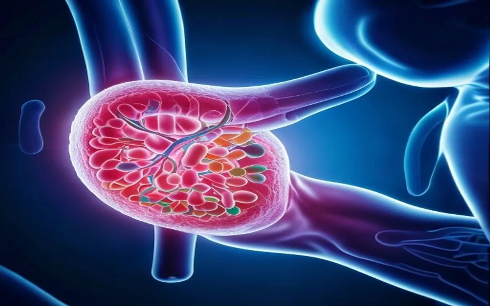
Diabetic nephropathy affects approximately 30–40% of people with diabetes and is the single largest driver of end-stage renal disease (ESRD) worldwide. [1] [2]
Where conventional management falls short. Tight glycemic control, RAAS blockade with ACE inhibitors or ARBs, and newer SGLT2 inhibitors and GLP-1 receptor agonists can slow the decline in kidney function. But even optimal medical therapy rarely halts progression entirely, and none of these approaches reverse the structural damage that has already accumulated — glomerulosclerosis, tubulointerstitial fibrosis, and podocyte dropout are considered irreversible by current standards.
The deeper problem is microvascular and inflammatory. Diabetic nephropathy is not simply a "sugar problem." Chronic hyperglycemia drives the formation of advanced glycation end-products (AGEs), activates protein kinase C and the polyol pathway, and generates sustained oxidative stress. This cascade triggers glomerular hyperfiltration, mesangial matrix expansion, podocyte injury, and a smoldering inflammatory infiltrate that progressively replaces functional nephrons with scar tissue. [3] [4]
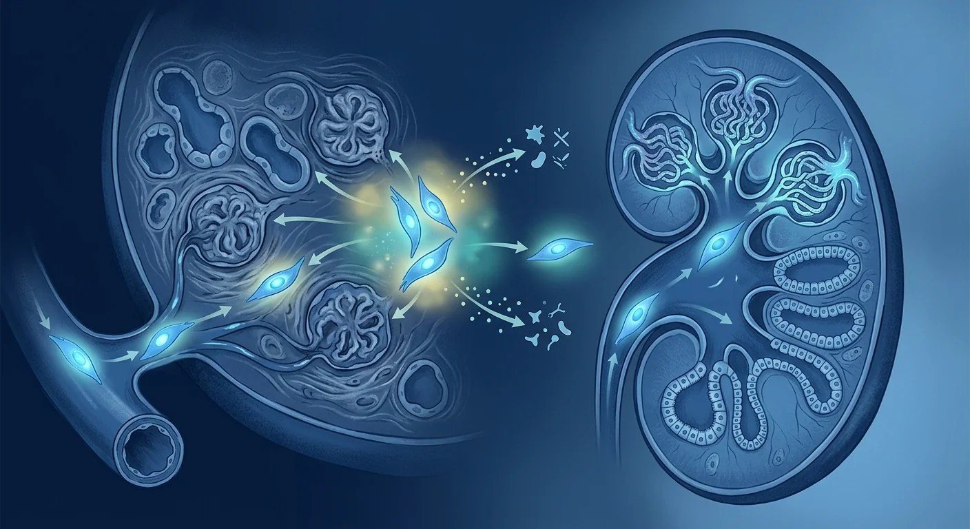
MSC therapy targets several of these drivers simultaneously. Rather than blocking a single pathway, mesenchymal stem cells are being investigated for their capacity to deliver a coordinated anti-inflammatory, anti-fibrotic, and pro-angiogenic signal directly to the injured renal microenvironment — a multi-target approach that matches the multi-factorial nature of diabetic kidney disease.
What is diabetic nephropathy — and why does it progress?
Diabetic nephropathy is a microvascular complication of diabetes characterized by progressive glomerular injury, proteinuria, and declining kidney function. It unfolds over years to decades, beginning with glomerular hyperfiltration and microalbuminuria before advancing to overt proteinuria and, ultimately, end-stage renal disease requiring dialysis or transplantation. The histological hallmarks are mesangial expansion, glomerular basement membrane thickening, podocyte foot-process effacement, and eventual nodular glomerulosclerosis (Kimmelstiel-Wilson lesions). [5]
The disease's relentless progression is driven by a self-reinforcing cycle: hyperglycemia and hemodynamic stress injure podocytes and glomerular endothelial cells, provoking local inflammation and the release of pro-fibrotic mediators such as TGF-β1. Activated myofibroblasts deposit extracellular matrix, which stiffens the tissue and further impairs perfusion, feeding back into more injury. Once a critical mass of nephrons is lost — typically estimated at 50–70% — the remaining nephrons hyperfilter to compensate, accelerating their own demise through what is essentially mechanical and metabolic burnout.
The clinical trajectory
Without intervention, diabetic nephropathy follows a predictable arc: microalbuminuria (30–300 mg/g creatinine) → macroalbuminuria (>300 mg/g) → declining eGFR → ESRD. The slope of eGFR decline is the single most important prognostic marker, and therapies that slow this slope — even modestly — translate into years of dialysis-free survival. MSCs are being studied as a way to flatten this curve, not to reverse it entirely.
Why mesenchymal stem cells are the leading candidate
MSCs are the most-studied cell type for diabetic nephropathy because they combine an excellent safety record with a biological toolkit that directly addresses the disease's core pathology. Unlike pharmacologic agents that target one receptor or pathway, MSCs sense the local inflammatory and hypoxic environment and respond by secreting a broad panel of paracrine factors — cytokines, growth factors, extracellular vesicles, and microRNAs — that collectively shift the renal microenvironment from pro-inflammatory and pro-fibrotic toward repair-oriented. [6]
The key mechanisms under investigation include:
- Anti-inflammatory reprogramming. MSCs suppress pro-inflammatory M1 macrophage polarization while promoting regulatory M2 and regulatory T-cell (Treg) phenotypes. They reduce local levels of TNF-α, IL-1β, and IL-6 — cytokines strongly implicated in diabetic glomerular injury. [7]
- Anti-fibrotic signaling. MSC-derived factors — notably hepatocyte growth factor (HGF), insulin-like growth factor-1 (IGF-1), and bone morphogenetic protein-7 (BMP-7) — directly antagonize TGF-β1/Smad-driven fibrogenesis, the central pathway of renal scarring. Preclinical studies show reduced collagen deposition and α-SMA expression in treated kidneys. [8]
- Podocyte protection. Podocytes are terminally differentiated cells with minimal regenerative capacity; their loss is a point of no return in DN. MSC-derived exosomes and soluble factors have been shown to reduce podocyte apoptosis, preserve slit-diaphragm protein expression (nephrin, podocin), and attenuate foot-process effacement in diabetic models. [9]
- Microvascular preservation. MSCs secrete vascular endothelial growth factor (VEGF) and angiopoietin-1, which support peritubular capillary integrity. Preserving the renal microvasculature is essential because capillary rarefaction is both a consequence and an accelerator of tubulointerstitial fibrosis. [10]
Critically, MSCs are not expected to differentiate into new nephrons. Their value proposition is paracrine, not structural — they act as temporary "living drug factories" that recalibrate the local signaling environment before being cleared, typically within days to weeks.
What the clinical trials actually show
The most advanced clinical data come from diabetic nephropathy — not coincidentally, but because it is the largest and best-characterized CKD population. The field's flagship product, rexlemestrocel-L (Mesoblast), is an allogeneic bone-marrow-derived MSC preparation studied specifically in type 2 diabetic patients with moderate-to-severe kidney impairment.
In a randomized, placebo-controlled phase 1b/2a trial, a single intravenous infusion of rexlemestrocel-L was well tolerated with no treatment-related serious adverse events. At 12 weeks, the treated group showed a stabilization or modest improvement in eGFR, while the placebo group continued to decline — a difference that, while not conclusive, was statistically significant on a post-hoc analysis. A subsequent phase 3 trial (DREAM) was designed to confirm these signals with a larger sample and harder endpoints. [11]
Other notable studies include:
- NEPHSTROM (EU Horizon 2020). A randomized controlled trial evaluating bone-marrow MSC therapy for diabetic kidney disease, with primary endpoints of safety and eGFR slope. Results have shown acceptable safety and encouraging biomarker signals, though full publication is ongoing. [12]
- Adipose-derived MSC pilots. Several small single-arm studies using autologous adipose-derived stromal vascular fraction or culture-expanded adipose MSCs have reported reductions in albuminuria and stable eGFR over 6–12 months in patients with diabetic nephropathy, though these remain uncontrolled and hypothesis-generating. [13]
- Umbilical cord MSC studies. Emerging trials from China and Southeast Asia are evaluating umbilical cord-derived MSCs for diabetic kidney disease, with some reporting reductions in serum creatinine and inflammatory markers. These data are preliminary and require confirmation in larger randomized designs.
The honest assessment
Phase 1/2 data consistently show that MSC therapy for diabetic nephropathy is safe and well tolerated — a necessary prerequisite. Some trials report encouraging signals on eGFR trajectory and albuminuria. None have yet demonstrated, in a large adequately powered phase 3 trial, that MSCs reduce the risk of ESRD, dialysis, or death. The field is at the "promising but not proven" stage, and the responsible description remains investigational.
How treatment response is measured
Clinicians and researchers track diabetic nephropathy progression with objective, validated biomarkers — the same endpoints used in MSC trials.
Delivery routes and practical considerations
Intravenous infusion is the dominant delivery route for diabetic nephropathy MSC trials. IV administration is minimally invasive, widely available, and leverages the natural homing of MSCs to sites of inflammation — including the renal microvasculature, where injury signals (SDF-1/CXCR4 axis, hyaluronic acid fragments, and chemokine gradients) direct a portion of the infused cells to the kidneys. [14]
Direct intra-arterial renal infusion has been explored in some studies, offering the theoretical advantage of higher first-pass renal delivery. However, it carries procedural risk (arterial access, contrast exposure) and has not been shown to produce superior outcomes versus IV in head-to-head comparisons. For most patients, IV remains the pragmatic default.
The question of repeat dosing is active and unresolved. MSCs are cleared within days to weeks, and the paracrine benefit is transient. Whether repeat infusions — at intervals of 3, 6, or 12 months — can sustain or augment the therapeutic signal is a key question for the next generation of trials.
Limitations and what patients should know
MSC therapy for diabetic nephropathy is not an approved treatment, and several important limitations deserve honest acknowledgment.
- No phase 3 confirmation. While early-phase signals are encouraging, the definitive trials that would establish efficacy — large, randomized, placebo-controlled studies with hard renal endpoints — have not yet reported positive results. The DREAM phase 3 trial is the most important pending dataset.
- Late-stage disease may be less responsive. Once glomerulosclerosis is advanced and nephron loss exceeds a critical threshold, the potential for MSC therapy to alter the trajectory diminishes. Most trials enroll patients with moderate (stage 3) rather than severe (stage 4–5) CKD.
- Variable MSC preparations. Differences in cell source (bone marrow vs adipose vs umbilical cord), culture conditions, passage number, and dosing protocols make cross-trial comparisons difficult. A positive result with one MSC product does not automatically apply to another.
- Cost and access. MSC therapy is not covered by standard insurance for diabetic nephropathy, and out-of-pocket costs are substantial. Patients considering treatment abroad should request published safety data and independent verification of cell product quality. [15]
"Diabetic nephropathy is arguably the condition where MSC therapy has the strongest biological rationale — because the pathology is simultaneously inflammatory, fibrotic, and microvascular, and MSCs address all three. The question is whether the paracrine signal is strong enough, sustained enough, and delivered early enough to change the clinical trajectory."
Frequently Asked Questions
Can stem cells reverse diabetic nephropathy?
No. The credible aim of MSC therapy in diabetic nephropathy is to slow or stabilize the decline in kidney function, not to reverse established scarring. Preclinical studies show reduced fibrosis and preserved nephron structure, but reversal of advanced glomerulosclerosis has not been demonstrated in humans. The realistic goal is to flatten the eGFR slope — delaying dialysis by months to years — not to restore a scarred kidney to normal function.
How much does stem cell therapy for diabetic nephropathy cost in Thailand?
Costs vary by clinic, cell source, and protocol but typically range from USD 8,000 to 25,000 per infusion in Bangkok. This is not covered by standard health insurance, and patients should verify what is included (cell characterization, follow-up monitoring, repeat dosing) before committing. VELAR Center provides transparent, itemized cost breakdowns during consultation.
What stage of diabetic nephropathy is best suited for MSC therapy?
Most clinical trials enroll patients with stage 2–3 CKD (eGFR 30–60 mL/min) and evidence of progressive decline despite optimal medical therapy. At this stage, enough functional nephrons remain for a paracrine intervention to plausibly make a difference. Advanced stage 4–5 disease (eGFR <30) is less studied and may benefit less, though individual consultation with a nephrologist is essential.
Is one infusion enough, or are repeat doses needed?
The available evidence does not definitively answer this. Single-infusion protocols have shown transient biomarker improvements in some trials, leading many researchers to hypothesize that repeat dosing — at 3–12 month intervals — may be needed to sustain the paracrine signal. This area is actively under investigation and remains speculative.
Are there any serious side effects?
Across multiple early-phase trials, MSC therapy has been well tolerated with no treatment-related serious adverse events reported. The most common minor effects include transient low-grade fever and infusion-related fatigue. Theoretical risks — including immunogenicity with repeat allogeneic dosing and rare thromboembolic events — are monitored but have not materialized as clinical problems in published diabetic nephropathy studies.
How does MSC therapy compare with SGLT2 inhibitors and finerenone?
SGLT2 inhibitors (empagliflozin, dapagliflozin) and the non-steroidal mineralocorticoid receptor antagonist finerenone are proven, guideline-recommended therapies that slow diabetic nephropathy progression. MSC therapy is not a replacement for these agents — it is an investigational adjunct that targets different mechanisms (inflammation, fibrosis, microvascular repair) and would, if proven effective, be used alongside optimized conventional care, not instead of it.
References
- Alicic RZ, Rooney MT, Tuttle KR. Diabetic kidney disease: challenges, progress, and possibilities. Clinical Journal of the American Society of Nephrology. 2017;12(12):2032-2045. doi:10.2215/CJN.11491116 ↩
- Thomas MC, Brownlee M, Susztak K, et al. Diabetic kidney disease. Nature Reviews Disease Primers. 2015;1:15018. doi:10.1038/nrdp.2015.18 ↩
- Brownlee M. Biochemistry and molecular cell biology of diabetic complications. Nature. 2001;414(6865):813-820. doi:10.1038/414813a ↩
- Sharma K, Ziyadeh FN. Hyperglycemia and diabetic kidney disease: the case for transforming growth factor-β as a key mediator. Diabetes. 1995;44(10):1139-1146. doi:10.2337/diab.44.10.1139 ↩
- Tervaert TW, Mooyaart AL, Amann K, et al. Pathologic classification of diabetic nephropathy. Journal of the American Society of Nephrology. 2010;21(4):556-563. doi:10.1681/ASN.2010010010 ↩
- Peired AJ, Sisti A, Romagnani P. Mesenchymal stem cell-based therapy for kidney disease: a review of clinical evidence. Stem Cells International. 2016;2016:4798639. doi:10.1155/2016/4798639 ↩
- Li Y, Liu J, Liao G, et al. Early intervention with mesenchymal stem cells prevents nephropathy in diabetic rats through amelioration of the inflammatory microenvironment. Stem Cell Research & Therapy. 2018;9(1):162. doi:10.1186/s13287-018-0906-6 ↩
- Lv S, Liu G, Wang J, et al. Mesenchymal stem cells transplantation ameliorates glomerular injury in streptozotocin-induced diabetic nephropathy in rats via inhibiting macrophage infiltration. Stem Cell Research & Therapy. 2020;11:174. doi:10.1186/s13287-020-01681-7 ↩
- Jin J, Shi Y, Gong J, et al. Exosome secreted from adipose-derived stem cells attenuates diabetic nephropathy by promoting autophagy flux and inhibiting apoptosis in podocyte. Stem Cell Research & Therapy. 2019;10(1):95. doi:10.1186/s13287-019-1177-1 ↩
- Eirin A, Zhu XY, Puranik AS, et al. Mesenchymal stem cell-derived extracellular vesicles attenuate kidney inflammation and fibrosis in a pig model of renal artery stenosis. Kidney International. 2017;91(1):119-131. doi:10.1016/j.kint.2016.09.005 ↩
- Perico N, Casiraghi F, Remuzzi G. Clinical translation of mesenchymal stromal cell therapies in kidney diseases. Journal of the American Society of Nephrology. 2018;29(1):361-375. doi:10.1681/ASN.2017070781 ↩
- Griffin TP, Martin WP, Islam N, O'Brien T, Griffin MD. The promise of mesenchymal stem cell therapy for diabetic kidney disease. Current Diabetes Reports. 2016;16(5):42. doi:10.1007/s11892-016-0734-6 ↩
- Villatoro AJ, Alcoholado C, Martín-Astorga MC, et al. Adipose tissue-derived mesenchymal stem cells: a new therapeutic approach for the management of diabetic nephropathy. World Journal of Diabetes. 2019;10(11):491-507. doi:10.4239/wjd.v10.i11.491 ↩
- Morigi M, Rota C, Remuzzi G. Mesenchymal stem cells in kidney repair. Methods in Molecular Biology. 2016;1416:89-107. doi:10.1007/978-1-4939-3584-0_5 ↩
- Marcheque J, Bussolati B, Csete M, Perin L. Concise reviews: stem cells and kidney regeneration: an update. Stem Cells Translational Medicine. 2019;8(1):82-92. doi:10.1002/sctm.18-0115 ↩
糖尿病肾病影响大约30–40%的糖尿病患者,是全球终末期肾病(ESRD)的最大驱动因素。[1] [2]
常规治疗的局限性。严格的血糖控制、使用ACE抑制剂或ARB类药物阻断RAAS系统,以及新型SGLT2抑制剂和GLP-1受体激动剂,可以减缓肾功能的下降速度。但即使是最优化的药物治疗也很少能完全阻止疾病进展,而这些方法都无法逆转已经积累的结构性损伤——肾小球硬化、肾小管间质纤维化和足细胞丢失在目前的标准下被认为是不可逆的。
更深层的问题是微血管和炎症性的。糖尿病肾病不仅仅是一个"血糖问题"。慢性高血糖驱动晚期糖基化终产物(AGEs)的形成,激活蛋白激酶C和多元醇通路,并产生持续的氧化应激。这一级联反应触发肾小球高滤过、系膜基质扩张、足细胞损伤和持续的炎症浸润,逐渐用瘢痕组织取代功能性肾单位。[3] [4]
间充质干细胞疗法同时针对多个驱动因素。间充质干细胞不是阻断单一通路,而是被研究用于向受损的肾脏微环境传递协调的抗炎、抗纤维化和促血管生成信号——这种多靶点方法与糖尿病肾病的多因素特性相匹配。
什么是糖尿病肾病——为什么它会进展?
糖尿病肾病是糖尿病的一种微血管并发症,以进行性肾小球损伤、蛋白尿和肾功能下降为特征。它在数年到数十年间逐步发展,开始于肾小球高滤过和微量白蛋白尿,随后进展为显性蛋白尿,最终发展为需要透析或移植的终末期肾病。组织学标志包括系膜扩张、肾小球基底膜增厚、足细胞足突融合,以及最终的结节性肾小球硬化(Kimmelstiel-Wilson病变)。[5]
疾病的持续性进展由一个自我强化的循环驱动:高血糖和血流动力学应激损伤足细胞和肾小球内皮细胞,引发局部炎症和促纤维化介质如TGF-β1的释放。活化的肌成纤维细胞沉积细胞外基质,使组织变硬并进一步损害灌注,反过来加剧损伤。一旦关键数量的肾单位丢失——通常估计为50–70%——剩余的肾单位通过高滤过来代偿,通过机械和代谢性耗竭加速自身的消亡。
临床发展轨迹
若不干预,糖尿病肾病遵循一个可预测的轨迹:微量白蛋白尿(30–300 mg/g肌酐)→ 大量白蛋白尿(>300 mg/g)→ eGFR下降 → 终末期肾病。eGFR下降的斜率是最重要的预后标志,即使适度减缓这一斜率,也能转化为数年的无透析生存期。MSC正在被研究用于拉平这一曲线,而非完全逆转。
为什么间充质干细胞是主要候选
MSC是糖尿病肾病研究最多的细胞类型,因为它们将优秀的安全记录与直接针对疾病核心病理的生物学工具包结合在一起。与针对单一受体或通路的药物不同,MSC感知局部的炎症和缺氧环境,并通过分泌广泛的旁分泌因子——细胞因子、生长因子、细胞外囊泡和microRNA——作出反应,这些因子共同将肾脏微环境从促炎和促纤维化转向修复方向。[6]
正在研究的关键机制包括:
- 抗炎重编程。MSC抑制促炎性M1巨噬细胞极化,同时促进调节性M2和调节性T细胞(Treg)表型。它们降低局部TNF-α、IL-1β和IL-6水平——这些细胞因子与糖尿病肾小球损伤密切相关。[7]
- 抗纤维化信号。MSC来源的因子——特别是肝细胞生长因子(HGF)、胰岛素样生长因子-1(IGF-1)和骨形态发生蛋白-7(BMP-7)——直接对抗TGF-β1/Smad驱动的纤维化,这是肾脏瘢痕形成的中心通路。临床前研究显示治疗后肾脏中胶原沉积和α-SMA表达减少。[8]
- 足细胞保护。足细胞是终末分化细胞,再生能力极低;它们的丢失是DN中的一个不可逆点。MSC来源的外泌体和可溶性因子已被证明可以减少足细胞凋亡,保留裂孔膜蛋白表达(nephrin, podocin),并在糖尿病模型中减轻足突融合。[9]
- 微血管保护。MSC分泌血管内皮生长因子(VEGF)和血管生成素-1,支持肾小管周围毛细血管的完整性。保护肾脏微血管至关重要,因为毛细血管稀疏既是肾小管间质纤维化的结果,也是其加速器。[10]
关键的是,MSC并不预期分化为新的肾单位。它们的价值主张是旁分泌的,而非结构性的——它们充当临时的"活体药物工厂",在被清除前(通常在数天到数周内)重新校准局部信号环境。
临床试验实际显示了什么
最先进的临床数据来自糖尿病肾病——这并非巧合,而是因为它是最大且特征最明确的CKD人群。该领域的旗舰产品rexlemestrocel-L(Mesoblast)是一种同种异体骨髓来源的MSC制剂,专门在伴有中重度肾功能损害的二型糖尿病患者中进行研究。
在一项随机、安慰剂对照的1b/2a期试验中,单次静脉输注rexlemestrocel-L耐受良好,未出现治疗相关的严重不良事件。在12周时,治疗组显示eGFR稳定或轻度改善,而安慰剂组继续下降——这一差异虽非决定性,但在事后分析中具有统计学意义。后续的3期试验(DREAM)旨在用更大的样本量和更硬的终点来确认这些信号。[11]
其他值得注意的研究包括:
- NEPHSTROM(欧盟地平线2020)。一项评估骨髓MSC治疗糖尿病肾病的随机对照试验,主要终点为安全性和eGFR斜率。结果显示了可接受的安全性和令人鼓舞的生物标志物信号,但完整发表仍在进行中。[12]
- 脂肪来源MSC试点研究。几项使用自体脂肪来源基质血管组分或培养扩增的脂肪MSC的小型单臂研究报告了在6–12个月内白蛋白尿减少和eGFR稳定,但这些研究仍是非对照和假设生成性的。[13]
- 脐带MSC研究。来自中国和东南亚的新兴试验正在评估脐带来源的MSC治疗糖尿病肾病,部分研究报告了血清肌酐和炎症标志物的降低。这些数据是初步的,需要在更大的随机设计中确认。
诚实评估
1/2期数据一致显示,MSC治疗糖尿病肾病是安全且耐受良好的——这是必要的前提条件。一些试验报告了在eGFR轨迹和白蛋白尿方面的令人鼓舞的信号。但尚未有任何试验在大型、充分把握度的3期试验中证明MSC能降低ESRD、透析或死亡的风险。该领域处于"有希望但未证实"的阶段,负责任的描述仍然是研究性的。
如何衡量治疗反应
临床医生和研究人员使用客观、经过验证的生物标志物追踪糖尿病肾病的进展——这些也是MSC试验中使用的终点。
递送途径与实际考量
静脉输注是糖尿病肾病MSC试验的主要递送途径。IV给药是微创的,广泛可用,并利用MSC向炎症部位的自然归巢能力——包括肾脏微血管系统,损伤信号(SDF-1/CXCR4轴、透明质酸片段和趋化因子梯度)将一部分输注细胞引导至肾脏。[14]
直接肾动脉内输注在一些研究中被探索,提供更高的首过肾脏递送的理论优势。然而,它带有手术风险(动脉通路、造影剂暴露),并且在头对头比较中未显示出优于IV的结果。对大多数患者来说,IV仍然是实用默认的选择。
重复给药的问题正在活跃研究中且尚未解决。MSC在数天到数周内被清除,旁分泌获益是短暂的。是否每3、6或12个月重复输注可以维持或增强治疗信号,是下一代试验的关键问题。
局限性与患者须知
MSC治疗糖尿病肾病不是已批准的疗法,几个重要局限性值得诚实承认。
- 无3期确认。虽然早期信号令人鼓舞,但能确立疗效的决定性试验——大型、随机、安慰剂对照、具有硬性肾脏终点的研究——尚未报告阳性结果。DREAM 3期试验是最重要的待定数据集。
- 晚期疾病可能反应较差。一旦肾小球硬化进展且肾单位丢失超过临界阈值,MSC治疗改变轨迹的潜力将下降。大多数试验招募中度(3期)而非重度(4–5期)CKD患者。
- MSC制备的可变性。细胞来源(骨髓vs脂肪vs脐带)、培养条件、传代次数和给药方案的差异使跨试验比较困难。一种MSC产品的阳性结果不会自动适用于另一种。
- 费用和可及性。MSC治疗糖尿病肾病不在标准保险覆盖范围内,自付费用相当可观。考虑出国治疗的患者应索取已发表的安全数据和细胞产品质量的独立验证。[15]
"糖尿病肾病可能是MSC治疗具有最强生物学合理性的疾病——因为其病理同时具有炎症性、纤维化和微血管性,而MSC同时针对这三者。问题是旁分泌信号是否足够强、足够持久、足够早期地传递,以改变临床轨迹。"
常见问题
干细胞能逆转糖尿病肾病吗?
不能。MSC治疗在糖尿病肾病中的可信目标是减缓或稳定肾功能下降,而非逆转已形成的瘢痕。临床前研究显示减少纤维化和保留肾单位结构,但高级别肾小球硬化的逆转尚未在人类中被证明。现实目标是将eGFR斜率拉平——将透析推迟数月至数年——而非将瘢痕化的肾脏恢复到正常功能。
在泰国治疗糖尿病肾病的干细胞费用是多少?
费用因诊所、细胞来源和方案而异,但在曼谷通常为每次输注8,000至25,000美元。这不被标准健康保险覆盖,患者应在承诺前核实包含的项目(细胞鉴定、随访监测、重复给药)。VELAR中心在咨询期间提供透明、逐项的费用明细。
什么阶段的糖尿病肾病最适合MSC治疗?
大多数临床试验招募2–3期CKD(eGFR 30–60 mL/min)且尽管最佳药物治疗仍有进展证据的患者。在此阶段,仍有足够的功能性肾单位供旁分泌干预发挥合理作用。4–5期晚期疾病(eGFR <30)研究较少且可能获益较少,但与肾病专家的个体化咨询是必要的。
一次输注足够吗,还是需要重复给药?
现有证据未明确回答此问题。单次输注方案在一些试验中显示短暂的生物标志物改善,导致许多研究人员假设每3–12个月重复给药可能是维持旁分泌信号所必需的。该领域正在积极研究中且仍然是推测性的。
有什么严重的副作用吗?
在多项早期试验中,MSC治疗耐受良好,未报告治疗相关的严重不良事件。最常见的轻微影响包括暂时性低热和输注相关疲劳。理论风险——包括重复同种异体给药的免疫原性和罕见的血栓栓塞事件——正在监测中,但在已发表的糖尿病肾病研究中尚未成为临床问题。
MSC疗法与SGLT2抑制剂和finerenone相比如何?
SGLT2抑制剂(empagliflozin, dapagliflozin)和非甾体盐皮质激素受体拮抗剂finerenone是已被证实、指南推荐的能够减缓糖尿病肾病进展的疗法。MSC疗法不是这些药物的替代品——它是一种针对不同机制(炎症、纤维化、微血管修复)的研究性辅助疗法,如果被证明有效,将与优化的常规治疗同时使用,而非取代之。
参考文献
- Alicic RZ, Rooney MT, Tuttle KR. Diabetic kidney disease: challenges, progress, and possibilities. Clinical Journal of the American Society of Nephrology. 2017;12(12):2032-2045. doi:10.2215/CJN.11491116 ↩
- Thomas MC, Brownlee M, Susztak K, et al. Diabetic kidney disease. Nature Reviews Disease Primers. 2015;1:15018. doi:10.1038/nrdp.2015.18 ↩
- Brownlee M. Biochemistry and molecular cell biology of diabetic complications. Nature. 2001;414(6865):813-820. doi:10.1038/414813a ↩
- Sharma K, Ziyadeh FN. Hyperglycemia and diabetic kidney disease: the case for transforming growth factor-β as a key mediator. Diabetes. 1995;44(10):1139-1146. doi:10.2337/diab.44.10.1139 ↩
- Tervaert TW, Mooyaart AL, Amann K, et al. Pathologic classification of diabetic nephropathy. Journal of the American Society of Nephrology. 2010;21(4):556-563. doi:10.1681/ASN.2010010010 ↩
- Peired AJ, Sisti A, Romagnani P. Mesenchymal stem cell-based therapy for kidney disease: a review of clinical evidence. Stem Cells International. 2016;2016:4798639. doi:10.1155/2016/4798639 ↩
- Li Y, Liu J, Liao G, et al. Early intervention with mesenchymal stem cells prevents nephropathy in diabetic rats through amelioration of the inflammatory microenvironment. Stem Cell Research & Therapy. 2018;9(1):162. doi:10.1186/s13287-018-0906-6 ↩
- Lv S, Liu G, Wang J, et al. Mesenchymal stem cells transplantation ameliorates glomerular injury in streptozotocin-induced diabetic nephropathy in rats via inhibiting macrophage infiltration. Stem Cell Research & Therapy. 2020;11:174. doi:10.1186/s13287-020-01681-7 ↩
- Jin J, Shi Y, Gong J, et al. Exosome secreted from adipose-derived stem cells attenuates diabetic nephropathy by promoting autophagy flux and inhibiting apoptosis in podocyte. Stem Cell Research & Therapy. 2019;10(1):95. doi:10.1186/s13287-019-1177-1 ↩
- Eirin A, Zhu XY, Puranik AS, et al. Mesenchymal stem cell-derived extracellular vesicles attenuate kidney inflammation and fibrosis in a pig model of renal artery stenosis. Kidney International. 2017;91(1):119-131. doi:10.1016/j.kint.2016.09.005 ↩
- Perico N, Casiraghi F, Remuzzi G. Clinical translation of mesenchymal stromal cell therapies in kidney diseases. Journal of the American Society of Nephrology. 2018;29(1):361-375. doi:10.1681/ASN.2017070781 ↩
- Griffin TP, Martin WP, Islam N, O'Brien T, Griffin MD. The promise of mesenchymal stem cell therapy for diabetic kidney disease. Current Diabetes Reports. 2016;16(5):42. doi:10.1007/s11892-016-0734-6 ↩
- Villatoro AJ, Alcoholado C, Martín-Astorga MC, et al. Adipose tissue-derived mesenchymal stem cells: a new therapeutic approach for the management of diabetic nephropathy. World Journal of Diabetes. 2019;10(11):491-507. doi:10.4239/wjd.v10.i11.491 ↩
- Morigi M, Rota C, Remuzzi G. Mesenchymal stem cells in kidney repair. Methods in Molecular Biology. 2016;1416:89-107. doi:10.1007/978-1-4939-3584-0_5 ↩
- Marcheque J, Bussolati B, Csete M, Perin L. Concise reviews: stem cells and kidney regeneration: an update. Stem Cells Translational Medicine. 2019;8(1):82-92. doi:10.1002/sctm.18-0115 ↩
يؤثر اعتلال الكلية السكري على حوالي 30-40% من مرضى السكري وهو المحرك الأكبر لمرض الكلى في المرحلة النهائية (ESRD) على مستوى العالم. [1] [2]
حدود العلاج التقليدي. يمكن للتحكم الصارم في نسبة السكر في الدم، وحصار نظام RAAS باستخدام مثبطات ACE أو ARBs، ومثبطات SGLT2 الحديثة ومنبهات مستقبلات GLP-1، أن تبطئ من انخفاض وظائف الكلى. ولكن حتى العلاج الدوائي الأمثل نادراً ما يوقف التقدم تماماً، ولا تعكس أي من هذه الأساليب الضرر الهيكلي المتراكم — حيث يُعتبر تصلب الكبيبات والتليف الأنبوبي الخلالي وفقدان الخلايا القدمية غير قابل للعكس وفقاً للمعايير الحالية.
المشكلة الأعمق هي وعائية دقيقة والتهابية. اعتلال الكلية السكري ليس مجرد "مشكلة سكر". يؤدي فرط سكر الدم المزمن إلى تكوين منتجات نهائية متقدمة للجليكيشن (AGEs)، وتنشيط بروتين كيناز C ومسار البوليول، وتوليد إجهاد تأكسدي مستمر. تؤدي هذه السلسلة إلى فرط الترشيح الكبيبي، وتوسع المطرس المسراق، وإصابة الخلايا القدمية، وارتشاح التهابي مستمر يستبدل النيفرونات الوظيفية بنسيج ندبي تدريجياً. [3] [4]
يستهدف علاج الخلايا الجذعية الوسيطة العديد من هذه المحركات في وقت واحد. بدلاً من حصار مسار واحد، يتم دراسة الخلايا الجذعية الوسيطة لقدرتها على توصيل إشارة منسقة مضادة للالتهابات ومضادة للتليف ومحفزة لتكوين الأوعية الدموية مباشرة إلى البيئة الدقيقة الكلوية المصابة — وهو نهج متعدد الأهداف يتناسب مع الطبيعة متعددة العوامل لمرض الكلى السكري.
ما هو اعتلال الكلية السكري — ولماذا يتقدم؟
اعتلال الكلية السكري هو مضاعفة وعائية دقيقة لمرض السكري تتميز بإصابة كبيبية تقدمية وبيلة بروتينية وانخفاض وظائف الكلى. يتطور على مدى سنوات إلى عقود، بدءاً من فرط الترشيح الكبيبي والبيلة الزلالية الدقيقة قبل التقدم إلى البيلة البروتينية الصريحة وفي النهاية إلى مرض الكلى في المرحلة النهائية الذي يتطلب الغسيل الكلوي أو الزرع. العلامات النسيجية هي توسع المطرس المسراق، وتسمك الغشاء القاعدي الكبيبي، وانمحاء العمليات القدمية للخلايا القدمية، والتصلب الكبيبي العقدي النهائي (آفات كيملستيل-ويلسون). [5]
يتم دفع التقدم المستمر للمرض بواسطة دورة ذاتية التعزيز: يؤدي فرط سكر الدم والإجهاد الديناميكي الدموي إلى إصابة الخلايا القدمية والخلايا البطانية الكبيبية، مما يثير التهاباً موضعياً وإفراز وسطاء محفزين للتليف مثل TGF-β1. تودع الخلايا الليفية العضلية المنشطة المطرس خارج الخلوي، مما يصلب النسيج ويزيد من ضعف التروية، مما يعود بتغذية راجعة إلى مزيد من الإصابة. بمجرد فقدان كتلة حرجة من النيفرونات — تقدر عادة بـ 50-70% — تقوم النيفرونات المتبقية بالترشيح المفرط للتعويض، مما يسرع من فنائها من خلال ما هو أساساً احتراق ميكانيكي واستقلابي.
المسار السريري
بدون تدخل، يتبع اعتلال الكلية السكري مساراً يمكن التنبؤ به: بيلة زلالية دقيقة (30-300 ملغ/غ كرياتينين) → بيلة زلالية كبيرة (>300 ملغ/غ) → انخفاض eGFR → الداء الكلوي بمراحله النهائية. ميل انخفاض eGFR هو العلامة الإنذارية الأكثر أهمية، والعلاجات التي تبطئ هذا الميل — حتى بشكل متواضع — تترجم إلى سنوات من البقاء على قيد الحياة بدون غسيل كلوي. يتم دراسة الخلايا الجذعية الوسيطة كوسيلة لتسطيح هذا المنحنى، وليس لعكسه بالكامل.
لماذا الخلايا الجذعية الوسيطة هي المرشح الرئيسي
الخلايا الجذعية الوسيطة هي نوع الخلايا الأكثر دراسة لاعتلال الكلية السكري لأنها تجمع بين سجل سلامة ممتاز ومجموعة أدوات بيولوجية تعالج مباشرة علم الأمراض الأساسي للمرض. على عكس العوامل الدوائية التي تستهدف مستقبلاً أو مساراً واحداً، تستشعر الخلايا الجذعية الوسيطة البيئة الالتهابية وناقصة الأكسجين المحلية وتستجيب بإفراز مجموعة واسعة من العوامل الباراكرينية — السيتوكينات وعوامل النمو والحويصلات خارج الخلوية والـmicroRNA — التي تحول مجتمعة البيئة الدقيقة الكلوية من محفزة للالتهاب والتليف نحو الإصلاح. [6]
تشمل الآليات الرئيسية قيد الدراسة:
- إعادة البرمجة المضادة للالتهابات. تثبط الخلايا الجذعية الوسيطة استقطاب البلاعم M1 المحفزة للالتهاب مع تعزيز الأنماط الظاهرية التنظيمية M2 والخلايا التائية التنظيمية (Treg). تقلل من المستويات المحلية لـ TNF-α وIL-1β وIL-6 — وهي سيتوكينات متورطة بقوة في الإصابة الكبيبية السكرية. [7]
- الإشارات المضادة للتليف. العوامل المشتقة من الخلايا الجذعية الوسيطة — وبشكل خاص عامل نمو الخلايا الكبدية (HGF) وعامل النمو الشبيه بالأنسولين-1 (IGF-1) والبروتين المخلق للعظم-7 (BMP-7) — ت antagonize مباشرة التليف المدفوع بـ TGF-β1/Smad، وهو المسار المركزي لتندب الكلى. تظهر الدراسات قبل السريرية انخفاض ترسب الكولاجين وتعبير α-SMA في الكلى المعالجة. [8]
- حماية الخلايا القدمية. الخلايا القدمية هي خلايا متمايزة نهائياً بقدرة تجديدية ضئيلة؛ فقدانها هو نقطة اللاعودة في اعتلال الكلية السكري. ثبت أن الإكسوزومات والعوامل القابلة للذوبان المشتقة من الخلايا الجذعية الوسيطة تقلل من موت الخلايا القدمية المبرمج، وتحافظ على تعبير بروتينات الحجاب الحاجز الشقي (nephrin, podocin)، وتخفف من انمحاء العمليات القدمية في النماذج السكرية. [9]
- الحفاظ على الأوعية الدقيقة. تفرز الخلايا الجذعية الوسيطة عامل نمو البطانة الوعائية (VEGF) والأنجيوبويتين-1، اللذين يدعمان سلامة الشعيرات الدموية حول الأنبوبية. الحفاظ على الأوعية الدقيقة الكلوية أمر ضروري لأن تخلخل الشعيرات الدموية هو نتيجة ومسرع للتليف الأنبوبي الخلالي. [10]
بشكل حاسم، ليس من المتوقع أن تتمايز الخلايا الجذعية الوسيطة إلى نيفرونات جديدة. اقتراح قيمتها هو باراكريني، وليس هيكلي — تعمل "كمصانع أدوية حية" مؤقتة تعيد معايرة بيئة الإشارات المحلية قبل أن يتم تطهيرها، عادة في غضون أيام إلى أسابيع.
ما تظهره التجارب السريرية فعلياً
أكثر البيانات السريرية تقدماً تأتي من اعتلال الكلية السكري — ليس من قبيل الصدفة، بل لأنه أكبر مجموعة من مرضى CKD وأفضلها توصيفاً. المنتج الرائد في المجال، rexlemestrocel-L (Mesoblast)، هو مستحضر خلايا جذعية وسيطة مستمدة من نخاع العظم خيفي تمت دراسته تحديداً في مرضى السكري من النوع الثاني الذين يعانون من اختلال كلوي متوسط إلى شديد.
في تجربة عشوائية مضبوطة بالغفل من المرحلة 1ب/2أ، تم تحمل تسريب وريدي واحد من rexlemestrocel-L بشكل جيد دون أحداث سلبية خطيرة متعلقة بالعلاج. في الأسبوع 12، أظهرت المجموعة المعالجة استقراراً أو تحسناً طفيفاً في eGFR، بينما استمرت مجموعة الغفل في الانخفاض — وهو فرق، على الرغم من أنه ليس قاطعاً، كان ذا دلالة إحصائية في تحليل لاحق. تم تصميم تجربة المرحلة 3 اللاحقة (DREAM) لتأكيد هذه الإشارات بعينة أكبر ونقاط نهاية أصلب. [11]
تشمل الدراسات البارزة الأخرى:
- NEPHSTROM (أفق 2020 للاتحاد الأوروبي). تجربة عشوائية مضبوطة تقيم علاج الخلايا الجذعية الوسيطة من نخاع العظم لمرض الكلى السكري، مع نقاط نهاية أولية للسلامة وميل eGFR. أظهرت النتائج سلامة مقبولة وإشارات مشجعة للمؤشرات الحيوية، على الرغم من أن النشر الكامل مستمر. [12]
- دراسات تجريبية للخلايا الجذعية الوسيطة المشتقة من الدهون. أبلغت عدة دراسات صغيرة أحادية الذراع باستخدام الجزء الوعائي السدوي المشتق من الدهون الذاتية أو الخلايا الجذعية الوسيطة الدهنية الموسعة بالاستزراع عن انخفاضات في البيلة الزلالية واستقرار eGFR على مدى 6-12 شهراً في مرضى اعتلال الكلية السكري، على الرغم من أنها لا تزال غير مضبوطة ومولدة للفرضيات. [13]
- دراسات الخلايا الجذعية الوسيطة من الحبل السري. تقوم تجارب ناشئة من الصين وجنوب شرق آسيا بتقييم الخلايا الجذعية الوسيطة المشتقة من الحبل السري لمرض الكلى السكري، مع تقارير عن انخفاضات في كرياتينين المصل وعلامات الالتهاب. هذه البيانات أولية وتتطلب تأكيداً في تصاميم عشوائية أكبر.
التقييم الصادق
تظهر بيانات المرحلة 1/2 باستمرار أن علاج الخلايا الجذعية الوسيطة لاعتلال الكلية السكري آمن وجيد التحمل — وهو شرط أساسي ضروري. تبلغ بعض التجارب عن إشارات مشجعة على مسار eGFR والبيلة الزلالية. لم تثبت أي منها بعد، في تجربة مرحلة 3 كبيرة ذات قوة كافية، أن الخلايا الجذعية الوسيطة تقلل من خطر الداء الكلوي بمراحله النهائية أو الغسيل الكلوي أو الوفاة. المجال في مرحلة "واعد لكن غير مثبت"، ويظل الوصف المسؤول استقصائياً.
كيف يتم قياس الاستجابة للعلاج
يتتبع الأطباء والباحثون تقدم اعتلال الكلية السكري باستخدام مؤشرات حيوية موضوعية ومعتمدة — وهي نفس نقاط النهاية المستخدمة في تجارب الخلايا الجذعية الوسيطة.
طرق التوصيل والاعتبارات العملية
التسريب الوريدي هو طريق التوصيل السائد في تجارب الخلايا الجذعية الوسيطة لاعتلال الكلية السكري. الإعطاء الوريدي هو طفيف التوغل، ومتوفر على نطاق واسع، ويستفيد من الانجذاب الطبيعي للخلايا الجذعية الوسيطة إلى مواقع الالتهاب — بما في ذلك الأوعية الدقيقة الكلوية، حيث توجه إشارات الإصابة (محور SDF-1/CXCR4، وشظايا حمض الهيالورونيك، ومدرجات الكيموكين) جزءاً من الخلايا المحقونة إلى الكلى. [14]
تم استكشاف التسريب داخل الشريان الكلوي المباشر في بعض الدراسات، مما يوفر الميزة النظرية لتوصيل كلوي أعلى في المرور الأول. ومع ذلك، فإنه يحمل مخاطر إجرائية (الوصول الشرياني، التعرض للصبغة) ولم يظهر نتائج متفوقة مقابل الوريدي في المقارنات المباشرة. بالنسبة لمعظم المرضى، يظل الوريدي هو الخيار الافتراضي العملي.
مسألة الجرعات المتكررة قيد الدراسة النشطة وغير محلولة. يتم تطهير الخلايا الجذعية الوسيطة في غضون أيام إلى أسابيع، والفائدة الباراكرينية عابرة. ما إذا كانت التسريبات المتكررة — على فترات 3 أو 6 أو 12 شهراً — يمكن أن تحافظ على الإشارة العلاجية أو تعززها هو سؤال رئيسي للجيل القادم من التجارب.
القيود وما يجب أن يعرفه المرضى
علاج الخلايا الجذعية الوسيطة لاعتلال الكلية السكري ليس علاجاً معتمداً، وتستحق العديد من القيود الهامة اعترافاً صادقاً.
- لا يوجد تأكيد من المرحلة 3. بينما الإشارات المبكرة مشجعة، فإن التجارب الحاسمة التي ستثبت الفعالية — دراسات كبيرة وعشوائية ومضبوطة بالغفل ذات نقاط نهاية كلوية صلبة — لم تبلغ عن نتائج إيجابية بعد. تجربة DREAM للمرحلة 3 هي أهم مجموعة بيانات معلقة.
- المرض المتقدم قد يكون أقل استجابة. بمجرد أن يصبح التصلب الكبيبي متقدماً ويتجاوز فقدان النيفرونات العتبة الحرجة، تتضاءل إمكانية علاج الخلايا الجذعية الوسيطة لتغيير المسار. تسجل معظم التجارب مرضى بمرض معتدل (المرحلة 3) بدلاً من الشديد (المرحلة 4-5).
- تباين مستحضرات الخلايا الجذعية الوسيطة. الاختلافات في مصدر الخلايا (نخاع العظم مقابل الدهون مقابل الحبل السري)، وظروف الاستزراع، ورقم المرور، وبروتوكولات الجرعات تجعل المقارنات عبر التجارب صعبة. النتيجة الإيجابية مع منتج خلايا جذعية وسيطة واحد لا تنطبق تلقائياً على آخر.
- التكلفة والوصول. علاج الخلايا الجذعية الوسيطة غير مغطى بالتأمين الصحي القياسي لاعتلال الكلية السكري، والتكاليف من الجيب كبيرة. يجب على المرضى الذين يفكرون في العلاج في الخارج طلب بيانات السلامة المنشورة والتحقق المستقل من جودة منتج الخلايا. [15]
"قد يكون اعتلال الكلية السكري هو الحالة التي يمتلك فيها علاج الخلايا الجذعية الوسيطة أقوى مبرر بيولوجي — لأن علم الأمراض التهابي وتليفي ووعائي دقيق في آن واحد، والخلايا الجذعية الوسيطة تعالج الثلاثة جميعاً. السؤال هو ما إذا كانت الإشارة الباراكرينية قوية بما فيه الكفاية، ومستدامة بما فيه الكفاية، ومقدمة مبكراً بما فيه الكفاية لتغيير المسار السريري."
الأسئلة الشائعة
هل يمكن للخلايا الجذعية عكس اعتلال الكلية السكري؟
لا. الهدف المعقول لعلاج الخلايا الجذعية الوسيطة في اعتلال الكلية السكري هو إبطاء أو تثبيت انخفاض وظائف الكلى، وليس عكس التندب المستقر. تظهر الدراسات قبل السريرية انخفاض التليف والحفاظ على بنية النيفرون، لكن عكس التصلب الكبيبي المتقدم لم يتم إثباته في البشر. الهدف الواقعي هو تسطيح منحنى eGFR — تأخير الغسيل الكلوي بأشهر إلى سنوات — وليس استعادة كلية متندبة إلى وظيفة طبيعية.
كم تكلفة علاج الخلايا الجذعية لاعتلال الكلية السكري في تايلاند؟
تختلف التكاليف حسب العيادة ومصدر الخلايا والبروتوكول ولكنها تتراوح عادة من 8,000 إلى 25,000 دولار أمريكي لكل تسريب في بانكوك. هذا غير مغطى بالتأمين الصحي القياسي، ويجب على المرضى التحقق مما هو مشمول (توصيف الخلايا، مراقبة المتابعة، الجرعات المتكررة) قبل الالتزام. يقدم مركز فيلار تفاصيل تكلفة شفافة ومفصلة أثناء الاستشارة.
ما هي مرحلة اعتلال الكلية السكري الأنسب لعلاج الخلايا الجذعية الوسيطة؟
تسجل معظم التجارب السريرية مرضى بمرض الكلى المزمن من المرحلة 2-3 (eGFR 30-60 مل/دقيقة) مع دليل على التقدم المستمر على الرغم من العلاج الطبي الأمثل. في هذه المرحلة، تبقى نيفرونات وظيفية كافية لتدخل باراكريني لإحداث فرق معقول. المرض المتقدم من المرحلة 4-5 (eGFR أقل من 30) أقل دراسة وقد يستفيد أقل، على الرغم من أن الاستشارة الفردية مع أخصائي كلى ضرورية.
هل تسريب واحد كافٍ، أم هناك حاجة لجرعات متكررة؟
الأدلة المتاحة لا تجيب على هذا بشكل قاطع. أظهرت بروتوكولات التسريب الواحد تحسينات عابرة في المؤشرات الحيوية في بعض التجارب، مما دفع العديد من الباحثين إلى افتراض أن الجرعات المتكررة — على فترات 3-12 شهراً — قد تكون ضرورية لاستدامة الإشارة الباراكرينية. هذا المجال قيد الدراسة النشطة ويظل تخمينياً.
هل هناك أي آثار جانبية خطيرة؟
عبر تجارب المرحلة المبكرة المتعددة، تم تحمل علاج الخلايا الجذعية الوسيطة بشكل جيد دون أحداث سلبية خطيرة متعلقة بالعلاج. تشمل الآثار الطفيفة الأكثر شيوعاً حمى منخفضة الدرجة عابرة وإرهاق متعلق بالتسريب. تتم مراقبة المخاطر النظرية — بما في ذلك المناعة مع الجرعات الخيفية المتكررة والأحداث الانصمامية الخثارية النادرة — لكنها لم تتحقق كمشاكل سريرية في دراسات اعتلال الكلية السكري المنشورة.
كيف يقارن علاج الخلايا الجذعية الوسيطة مع مثبطات SGLT2 والفينيرينون؟
مثبطات SGLT2 (empagliflozin, dapagliflozin) ومضاد مستقبلات القشرانيات المعدنية غير الستيرويدي finerenone هي علاجات مثبتة وموصى بها في الإرشادات تبطئ تقدم اعتلال الكلية السكري. علاج الخلايا الجذعية الوسيطة ليس بديلاً عن هذه العوامل — إنه مساعد استقصائي يستهدف آليات مختلفة (الالتهاب، التليف، إصلاح الأوعية الدقيقة) وسيتم استخدامه، إذا ثبتت فعاليته، إلى جانب الرعاية التقليدية المثلى، وليس بدلاً منها.
المراجع
- Alicic RZ, Rooney MT, Tuttle KR. Diabetic kidney disease: challenges, progress, and possibilities. Clinical Journal of the American Society of Nephrology. 2017;12(12):2032-2045. doi:10.2215/CJN.11491116 ↩
- Thomas MC, Brownlee M, Susztak K, et al. Diabetic kidney disease. Nature Reviews Disease Primers. 2015;1:15018. doi:10.1038/nrdp.2015.18 ↩
- Brownlee M. Biochemistry and molecular cell biology of diabetic complications. Nature. 2001;414(6865):813-820. doi:10.1038/414813a ↩
- Sharma K, Ziyadeh FN. Hyperglycemia and diabetic kidney disease: the case for transforming growth factor-β as a key mediator. Diabetes. 1995;44(10):1139-1146. doi:10.2337/diab.44.10.1139 ↩
- Tervaert TW, Mooyaart AL, Amann K, et al. Pathologic classification of diabetic nephropathy. Journal of the American Society of Nephrology. 2010;21(4):556-563. doi:10.1681/ASN.2010010010 ↩
- Peired AJ, Sisti A, Romagnani P. Mesenchymal stem cell-based therapy for kidney disease: a review of clinical evidence. Stem Cells International. 2016;2016:4798639. doi:10.1155/2016/4798639 ↩
- Li Y, Liu J, Liao G, et al. Early intervention with mesenchymal stem cells prevents nephropathy in diabetic rats through amelioration of the inflammatory microenvironment. Stem Cell Research & Therapy. 2018;9(1):162. doi:10.1186/s13287-018-0906-6 ↩
- Lv S, Liu G, Wang J, et al. Mesenchymal stem cells transplantation ameliorates glomerular injury in streptozotocin-induced diabetic nephropathy in rats via inhibiting macrophage infiltration. Stem Cell Research & Therapy. 2020;11:174. doi:10.1186/s13287-020-01681-7 ↩
- Jin J, Shi Y, Gong J, et al. Exosome secreted from adipose-derived stem cells attenuates diabetic nephropathy by promoting autophagy flux and inhibiting apoptosis in podocyte. Stem Cell Research & Therapy. 2019;10(1):95. doi:10.1186/s13287-019-1177-1 ↩
- Eirin A, Zhu XY, Puranik AS, et al. Mesenchymal stem cell-derived extracellular vesicles attenuate kidney inflammation and fibrosis in a pig model of renal artery stenosis. Kidney International. 2017;91(1):119-131. doi:10.1016/j.kint.2016.09.005 ↩
- Perico N, Casiraghi F, Remuzzi G. Clinical translation of mesenchymal stromal cell therapies in kidney diseases. Journal of the American Society of Nephrology. 2018;29(1):361-375. doi:10.1681/ASN.2017070781 ↩
- Griffin TP, Martin WP, Islam N, O'Brien T, Griffin MD. The promise of mesenchymal stem cell therapy for diabetic kidney disease. Current Diabetes Reports. 2016;16(5):42. doi:10.1007/s11892-016-0734-6 ↩
- Villatoro AJ, Alcoholado C, Martín-Astorga MC, et al. Adipose tissue-derived mesenchymal stem cells: a new therapeutic approach for the management of diabetic nephropathy. World Journal of Diabetes. 2019;10(11):491-507. doi:10.4239/wjd.v10.i11.491 ↩
- Morigi M, Rota C, Remuzzi G. Mesenchymal stem cells in kidney repair. Methods in Molecular Biology. 2016;1416:89-107. doi:10.1007/978-1-4939-3584-0_5 ↩
- Marcheque J, Bussolati B, Csete M, Perin L. Concise reviews: stem cells and kidney regeneration: an update. Stem Cells Translational Medicine. 2019;8(1):82-92. doi:10.1002/sctm.18-0115 ↩