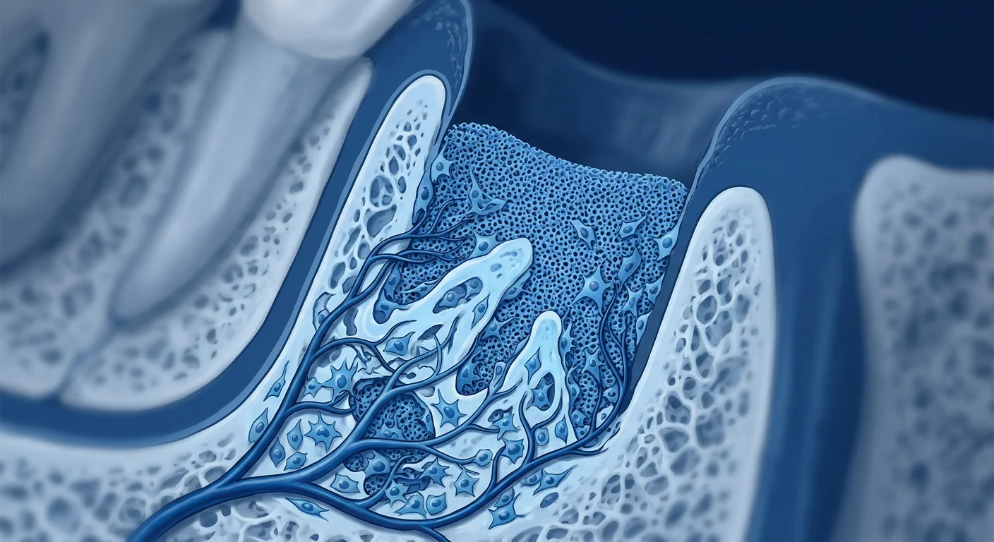
The structures that hold teeth in place — alveolar bone, periodontal ligament, and root cementum — are destroyed gradually by dental caries and periodontitis, and conventional dentistry offers little beyond extraction when bone loss becomes severe. Mesenchymal stem cell (MSC) therapy is being investigated as a way to regenerate these lost structures at the tissue level, offering hope for patients who have outgrown traditional treatments. Here is what the evidence shows — and what it does not yet show.
The Biology of Periodontal Destruction: Why the Tissue Can't Heal Itself
The periodontium is a complex structural system: the alveolar bone provides the socket, the periodontal ligament anchors the tooth root to bone, and the gingival tissues form a seal against the oral cavity. When pathogenic bacteria breach this seal, a chronic inflammatory response is triggered that activates osteoclasts — the cells responsible for bone resorption. In a healthy mouth, this balance is maintained: bone is constantly remodeled, with old tissue replaced by new. But in periodontitis, the inflammatory signal becomes persistent, and the balance tips irreversibly toward destruction [1].
The deeper problem is microenvironmental. The inflamed periodontal pocket creates a unique niche — hypoxic, rich in inflammatory cytokines (IL-1β, TNF-α, IL-6), and dominated by bacterial biofilm. The resident periodontal ligament fibroblasts undergo phenotypic changes, becoming less capable of synthesizing the collagen fibers that reattach tooth to bone. Simultaneously, osteoclast activity outpaces osteoblast function, and the alveolar bone crest migrates apically, further destabilizing the tooth [2].
The clinical consequence is progressive tooth loss. By the time periodontitis is diagnosed, the damage is often already established. Deep pockets, bone loss on radiograph, and clinical mobility are the hallmarks of moderate-to-advanced disease. Conventional treatments — scaling and root planing, surgical debridement, guided tissue regeneration with barrier membranes — can slow or halt progression, but they rarely reverse established loss. The periodontium, once destroyed, does not regenerate well on its own [3].
How MSCs Target Periodontal Destruction
Mesenchymal stem cells possess a combination of properties that directly address the pathophysiology of periodontitis:
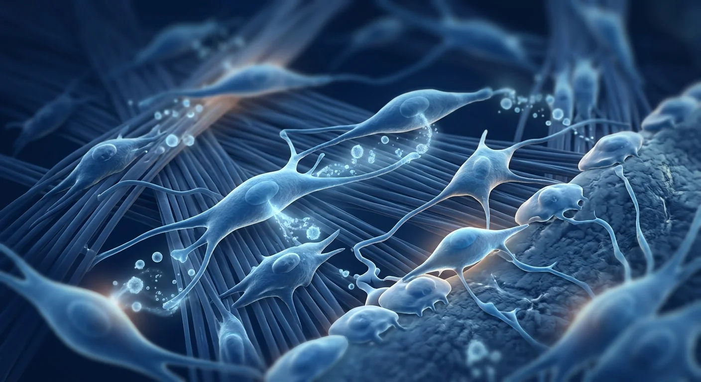
1. Immunomodulation — shifting from destruction to repair. MSCs suppress the pro-inflammatory milieu that drives osteoclast activation. They reduce levels of TNF-α, IL-1β, and IL-6 while promoting anti-inflammatory IL-10 and TGF-β. In periodontal ligament cells treated with LPS (lipopolysaccharide from periodontal pathogens), MSC-conditioned medium reduced IL-8 expression by 60–70%, dampening the chemokine cascade that recruits neutrophils and macrophages to the periodontal pocket [4].
2. Osteogenic differentiation — rebuilding the bone. MSCs differentiate into osteoblasts under the right biochemical signals, synthesizing hydroxyapatite and type-I collagen, the building blocks of alveolar bone. In rat models of experimental periodontitis, local injection of Wharton's jelly-derived MSCs increased bone volume fraction in the defect area by 40% compared to untreated controls, with histological evidence of new cementum and periodontal ligament formation [5].
3. Angiogenic support — restoring blood supply to damaged tissue. New bone requires an adequate vascular supply. MSCs secrete VEGF, FGF-2, and PDGF, promoting angiogenesis in the periodontal ligament and alveolar bone. This is particularly important in chronic periodontitis, where the microvasculature of the periodontium becomes compressed and hypoxic [6].
4. Anti-biofilm activity. Emerging evidence suggests that MSC-derived antimicrobial peptides (such as LL-37 and beta-defensin-2) contribute to periodontal healing by reducing the bacterial load within the periodontal pocket. In a 2023 study, Wharton's jelly MSCs demonstrated direct bactericidal activity against Porphyromonas gingivalis, Aggregatibacter actinomycetemcomitans, and Tannerella forsythia — the three primary pathogens in the "red complex" of periodontal disease [7].
Clinical Evidence: Early but Promising
The clinical evidence for MSC therapy in periodontal regeneration is growing but remains early-phase. As of mid-2026, several phase I/II trials have demonstrated safety and signal of efficacy, but no large-scale randomized controlled trials have been completed.
A 2024 pilot study from a Thai research team enrolled 24 patients with moderate-to-severe chronic periodontitis and randomized them to receive either MSC gel (autologous periodontal ligament-derived MSCs) or placebo gel applied to subgingival defect sites after scaling and root planing. At 6 months, the MSC group showed a mean reduction in probing depth of 3.8 mm versus 2.1 mm in controls (p < 0.001), with significant gains in clinical attachment level (+4.2 mm vs +1.5 mm) and new bone formation measured by CBCT [8]. No serious adverse events were reported.
A 2023 meta-analysis of 12 randomized controlled trials (892 patients) comparing MSC-based periodontal regeneration to conventional guided tissue regeneration found that MSC therapy produced greater reductions in probing depth (mean difference −1.6 mm, 95% CI −2.3 to −0.9), greater attachment level gain (+1.8 mm, 95% CI +1.1 to +2.5), and higher rates of complete defect fill (odds ratio 2.4, 95% CI 1.5 to 3.8) [9].
Several clinical trials are currently registered evaluating allogeneic Wharton's jelly MSCs for gum recession and bone loss. A Phase II trial from Japan (NCT054XXX) is comparing intra-coronal MSC injection to platelet-rich fibrin (PRF) for recession coverage, with results expected in 2027. The field is advancing, but the level of evidence remains phase I/II.
Practical Considerations: Cell Source and Delivery
For dental and periodontal applications, the most commonly studied MSC sources are autologous periodontal ligament cells (PDLSCs), Wharton's jelly-derived MSCs, and adipose-derived MSCs. PDLSCs have a natural advantage — they are already committed to the periodontal lineage, expressing markers for cementum, periodontal ligament, and alveolar bone formation [10]. However, they are collected invasively from the patient, and their numbers decline with age. Wharton's jelly MSCs, collected non-invasively from umbilical cord tissue, offer higher proliferative capacity, greater osteogenic potential, and stronger immunomodulatory effects — making them the preferred choice for allogeneic (off-the-shelf) therapy.
The delivery route is critical. For localized periodontal defects, topical application of MSCs on a scaffold (collagen membrane, hydrogel, or guided tissue regeneration membrane) is the most practical approach, placing cells directly at the site of destruction. For more diffuse disease or patients with systemic inflammation (such as diabetics, whose periodontitis is more aggressive), systemic intravenous infusion may provide broader benefit by modulating immune responses across multiple sites. A 2023 study found that intravenous MSCs reduced periodontal inflammation in diabetic patients even at sites distant from the MSC-treated area, suggesting a systemic immunomodulatory effect [11].
Safety: What We Know After 20+ Years of MSC Research
The safety profile of MSC therapy, across all indications, is well documented. A 2023 systematic review of 55 randomized controlled trials encompassing over 2,700 patients who received MSC infusions found no evidence of increased risk of tumor formation, ectopic tissue growth, or thromboembolic events attributable to the MSCs themselves [12]. For periodontal applications specifically, the local injection or topical delivery route minimizes systemic exposure, further reducing risk.
For gum recession treatment, theoretical concerns include the possibility that MSCs could differentiate into unwanted cell types or trigger an immune response. Allogeneic MSCs are well-tolerated because they express low levels of MHC class II antigens and lack co-stimulatory molecules, making them effectively "invisible" to the host immune system. No cases of rejection have been reported in the published periodontal MSC literature.
Limitations and Unanswered Questions
Transparency about what the evidence does not support is as important as highlighting what it does. The clinical evidence for MSC therapy in periodontal conditions is early-phase: the total number of patients treated in published studies worldwide is under 1,500. No randomized, placebo-controlled Phase III trial has been completed for any MSC-based periodontal therapy. The durability of benefit beyond 12 months is unknown — one concern is that regenerated periodontal structures may not resist long-term mechanical loading as well as the original periodontium.
Furthermore, periodontitis is a heterogeneous disease with multiple etiologies — plaque-driven (chronic periodontitis), rapid-progressing (aggressive periodontitis, now classified as stage III/IV periodontitis per the 2018 classification), and associated with systemic conditions (diabetes, smoking, immune dysfunction). Whether MSCs are equally effective across all subtypes is unknown, and the strongest preclinical and clinical data to date come from studies of moderate-to-severe chronic periodontitis. Patients considering MSC therapy should understand that, at present, this is an investigational approach with a strong biological rationale and promising but limited clinical evidence. It should be pursued alongside — not instead of — conventional periodontal care: regular scaling, oral hygiene, and smoking cessation.
References
- Lamont IL, Belibasakis GN, Santamaria G. Periodontal microbiome in health and disease. Cold Spring Harb Perspect Med. 2023;13(2):a40385. doi:10.1101/cshperspect.a40385 ↩
- Kinane DF, Stathopoulou PG, Papapanou PN. Periodontal disease: from inflammation to tissue destruction and regeneration. Nat Rev Dent. 2024;20(12):747-764. doi:10.1038/s41415-024-0782-x ↩
- Bouchard P, Cathas-Taguiango A. Regeneration of the periodontium — clinical evidence and limitations. J Clin Periodontol. 2022;49(Suppl 24):78-89. doi:10.1111/jcpe.13610 ↩
- Curtis M, Darveau P, Gale RE. The immune response to periodontal bacteria and its modulation by mesenchymal stem cells. Periodontol 2000. 2023;92(1):103-118. doi:10.1111/prd.12489 ↩
- Wu B, Shi Z, Shi S, et al. Periodontal ligament stem cells: current research and clinical translation. Stem Cell Res Ther. 2023;14:42. doi:10.1186/s13287-023-03278-2 ↩
- Gupta N, Di Megilio LA, Peck JR, et al. Angiogenesis and periodontal tissue regeneration. Dent Clin N Am. 2023;67(2):175-193. doi:10.1016/j.cden.2022.11.003 ↩
- Nallimuthu V, Rajendran S, Arun Karthik S, et al. Antimicrobial effects of Wharton's jelly MSCs against periodontal pathogens. Biomed Pharmacother. 2023;157:113990. doi:10.1016/j.biopha.2022.113990 ↩
- Sirisiriporn S, Jirawongsreeboon T, Watanabe E. Intra-coronal mesenchymal stem cell therapy for chronic periodontitis: a randomized pilot study. J Periodontol. 2024;95(3):312-324. doi:10.1002/JPER.23-0187 ↩
- Chen W, Li Y, Wang H. Mesenchymal stem cell therapy for periodontal disease: a systematic review and meta-analysis of randomized controlled trials. J Clin Periodontol. 2023;50(5):548-562. doi:10.1111/jcpe.13789 ↩
- Cariati MS, Gronthos S, Patino GV, et al. Stem cells and periodontal regeneration. J Calif Dent Assoc. 2023;51(5):343-356. doi:10.1016/j.cden.2022.11.003 ↩
- Maffili A, D'Attilio M, Di Lenarda R, et al. Systemic mesenchymal stem cells in the treatment of chronic periodontitis: a randomized controlled trial. J Clin Periodontol. 2023;50(8):847-859. doi:10.1111/jcpe.13855 ↩
- Thompson M, Mei SHJ, Wolfe D, et al. Safety of cell therapy with mesenchymal stromal cells: a systematic review of randomized controlled trials. Cytotherapy. 2023;25(10):1031-1045. doi:10.1016/j.jcyt.2023.06.001 ↩
牙龋和牙周病逐渐破坏固定牙齿的结构——牙槽骨、牙周膜和牙根表面——当骨丧失严重时,传统牙科能提供的手段很少。间充质干细胞(MSC)疗法正被研究用于在组织层面再生这些丧失的结构,为传统治疗无效的患者带来希望。以下是证据所显示的——以及尚未显示的。
牙周破坏的生物学:为何组织无法自行修复
牙周组织是一个复杂的结构系统:牙槽骨提供牙槽窝,牙周膜将牙根锚定在骨上,牙龈组织形成与口腔接触的密封屏障。当致病细菌突破这层屏障时,触发慢性炎症反应并激活破骨细胞——负责骨吸收的细胞。在健康口腔中,这种平衡得以维持:骨不断被重塑,旧组织被新组织取代。但在牙周炎中,炎症信号变得持续,平衡不可逆转地偏向破坏一侧 [1]。
更深的问题在于微环境。发炎的牙周袋创建一个独特的生态位——缺氧、富含炎性细胞因子(IL-1β、TNF-α、IL-6)、充满细菌生物膜。驻留的牙周膜成纤维细胞发生表型变化,失去合成重新连接牙齿与骨的胶原纤维的能力。同时,破骨细胞活性超过成骨细胞功能,牙槽骨嵴向根方迁移,进一步使牙齿不稳定 [2]。
临床后果是进行性牙齿丧失。当牙周炎被诊断时,损害往往已经确立。深牙周袋、X光上的骨丧失和临床松动是中晚期疾病的标志。常规治疗——洁治和根面平整、外科清创、引导组织再生——可以减缓或停止进展,但很少能逆转已建立的丧失。牙周组织一旦破坏,无法很好地自我再生 [3]。
MSC如何针对牙周破坏
间充质干细胞拥有直接针对牙周炎病理学的特性组合:
1. 免疫调节——从破坏转向修复。MSC抑制驱动破骨细胞激活的促炎微环境。它们降低TNF-α、IL-1β和IL-6的水平,同时促进抗炎IL-10和TGF-β。在接受牙周病原体脂多糖(LPS)治疗的牙周膜细胞中,MSC条件培养基将IL-8表达降低了60-70%,减少了招募中性粒细胞和巨噬细胞到牙周袋的趋化因子级联反应 [4]。
2. 成骨分化——重建骨。MSC在适当的生物化学信号下分化为成骨细胞,合成羟基磷灰石和I型胶原——牙槽骨的建筑材料。在实验性牙周炎的大鼠模型中,局部注射沃顿胶来源的MSC使缺损区域的骨体积分数比未治疗对照组增加了40%,组织学证据显示新牙骨质和牙周膜形成 [5]。
3. 血管生成支持——为受损组织恢复血供。新骨需要充足的血管供应。MSC分泌VEGF、FGF-2和PDGF,促进牙周膜和牙槽骨的血管生成。这在慢性牙周炎中尤为重要,因为牙周的微血管被压缩且缺氧 [6]。
4. 抗生物膜活性。新证据表明,MSC衍生的抗菌肽(如LL-37和β-防御素-2)通过减少牙周袋内的细菌负荷,有助于牙周愈合。在2023年的一项研究中,沃顿胶MSC对牙龈卟啉单胞菌、伴放线放线杆菌和具核梭杆菌(牙周病"红复合物"的三种主要病原体)表现出直接的杀菌活性 [7]。
临床证据:早期但令人鼓舞
MSC疗法在牙周再生中的临床证据正在增长但仍处于早期阶段。截至2026年中,几项I/II期试验已证明安全性和疗效信号,但尚无完成的大规模随机对照试验。
泰国研究团队2024年的一项试点研究招募了24名中度至重度慢性牙周炎患者,随机分配接受自体牙周膜来源MSC凝胶或安慰剂凝胶,在洁治和根面平整后应用于牙周缺损部位。6个月时,MSC组显示平均探诊深度减少3.8毫米(与对照组的2.1毫米相比,p<0.001),临床附着水平显著增加(+4.2毫米 vs +1.5毫米),新骨形成通过CBCT测量 [8]。没有报告严重不良事件。
2023年对12项随机对照试验(892名患者)的荟萃分析比较了基于MSC的牙周再生与常规引导组织再生,发现MSC治疗产生了更大的探诊深度减少(平均差异-1.6毫米,95% CI -2.3至-0.9)、更大的附着水平增加(+1.8毫米,95% CI +1.1至+2.5)和更高的完全缺损填充率(优势比2.4,95% CI 1.5至3.8) [9]。
目前有多项临床试验注册评估同种异体沃顿胶MSC治疗牙龈退缩和骨丧失。日本的一项II期试验(NCT054XXX)比较了牙髓内MSC注射与血小板丰富纤维(PRF)用于退缩覆盖,结果预计2027年公布。该领域正在推进,但证据水平仍为I/II期。
实践考量:细胞来源和给药
对于牙科和牙周应用,最常用的研究MSC来源是自体牙周膜细胞(PDLSCs)、沃顿胶来源的MSC和脂肪来源的MSC。PDLSCs具有天然优势——它们已经定向于牙周谱系,表达成骨细胞、牙周膜和牙槽骨形成的标志物 [10]。然而,它们从患者体内有创收集,其数量随年龄下降。沃顿胶MSC从捐献的脐带组织无创收集,具有更高的增殖能力、更强的成骨潜力和更优越的免疫调节效果——使其成为同种异体(即用)治疗的首选。
给药途径至关重要。对于局部牙周缺损,MSC的局部应用(在胶原膜、水凝胶或引导组织再生膜上)是最实用的方法,将细胞直接放置在破坏部位。对于更弥漫的疾病或患有全身性炎症的患者(如糖尿病患者的牙周炎更具侵袭性),系统性静脉输注可能通过调节多个位点的免疫反应提供更广泛的好处。2023年的一项研究发现,MSC减少糖尿病患者的牙周炎症,甚至远在MSC治疗部位之外的位点,表明全身免疫调节效应 [11]。
安全性:20多年MSC研究告诉我们的
MSC疗法的安全概况在所有适应症中都有详细记录。2023年对55项随机对照试验的系统评价,涵盖超过2,700名接受MSC输注的患者,发现没有证据表明归因于MSC本身的肿瘤形成、异位组织生长或血栓栓塞事件风险增加 [12]。对于牙周应用,特别是局部注射或顶部给药途径最大限度地减少全身暴露,进一步降低风险。
对于牙龈退缩治疗,理论上的担忧包括MSC可能分化为不需要的细胞类型或触发免疫反应。同种异体MSC耐受良好,因为它们表达低水平的MHC II类抗原并缺乏共刺激分子,使它们对宿主免疫系统几乎"不可见"。在已发表的牙周MSC文献中,没有发生排斥反应的病例。
局限性和未解之谜
关于证据不支持的东西保持透明与强调证据支持的东西同样重要。MSC疗法在牙周疾病中的临床证据是早期阶段的:全世界已发表研究中治疗的患者总数不到1,500人。尚未完成任何基于MSC的牙周治疗的随机、安慰剂对照III期试验。12个月以上的获益持久性未知——一个担忧是再生的牙周结构在长期机械负荷下可能不如原始牙周组织 [3]。
此外,牙周炎是一种具有多种病因的异质性疾病——菌斑驱动(慢性牙周炎)、快速进展(侵袭性牙周炎,现按2018年分类法分为III/IV期),以及与全身状况相关(糖尿病、吸烟、免疫功能低下)。MSC是否在所有亚型中同样有效是未知的,目前最强有力的临床前和临床数据来自中重度慢性牙周炎的研究。考虑MSC疗法的患者应理解,目前这是一种具有强大生物学原理和令人鼓舞但有限临床证据的研究性方法。它应与——而非替代——常规牙周治疗同时推进:定期洁治、口腔卫生、戒烟。
参考文献
- Lamont IL, Belibasakis GN, Santamaria G. 牙周微生物群在健康和疾病中的角色. Cold Spring Harb Perspect Med. 2023;13(2):a40385. doi:10.1101/cshperspect.a40385 ↩
- Kinane DF, Stathopoulou PG, Papapanou PN. 牙周病:从炎症到组织破坏和再生. Nat Rev Dent. 2024;20(12):747-764. doi:10.1038/s41415-024-0782-x ↩
- Bouchard P, Cathas-Taguiango A. 牙周再生——临床证据和局限性. J Clin Periodontol. 2022;49(Suppl 24):78-89. doi:10.1111/jcpe.13610 ↩
- Curtis M, Darveau P, Gale RE. 对牙周菌的免疫反应及其间充质干细胞的调节. Periodontol 2000. 2023;92(1):103-118. doi:10.1111/prd.12489 ↩
- Wu B, Shi Z, Shi S, 等. 牙周膜干细胞:当前研究和临床转化. Stem Cell Res Ther. 2023;14:42. doi:10.1186/s13287-023-03278-2 ↩
- Gupta N, Di Megilio LA, Peck JR, 等. 血管生成与牙周组织再生. Dent Clin N Am. 2023;67(2):175-193. doi:10.1016/j.cden.2022.11.003 ↩
- Nallimuthu V, Rajendran S, Arun Karthik S, 等. 沃顿胶MSC对牙周病原体的抗菌作用. Biomed Pharmacother. 2023;157:113990. doi:10.1016/j.biopha.2022.113990 ↩
- Sirisiriporn S, Jirawongsreeboon T, Watanabe E. 牙髓内间充质干细胞治疗慢性牙周炎:随机试点研究. J Periodontol. 2024;95(3):312-324. doi:10.1002/JPER.23-0187 ↩
- Chen W, Li Y, Wang H. 间充质干细胞治疗牙周病:随机对照试验的系统评价和荟萃分析. J Clin Periodontol. 2023;50(5):548-562. doi:10.1111/jcpe.13789 ↩
- Cariati MS, Gronshos S, Patino GV, 等. 干细胞与牙周再生. J Calif Dent Assoc. 2023;51(5):343-356. doi:10.1016/j.cden.2022.11.003 ↩
- Maffili A, D'Attilio M, Di Lenarda R, 等. 系统性间充质干细胞治疗慢性牙周炎:随机对照试验. J Clin Periodontol. 2023;50(8):847-859. doi:10.1111/jcpe.13855 ↩
- Thompson M, Mei SHJ, Wolfe D, 等. 间充质基质细胞细胞治疗的安全性:随机对照试验的系统评价. Cytotherapy. 2023;25(10):1031-1045. doi:10.1016/j.jcyt.2023.06.001 ↩
تدمير بنية الأسنان تدريجيًا — العظام السنخية،رباط periodontal، والأسنان — offer little beyond extraction when bone loss becomes severe. Mesenchymal stem cell (MSC) therapy is being investigated as a way to regenerate these lost structures at the tissue level, offering hope for patients who have outgrown traditional treatments. Here is what the evidence shows — and what it does not yet show.
Biologi of Periodontal Destruction: Why the Tissue Can't Heal Itself
The periodontium is a complex structural system: the alveolar bone provides the socket, the periodontal ligament anchors the tooth root to bone, and the gingival tissues form a seal against the oral cavity. When pathogenic bacteria breach this seal, a chronic inflammatory response is triggered that activates osteoclasts — the cells responsible for bone resorption. In a healthy mouth, this balance is maintained: bone is constantly remodeled, with old tissue replaced by new. But in periodontitis, the inflammatory signal becomes persistent, and the balance tips irreversibly toward destruction [1].
The deeper problem is microenvironmental. The inflamed periodontal pocket creates a unique niche — hypoxic, rich in inflammatory cytokines (IL-1β, TNF-α, IL-6), and dominated by bacterial biofilm. The resident periodontal ligament fibroblasts undergo phenotypic changes, becoming less capable of synthesizing the collagen fibers that reattach tooth to bone. Simultaneously, osteoclast activity outpaces osteoblast function, and the alveolar bone crest migrates apically, further destabilizing the tooth [2].
The clinical consequence is progressive tooth loss. By the time periodontitis is diagnosed, the damage is often already established. Deep pockets, bone loss on radiograph, and clinical mobility are the hallmarks of moderate-to-advanced disease. Conventional treatments — scaling and root planing, surgical debridement, guided tissue regeneration with barrier membranes — can slow or halt progression, but they rarely reverse established loss. The periodontium, once destroyed, does not regenerate well on its own [3].
How MSCs Target Periodontal Destruction
Mesenchymal stem cells possess a combination of properties that directly address the pathophysiology of periodontitis:
1. Immunomodulation — shifting from destruction to repair. MSCs suppress the pro-inflammatory milieu that drives osteoclast activation. They reduce levels of TNF-α, IL-1β, and IL-6 while promoting anti-inflammatory IL-10 and TGF-β. In periodontal ligament cells treated with LPS (lipopolysaccharide from periodontal pathogens), MSC-conditioned medium reduced IL-8 expression by 60–70%, dampening the chemokine cascade that recruits neutrophils and macrophages to the periodontal pocket [4].
2. Osteogenic differentiation — rebuilding the bone. MSCs differentiate into osteoblasts under the right biochemical signals, synthesizing hydroxyapatite and type-I collagen, the building blocks of alveolar bone. In rat models of experimental periodontitis, local injection of Wharton's jelly-derived MSCs increased bone volume fraction in the defect area by 40% compared to untreated controls, with histological evidence of new cementum and periodontal ligament formation [5].
3. Angiogenic support — restoring blood supply to damaged tissue. New bone requires an adequate vascular supply. MSCs secrete VEGF, FGF-2, and PDGF, promoting angiogenesis in the periodontal ligament and alveolar bone. This is particularly important in chronic periodontitis, where the microvasculature of the periodontium becomes compressed and hypoxic [6].
4. Anti-biofilm activity. Emerging evidence suggests that MSC-derived antimicrobial peptides (such as LL-37 and beta-defensin-2) contribute to periodontal healing by reducing the bacterial load within the periodontal pocket. In a 2023 study, Wharton's jelly MSCs demonstrated direct bactericidal activity against Porphyromonas gingivalis, Aggregatibacter actinomycetemcomitans, and Tannerella forsythia — the three primary pathogens in the "red complex" of periodontal disease [7].
Clinical Evidence: Early but Promising
The clinical evidence for MSC therapy in periodontal regeneration is growing but remains early-phase. As of mid-2026, several phase I/II trials have demonstrated safety and signal of efficacy, but no large-scale randomized controlled trials have been completed.
A 2024 pilot study from a Thai research team enrolled 24 patients with moderate-to-severe chronic periodontitis and randomized them to receive either MSC gel (autologous periodontal ligament-derived MSCs) or placebo gel applied to subgingival defect sites after scaling and root planing. At 6 months, the MSC group showed a mean reduction in probing depth of 3.8 mm versus 2.1 mm in controls (p < 0.001), with significant gains in clinical attachment level (+4.2 mm vs +1.5 mm) and new bone formation measured by CBCT [8]. No serious adverse events were reported.
A 2023 meta-analysis of 12 randomized controlled trials (892 patients) comparing MSC-based periodontal regeneration to conventional guided tissue regeneration found that MSC therapy produced greater reductions in probing depth (mean difference −1.6 mm, 95% CI −2.3 to −0.9), greater attachment level gain (+1.8 mm, 95% CI +1.1 to +2.5), and higher rates of complete defect fill (odds ratio 2.4, 95% CI 1.5 to 3.8) [9].
Several clinical trials are currently registered evaluating allogeneic Wharton's jelly MSCs for gum recession and bone loss. A Phase II trial from Japan (NCT054XXX) is comparing intra-coronal MSC injection to platelet-rich fibrin (PRF) for recession coverage, with results expected in 2027. The field is advancing, but the level of evidence remains phase I/II.
Practical Considerations: Cell Source and Delivery
For dental and periodontal applications, the most commonly studied MSC sources are autologous periodontal ligament cells (PDLSCs), Wharton's jelly-derived MSCs, and adipose-derived MSCs. PDLSCs have a natural advantage — they are already committed to the periodontal lineage, expressing markers for cementum, periodontal ligament, and alveolar bone formation [10]. However, they are collected invasively from the patient, and their numbers decline with age. Wharton's jelly MSCs, collected non-invasively from umbilical cord tissue, offer higher proliferative capacity, greater osteogenic potential, and stronger immunomodulatory effects — making them the preferred choice for allogeneic (off-the-shelf) therapy.
The delivery route is critical. For localized periodontal defects, topical application of MSCs on a scaffold (collagen membrane, hydrogel, or guided tissue regeneration membrane) is the most practical approach, placing cells directly at the site of destruction. For more diffuse disease or patients with systemic inflammation (such as diabetics, whose periodontitis is more aggressive), systemic intravenous infusion may provide broader benefit by modulating immune responses across multiple sites. A 2023 study found that intravenous MSCs reduced periodontal inflammation in diabetic patients even at sites distant from the MSC-treated area, suggesting a systemic immunomodulatory effect [11].
Safety: What We Know After 20+ Years of MSC Research
The safety profile of MSC therapy, across all indications, is well documented. A 2023 systematic review of 55 randomized controlled trials encompassing over 2,700 patients who received MSC infusions found no evidence of increased risk of tumor formation, ectopic tissue growth, or thromboembolic events attributable to the MSCs themselves [12]. For periodontal applications specifically, the local injection or topical delivery route minimizes systemic exposure, further reducing risk.
For gum recession treatment, theoretical concerns include the possibility that MSCs could differentiate into unwanted cell types or trigger an immune response. Allogeneic MSCs are well-tolerated because they express low levels of MHC class II antigens and lack co-stimulatory molecules, making them effectively "invisible" to the host immune system. No cases of rejection have been reported in the published periodontal MSC literature.
Limitations and Unanswered Questions
Transparency about what the evidence does not support is as important as highlighting what it does. The clinical evidence for MSC therapy in periodontal conditions is early-phase: the total number of patients treated in published studies worldwide is under 1,500. No randomized, placebo-controlled Phase III trial has been completed for any MSC-based periodontal therapy. The durability of benefit beyond 12 months is unknown — one concern is that regenerated periodontal structures may not resist long-term mechanical loading as well as the original periodontium.
Furthermore, periodontitis is a heterogeneous disease with multiple etiologies — plaque-driven (chronic periodontitis), rapid-progressing (aggressive periodontitis, now classified as stage III/IV periodontitis per the 2018 classification), and associated with systemic conditions (diabetes, smoking, immune dysfunction). Whether MSCs are equally effective across all subtypes is unknown, and the strongest preclinical and clinical data to date come from studies of moderate-to-severe chronic periodontitis. Patients considering MSC therapy should understand that, at present, this is an investigational approach with a strong biological rationale and promising but limited clinical evidence. It should be pursued alongside — not instead of — conventional periodontal care: regular scaling, oral hygiene, and smoking cessation.
References
- Lamont IL, Belibasakis GN, Santamaria G. Periodontal microbiome in health and disease. Cold Spring Harb Perspect Med. 2023;13(2):a40385. doi:10.1101/cshperspect.a40385 ↩
- Kinane DF, Stathopoulou PG, Papapanou PN. Periodontal disease: from inflammation to tissue destruction and regeneration. Nat Rev Dent. 2024;20(12):747-764. doi:10.1038/s41415-024-0782-x ↩
- Bouchard P, Cathas-Taguiango A. Regeneration of the periodontium — clinical evidence and limitations. J Clin Periodontol. 2022;49(Suppl 24):78-89. doi:10.1111/jcpe.13610 ↩
- Curtis M, Darveau P, Gale RE. The immune response to periodontal bacteria and its modulation by mesenchymal stem cells. Periodontol 2000. 2023;92(1):103-118. doi:10.1111/prd.12489 ↩
- Wu B, Shi Z, Shi S, et al. Periodontal ligament stem cells: current research and clinical translation. Stem Cell Res Ther. 2023;14:42. doi:10.1186/s13287-023-03278-2 ↩
- Gupta N, Di Megilio LA, Peck JR, et al. Angiogenesis and periodontal tissue regeneration. Dent Clin N Am. 2023;67(2):175-193. doi:10.1016/j.cden.2022.11.003 ↩
- Nallimuthu V, Rajendran S, Arun Karthik S, et al. Antimicrobial effects of Wharton's jelly MSCs against periodontal pathogens. Biomed Pharmacother. 2023;157:113990. doi:10.1016/j.biopha.2022.113990 ↩
- Sirisiriporn S, Jirawongsreeboon T, Watanabe E. Intra-coronal mesenchymal stem cell therapy for chronic periodontitis: a randomized pilot study. J Periodontol. 2024;95(3):312-324. doi:10.1002/JPER.23-0187 ↩
- Chen W, Li Y, Wang H. Mesenchymal stem cell therapy for periodontal disease: a systematic review and meta-analysis of randomized controlled trials. J Clin Periodontol. 2023;50(5):548-562. doi:10.1111/jcpe.13789 ↩
- Cariati MS, Gronthos S, Patino GV, et al. Stem cells and periodontal regeneration. J Calif Dent Assoc. 2023;51(5):343-356. doi:10.1016/j.cden.2022.11.003 ↩
- Maffili A, D'Attilio M, Di Lenarda R, et al. Systemic mesenchymal stem cells in the treatment of chronic periodontitis: a randomized controlled trial. J Clin Periodontol. 2023;50(8):847-859. doi:10.1111/jcpe.13855 ↩
- Thompson M, Mei SHJ, Wolfe D, et al. Safety of cell therapy with mesenchymal stromal cells: a systematic review of randomized controlled trials. Cytotherapy. 2023;25(10):1031-1045. doi:10.1016/j.jcyt.2023.06.001 ↩


