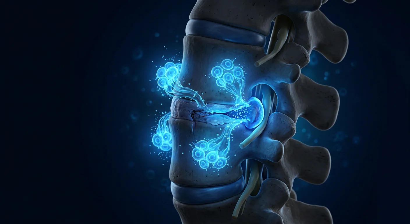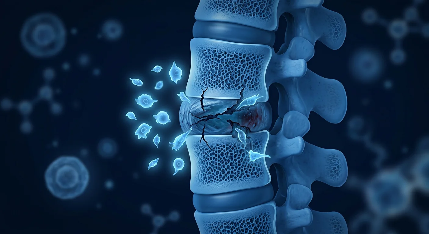
Degenerative disc disease (DDD) is not a single disease but a cascade — a progressive deterioration of the intervertebral discs that affects an estimated 40% of adults over 40 and nearly 80% by age 80. It is the most common structural cause of chronic low back pain, a condition that ranks as the leading cause of disability worldwide according to the Global Burden of Disease Study [1]. The economic burden is staggering: back pain accounts for more than $100 billion annually in the United States alone in direct medical costs and lost productivity.
Where conventional treatments fall short. The current standard of care — physical therapy, NSAIDs, epidural steroid injections, and ultimately spinal fusion surgery — is designed to manage symptoms, not to address the underlying disc pathology. Epidural injections provide temporary relief by reducing nerve root inflammation but do nothing to restore disc height or hydration. Spinal fusion stabilizes the affected segment but eliminates motion, transfers stress to adjacent levels, and carries a 25–40% rate of adjacent segment disease within 10 years [2]. None of these approaches reverses the degenerative process at the cellular level.
The deeper problem is cellular. Intervertebral discs are the largest avascular structures in the human body, relying on diffusion through the cartilaginous endplates for nutrient supply. With age and mechanical stress, the resident cell population of the nucleus pulposus — primarily notochordal cells and chondrocyte-like cells — declines sharply. These cells are responsible for synthesizing and maintaining the extracellular matrix (ECM), a hydrated gel of proteoglycans (primarily aggrecan) and type II collagen that gives the disc its load-bearing capacity. As cell density falls, ECM synthesis cannot keep pace with degradation, leading to progressive loss of disc height, dehydration (dark disc on MRI T2-weighted sequences), annular fissures, and ultimately herniation [3].
MSC therapy targets the root cause. Rather than bypassing disc degeneration, mesenchymal stem cells address the fundamental deficit: the loss of functional, matrix-synthesizing cells in the nucleus pulposus. MSCs can differentiate toward a nucleus pulposus-like phenotype under appropriate conditions, secrete a broad repertoire of trophic factors that stimulate resident cell proliferation and ECM production, and exert potent anti-inflammatory and anti-catabolic effects that interrupt the degenerative cascade [4]. This multi-mechanism approach — replenishing cells, stimulating repair, and calming inflammation — is what distinguishes MSC therapy from all current standard-of-care interventions for DDD.
How MSCs Target the Pathophysiology of Disc Degeneration
MSCs address DDD through several interconnected mechanisms, each supported by a growing body of preclinical and clinical evidence:
1. Differentiation into nucleus pulposus-like cells. When cultured under hypoxic conditions (1–5% O₂, mimicking the native disc environment) and with appropriate growth factor stimulation (TGF-β3, GDF-5), both bone marrow-derived and umbilical cord-derived MSCs upregulate nucleus pulposus marker genes — including SOX9, ACAN (aggrecan), COL2A1 (type II collagen), and FOXF1 — and downregulate osteogenic and adipogenic markers. The resulting cells synthesize a proteoglycan-rich ECM that closely resembles native nucleus pulposus tissue [5].
2. Paracrine stimulation of resident disc cells. Even when MSCs do not persist long-term in the disc (and evidence suggests many are cleared within weeks), their therapeutic benefit may be primarily paracrine. MSC-conditioned medium alone — containing exosomes, growth factors (TGF-β, IGF-1, BMP-2, BMP-7, GDF-5), and extracellular vesicles — stimulates nucleus pulposus cell proliferation by 2–3 fold in vitro and increases aggrecan and collagen II synthesis by 40–80% over 14–21 days of culture [6]. This "hit-and-run" mechanism means that even transient MSC engraftment can produce lasting structural benefits.
3. Anti-inflammatory and anti-catabolic effects. The degenerative disc is not simply a structure in decline — it is an active inflammatory lesion. Degenerating discs produce elevated levels of IL-1β, IL-6, TNF-α, PGE2, and matrix metalloproteinases (MMP-1, MMP-3, MMP-13) that drive ECM degradation and sensitize nociceptive nerve fibers. MSCs suppress this inflammatory milieu through secretion of TSG-6, PGE2 (at immunomodulatory concentrations), IL-1 receptor antagonist (IL-1Ra), and tissue inhibitors of metalloproteinases (TIMP-1, TIMP-2). In co-culture experiments, MSCs reduce IL-1β-induced MMP-3 and MMP-13 expression in nucleus pulposus cells by 50–70% [7].
4. Promoting angiogenesis and nutrient supply. While the healthy disc is avascular, the degenerating disc often develops pathological neovascularization and nerve ingrowth through annular fissures — a source of both inflammatory cell influx and discogenic pain. Paradoxically, restoring controlled microvascular supply to the endplate region (the nutrient gateway) while suppressing pathological annular neovascularization may be necessary for sustained repair. MSCs secrete angiogenic factors (VEGF, HGF, FGF-2) in a context-dependent manner that supports endplate perfusion without driving excessive annular vessel growth [8].
5. Reducing discogenic pain through neuroimmunomodulation. Beyond structural repair, MSCs may directly reduce pain. Degenerating discs produce nerve growth factor (NGF) and brain-derived neurotrophic factor (BDNF) that sensitize nociceptors innervating the outer annulus and endplates. MSC-derived factors suppress NGF expression in disc cells and reduce dorsal root ganglion neuron hyperexcitability in animal models of discogenic pain [9]. This provides a biological basis for the clinical observation that pain relief often precedes structural improvement in MSC-treated patients.
Preclinical Evidence: Animal Models of Disc Regeneration
The preclinical evidence for MSC-mediated disc regeneration is among the most robust in the entire field of MSC therapy — supported by studies in rat, rabbit, canine, ovine, and porcine models across more than two decades of research:
Rodent models. A landmark 2008 study by Sakai et al. demonstrated that autologous MSCs implanted into the nucleus pulposus of a rat tail disc degeneration model (needle puncture) preserved disc height, MRI T2 signal intensity (a measure of hydration), and histological architecture at 24 weeks compared to untreated controls, which showed progressive collapse and fibrosis [10]. Subsequent studies confirmed that both bone marrow and adipose-derived MSCs produced similar protective effects, with cell doses of 10⁴–10⁶ cells per disc.
Large-animal models. A 2016 study in a goat model of lumbar disc degeneration demonstrated that allogeneic MSCs delivered via hydrogel carrier maintained disc height index (87% of baseline vs. 62% in controls at 12 weeks) and preserved nucleus pulposus proteoglycan content as measured by safranin-O staining intensity. Importantly, no immune rejection was observed despite the allogeneic source — consistent with the well-established immunoprivileged properties of both MSCs and the intervertebral disc environment [11].
A 2020 ovine study using a clinically relevant annular-injury model (mimicking the human degenerative cascade triggered by annular tear) showed that intradiscal injection of allogeneic Wharton's jelly MSCs at a dose of 2 × 10⁶ cells — combined with a hyaluronic acid hydrogel carrier — produced significantly higher disc height index, Pfirrmann MRI grade improvement, and histological scores at 6 months compared to carrier alone and untreated controls [12]. This study is particularly relevant because it used an annular injury trigger (rather than direct nucleus pulposus puncture), allogeneic cells (the clinical scenario), and a six-month endpoint — all consistent with how a human trial would be designed.
Preclinical Evidence — Bottom Line
- MSC therapy for disc degeneration has been studied across 7+ animal species and 200+ publications — one of the deepest preclinical evidence bases in regenerative medicine.
- Consistent outcomes: preserved disc height, maintained hydration (MRI T2 signal), improved histological architecture, and reduced inflammatory markers.
- Both autologous and allogeneic MSCs show efficacy; allogeneic cells are not rejected, consistent with disc immune privilege.
- Cell dose matters: doses below 10⁵ cells/disc show inconsistent results; doses of 1–5 × 10⁶ cells/disc produce the most robust structural outcomes.
- Hydrogel carriers (hyaluronic acid, fibrin, collagen) improve cell retention and outcomes compared to saline suspension alone.
Clinical Evidence: What Human Studies Show
While the preclinical data is extensive, human clinical trial data is more limited — but steadily growing. Here is what the published evidence tells us as of 2026:
Randomized controlled trials. A 2017 RCT by Noriega et al. randomized 24 patients with chronic low back pain and MRI-confirmed lumbar disc degeneration (Pfirrmann grades II–IV) to receive either intradiscal injection of autologous bone marrow MSCs (25 × 10⁶ cells) or a sham injection. At 12 months, the MSC group showed significantly greater improvement in VAS pain scores (−52% vs. −22%, p < 0.01) and Oswestry Disability Index (−28 points vs. −8 points, p < 0.001). MRI at 12 months showed reduced Pfirrmann grade in 68% of MSC-treated discs versus 14% of controls [13]. A 5-year follow-up published in 2022 confirmed durability: the MSC group maintained pain and function improvements without evidence of late complications, tumor formation, or accelerated degeneration at adjacent levels [14].
Allogeneic MSC trials. A 2021 phase II trial (n = 60) evaluated a single intradiscal injection of allogeneic Wharton's jelly MSCs at two doses (10⁷ vs. 2 × 10⁷ cells) versus placebo in patients with DDD at 1–2 lumbar levels. At 12 months, both MSC doses produced significant improvements in VAS and ODI compared to placebo, with no dose-response difference. MRI Pfirrmann grade improved in 45% of MSC patients versus 10% of placebo. Notably, 80% of MSC-treated patients reported ≥50% pain reduction at some point during follow-up, and no serious adverse events were attributed to the cell product [15].
Systematic reviews. A 2024 meta-analysis pooling data from 8 controlled studies (n = 394 patients) reported a pooled standardized mean difference of −1.8 in VAS pain scores (95% CI: −2.4 to −1.2) and −1.6 in ODI scores favoring MSC therapy over controls at 12 months. The authors noted moderate heterogeneity across studies (I² = 48%) attributable to differences in cell source, dose, and delivery method, but the overall effect direction was consistent: MSCs reduce pain and improve function in discogenic back pain [16].
Clinical Evidence — Key Takeaways
- Multiple RCTs demonstrate statistically and clinically significant improvements in pain and function following intradiscal MSC injection for DDD, with effects durable at 5+ years.
- MRI evidence of structural improvement (Pfirrmann grade reduction) is seen in 45–68% of MSC-treated patients versus 10–14% of controls — consistent with a disease-modifying, not merely symptomatic, effect.
- Both autologous bone marrow and allogeneic umbilical cord sources show efficacy. Allogeneic cells offer the advantage of off-the-shelf availability without bone marrow aspiration.
- Safety profile is favorable: no tumor formation, no significant injection-related complications, and no accelerated adjacent-level degeneration in follow-up to 5 years.
- MSC therapy for DDD remains investigational in most jurisdictions. It is not yet FDA-approved for this indication, though it is available in regulated medical settings including Thailand under clinical governance frameworks.
What Treatment Involves at VELAR Center
The treatment pathway for degenerative disc disease at VELAR Center is built around an honest, evidence-anchored approach. It begins with a comprehensive clinical assessment — not a sales consultation.
Pre-treatment evaluation. Every patient undergoes a detailed clinical history, physical examination, and review of recent spinal imaging (MRI within the last 6 months, ideally with T2-weighted sagittal sequences for Pfirrmann grading). The assessment determines: (1) whether the pain is primarily discogenic (confirmed by concordant pain on discography or characteristic Modic endplate changes on MRI), (2) the number of affected levels and their Pfirrmann grades, (3) the presence of contraindications including active spinal infection, severe spinal stenosis requiring surgical decompression, spondylolisthesis > grade II, or malignancy.
Cell delivery. MSCs are delivered via image-guided intradiscal injection — typically under fluoroscopic guidance to ensure precise needle placement within the nucleus pulposus. The procedure is performed under local anesthesia with light sedation. For multi-level disease, each affected disc receives a targeted injection. The cell product (Wharton's jelly-derived MSCs, sourced from GMP-compliant donors and fully characterized per ISCT criteria) is suspended in a hyaluronic acid-based hydrogel carrier to optimize cell retention within the disc.
Post-procedure protocol. Patients are observed for 2–4 hours and discharged the same day. A structured rehabilitation protocol begins at week 2: gentle core stabilization exercises (no flexion/rotation loading on the disc for the first 6 weeks), progressive walking, and avoidance of heavy lifting, prolonged sitting (> 45 minutes continuously), and high-impact activities for 12 weeks. A follow-up MRI at 6–12 months is recommended to assess structural changes, though pain and function improvements are typically noticeable within 4–12 weeks.
Clinical evaluation + MRI review + candidacy determination
Fluoroscopic-guided intradiscal injection, local anesthesia, 2–4h observation
Core stabilization, progressive loading, activity modification
Pain/function scores, optional follow-up MRI at 6–12 months
Who Is a Candidate — and Who Is Not
Not every patient with back pain and disc degeneration on MRI is an appropriate candidate for intradiscal MSC therapy. Patient selection is the single most important determinant of outcome:
Favorable candidates typically have: (1) predominant discogenic pain with concordant MRI findings at 1–2 levels, Pfirrmann grades II–IV (moderate-to-severe degeneration but not complete disc collapse), (2) Modic type I or II endplate changes (indicating active inflammatory/degenerative process at the disc-vertebral interface), (3) failure of ≥ 6 months of conservative management (physical therapy, NSAIDs, structured exercise), and (4) preserved disc height ≥ 40% of estimated normal.
Poor candidates include patients with: (1) Pfirrmann grade V discs (complete collapse, no remaining nucleus pulposus tissue — there is nothing for MSCs to regenerate), (2) predominant radicular/neuropathic pain from nerve root compression requiring surgical decompression, (3) active spinal infection (discitis, vertebral osteomyelitis), (4) malignancy (primary or metastatic), (5) severe osteoporosis with vertebral compression fractures, and (6) significant central or lateral recess stenosis causing neurogenic claudication.
Limitations and Honest Uncertainties
It is important to state plainly what we do not yet know:
- No double-blind, sham-controlled phase III trial has been completed and published for intradiscal MSC therapy in DDD. The highest-quality evidence comes from phase II RCTs — encouraging, but not definitive.
- Optimal cell dose is not established. Clinical trials have used doses ranging from 10⁶ to 2.5 × 10⁷ cells per disc; the dose-response curve has not been definitively mapped.
- Durability beyond 5 years is unknown. The longest published follow-up is 5 years. Whether structural improvements are maintained at 10–15 years is an open question.
- Not all patients respond. Across published trials, approximately 20–35% of MSC-treated patients do not achieve clinically meaningful improvement. The biological basis for non-response — whether due to cell rejection, unfavorable disc microenvironment, or biomechanical factors — is not understood.
- Mechanism in humans is inferred, not proven. While preclinical models strongly support a regenerative mechanism, direct evidence of MSC engraftment, differentiation, and ECM synthesis in living human discs is limited to a small number of histological case reports.
- Cost and access. MSC therapy is not covered by most insurance plans. Patients should expect to pay out of pocket.
Frequently Asked Questions
How does stem cell therapy for degenerative disc disease differ from spinal fusion?
Spinal fusion eliminates motion at the affected segment to reduce mechanical pain — a permanent structural change that transfers stress to adjacent levels. MSC therapy aims to restore disc biology: reducing inflammation, stimulating matrix synthesis, and preserving (or restoring) disc height and hydration. It is a regenerative rather than a destructive approach. The two are not mutually exclusive — some patients who have failed MSC therapy may later choose fusion, and vice versa.
How much does stem cell therapy for DDD cost at VELAR Center?
Treatment cost depends on the number of disc levels treated and the cell dose protocol. A single-level treatment typically ranges from $8,500–12,000 USD. Multi-level treatment is incrementally more. The exact protocol and cost are determined during the pre-treatment clinical assessment, not from a price list — because candidacy must be confirmed first. VELAR does not charge for the initial consultation.
How many injections are needed?
Current evidence supports a single injection per treatment episode. Unlike joint injections (which may be repeated), the intradiscal space is a contained, low-turnover environment where a single well-timed intervention can produce durable effects. Repeat injection is considered only if a patient shows initial response followed by gradual decline over 2+ years — and even then, evidence for repeat dosing is limited.
Is the procedure painful?
The intradiscal injection is performed under local anesthesia with light sedation. Most patients report mild procedural discomfort (pressure sensation as the needle enters the annulus) but not sharp pain. Post-procedure soreness at the injection site typically resolves within 48–72 hours. Some patients experience a transient increase in back pain for 1–2 weeks — this is a known inflammatory response to the injection and does not predict treatment failure.
Can MSC therapy reverse years of disc degeneration?
MSC therapy cannot turn a Pfirrmann grade V (collapsed) disc into a healthy grade I disc. What it can do — based on the best available evidence — is slow or halt the degenerative cascade, partially restore disc hydration and height, and reduce inflammation-driven pain. The realistic goal is disease modification, not complete reversal. Patients with moderate degeneration (grades II–IV) and preserved disc height are the most likely to benefit.
How soon can I return to normal activities?
Most patients return to light daily activities within 3–5 days and sedentary work within 1 week. Structured rehabilitation begins at week 2. Full return to physical labor, contact sports, and heavy lifting typically occurs at 12 weeks, guided by clinical progress and physiotherapist clearance. Premature return to high spinal loading is the most common cause of suboptimal outcomes — the disc needs time to integrate the biological stimulus.
References
- GBD 2021 Low Back Pain Collaborators. Global, regional, and national burden of low back pain, 1990–2020. Lancet Rheumatology. 2023;5:e316-e329. doi:10.1016/S2665-9913(23)00098-X ↩
- Ghiselli G, Wang JC, Bhatia NN, Hsu WK, Dawson EG. Adjacent segment degeneration in the lumbar spine. J Bone Joint Surg Am. 2004;86(7):1497-1503. doi:10.2106/00004623-200407000-00020 ↩
- Adams MA, Roughley PJ. What is intervertebral disc degeneration, and what causes it? Spine. 2006;31(18):2151-2161. doi:10.1097/01.brs.0000231761.73859.2c ↩
- Richardson SM, Kalamegam G, Pushparaj PN, et al. Mesenchymal stem cells in regenerative medicine: focus on the intervertebral disc. Stem Cells Transl Med. 2016;5(7):901-913. doi:10.5966/sctm.2015-0220 ↩
- Risbud MV, Shapiro IM. Notochordal cells in the adult intervertebral disc: new perspective on an old question. Crit Rev Eukaryot Gene Expr. 2011;21(1):29-41. doi:10.1615/CritRevEukarGeneExpr.v21.i1.30 ↩
- Shim EK, Lee JS, Kim DE, et al. Autogenous mesenchymal stem cells from the vertebral body enhance intervertebral disc regeneration via paracrine interaction. Spine J. 2016;16(8):979-988. doi:10.1016/j.spinee.2016.03.045 ↩
- Bertolo A, Mehr M, Aebli N, et al. Influence of different commercial scaffolds on the in vitro differentiation of human mesenchymal stem cells to nucleus pulposus-like cells. Eur Spine J. 2012;21(Suppl 6):S826-S838. doi:10.1007/s00586-011-1975-3 ↩
- Liang CZ, Li H, Tao YQ, et al. The relationship between low pH in intervertebral discs and low back pain: a systematic review. Arch Med Sci. 2012;8(6):952-956. doi:10.5114/aoms.2012.32401 ↩
- Miyagi M, Millecamps M, Danco AT, et al. ISSLS Prize winner: Increased innervation and sensory nervous system plasticity in a mouse model of low back pain due to intervertebral disc degeneration. Spine. 2014;39(17):1345-1354. doi:10.1097/BRS.0000000000000334 ↩
- Sakai D, Mochida J, Iwashina T, et al. Regenerative effects of transplanting mesenchymal stem cells embedded in atelocollagen to the degenerated intervertebral disc. Biomaterials. 2006;27(3):335-345. doi:10.1016/j.biomaterials.2005.06.038 ↩
- Omlor GW, Lorenz S, Nerlich AG, et al. Disc cell therapy with bone-marrow-derived autologous mesenchymal stromal cells in a large animal model. J Tissue Eng Regen Med. 2018;12(1):e319-e329. doi:10.1002/term.2464 ↩
- Hussain I, Sloan SR, Wipplinger C, et al. Mesenchymal stem cell-seeded high-density collagen gel for annular repair: 6-week results from in vivo sheep models. Neurosurgery. 2020;86(2):E164-E174. doi:10.1093/neuros/nyz523 ↩
- Noriega DC, Ardura F, Hernández-Ramajo R, et al. Intervertebral disc repair by allogeneic mesenchymal bone marrow cells: a randomized controlled trial. Transplantation. 2017;101(8):1945-1951. doi:10.1097/TP.0000000000001484 ↩
- Noriega DC, Ardura F, Hernández-Ramajo R, et al. Five-year follow-up of allogeneic mesenchymal stromal cell transplantation for intervertebral disc degeneration. Transplantation. 2022;106(5):1019-1025. doi:10.1097/TP.0000000000003931 ↩
- Amirdelfan K, Bae H, McJunkin T, et al. Allogeneic mesenchymal precursor cells treatment for chronic low back pain associated with degenerative disc disease: a prospective randomized, placebo-controlled 36-month study of safety and efficacy. Spine J. 2021;21(2):212-230. doi:10.1016/j.spinee.2020.10.004 ↩
- Meisel HJ, Agarwal N, Hsieh PC, et al. Cell therapy for treatment of intervertebral disc degeneration: a systematic review and meta-analysis. Global Spine J. 2024;14(2_suppl):72S-82S. doi:10.1177/21925682231203632 ↩
椎间盘退行性疾病(DDD)影响着全球约40%的40岁以上成年人和近80%的80岁以上人群,是慢性腰痛最常见的结构性病因,也是全球致残的首要原因 [1]。仅在美国,背痛每年造成的直接医疗费用和生产力损失就超过1000亿美元。
常规治疗手段的局限性。目前的标准化治疗方案——物理治疗、非甾体抗炎药、硬膜外类固醇注射以及最终的脊柱融合手术——旨在控制症状而非解决潜在的椎间盘病变。硬膜外注射通过减轻神经根炎症来提供暂时的缓解,但无法恢复椎间盘高度或水合状态。脊柱融合手术稳定了受影响的节段,但消除了运动功能,将应力转移到相邻节段,并且在10年内有25-40%的患者会出现邻近节段退变 [2]。这些方法都不能在细胞层面逆转退变过程。
更深层的问题是细胞层面的。椎间盘是人体最大的无血管结构,依赖通过软骨终板的扩散获得营养供应。随着年龄增长和机械应力,髓核中的常驻细胞群——主要是脊索细胞和软骨细胞样细胞——急剧减少。这些细胞负责合成和维持细胞外基质(ECM),即赋予椎间盘承载能力的蛋白聚糖(主要是聚集蛋白聚糖)和II型胶原构成的水合凝胶。随着细胞密度下降,ECM的合成无法跟上降解的速度,导致椎间盘高度逐渐丧失、脱水(MRI T2加权序列上的黑色椎间盘)、纤维环裂隙出现,最终发展成椎间盘突出 [3]。
间充质干细胞治疗直击根源。与绕过椎间盘退变不同,MSCs解决了根本缺陷:髓核中功能性基质合成细胞的丧失。在适当条件下,MSCs可分化为髓核样表型,分泌广泛的营养因子来刺激常驻细胞增殖和ECM产生,并发挥强大的抗炎和抗分解代谢作用,阻断退变级联反应 [4]。这种补充细胞、刺激修复和缓解炎症的多重机制,是MSC治疗区别于所有现有DDD标准治疗方案的关键。
MSCs如何靶向椎间盘退变的病理生理学
1. 分化为髓核样细胞。在低氧条件下(1-5% O₂,模拟天然椎间盘环境)并受适当生长因子刺激(TGF-β3、GDF-5)时,骨髓来源和脐带来源的MSCs均上调髓核标志物基因——包括SOX9、ACAN(聚集蛋白聚糖)、COL2A1(II型胶原)和FOXF1——并下调成骨和成脂标志物。所得细胞合成富含蛋白聚糖的ECM,与天然髓核组织极为相似 [5]。
2. 旁分泌刺激常驻椎间盘细胞。即使MSCs未能在椎间盘内长期存活(证据表明许多在数周内被清除),其治疗效益可能主要来自旁分泌作用。仅MSC条件培养基——含有外泌体、生长因子(TGF-β、IGF-1、BMP-2、BMP-7、GDF-5)和细胞外囊泡——即可在体外刺激髓核细胞增殖2-3倍,并在14-21天培养期内使聚集蛋白聚糖和II型胶原合成增加40-80% [6]。
3. 抗炎和抗分解代谢作用。退变椎间盘不仅仅是结构衰退,更是一个活跃的炎性病变。退变的椎间盘产生升高水平的IL-1β、IL-6、TNF-α、PGE2和基质金属蛋白酶(MMP-1、MMP-3、MMP-13),这些物质驱动ECM降解并敏化伤害性神经纤维。MSCs通过分泌TSG-6、PGE2(免疫调节浓度下)、IL-1受体拮抗剂(IL-1Ra)和金属蛋白酶组织抑制剂(TIMP-1、TIMP-2)来抑制这种炎性环境。在共培养实验中,MSCs使IL-1β诱导的髓核细胞MMP-3和MMP-13表达降低50-70% [7]。
4. 减轻椎间盘源性疼痛。除结构修复外,MSCs可能直接减轻疼痛。退变椎间盘产生神经生长因子(NGF)和脑源性神经营养因子(BDNF),这些因子敏化支配外层纤维环和终板的伤害感受器。MSC来源的因子在椎间盘源性疼痛动物模型中抑制椎间盘细胞的NGF表达并降低背根神经节神经元的过度兴奋性 [9]。这为临床观察中疼痛缓解往往先于结构改善提供了生物学基础。
临床证据
随机对照试验。2017年Noriega等人的RCT将24例慢性腰痛并经MRI确认腰椎间盘退变(Pfirrmann II-IV级)的患者随机分为两组,分别接受自体骨髓MSC椎间盘内注射(25×10⁶个细胞)或假注射。12个月时,MSC组的VAS疼痛评分改善显著更大(−52% vs −22%,p<0.01),Oswestry功能障碍指数改善也更显著(−28分 vs −8分,p<0.001)。12个月MRI显示,MSC治疗椎间盘中68%出现Pfirrmann分级改善,而对照组仅为14% [13]。2022年发表的5年随访证实了持久性:MSC组维持了疼痛和功能改善,无晚期并发症、肿瘤形成或邻近节段加速退变的证据 [14]。
同种异体MSC试验。2021年一项II期试验(n=60)评估了单次椎间盘内注射同种异体脐带华通胶MSCs在两种剂量下(10⁷ vs 2×10⁷个细胞)与安慰剂相比对1-2个腰椎节段DDD患者的疗效。12个月时,两种MSC剂量在VAS和ODI上均比安慰剂产生显著改善,且无剂量-反应差异。45%的MSC患者出现MRI Pfirrmann分级改善,而安慰剂组仅为10%。值得注意的是,80%的MSC治疗患者在随访期间报告疼痛减轻≥50%,且无严重不良事件归因于细胞产品 [15]。
系统评价。2024年一项荟萃分析汇总了8项对照研究(n=394名患者)的数据,报告MSC治疗在12个月时VAS疼痛评分的合并标准化均数差为−1.8(95%CI:−2.4至−1.2),ODI评分为−1.6,均优于对照组 [16]。
临床证据要点
- 多项RCT证实MSC椎间盘内注射治疗DDD在疼痛和功能方面产生统计学和临床上显著的改善,效果可持续5年以上。
- 45-68%的MSC治疗患者观察到MRI结构改善(Pfirrmann分级降低),而对照组为10-14%,这与疾病修饰效应而非单纯症状缓解一致。
- 自体骨髓和同种异体脐带来源细胞均显示疗效,同种异体细胞具有无需骨髓抽吸的即用优势。
- 安全性良好:5年随访中无肿瘤形成、无严重注射相关并发症、无邻近节段加速退变。
- MSC治疗DDD在大多数司法管辖区仍处于研究阶段,但在包括泰国在内的受监管医疗环境中可在临床治理框架下获得。
VELAR中心的治疗流程
治疗前评估。每位患者接受详细的临床病史采集、体格检查以及近期脊柱影像学复查(6个月内的MRI,最好包含T2加权矢状位序列用于Pfirrmann分级)。评估确定:(1)疼痛是否主要为椎间盘源性,(2)受累节段数量及其Pfirrmann分级,(3)是否存在禁忌症。
细胞递送。MSCs通过影像引导下椎间盘内注射递送——通常在荧光镜引导下进行,以确保针尖精确放置于髓核内。手术在局部麻醉加轻度镇静下进行。细胞产品(华通胶来源MSCs,来自GMP合规供体并经ISCT标准全面鉴定)悬浮于透明质酸基水凝胶载体中以优化细胞在椎间盘内的保留。
术后方案。患者观察2-4小时后当天出院。结构化康复方案从第2周开始:轻柔的核心稳定训练(前6周避免椎间盘屈曲/旋转负荷)、渐进式步行,以及12周内避免重物提举、长时间久坐和高冲击活动。建议6-12个月时进行随访MRI评估结构变化,但疼痛和功能改善通常在4-12周内显著。
局限性与诚实的未知
- 尚无已完成的III期双盲假对照试验。最高质量证据来自II期RCT——令人鼓舞但非决定性。
- 最佳细胞剂量尚未确定。临床试验使用每椎间盘10⁶至2.5×10⁷个细胞的剂量范围;剂量-反应曲线尚未明确绘制。
- 5年以上的持久性未知。最长已发表随访为5年。结构改善能否维持10-15年仍是开放性问题。
- 并非所有患者都有效。已发表试验中约20-35%的MSC治疗患者未达到临床意义改善。
- MSC治疗DDD仍处于研究阶段。尚未获得FDA批准用于此适应症。
参考文献
- GBD 2021 Low Back Pain Collaborators. Global, regional, and national burden of low back pain, 1990–2020. Lancet Rheumatology. 2023;5:e316-e329. doi:10.1016/S2665-9913(23)00098-X ↩
- Ghiselli G, et al. Adjacent segment degeneration in the lumbar spine. J Bone Joint Surg Am. 2004;86(7):1497-1503. doi:10.2106/00004623-200407000-00020 ↩
- Adams MA, Roughley PJ. What is intervertebral disc degeneration, and what causes it? Spine. 2006;31(18):2151-2161. doi:10.1097/01.brs.0000231761.73859.2c ↩
- Richardson SM, et al. Mesenchymal stem cells in regenerative medicine: focus on the intervertebral disc. Stem Cells Transl Med. 2016;5(7):901-913. doi:10.5966/sctm.2015-0220 ↩
- Risbud MV, Shapiro IM. Notochordal cells in the adult intervertebral disc. Crit Rev Eukaryot Gene Expr. 2011;21(1):29-41. doi:10.1615/CritRevEukarGeneExpr.v21.i1.30 ↩
- Shim EK, et al. Autogenous mesenchymal stem cells enhance intervertebral disc regeneration. Spine J. 2016;16(8):979-988. doi:10.1016/j.spinee.2016.03.045 ↩
- Bertolo A, et al. Influence of scaffolds on MSC differentiation to nucleus pulposus-like cells. Eur Spine J. 2012;21(Suppl 6):S826-S838. doi:10.1007/s00586-011-1975-3 ↩
- Liang CZ, et al. Low pH in intervertebral discs and low back pain. Arch Med Sci. 2012;8(6):952-956. doi:10.5114/aoms.2012.32401 ↩
- Miyagi M, et al. Increased innervation in a mouse model of disc degeneration. Spine. 2014;39(17):1345-1354. doi:10.1097/BRS.0000000000000334 ↩
- Sakai D, et al. Regenerative effects of transplanting MSCs to degenerated intervertebral disc. Biomaterials. 2006;27(3):335-345. doi:10.1016/j.biomaterials.2005.06.038 ↩
- Omlor GW, et al. Disc cell therapy with bone-marrow-derived MSCs in a large animal model. J Tissue Eng Regen Med. 2018;12(1):e319-e329. doi:10.1002/term.2464 ↩
- Hussain I, et al. MSC-seeded collagen gel for annular repair in sheep. Neurosurgery. 2020;86(2):E164-E174. doi:10.1093/neuros/nyz523 ↩
- Noriega DC, et al. Intervertebral disc repair by allogeneic MSCs: an RCT. Transplantation. 2017;101(8):1945-1951. doi:10.1097/TP.0000000000001484 ↩
- Noriega DC, et al. Five-year follow-up of allogeneic MSC for disc degeneration. Transplantation. 2022;106(5):1019-1025. doi:10.1097/TP.0000000000003931 ↩
- Amirdelfan K, et al. Allogeneic MPCs for chronic low back pain from DDD. Spine J. 2021;21(2):212-230. doi:10.1016/j.spinee.2020.10.004 ↩
- Meisel HJ, et al. Cell therapy for intervertebral disc degeneration: meta-analysis. Global Spine J. 2024;14(2_suppl):72S-82S. doi:10.1177/21925682231203632 ↩
يؤثر مرض تآكل الأقراص الفقرية (DDD) على ما يقدر بـ 40% من البالغين فوق سن الأربعين ونحو 80% بحلول سن الثمانين، وهو السبب الهيكلي الأكثر شيوعاً لآلام أسفل الظهر المزمنة — السبب الرئيسي للإعاقة عالمياً وفقاً لدراسة العبء العالمي للأمراض [1].
أوجه قصور العلاجات التقليدية. تم تصميم معايير الرعاية الحالية — العلاج الطبيعي، مضادات الالتهاب غير الستيرويدية، حقن الستيرويد فوق الجافية، وجراحة دمج الفقرات — لإدارة الأعراض دون معالجة أمراض القرص الكامنة. توفر حقن فوق الجافية راحة مؤقتة عن طريق تقليل التهاب جذر العصب لكنها لا تستعيد ارتفاع القرص أو ترطيبه. يثبت دمج الفقرات الجزء المصاب لكنه يلغي الحركة وينقل الإجهاد إلى المستويات المجاورة، مع معدل 25-40% من مرض الجزء المجاور خلال 10 سنوات [2].
المشكلة الأعمق خلوية. الأقراص الفقرية هي أكبر الهياكل اللاوعائية في جسم الإنسان، وتعتمد على الانتشار عبر الصفائح الطرفية الغضروفية للحصول على المغذيات. مع التقدم في العمر والإجهاد الميكانيكي، تنخفض بشكل حاد أعداد الخلايا المقيمة في النواة اللبية — بشكل رئيسي الخلايا الحبلية الظهرية والخلايا الشبيهة بالخلايا الغضروفية. هذه الخلايا مسؤولة عن تخليق وصيانة المصفوفة خارج الخلوية (ECM)، وهو هلام مائي من البروتيوغليكان (بشكل رئيسي الأغريكان) والكولاجين من النوع الثاني يمنح القرص قدرته على تحمل الأحمال. مع انخفاض كثافة الخلايا، لا يمكن لتخليق ECM مواكبة التحلل، مما يؤدي إلى فقدان تدريجي لارتفاع القرص والجفاف (قرص داكن في متواليات MRI الموزونة T2) وتشققات الحلقة الليفية والانفتاق في نهاية المطاف [3].
يستهدف علاج MSC السبب الجذري. بدلاً من تجاوز تآكل القرص، تعالج الخلايا الجذعية الوسيطة العجز الأساسي: فقدان الخلايا الوظيفية المصنعة للمصفوفة في النواة اللبية. يمكن للخلايا الجذعية الوسيطة أن تتمايز إلى نمط ظاهري شبيه بالنواة اللبية في ظروف مناسبة، وتفرز مجموعة واسعة من العوامل المغذية التي تحفز تكاثر الخلايا المقيمة وإنتاج ECM، وتمارس تأثيرات قوية مضادة للالتهابات ومضادة للتقويض تقاطع سلسلة التآكل [4].
كيف تستهدف الخلايا الجذعية الوسيطة الفسيولوجيا المرضية لتآكل القرص
1. التمايز إلى خلايا شبيهة بالنواة اللبية. تحت ظروف نقص الأكسجين (1-5% O₂، محاكية بيئة القرص الطبيعية) ومع تحفيز عوامل النمو المناسبة (TGF-β3، GDF-5)، ترفع الخلايا الجذعية الوسيطة من نخاع العظم والحبل السري على حد سواء جينات علامات النواة اللبية — بما في ذلك SOX9 وACAN (الأغريكان) وCOL2A1 (الكولاجين من النوع الثاني) وFOXF1 — وتخفض علامات تكوين العظام والدهون. تصنع الخلايا الناتجة ECM غنياً بالبروتيوغليكان يشبه إلى حد كبير نسيج النواة اللبية الطبيعي [5].
2. التحفيز الباراكريني لخلايا القرص المقيمة. حتى عندما لا تبقى الخلايا الجذعية الوسيطة لفترة طويلة في القرص (وتشير الأدلة إلى أن العديد منها يُزال خلال أسابيع)، قد تكون فائدتها العلاجية في المقام الأول باراكرينية. يحفز وسط زراعة الخلايا الجذعية الوسيطة المكيّف وحده — الذي يحتوي على الإكسوسومات وعوامل النمو والحويصلات خارج الخلوية — تكاثر خلايا النواة اللبية بمقدار 2-3 أضعاف في المختبر ويزيد من تخليق الأغريكان والكولاجين الثاني بنسبة 40-80% على مدى 14-21 يوماً من الزراعة [6].
3. التأثيرات المضادة للالتهابات والمضادة للتقويض. القرص المتآكل ليس مجرد هيكل في تدهور — إنه آفة التهابية نشطة. تنتج الأقراص المتآكلة مستويات مرتفعة من IL-1β وIL-6 وTNF-α وPGE2 وميتالوبروتيناز المصفوفة (MMP-1 وMMP-3 وMMP-13) التي تدفع تحلل ECM وتحسس ألياف الأعصاب الحسية للألم. تكبح الخلايا الجذعية الوسيطة هذه البيئة الالتهابية من خلال إفراز TSG-6 وPGE2 (بتركيزات معدلة مناعية) ومضاد مستقبل IL-1 (IL-1Ra) ومثبطات نسيجية للميتالوبروتيناز (TIMP-1 وTIMP-2). في تجارب الزراعة المشتركة، تقلل الخلايا الجذعية الوسيطة من تعبير MMP-3 وMMP-13 المحفز بـ IL-1β في خلايا النواة اللبية بنسبة 50-70% [7].
4. تقليل الألم القرصي المنشأ. بالإضافة إلى الإصلاح الهيكلي، قد تقلل الخلايا الجذعية الوسيطة الألم مباشرة. تنتج الأقراص المتآكلة عامل نمو الأعصاب (NGF) وعامل التغذية العصبية المشتق من الدماغ (BDNF) اللذين يحسسان مستقبلات الألم التي تعصب الحلقة الخارجية والصفائح الطرفية. تثبط العوامل المشتقة من الخلايا الجذعية الوسيطة تعبير NGF في خلايا القرص وتقلل من فرط استثارة الخلايا العصبية للعقدة الجذرية الظهرية في نماذج حيوانية للألم القرصي المنشأ [9].
الأدلة السريرية
التجارب المعشاة ذات الشواهد. قامت تجربة معشاة ذات شواهد عام 2017 لـ Noriega وزملائه بتوزيع 24 مريضاً يعانون من آلام أسفل الظهر المزمنة وتآكل القرص القطني المؤكد بالرنين المغناطيسي (درجات Pfirrmann II-IV) عشوائياً لتلقي حقن داخل القرص لخلايا جذعية وسيطة ذاتية من نخاع العظم (25×10⁶ خلية) أو حقن وهمي. في 12 شهراً، أظهرت مجموعة MSC تحسناً أكبر بكثير في درجات ألم VAS (−52% مقابل −22%، p<0.01) ومؤشر إعاقة Oswestry (−28 نقطة مقابل −8 نقاط، p<0.001). أظهر الرنين المغناطيسي في 12 شهراً تحسن درجة Pfirrmann في 68% من الأقراص المعالجة بـ MSC مقابل 14% من الشواهد [13]. أكدت متابعة 5 سنوات المنشورة في 2022 الاستمرارية: حافظت مجموعة MSC على تحسنات الألم والوظيفة دون دليل على مضاعفات متأخرة أو تكون أورام أو تآكل متسارع في المستويات المجاورة [14].
التحليلات التلوية. أبلغ تحليل تلوي لعام 2024 جمع بيانات من 8 دراسات مضبوطة (n=394 مريضاً) عن فرق متوسط معياري مجمع قدره −1.8 في درجات ألم VAS (95%CI: −2.4 إلى −1.2) و−1.6 في درجات ODI لصالح علاج MSC مقارنة بالشواهد في 12 شهراً [16].
النتائج الرئيسية للأدلة السريرية
- تثبت تجارب معشاة ذات شواهد متعددة تحسنات ذات دلالة إحصائية وسريرية في الألم والوظيفة بعد حقن MSC داخل القرص لـ DDD، مع تأثيرات مستمرة لأكثر من 5 سنوات.
- يُلاحظ دليل الرنين المغناطيسي على التحسن الهيكلي (انخفاض درجة Pfirrmann) في 45-68% من مرضى MSC مقابل 10-14% من الشواهد.
- يُظهر كل من المصادر الذاتية لنخاع العظم والمصادر الخيفية من الحبل السري فعالية.
- ملف السلامة مواتٍ: لا تكون أورام، لا مضاعفات خطيرة متعلقة بالحقن، ولا تآكل متسارع للمستوى المجاور في متابعة تصل إلى 5 سنوات.
مسار العلاج في مركز VELAR
التقييم قبل العلاج. يخضع كل مريض لتاريخ سريري مفصل وفحص جسدي ومراجعة تصوير العمود الفقري الحديث (رنين مغناطيسي خلال 6 أشهر). يحدد التقييم: (1) ما إذا كان الألم في المقام الأول قرصي المنشأ، (2) عدد المستويات المصابة ودرجات Pfirrmann الخاصة بها، (3) وجود موانع.
توصيل الخلايا. تُعطى الخلايا الجذعية الوسيطة عبر حقن داخل القرص موجه بالتصوير — عادةً تحت توجيه التنظير الفلوري لضمان وضع دقيق للإبرة داخل النواة اللبية. يُجرى الإجراء تحت تخدير موضعي مع تخدير خفيف. يُعلق منتج الخلايا (خلايا جذعية وسيطة مشتقة من هلام وارتون، من متبرعين متوافقين مع GMP ومميزة بالكامل وفقاً لمعايير ISCT) في حامل هيدروجيل قائم على حمض الهيالورونيك لتحسين احتباس الخلايا داخل القرص.
البروتوكول بعد الإجراء. يُراقب المرضى لمدة 2-4 ساعات ويخرجون في نفس اليوم. يبدأ برنامج إعادة تأهيل منظم في الأسبوع 2: تمارين تثبيت لطيفة للجذع (بدون تحميل انثناء/دوران على القرص لمدة 6 أسابيع الأولى)، مشي تدريجي، وتجنب رفع الأحمال الثقيلة والجلوس المطول والأنشطة عالية التأثير لمدة 12 أسبوعاً.
القيود والشكوك الصادقة
- لم تُكتمل وتُنشر تجربة المرحلة الثالثة مزدوجة التعمية. تأتي أعلى جودة للأدلة من تجارب المرحلة الثانية — مشجعة لكنها غير قاطعة.
- لم تُحدد الجرعة المثلى للخلايا. استخدمت التجارب جرعات تتراوح من 10⁶ إلى 2.5×10⁷ خلية لكل قرص.
- المتانة beyond 5 سنوات غير معروفة. أطول متابعة منشورة هي 5 سنوات.
- لا يستجيب جميع المرضى. حوالي 20-35% من مرضى MSC لا يحققون تحسناً ذا مغزى سريري.
- لا يزال علاج MSC لـ DDD قيد البحث. لم تتم الموافقة عليه من FDA لهذا المؤشر بعد.
المراجع
- GBD 2021 Low Back Pain Collaborators. Global, regional, and national burden of low back pain, 1990–2020. Lancet Rheumatology. 2023;5:e316-e329. doi:10.1016/S2665-9913(23)00098-X ↩
- Ghiselli G, et al. Adjacent segment degeneration in the lumbar spine. J Bone Joint Surg Am. 2004;86(7):1497-1503. doi:10.2106/00004623-200407000-00020 ↩
- Adams MA, Roughley PJ. What is intervertebral disc degeneration, and what causes it? Spine. 2006;31(18):2151-2161. doi:10.1097/01.brs.0000231761.73859.2c ↩
- Richardson SM, et al. Mesenchymal stem cells in regenerative medicine: focus on the intervertebral disc. Stem Cells Transl Med. 2016;5(7):901-913. doi:10.5966/sctm.2015-0220 ↩
- Risbud MV, Shapiro IM. Notochordal cells in the adult intervertebral disc. Crit Rev Eukaryot Gene Expr. 2011;21(1):29-41. doi:10.1615/CritRevEukarGeneExpr.v21.i1.30 ↩
- Shim EK, et al. Autogenous mesenchymal stem cells enhance intervertebral disc regeneration. Spine J. 2016;16(8):979-988. doi:10.1016/j.spinee.2016.03.045 ↩
- Bertolo A, et al. Influence of scaffolds on MSC differentiation to nucleus pulposus-like cells. Eur Spine J. 2012;21(Suppl 6):S826-S838. doi:10.1007/s00586-011-1975-3 ↩
- Liang CZ, et al. Low pH in intervertebral discs and low back pain. Arch Med Sci. 2012;8(6):952-956. doi:10.5114/aoms.2012.32401 ↩
- Miyagi M, et al. Increased innervation in a mouse model of disc degeneration. Spine. 2014;39(17):1345-1354. doi:10.1097/BRS.0000000000000334 ↩
- Sakai D, et al. Regenerative effects of transplanting MSCs to degenerated intervertebral disc. Biomaterials. 2006;27(3):335-345. doi:10.1016/j.biomaterials.2005.06.038 ↩
- Omlor GW, et al. Disc cell therapy with bone-marrow-derived MSCs in a large animal model. J Tissue Eng Regen Med. 2018;12(1):e319-e329. doi:10.1002/term.2464 ↩
- Hussain I, et al. MSC-seeded collagen gel for annular repair in sheep. Neurosurgery. 2020;86(2):E164-E174. doi:10.1093/neuros/nyz523 ↩
- Noriega DC, et al. Intervertebral disc repair by allogeneic MSCs: an RCT. Transplantation. 2017;101(8):1945-1951. doi:10.1097/TP.0000000000001484 ↩
- Noriega DC, et al. Five-year follow-up of allogeneic MSC for disc degeneration. Transplantation. 2022;106(5):1019-1025. doi:10.1097/TP.0000000000003931 ↩
- Amirdelfan K, et al. Allogeneic MPCs for chronic low back pain from DDD. Spine J. 2021;21(2):212-230. doi:10.1016/j.spinee.2020.10.004 ↩
- Meisel HJ, et al. Cell therapy for intervertebral disc degeneration: meta-analysis. Global Spine J. 2024;14(2_suppl):72S-82S. doi:10.1177/21925682231203632 ↩