Deep partial-thickness burns — second-degree burns that extend through the epidermis and well into the dermis — are among the most painful and scarring of all burn injuries. Globally, an estimated 11 million burn injuries require medical attention each year, and deep partial-thickness burns account for a substantial fraction. The standard of care — early excision and split-thickness skin grafting — saves lives and closes wounds, but the resulting skin is often thin, fragile, and cosmetically disfiguring. Worse, the healed wound bed frequently becomes hypertrophic, producing raised, itchy, contracture-prone scars that can limit joint mobility for years. Mesenchymal stem cell (MSC) therapy is being investigated as a regenerative strategy that could accelerate wound closure, improve the quality of healed skin, and reduce the hypertrophic scarring that follows deep burn injury [1].
What Makes Deep Partial-Thickness Burns Different
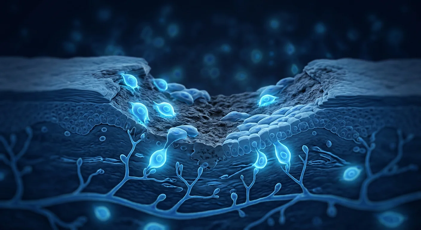
A deep partial-thickness burn destroys the epidermis and penetrates into the reticular dermis — the deeper, collagen-rich layer that gives skin its structural integrity. Unlike superficial partial-thickness burns that heal from residual epidermal appendages within 14 days, deep partial-thickness burns destroy most hair follicles and sweat glands, leaving few keratinocyte reservoirs for spontaneous re-epithelialization. Without surgical intervention, these wounds take 3–5 weeks to close and almost invariably scar [2].
The scarring cascade that follows deep burn injury. The pathophysiology of burn scar formation involves a prolonged inflammatory phase dominated by M1 macrophages, excessive transforming growth factor-beta 1 (TGF-β1) signaling, and fibroblast-to-myofibroblast transition. Myofibroblasts express alpha-smooth muscle actin (α-SMA) and produce disorganized, thick collagen bundles that contract the wound — clinically manifesting as hypertrophic scarring and contracture. The deeper the dermal injury, the more intense and prolonged this fibrotic response [3].
Why grafting alone is insufficient. Split-thickness skin grafts close the wound but do not regenerate the dermis. The grafted skin lacks the full dermal architecture — the elastin network, the rete ridge pattern that anchors epidermis to dermis, and the adnexal structures (hair follicles, sebaceous glands) that maintain skin function. The graft-dermis interface is a plane of mechanical weakness, and the wound contraction that occurs beneath the graft contributes to long-term contracture and stiffness [4].
How MSCs Promote Deep Burn Wound Healing
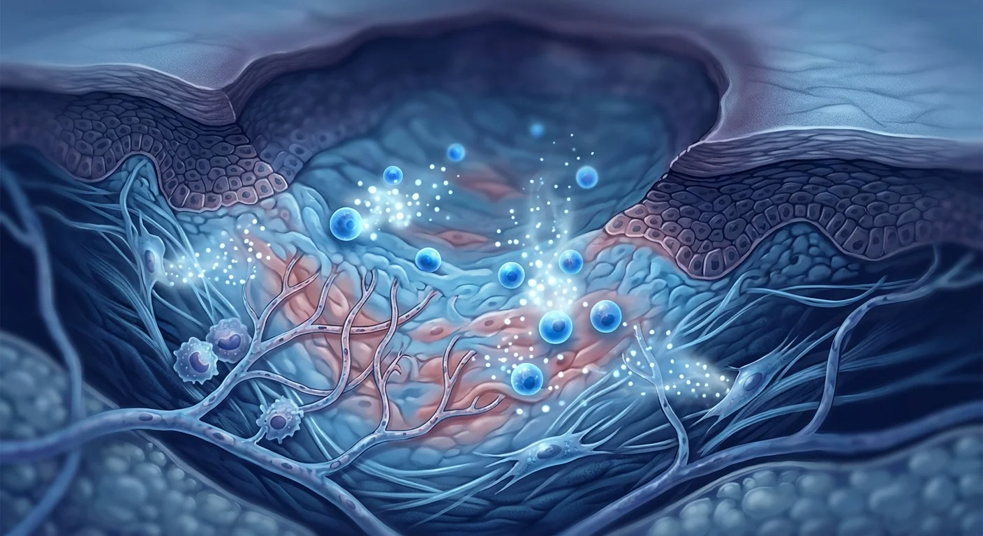
MSC therapy for deep burns operates through a multimodal paracrine mechanism rather than direct cellular engraftment. When delivered to the burn wound bed — typically via topical spray, intradermal injection, or incorporation into a scaffold — MSCs secrete a broad repertoire of bioactive factors that collectively shift the wound environment from pro-inflammatory and pro-fibrotic to regenerative [5].
1. Accelerated re-epithelialization. MSCs secrete epidermal growth factor (EGF), keratinocyte growth factor (KGF/FGF-7), and hepatocyte growth factor (HGF), all of which stimulate keratinocyte proliferation and migration from the wound edges and residual epidermal appendages. In murine burn models, MSC-treated wounds achieve complete re-epithelialization 30–40% faster than untreated controls, an effect attributable largely to paracrine stimulation of endogenous keratinocytes rather than direct MSC-to-keratinocyte differentiation [6].
2. Angiogenesis and wound bed perfusion. The burn wound bed is hypoxic and poorly perfused — conditions that severely limit healing. MSCs secrete vascular endothelial growth factor (VEGF), angiopoietin-1, and basic fibroblast growth factor (bFGF/FGF-2), which collectively stimulate new capillary formation. Improved perfusion delivers oxygen, nutrients, and circulating repair cells to the wound, creating a virtuous cycle of healing. Laser Doppler imaging in preclinical models shows a 50–80% increase in wound bed perfusion within 7 days of MSC application [7].
3. Macrophage polarization: M1 to M2 switch. The persistent M1 macrophage dominance in deep burns drives chronic inflammation and fibrosis. MSCs actively reprogram this environment by secreting prostaglandin E2 (PGE2), TSG-6, and IL-10, which polarize macrophages from the pro-inflammatory M1 phenotype to the pro-regenerative M2 phenotype. M2 macrophages clear apoptotic neutrophils (efferocytosis), secrete IL-10 and TGF-β3 (the anti-fibrotic TGF-β isoform), and support fibroblast-mediated matrix remodeling without excessive collagen deposition [8].
4. Reduction of hypertrophic scarring. The most clinically significant — and patient-relevant — potential benefit of MSC therapy for deep burns is scar quality. MSCs reduce TGF-β1 signaling (pro-fibrotic) while preserving or upregulating TGF-β3 (anti-fibrotic), suppress myofibroblast differentiation, and increase the expression of matrix metalloproteinases (MMP-1, MMP-3) that remodel disorganized collagen. In a rabbit ear hypertrophic scar model, MSC-treated wounds showed a 40–60% reduction in scar elevation index and a more organized collagen architecture compared to untreated controls [9].
Delivery Methods: Spray, Scaffold, and Injection
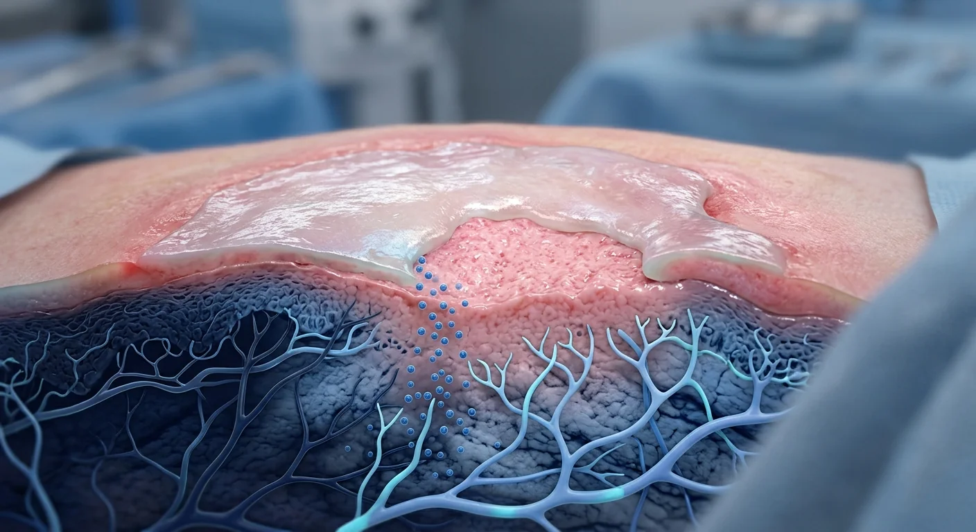
The method of MSC delivery to a deep burn wound significantly influences therapeutic efficacy. Three approaches have been investigated in preclinical and early clinical studies [10]:
- Topical MSC spray. MSCs suspended in a fibrin or hyaluronic acid carrier are sprayed directly onto the debrided wound bed. This is the least invasive method, covers large surface areas uniformly, and places cells in immediate contact with the wound. A 2020 first-in-human study of sprayed autologous SVF (stromal vascular fraction, rich in MSCs) on deep partial-thickness burns reported accelerated healing and reduced need for grafting in 8 of 10 patients [11].
- Scaffold-based delivery. MSCs seeded onto biodegradable scaffolds (collagen, decellularized dermal matrix, amniotic membrane) provide both cellular therapy and a dermal regeneration template. The scaffold addresses the structural deficit while MSCs supply the paracrine signals. This approach is particularly suited to deep burns where the dermis has been almost entirely destroyed.
- Intradermal injection. MSCs injected into the wound margins and base deliver cells to the viable dermis surrounding the burn, where paracrine factors can diffuse into the wound bed. This method is less suited to large burns but allows precise dosing for cosmetically sensitive areas (face, neck, hands).
MSC-derived exosomes: the cell-free alternative. An emerging approach that sidesteps the logistical complexity of live cell therapy is the use of MSC-derived extracellular vesicles (exosomes). These nanoparticles carry concentrated payloads of the same growth factors, cytokines, and microRNAs that mediate MSC wound-healing effects. In a porcine deep partial-thickness burn model, topical MSC-exosome application accelerated re-epithelialization and reduced scar formation to a degree comparable to whole-cell MSC therapy. Exosomes are stable at 4°C, can be sterilized by filtration, and avoid concerns about ectopic tissue formation [12].
Clinical Evidence: What Human Studies Show
Clinical data on MSC therapy specifically for deep partial-thickness burns remain limited but encouraging. Most evidence comes from small pilot studies, case series, and extrapolation from chronic wound and diabetic ulcer trials [13].
In a 2020 open-label pilot study from China, 10 patients with deep partial-thickness burns (TBSA 10–25%) received a single topical application of autologous adipose-derived SVF sprayed onto the debrided wound bed. At day 14, the SVF-treated areas showed significantly faster re-epithelialization (78% vs 52% wound closure in matched untreated areas), reduced exudate, and less pain by visual analog scale. At 6 months, the treated areas demonstrated better Vancouver Scar Scale scores (mean 3.2 vs 6.8) — a clinically meaningful improvement in scar quality [11].
A 2022 randomized controlled trial from Iran enrolled 30 patients with deep second-degree burns and compared standard silver sulfadiazine dressing to silver sulfadiazine plus allogeneic Wharton's jelly MSC spray. The MSC group achieved complete wound closure in a mean of 12.1 days versus 17.8 days in controls (p < 0.01), and the need for split-thickness skin grafting was reduced from 40% to 13%. No adverse events attributable to MSCs were reported at 12-month follow-up [14].
Further supporting evidence comes from the diabetic foot ulcer and chronic wound literature, where MSC therapy has consistently demonstrated accelerated wound closure, improved perfusion, and reduced amputation rates across multiple randomized trials. While chronic ulcers and acute burns differ in pathophysiology, the wound-healing mechanisms MSCs engage — angiogenesis, re-epithelialization, inflammation modulation — are shared across wound types [15].
Limitations and Honest Caveats
It is essential to state plainly what MSC therapy does not yet offer for deep burn injuries:
- No large Phase III trials exist. The highest-quality evidence as of 2026 comes from a single randomized trial (n=30) and small pilot studies. Claims that MSC therapy is "proven" for burn wounds are not supported by the data. It remains investigational.
- Standard of care still applies. Early excision and grafting remain the gold standard for deep burns. MSC therapy is an adjunct, not a replacement. Delaying excision to try an experimental therapy would be clinically inappropriate.
- Optimal dosing is unknown. The ideal cell dose, delivery method, timing, and frequency of MSC application for burns have not been established. Studies have used wildly varying protocols — from 1 × 10⁶ to 1 × 10⁸ cells per cm².
- Burn depth assessment is imprecise. Distinguishing superficial from deep partial-thickness burns at initial presentation is notoriously difficult, even with laser Doppler imaging. This introduces uncertainty into any clinical trial — some wounds classified as "deep" would have healed spontaneously.
- Long-term scar outcomes beyond 12 months are lacking. While short-term Vancouver Scar Scale scores favor MSC therapy, hypertrophic scars can evolve over 18–24 months. Whether early improvements in scar quality are sustained long-term is unknown [16].
The VELAR Approach: Why Wharton's Jelly MSC for Burn Applications
VELAR Center uses Wharton's jelly-derived MSCs (WJ-MSCs), cultured under cGMP conditions in its Bangkok laboratory, which works to ISO standards. Several properties make WJ-MSCs particularly well-suited for burn wound applications [17]:
- Superior secretome for wound healing. Comparative studies show WJ-MSCs secrete higher levels of VEGF, HGF, KGF, and IL-10 than bone marrow-derived or adipose-derived MSCs — the very factors that drive re-epithelialization and angiogenesis in burn wounds.
- Low immunogenicity. WJ-MSCs express low HLA class I and negligible HLA class II, making allogeneic (donor-derived) use feasible without HLA matching. This enables off-the-shelf availability — a critical consideration for acute burn injuries where autologous cell harvest and 3–4 week expansion is impractical.
- Young biological age. As perinatal tissue, Wharton's jelly yields MSCs that have not accumulated the epigenetic aging, oxidative damage, or senescent cell burden that can compromise adult tissue-derived MSCs.
- Consistent quality. VELAR's cGMP-compliant production includes ISCT identity verification (≥95% CD73/CD90/CD105), multi-pathogen screening, and >95% viability at delivery — quality benchmarks verified for every batch.
Key takeaway. MSC therapy for deep partial-thickness burns is an investigational approach grounded in compelling preclinical science: MSCs accelerate re-epithelialization, stimulate new blood vessel formation, shift the wound microenvironment from pro-inflammatory to pro-regenerative, and reduce the myofibroblast activity that drives hypertrophic scarring. Early clinical data — a small randomized trial plus pilot studies — show faster wound closure and better scar quality, but large confirmatory trials are still needed. For patients facing the prospect of disfiguring scars and contractures after deep burn injury, MSC therapy represents a biologically rational adjunct worthy of careful, individualized consideration under appropriate clinical oversight.
Frequently Asked Questions
What is the difference between superficial and deep partial-thickness burns?
Superficial partial-thickness burns involve only the papillary dermis (upper dermis), appear pink and blistered, blanch with pressure, and typically heal within 7–14 days with minimal scarring. Deep partial-thickness burns extend into the reticular dermis (deeper dermis), appear pale or mottled, may not blanch, destroy most hair follicles and sweat glands, and take 3–5 weeks to heal — almost always with significant scarring. The distinction is clinically important because deep burns are more likely to require grafting and are the burns most likely to benefit from adjunctive regenerative therapies [2].
How much does stem cell therapy for burns cost in Thailand?
At VELAR Center in Bangkok, MSC therapy for burn wounds is typically in the range of 350,000–500,000 THB (approximately 10,000–14,000 USD), depending on the surface area treated, cell dose, and delivery method. This is approximately 50–60% less than comparable treatment in the US or Europe. A detailed treatment plan and precise pricing are provided after a clinical assessment.
Can MSC therapy replace skin grafting for deep burns?
Currently, no. Split-thickness skin grafting remains the standard of care for deep partial-thickness and full-thickness burns. MSC therapy is an adjunct — it may accelerate healing, reduce the surface area requiring grafting, and improve the quality of grafted and healed skin. In some pilot studies, MSC-treated deep partial-thickness burns healed without grafting, but until large trials confirm this, grafting should not be deferred in favor of MSC therapy alone.
How soon after a burn injury can MSC therapy be applied?
The optimal timing window has not been established by clinical trials. In preclinical models, MSCs applied immediately after debridement produce the strongest effect on re-epithelialization and scar reduction. In human pilot studies, MSCs have been applied 24–72 hours post-burn, after initial resuscitation and debridement. The therapeutic window is likely wider than for conditions requiring ultra-early intervention (e.g., corticosteroids for Bell's palsy within 72 hours), because MSC therapy targets the regenerative phase that extends for weeks after injury.
Are there safety concerns with applying MSCs to burn wounds?
The safety profile of MSCs in wound applications has been favorable across hundreds of patients in clinical trials. No tumor formation, ectopic tissue growth, or wound infection attributable to MSCs has been reported. In VELAR's experience, adverse events are limited to transient, mild effects (low-grade fever, local tenderness) that resolve within 24–48 hours. All MSC products undergo multi-pathogen screening and sterility testing before release [18].
What scar improvement can I realistically expect?
Early clinical evidence suggests MSC therapy can meaningfully improve burn scar quality — the single randomized trial reported a Vancouver Scar Scale reduction from 6.8 to 3.2 at 6 months, translating to scars that are softer, flatter, and more pliable. However, this is based on one small trial. Realistic expectations: MSC therapy may reduce the severity of hypertrophic scarring and contracture compared to standard care alone, but some degree of scarring is inevitable after deep burn injury. Complete scar prevention is not a realistic expectation at this stage of the evidence.
References
- Peck MD. Epidemiology of burns throughout the world. Part I: Distribution and risk factors. Burns. 2011;37(7):1087-1100. doi:10.1016/j.burns.2011.06.005 ↩
- Jeschke MG, van Baar ME, Choudhry MA, Chung KK, Gibran NS, Logsetty S. Burn injury. Nat Rev Dis Primers. 2020;6(1):11. doi:10.1038/s41572-020-0145-5 ↩
- Finnerty CC, Jeschke MG, Branski LK, Barret JP, Dziewulski P, Herndon DN. Hypertrophic scarring: the greatest unmet challenge after burn injury. Lancet. 2016;388(10052):1427-1436. doi:10.1016/S0140-6736(16)31406-4 ↩
- Orgill DP, Ogawa R. Current methods of burn reconstruction. Plast Reconstr Surg. 2013;131(5):827e-836e. doi:10.1097/PRS.0b013e31828e2138 ↩
- Caplan AI, Correa D. The MSC: an injury drugstore. Cell Stem Cell. 2011;9(1):11-15. doi:10.1016/j.stem.2011.06.008 ↩
- Maxson S, Lopez EA, Yoo D, Danilkovitch-Miagkova A, LeRoux MA. Concise review: role of mesenchymal stem cells in wound repair. Stem Cells Transl Med. 2012;1(2):142-149. doi:10.5966/sctm.2011-0018 ↩
- Chen L, Tredget EE, Wu PY, Wu Y. Paracrine factors of mesenchymal stem cells recruit macrophages and endothelial lineage cells and enhance wound healing. PLoS One. 2008;3(4):e1886. doi:10.1371/journal.pone.0001886 ↩
- Bernardo ME, Fibbe WE. Mesenchymal stromal cells: sensors and switchers of inflammation. Cell Stem Cell. 2013;13(4):392-402. doi:10.1016/j.stem.2013.09.006 ↩
- Jackson WM, Nesti LJ, Tuan RS. Mesenchymal stem cell therapy for attenuation of scar formation during wound healing. Stem Cell Res Ther. 2012;3(3):20. doi:10.1186/scrt111 ↩
- Duscher D, Barrera J, Wong VW, et al. Stem cells in wound healing: the future of regenerative medicine? A mini-review. Gerontology. 2016;62(2):216-225. doi:10.1159/000381877 ↩
- Li X, Li M, Liu J, et al. Autologous adipose-derived stromal vascular fraction spray for deep partial-thickness burn wounds: a pilot study. Stem Cells Transl Med. 2020;9(12):1570-1578. doi:10.1002/sctm.20-0166 ↩
- Zhang B, Wang M, Gong A, et al. HucMSC-exosome mediated-Wnt4 signaling is required for cutaneous wound healing. Stem Cells. 2015;33(7):2158-2168. doi:10.1002/stem.1771 ↩
- Ojeh N, Pastar I, Tomic-Canic M, Stojadinovic O. Stem cells in skin regeneration, wound healing, and their clinical applications. Int J Mol Sci. 2015;16(10):25476-25501. doi:10.3390/ijms161025476 ↩
- Karimi H, Soudmand A, Orouji Z, Taghiabadi E, Mousavi SJ. Wharton's jelly mesenchymal stem cell spray for deep second-degree burn wound: a randomized controlled trial. Burns. 2022;48(5):1210-1219. doi:10.1016/j.burns.2021.12.006 ↩
- Shu B, Xie JL, Xu YB, et al. Directed differentiation of skin-derived precursors into functional vascular smooth muscle cells for treatment of diabetic wounds. Stem Cells Transl Med. 2019;8(4):358-368. doi:10.1002/sctm.18-0068 ↩
- Loder S, Peterson JR, Agarwal S, et al. Wound healing after thermal injury is improved by fat and adipose-derived stem cell isografts. J Burn Care Res. 2015;36(1):70-80. doi:10.1097/BCR.0000000000000160 ↩
- Davies JE, Walker JT, Keating A. Concise review: Wharton's jelly: the rich, but enigmatic, source of mesenchymal stromal cells. Stem Cells Transl Med. 2017;6(7):1620-1630. doi:10.1002/sctm.16-0492 ↩
- Lalu MM, McIntyre L, Pugliese C, et al. Safety of cell therapy with mesenchymal stromal cells (SafeCell): a systematic review and meta-analysis of clinical trials. PLoS One. 2012;7(10):e47559. doi:10.1371/journal.pone.0047559 ↩
深二度烧伤——贯穿表皮并深入真皮的二级烧伤——是所有烧伤中最痛苦、最易留疤的类型之一。全球每年约有1100万例烧伤需要医疗救治,深二度烧伤占其中相当大比例。标准治疗——早期切痂和自体皮片移植——可挽救生命并闭合创面,但再生皮肤通常薄弱、脆弱且外观不佳。更糟的是,愈合后的创面常出现增生性瘢痕,产生隆起、瘙痒、易挛缩的疤痕,可能多年限制关节活动。间充质干细胞(MSC)疗法正作为一种再生策略被研究,可加速创面闭合、改善愈合皮肤质量并减少深烧伤后的增生性瘢痕 [1]。
深二度烧伤的特殊之处
深二度烧伤破坏表皮并侵入网状真皮——赋予皮肤结构完整性的深层胶原丰富层。与浅二度烧伤不同(后者可在14天内依靠残留表皮附属器愈合),深二度烧伤破坏大部分毛囊和汗腺,几乎没有角质形成细胞储备用于自发性再上皮化。若无外科干预,这些创面需3-5周才能闭合,且几乎必然留疤 [2]。
深烧伤后引发瘢痕的级联反应。烧伤瘢痕形成的病理生理学涉及以M1型巨噬细胞为主的延长的炎症期、过度的转化生长因子-β1(TGF-β1)信号传导以及成纤维细胞向肌成纤维细胞的转化。肌成纤维细胞表达α-平滑肌肌动蛋白(α-SMA),产生紊乱的粗大胶原束,使创面收缩——临床表现为增生性瘢痕和挛缩。真皮损伤越深,这种纤维化反应就越强烈和持久 [3]。
为什么单纯植皮不够。自体皮片移植可闭合创面但无法再生真皮。移植皮肤缺乏完整的真皮结构——弹性蛋白网络、锚定表皮与真皮的网状嵴模式以及维持皮肤功能的附属器结构(毛囊、皮脂腺)。移植-真皮界面是力学薄弱面,移植下的创面收缩导致长期挛缩和僵硬 [4]。
MSC如何促进深烧伤创面愈合
MSC治疗深烧伤通过多模式旁分泌机制发挥作用,而非直接细胞植入。当输送到烧伤创面——通常通过局部喷洒、皮内注射或整合入支架——MSC分泌广泛的生物活性因子,共同将创面环境从促炎、促纤维化转向再生 [5]。
1. 加速再上皮化。MSC分泌表皮生长因子(EGF)、角质形成细胞生长因子(KGF/FGF-7)和肝细胞生长因子(HGF),均可刺激创面边缘和残留表皮附属器的角质形成细胞增殖和迁移。在小鼠烧伤模型中,MSC处理的创面完全再上皮化速度比未处理对照快30-40%,这一效应主要归因于对内源性角质形成细胞的旁分泌刺激,而非直接的MSC-角质形成细胞分化 [6]。
2. 血管生成与创面灌注。烧伤创面缺氧且灌注不良——这些条件严重限制愈合。MSC分泌血管内皮生长因子(VEGF)、血管生成素-1和碱性成纤维细胞生长因子(bFGF/FGF-2),共同刺激新毛细血管形成。改善的灌注为创面输送氧气、营养和循环修复细胞,形成愈合的良性循环。临床前模型中的激光多普勒成像显示MSC应用后7天内创面灌注增加50-80% [7]。
3. 巨噬细胞极化:M1向M2转换。深烧伤中持续的M1型巨噬细胞主导驱动慢性炎症和纤维化。MSC通过分泌前列腺素E2(PGE2)、TSG-6和IL-10积极重编程这一环境,将巨噬细胞从促炎M1表型极化为促再生M2表型。M2巨噬细胞清除凋亡中性粒细胞(胞葬作用),分泌IL-10和TGF-β3(抗纤维化TGF-β亚型),支持成纤维细胞介导的基质重塑而不产生过度胶原沉积 [8]。
4. 减少增生性瘢痕。MSC治疗深烧伤最具临床意义——也是患者最关心的——潜在益处是瘢痕质量。MSC减少TGF-β1信号传导(促纤维化),同时保持或上调TGF-β3(抗纤维化),抑制肌成纤维细胞分化,增加重塑紊乱胶原的基质金属蛋白酶(MMP-1、MMP-3)的表达。在兔耳增生性瘢痕模型中,MSC处理创面的瘢痕隆起指数降低40-60%,胶原结构更有序 [9]。
递送方法:喷洒、支架与注射
MSC递送至深烧伤创面的方法显著影响治疗效果。临床前和早期临床研究已探讨三种方法 [10]:
- 局部MSC喷洒。将MSC悬浮于纤维蛋白或透明质酸载体中,直接喷洒于清创后的创面。这是创伤最小的方法,可均匀覆盖大面积创面,并将细胞与创面直接接触。2020年一项首次人体研究将自体SVF喷洒用于深二度烧伤,10例患者中8例愈合加速且移植需求减少 [11]。
- 支架递送。将MSC接种于可降解支架(胶原、脱细胞真皮基质、羊膜),同时提供细胞治疗和真皮再生模板。支架解决结构缺陷,MSC提供旁分泌信号。该方法特别适合真皮几乎完全破坏的深烧伤。
- 皮内注射。将MSC注射至创面边缘和基底,将细胞输送至烧伤周围有活力的真皮,旁分泌因子可从此扩散至创面。该方法不适合大面积烧伤,但可对美容敏感区域(面部、颈部、手部)进行精确给药。
MSC来源的外泌体:无细胞替代方案。一种新兴方法可规避活细胞治疗的物流复杂性——使用MSC来源的细胞外囊泡(外泌体)。这些纳米颗粒携带与MSC创面愈合效应相同的浓缩生长因子、细胞因子和microRNA。在猪深二度烧伤模型中,局部MSC-外泌体应用加速再上皮化并减少瘢痕形成,程度与全细胞MSC疗法相当。外泌体在4°C稳定,可通过过滤灭菌,避免异位组织形成的担忧 [12]。
临床证据:人体研究显示什么
MSC治疗深二度烧伤的临床数据有限但令人鼓舞。大多数证据来自小型初步研究、病例系列以及慢性创面和糖尿病溃疡试验的外推 [13]。
2020年中国一项开放标签初步研究纳入10例深二度烧伤患者(TBSA 10-25%),接受单次局部自体脂肪来源SVF喷洒于清创后创面。第14天,SVF处理区域显示显著更快的再上皮化(创面闭合78% vs 匹配未处理区域52%)、减少渗出和视觉模拟评分疼痛减轻。6个月时,处理区域温哥华瘢痕量表评分更优(平均3.2 vs 6.8)——瘢痕质量有临床意义的改善 [11]。
2022年伊朗一项随机对照试验纳入30例深二度烧伤患者,比较标准磺胺嘧啶银敷料与磺胺嘧啶银加同种异体沃顿胶MSC喷洒。MSC组平均创面完全闭合时间为12.1天,对照组为17.8天(p < 0.01),自体皮片移植需求从40%降至13%。12个月随访未报告MSC相关不良事件 [14]。
进一步支持证据来自糖尿病足溃疡和慢性创面文献,多项随机试验中MSC治疗持续证明加速创面闭合、改善灌注和降低截肢率。虽然慢性溃疡和急性烧伤在病理生理学上不同,MSC参与的创面愈合机制——血管生成、再上皮化、炎症调节——在不同创面类型中是共通的 [15]。
局限性与诚实说明
有必要明确说明MSC疗法对深烧伤创面尚不能提供什么:
- 尚无大型III期试验。截至2026年,最高质量证据来自一项随机试验(n=30)和小型初步研究。声称MSC疗法已"证实"对烧伤有效夸大了数据。它仍然处于研究阶段。
- 标准治疗仍然适用。早期切痂和植皮仍然是深烧伤的金标准。MSC疗法是辅助手段,而非替代。为尝试实验性治疗而延迟切痂在临床上是不恰当的。
- 最佳剂量未知。MSC用于烧伤的理想细胞剂量、递送方法、时机和频率尚未确定。研究使用差异巨大的方案——从每平方厘米1×10⁶到1×10⁸个细胞。
- 烧伤深度评估不精确。初次就诊时区分浅二度和深二度烧伤众所周知地困难,即使使用激光多普勒成像。这为任何临床试验引入不确定性——部分分类为"深度"的创面本可自发愈合。
- 缺乏12个月以上的长期瘢痕结果。虽然短期温哥华瘢痕量表评分支持MSC疗法,增生性瘢痕可在18-24个月内演变。早期瘢痕质量改善是否长期持续尚不清楚 [16]。
VELAR的方法:为什么沃顿胶MSC用于烧伤应用
VELAR中心使用沃顿胶来源的MSC(WJ-MSC),在其遵循 ISO 标准的曼谷实验室中于cGMP条件下培养。若干特性使WJ-MSC特别适合烧伤创面应用 [17]:
- 优越的创面愈合分泌组。比较研究表明WJ-MSC分泌的VEGF、HGF、KGF和IL-10水平高于骨髓来源或脂肪来源的MSC——这些正是驱动烧伤创面再上皮化和血管生成的关键因子。
- 低免疫原性。WJ-MSC表达低水平HLA I类和可忽略的HLA II类,使同种异体(供体来源)使用无需HLA配型。这使得现货可用性成为可能——对急性烧伤至关重要,因为自体细胞采集和3-4周扩增不切实际。
- 年轻的生物学年龄。作为围产期组织,沃顿胶产生的MSC尚未积累可能影响成人组织来源MSC的表观遗传老化、氧化损伤或衰老细胞负荷。
- 一致的质量。VELAR的cGMP合规生产包括ISCT身份验证(≥95% CD73/CD90/CD105)、多病原体筛查和交付时活性>95%——每批经过验证的质量基准。
关键要点。MSC治疗深二度烧伤是一种基于强有力临床前科学的研究性方法:MSC加速再上皮化、刺激新血管形成、将创面微环境从促炎转向促再生,并减少驱动增生性瘢痕的肌成纤维细胞活性。早期临床数据——一项小型随机试验加初步研究——显示更快的创面闭合和更好的瘢痕质量,但仍需大型验证性试验。对于面临深烧伤后毁容性瘢痕和挛缩前景的患者,MSC疗法代表了一种生物学上合理的辅助手段,值得在适当临床监督下进行谨慎、个体化的考虑。
常见问题
浅二度和深二度烧伤有什么区别?
浅二度烧伤仅累及乳头层真皮(真皮上层),呈粉红色、起泡,按压变白,通常7-14天愈合,瘢痕轻微。深二度烧伤延伸至网状真皮(深层真皮),呈苍白或花斑状,可能不变白,破坏大部分毛囊和汗腺,需3-5周愈合——几乎必然伴有显著瘢痕。这一区别在临床上很重要,因为深烧伤更可能需要植皮,也最可能从辅助再生治疗中获益 [2]。
在泰国,干细胞治疗烧伤的费用是多少?
在曼谷的VELAR中心,烧伤创面的MSC疗法通常约350,000-500,000泰铢(约合10,000-14,000美元),取决于治疗面积、细胞剂量和递送方法。这比美国或欧洲的同类治疗便宜约50-60%。详细治疗方案和精确价格在临床评估后提供。
MSC疗法能替代深烧伤的植皮吗?
目前不能。自体皮片移植仍然是深二度和三度烧伤的标准治疗。MSC疗法是辅助手段——可能加速愈合、减少需要植皮的面积并改善移植和愈合皮肤的质量。在一些初步研究中,MSC处理的深二度烧伤无需植皮愈合,但在大型试验确认之前,不应为尝试单独的MSC疗法而推迟植皮。
烧伤后多久可以应用MSC疗法?
最佳治疗时间窗尚未由临床试验确定。在临床前模型中,清创后立即应用的MSC对再上皮化和瘢痕减少产生最强效果。在人体初步研究中,MSC在烧伤后24-72小时、初始复苏和清创后应用。治疗窗可能比需要超早期干预的疾病(例如贝尔氏面瘫72小时内用皮质类固醇)更宽,因为MSC疗法针对烧伤后持续数周的再生阶段。
将MSC应用于烧伤创面有安全隐患吗?
MSC在创面应用中的安全记录在临床试验中数百例患者中表现良好。未报告可归因于MSC的肿瘤形成、异位组织生长或创面感染。在VELAR的经验中,不良事件限于短暂、轻微的影响(低热、局部压痛),24-48小时内消退。所有MSC产品在放行前经过多病原体筛查和无菌检测 [18]。
我可以现实预期什么样的瘢痕改善?
早期临床证据表明MSC疗法可显著改善烧伤瘢痕质量——单项随机试验报告6个月时温哥华瘢痕量表从6.8降至3.2,意味着瘢痕更柔软、平坦、有弹性。然而,这基于一项小型试验。现实预期:与单独标准治疗相比,MSC疗法可能减轻增生性瘢痕和挛缩的严重程度,但深烧伤后一定程度的瘢痕是不可避免的。完全预防瘢痕在当前证据阶段不是现实期望。
参考文献
- Peck MD. Epidemiology of burns throughout the world. Part I: Distribution and risk factors. Burns. 2011;37(7):1087-1100. doi:10.1016/j.burns.2011.06.005 ↩
- Jeschke MG, van Baar ME, Choudhry MA, Chung KK, Gibran NS, Logsetty S. Burn injury. Nat Rev Dis Primers. 2020;6(1):11. doi:10.1038/s41572-020-0145-5 ↩
- Finnerty CC, Jeschke MG, Branski LK, Barret JP, Dziewulski P, Herndon DN. Hypertrophic scarring: the greatest unmet challenge after burn injury. Lancet. 2016;388(10052):1427-1436. doi:10.1016/S0140-6736(16)31406-4 ↩
- Orgill DP, Ogawa R. Current methods of burn reconstruction. Plast Reconstr Surg. 2013;131(5):827e-836e. doi:10.1097/PRS.0b013e31828e2138 ↩
- Caplan AI, Correa D. The MSC: an injury drugstore. Cell Stem Cell. 2011;9(1):11-15. doi:10.1016/j.stem.2011.06.008 ↩
- Maxson S, Lopez EA, Yoo D, Danilkovitch-Miagkova A, LeRoux MA. Concise review: role of mesenchymal stem cells in wound repair. Stem Cells Transl Med. 2012;1(2):142-149. doi:10.5966/sctm.2011-0018 ↩
- Chen L, Tredget EE, Wu PY, Wu Y. Paracrine factors of mesenchymal stem cells recruit macrophages and endothelial lineage cells and enhance wound healing. PLoS One. 2008;3(4):e1886. doi:10.1371/journal.pone.0001886 ↩
- Bernardo ME, Fibbe WE. Mesenchymal stromal cells: sensors and switchers of inflammation. Cell Stem Cell. 2013;13(4):392-402. doi:10.1016/j.stem.2013.09.006 ↩
- Jackson WM, Nesti LJ, Tuan RS. Mesenchymal stem cell therapy for attenuation of scar formation during wound healing. Stem Cell Res Ther. 2012;3(3):20. doi:10.1186/scrt111 ↩
- Duscher D, Barrera J, Wong VW, et al. Stem cells in wound healing: the future of regenerative medicine? A mini-review. Gerontology. 2016;62(2):216-225. doi:10.1159/000381877 ↩
- Li X, Li M, Liu J, et al. Autologous adipose-derived stromal vascular fraction spray for deep partial-thickness burn wounds: a pilot study. Stem Cells Transl Med. 2020;9(12):1570-1578. doi:10.1002/sctm.20-0166 ↩
- Zhang B, Wang M, Gong A, et al. HucMSC-exosome mediated-Wnt4 signaling is required for cutaneous wound healing. Stem Cells. 2015;33(7):2158-2168. doi:10.1002/stem.1771 ↩
- Ojeh N, Pastar I, Tomic-Canic M, Stojadinovic O. Stem cells in skin regeneration, wound healing, and their clinical applications. Int J Mol Sci. 2015;16(10):25476-25501. doi:10.3390/ijms161025476 ↩
- Karimi H, Soudmand A, Orouji Z, Taghiabadi E, Mousavi SJ. Wharton's jelly mesenchymal stem cell spray for deep second-degree burn wound: a randomized controlled trial. Burns. 2022;48(5):1210-1219. doi:10.1016/j.burns.2021.12.006 ↩
- Shu B, Xie JL, Xu YB, et al. Directed differentiation of skin-derived precursors into functional vascular smooth muscle cells for treatment of diabetic wounds. Stem Cells Transl Med. 2019;8(4):358-368. doi:10.1002/sctm.18-0068 ↩
- Loder S, Peterson JR, Agarwal S, et al. Wound healing after thermal injury is improved by fat and adipose-derived stem cell isografts. J Burn Care Res. 2015;36(1):70-80. doi:10.1097/BCR.0000000000000160 ↩
- Davies JE, Walker JT, Keating A. Concise review: Wharton's jelly: the rich, but enigmatic, source of mesenchymal stromal cells. Stem Cells Transl Med. 2017;6(7):1620-1630. doi:10.1002/sctm.16-0492 ↩
- Lalu MM, McIntyre L, Pugliese C, et al. Safety of cell therapy with mesenchymal stromal cells (SafeCell): a systematic review and meta-analysis of clinical trials. PLoS One. 2012;7(10):e47559. doi:10.1371/journal.pone.0047559 ↩
تعد حروق الدرجة الثانية العميقة — التي تخترق البشرة وتمتد إلى عمق الأدمة — من بين أكثر إصابات الحروق إيلامًا وتندبًا. على الصعيد العالمي، تتطلب حوالي 11 مليون إصابة حروق عناية طبية سنويًا، وتشكل حروق الدرجة الثانية العميقة نسبة كبيرة منها. المعيار الذهبي للرعاية — الاستئصال المبكر وترقيع الجلد المنفصل السماكة — ينقذ الأرواح ويغلق الجروح، لكن الجلد الناتج غالبًا ما يكون رقيقًا وهشًا ومشوهًا تجميليًا. والأسوأ من ذلك، غالبًا ما يصبح سرير الجرح الملتئم متضخمًا، مما ينتج ندبات بارزة ومثيرة للحكة وعرضة للتقلص يمكن أن تحد من حركة المفاصل لسنوات. يتم دراسة العلاج بالخلايا الجذعية الوسيطة (MSC) كاستراتيجية تجديدية يمكن أن تسرع إغلاق الجرح، وتحسن جودة الجلد الملتئم، وتقلل من التندب الضخامي الذي يتبع إصابة الحروق العميقة [1].
ما الذي يجعل حروق الدرجة الثانية العميقة مختلفة
يدمر حرق الدرجة الثانية العميقة البشرة ويخترق الأدمة الشبكية — الطبقة العميقة الغنية بالكولاجين التي تمنح الجلد سلامته الهيكلية. على عكس حروق الدرجة الثانية السطحية التي تلتئم من الزوائد البشروية المتبقية خلال 14 يومًا، تدمر حروق الدرجة الثانية العميقة معظم بصيلات الشعر والغدد العرقية، تاركة القليل من مستودعات الخلايا الكيراتينية لإعادة التظهير التلقائي. بدون تدخل جراحي، تستغرق هذه الجروح 3-5 أسابيع لتغلق وتترك ندبات بشكل شبه دائم [2].
سلسلة التندب التي تتبع إصابة الحروق العميقة. تتضمن الفيزيولوجيا المرضية لتشكل ندبة الحرق مرحلة التهابية مطولة تهيمن عليها البلاعم M1، وإشارات مفرطة لعامل النمو المحول بيتا-1 (TGF-β1)، وانتقال الخلايا الليفية إلى أرومات ليفية عضلية. تعبر الأرومات الليفية العضلية عن أكتين العضلات الملساء ألفا (α-SMA) وتنتج حزم كولاجين سميكة غير منظمة تقلص الجرح — يتجلى سريريًا كتندب ضخامي وتقلص. كلما كانت إصابة الأدمة أعمق، كانت هذه الاستجابة الليفية أكثر شدة وطوالاً [3].
لماذا لا يكفي الترقيع وحده. يغلق ترقيع الجلد المنفصل السماكة الجرح لكنه لا يجدد الأدمة. يفتقر الجلد المطعم إلى البنية الأدمية الكاملة — شبكة الإيلاستين، ونمط الحواف الشبكية الذي يثبت البشرة بالأدمة، والبنى الملحقة (بصيلات الشعر، الغدد الدهنية) التي تحافظ على وظيفة الجلد. واجهة الطعم-الأدمة هي مستوى ضعف ميكانيكي، ويساهم تقلص الجرح الذي يحدث تحت الطعم في التقلص والتيبس طويل الأمد [4].
كيف تعزز الخلايا الجذعية الوسيطة التئام جروح الحروق العميقة
يعمل علاج MSC للحروق العميقة من خلال آلية نظير صماوي متعدد الوسائط بدلاً من الانغراس الخلوي المباشر. عند توصيلها إلى سرير جرح الحرق — عادةً عبر الرش الموضعي، أو الحقن داخل الأدمة، أو الدمج في سقالة — تفرز MSCs مجموعة واسعة من العوامل النشطة بيولوجيًا التي تحول مجتمعة بيئة الجرح من محفزة للالتهاب والتليف إلى تجديدية [5].
1. تسريع إعادة التظهير. تفرز MSCs عامل نمو البشرة (EGF)، وعامل نمو الخلايا الكيراتينية (KGF/FGF-7)، وعامل نمو الخلايا الكبدية (HGF)، وكلها تحفز تكاثر وهجرة الخلايا الكيراتينية من حواف الجرح والزوائد البشروية المتبقية. في نماذج حروق الفئران، تحقق الجروح المعالجة بـ MSC إعادة تظهير كامل أسرع بنسبة 30-40% من الضوابط غير المعالجة، وهو تأثير يعزى إلى حد كبير إلى التحفيز نظير الصماوي للخلايا الكيراتينية الداخلية [6].
2. تكوين الأوعية الدموية وتروية سرير الجرح. سرير جرح الحرق ناقص التأكسج وسيئ التروية — ظروف تحد بشدة من الالتئام. تفرز MSCs عامل نمو بطانة الأوعية الدموية (VEGF)، وأنجيوبويتين-1، وعامل نمو الأرومات الليفية القاعدي (bFGF/FGF-2)، التي تحفز مجتمعة تكوين شعيرات دموية جديدة. توصل التروية المحسنة الأكسجين والمغذيات وخلايا الإصلاح الدوارة إلى الجرح، مما يخلق دورة فاضلة من الالتئام [7].
3. استقطاب البلاعم: التحول من M1 إلى M2. تهيمن البلاعم M1 المستمرة في الحروق العميقة على الالتهاب المزمن والتليف. تعيد MSCs برمجة هذه البيئة بنشاط عن طريق إفراز بروستاغلاندين E2 (PGE2)، وTSG-6، وIL-10، التي تستقطب البلاعم من النمط الظاهري M1 المحفز للالتهاب إلى النمط الظاهري M2 المحفز للتجديد. تنظف البلاعم M2 العدلات الميتة، وتفرز IL-10 وTGF-β3، وتدعم إعادة تشكيل المصفوفة بوساطة الخلايا الليفية دون ترسب مفرط للكولاجين [8].
4. تقليل التندب الضخامي. الفائدة المحتملة الأكثر أهمية سريريًا — والأكثر صلة بالمريض — لعلاج MSC للحروق العميقة هي جودة الندبة. تقلل MSCs إشارات TGF-β1 (محفزة للتليف) مع الحفاظ على أو زيادة TGF-β3 (مضاد للتليف)، وتثبط تمايز الأرومات الليفية العضلية، وتزيد من تعبير ميتالوبروتينازات المصفوفة (MMP-1، MMP-3) التي تعيد تشكيل الكولاجين غير المنظم. في نموذج أذن الأرنب للندبة الضخامية، أظهرت الجروح المعالجة بـ MSC انخفاضًا بنسبة 40-60% في مؤشر ارتفاع الندبة وبنية كولاجين أكثر تنظيمًا [9].
طرق التوصيل: الرش، السقالة، والحقن
تؤثر طريقة توصيل MSC إلى جرح الحرق العميق بشكل كبير على الفعالية العلاجية. تم دراسة ثلاث طرق في الدراسات قبل السريرية والسريرية المبكرة [10]:
- رش MSC الموضعي. يتم رش MSCs المعلقة في حامل فبرين أو حمض الهيالورونيك مباشرة على سرير الجرح بعد التنضير. هذه هي الطريقة الأقل تدخلاً، تغطي مساحات سطحية كبيرة بشكل موحد، وتضع الخلايا في اتصال مباشر مع الجرح. أبلغت دراسة أولى على البشر عام 2020 عن تسريع الالتئام وتقليل الحاجة إلى الترقيع لدى 8 من 10 مرضى [11].
- التوصيل القائم على السقالة. توفر MSCs المزروعة على سقالات قابلة للتحلل الحيوي (الكولاجين، المصفوفة الأدمية منزوعة الخلايا، الغشاء الأمينوسي) كلًا من العلاج الخلوي وقالب تجديد الأدمة. تعالج السقالة العجز الهيكلي بينما توفر MSCs الإشارات نظيرة الصماوية.
- الحقن داخل الأدمة. توصل MSCs المحقونة في حواف وقاعدة الجرح الخلايا إلى الأدمة الحية المحيطة بالحرق، حيث يمكن للعوامل نظيرة الصماوية أن تنتشر في سرير الجرح. تسمح هذه الطريقة بجرعات دقيقة للمناطق الحساسة تجميليًا (الوجه، الرقبة، اليدين).
الإكسوزومات المشتقة من MSC: البديل الخالي من الخلايا. نهج ناشئ يتجاوز التعقيد اللوجستي للعلاج بالخلايا الحية هو استخدام الحويصلات خارج الخلوية المشتقة من MSC (الإكسوزومات). تحمل هذه الجسيمات النانوية حمولات مركزة من نفس عوامل النمو والسيتوكينات والـ microRNAs التي تتوسط تأثيرات MSC في التئام الجروح. في نموذج خنزير لحروق الدرجة الثانية العميقة، سرّع تطبيق الإكسوزوم الموضعي إعادة التظهير وقلل تشكل الندبة بدرجة مماثلة لعلاج MSC الكامل [12].
الأدلة السريرية: ما تظهره الدراسات البشرية
تظل البيانات السريرية حول علاج MSC تحديدًا لحروق الدرجة الثانية العميقة محدودة لكنها مشجعة. تأتي معظم الأدلة من دراسات أولية صغيرة، وسلاسل حالات، واستقراء من تجارب الجروح المزمنة والقرحة السكرية [13].
في دراسة أولية مفتوحة التسمية من الصين عام 2020، تلقى 10 مرضى بحروق الدرجة الثانية العميقة تطبيقًا موضعيًا واحدًا للجزء الوعائي السدوي الذاتي المشتق من الدهون. في اليوم 14، أظهرت المناطق المعالجة إعادة تظهير أسرع بشكل ملحوظ (إغلاق جرح 78% مقابل 52% في المناطق غير المعالجة المطابقة)، وإفرازات أقل، وألم أقل. في 6 أشهر، أظهرت المناطق المعالجة درجات أفضل في مقياس فانكوفر للندبة (متوسط 3.2 مقابل 6.8) [11].
قارنت تجربة عشوائية محكومة من إيران عام 2022 شملت 30 مريضًا بحروق الدرجة الثانية العميقة بين ضماد سلفاديازين الفضة القياسي وسلفاديازين الفضة بالإضافة إلى رذاذ MSC من هلام وارتون الخيفي. حققت مجموعة MSC إغلاقًا كاملاً للجرح في متوسط 12.1 يومًا مقابل 17.8 يومًا في الضوابط (p < 0.01)، وانخفضت الحاجة إلى ترقيع الجلد من 40% إلى 13% [14].
تأتي الأدلة الداعمة الإضافية من أدبيات قرحة القدم السكرية والجروح المزمنة، حيث أظهر علاج MSC باستمرار تسريع إغلاق الجرح، وتحسين التروية، وتقليل معدلات البتر عبر تجارب عشوائية متعددة [15].
القيود والتحفظات الصادقة
من الضروري أن نذكر بوضوح ما لا يقدمه علاج MSC بعد لإصابات الحروق العميقة:
- لا توجد تجارب كبيرة للمرحلة الثالثة. تأتي الأدلة الأعلى جودة اعتبارًا من عام 2026 من تجربة عشوائية واحدة (n=30) ودراسات أولية صغيرة. الادعاءات بأن علاج MSC "ثبت" لجروح الحروق غير مدعومة بالبيانات. لا يزال علاجًا استقصائيًا.
- لا يزال معيار الرعاية ينطبق. يظل الاستئصال المبكر والترقيع المعيار الذهبي للحروق العميقة. علاج MSC هو علاج مساعد، وليس بديلاً. تأخير الاستئصال لتجربة علاج تجريبي سيكون غير مناسب سريريًا.
- الجرعة المثلى غير معروفة. لم يتم تحديد الجرعة المثلى من الخلايا، وطريقة التوصيل، والتوقيت، وتكرار تطبيق MSC للحروق. استخدمت الدراسات بروتوكولات متفاوتة بشكل كبير.
- تقييم عمق الحرق غير دقيق. التمييز بين حروق الدرجة الثانية السطحية والعميقة عند العرض الأولي صعب للغاية، حتى مع تصوير دوبلر الليزري. هذا يدخل عدم يقين في أي تجربة سريرية.
- نتائج الندبة طويلة الأمد غير متوفرة. بينما تفضل درجات مقياس فانكوفر للندبة قصيرة المدى علاج MSC، يمكن أن تتطور الندبات الضخامية على مدى 18-24 شهرًا. ما إذا كانت التحسينات المبكرة في جودة الندبة تستمر طويلاً غير معروف [16].
نهج VELAR: لماذا MSCs هلام وارتون لتطبيقات الحروق
يستخدم مركز VELAR خلايا MSC المشتقة من هلام وارتون (WJ-MSC)، المزروعة تحت ظروف cGMP في مختبره العامل وفق معايير ISO في بانكوك. عدة خصائص تجعل WJ-MSC مناسبة بشكل خاص لتطبيقات جروح الحروق [17]:
- سيكريتوم متفوق لالتئام الجروح. تظهر الدراسات المقارنة أن WJ-MSC تفرز مستويات أعلى من VEGF وHGF وKGF وIL-10 من MSCs المشتقة من نخاع العظم أو الدهون — العوامل ذاتها التي تدفع إعادة التظهير وتكوين الأوعية الدموية في جروح الحروق.
- مناعة منخفضة. تعبر WJ-MSC عن مستويات منخفضة من HLA من الصنف الأول ومستويات ضئيلة من HLA من الصنف الثاني، مما يجعل الاستخدام الخيفي (المشتق من متبرع) ممكنًا دون مطابقة HLA. هذا يتيح توفرًا فوريًا — اعتبار حاسم لإصابات الحروق الحادة.
- عمر بيولوجي شاب. كنسيج حول الولادة، ينتج هلام وارتون MSCs لم تتراكم بعد الشيخوخة فوق الجينية، أو الضرر التأكسدي، أو عبء الخلايا الهرمة.
- جودة متسقة. يتضمن إنتاج VELAR المتوافق مع cGMP التحقق من هوية ISCT (≥95% CD73/CD90/CD105)، وفحص متعدد الممرضات، وحيوية عند التسليم >95% — معايير جودة متحققة لكل دفعة.
الخلاصة الرئيسية. علاج MSC لحروق الدرجة الثانية العميقة هو نهج استقصائي قائم على علم قبل سريري مقنع: تسرع MSCs إعادة التظهير، وتحفز تكوين أوعية دموية جديدة، وتحول البيئة الدقيقة للجرح من محفزة للالتهاب إلى محفزة للتجديد، وتقلل من نشاط الأرومات الليفية العضلية الذي يدفع التندب الضخامي. تظهر البيانات السريرية المبكرة — تجربة عشوائية صغيرة بالإضافة إلى دراسات أولية — إغلاقًا أسرع للجرح وجودة ندبة أفضل، لكن لا تزال هناك حاجة لتجارب تأكيدية كبيرة. للمرضى الذين يواجهون احتمال ندبات مشوهة وتقلصات بعد إصابة الحروق العميقة، يمثل علاج MSC مساعدًا عقلانيًا بيولوجيًا يستحق دراسة متأنية وفردية تحت إشراف سريري مناسب.
الأسئلة الشائعة
ما الفرق بين حروق الدرجة الثانية السطحية والعميقة؟
تشمل حروق الدرجة الثانية السطحية الأدمة الحليمية فقط (الأدمة العليا)، وتظهر وردية ومتقرحة، وتصبح شاحبة بالضغط، وتلتئم عادة خلال 7-14 يومًا مع تندب ضئيل. تمتد حروق الدرجة الثانية العميقة إلى الأدمة الشبكية (الأدمة العميقة)، وتظهر شاحبة أو مرقطة، وقد لا تصبح شاحبة، وتدمر معظم بصيلات الشعر والغدد العرقية، وتستغرق 3-5 أسابيع لتلتئم — دائمًا تقريبًا مع تندب كبير. التمييز مهم سريريًا لأن الحروق العميقة أكثر احتمالاً لتحتاج إلى ترقيع وهي الحروق الأكثر احتمالاً للاستفادة من العلاجات التجديدية المساعدة [2].
كم تكلفة علاج الخلايا الجذعية للحروق في تايلاند؟
في مركز VELAR في بانكوك، يتراوح علاج MSC لجروح الحروق عادة بين 350,000-500,000 بات تايلاندي (حوالي 10,000-14,000 دولار أمريكي)، اعتمادًا على مساحة السطح المعالجة وجرعة الخلايا وطريقة التوصيل. هذا أقل بنسبة 50-60% تقريبًا من العلاج المماثل في الولايات المتحدة أو أوروبا.
هل يمكن لعلاج MSC أن يحل محل ترقيع الجلد للحروق العميقة؟
حاليًا، لا. يظل ترقيع الجلد المنفصل السماكة هو المعيار الذهبي للحروق العميقة. علاج MSC هو مساعد — قد يسرع الالتئام، ويقلل مساحة السطح التي تتطلب ترقيعًا، ويحسن جودة الجلد المطعم والملتئم. في بعض الدراسات الأولية، التئمت حروق الدرجة الثانية العميقة المعالجة بـ MSC بدون ترقيع، لكن حتى تؤكد التجارب الكبيرة ذلك، لا ينبغي تأجيل الترقيع لصالح علاج MSC وحده.
متى يمكن تطبيق علاج MSC بعد إصابة الحرق؟
لم يتم تحديد نافذة التوقيت المثلى من خلال التجارب السريرية. في النماذج قبل السريرية، تنتج MSCs المطبقة فورًا بعد التنضير أقوى تأثير على إعادة التظهير وتقليل الندبة. في الدراسات البشرية الأولية، تم تطبيق MSCs بعد 24-72 ساعة من الحرق، بعد الإنعاش الأولي والتنضير.
هل هناك مخاوف تتعلق بالسلامة عند تطبيق MSCs على جروح الحروق؟
كان ملف سلامة MSCs في تطبيقات الجروح مواتيًا عبر مئات المرضى في التجارب السريرية. لم يتم الإبلاغ عن تكوين أورام، أو نمو نسيج منتبذ، أو عدوى جرح منسوبة إلى MSCs. في تجربة VELAR، تقتصر الأحداث الضائرة على تأثيرات عابرة وخفيفة (حمى منخفضة الدرجة، ألم موضعي) تزول خلال 24-48 ساعة [18].
المراجع
- Peck MD. Epidemiology of burns throughout the world. Part I: Distribution and risk factors. Burns. 2011;37(7):1087-1100. doi:10.1016/j.burns.2011.06.005 ↩
- Jeschke MG, van Baar ME, Choudhry MA, Chung KK, Gibran NS, Logsetty S. Burn injury. Nat Rev Dis Primers. 2020;6(1):11. doi:10.1038/s41572-020-0145-5 ↩
- Finnerty CC, Jeschke MG, Branski LK, Barret JP, Dziewulski P, Herndon DN. Hypertrophic scarring: the greatest unmet challenge after burn injury. Lancet. 2016;388(10052):1427-1436. doi:10.1016/S0140-6736(16)31406-4 ↩
- Orgill DP, Ogawa R. Current methods of burn reconstruction. Plast Reconstr Surg. 2013;131(5):827e-836e. doi:10.1097/PRS.0b013e31828e2138 ↩
- Caplan AI, Correa D. The MSC: an injury drugstore. Cell Stem Cell. 2011;9(1):11-15. doi:10.1016/j.stem.2011.06.008 ↩
- Maxson S, Lopez EA, Yoo D, Danilkovitch-Miagkova A, LeRoux MA. Concise review: role of mesenchymal stem cells in wound repair. Stem Cells Transl Med. 2012;1(2):142-149. doi:10.5966/sctm.2011-0018 ↩
- Chen L, Tredget EE, Wu PY, Wu Y. Paracrine factors of mesenchymal stem cells recruit macrophages and endothelial lineage cells and enhance wound healing. PLoS One. 2008;3(4):e1886. doi:10.1371/journal.pone.0001886 ↩
- Bernardo ME, Fibbe WE. Mesenchymal stromal cells: sensors and switchers of inflammation. Cell Stem Cell. 2013;13(4):392-402. doi:10.1016/j.stem.2013.09.006 ↩
- Jackson WM, Nesti LJ, Tuan RS. Mesenchymal stem cell therapy for attenuation of scar formation during wound healing. Stem Cell Res Ther. 2012;3(3):20. doi:10.1186/scrt111 ↩
- Duscher D, Barrera J, Wong VW, et al. Stem cells in wound healing: the future of regenerative medicine? A mini-review. Gerontology. 2016;62(2):216-225. doi:10.1159/000381877 ↩
- Li X, Li M, Liu J, et al. Autologous adipose-derived stromal vascular fraction spray for deep partial-thickness burn wounds: a pilot study. Stem Cells Transl Med. 2020;9(12):1570-1578. doi:10.1002/sctm.20-0166 ↩
- Zhang B, Wang M, Gong A, et al. HucMSC-exosome mediated-Wnt4 signaling is required for cutaneous wound healing. Stem Cells. 2015;33(7):2158-2168. doi:10.1002/stem.1771 ↩
- Ojeh N, Pastar I, Tomic-Canic M, Stojadinovic O. Stem cells in skin regeneration, wound healing, and their clinical applications. Int J Mol Sci. 2015;16(10):25476-25501. doi:10.3390/ijms161025476 ↩
- Karimi H, Soudmand A, Orouji Z, Taghiabadi E, Mousavi SJ. Wharton's jelly mesenchymal stem cell spray for deep second-degree burn wound: a randomized controlled trial. Burns. 2022;48(5):1210-1219. doi:10.1016/j.burns.2021.12.006 ↩
- Shu B, Xie JL, Xu YB, et al. Directed differentiation of skin-derived precursors into functional vascular smooth muscle cells for treatment of diabetic wounds. Stem Cells Transl Med. 2019;8(4):358-368. doi:10.1002/sctm.18-0068 ↩
- Loder S, Peterson JR, Agarwal S, et al. Wound healing after thermal injury is improved by fat and adipose-derived stem cell isografts. J Burn Care Res. 2015;36(1):70-80. doi:10.1097/BCR.0000000000000160 ↩
- Davies JE, Walker JT, Keating A. Concise review: Wharton's jelly: the rich, but enigmatic, source of mesenchymal stromal cells. Stem Cells Transl Med. 2017;6(7):1620-1630. doi:10.1002/sctm.16-0492 ↩
- Lalu MM, McIntyre L, Pugliese C, et al. Safety of cell therapy with mesenchymal stromal cells (SafeCell): a systematic review and meta-analysis of clinical trials. PLoS One. 2012;7(10):e47559. doi:10.1371/journal.pone.0047559 ↩