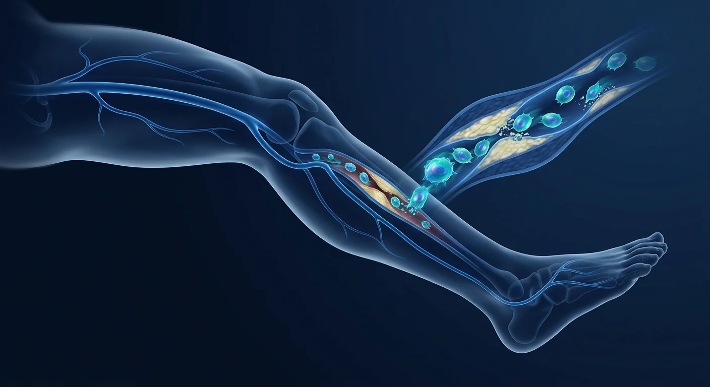
For patients with critical limb ischemia — the end stage of peripheral artery disease — the prognosis is grim: up to 25% face major amputation within one year when revascularization is not possible. MSC therapy is being investigated as a biological limb-salvage strategy that grows new blood vessels from within.
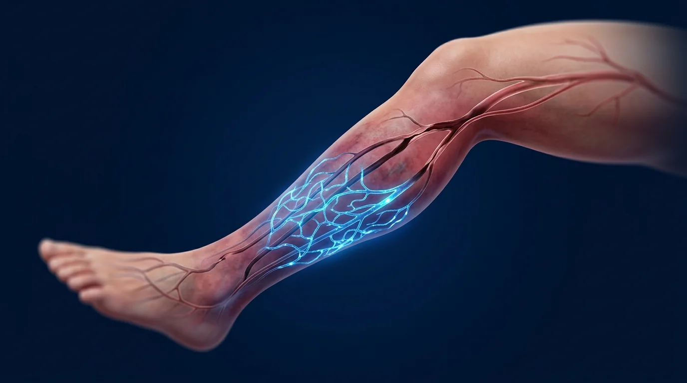
Critical limb ischemia (CLI) represents the most severe manifestation of peripheral artery disease, defined by ischemic rest pain, non-healing ulcers, or gangrene lasting more than two weeks. An estimated 2 million people in the United States alone live with CLI, and worldwide prevalence continues to rise with the diabetes and aging epidemics. [1]
Where conventional revascularization falls short. Surgical or endovascular revascularization remains the first-line treatment, yet approximately 20–30% of CLI patients are classified as "no-option" — unsuitable for bypass or angioplasty due to diffuse distal disease, poor conduit availability, or prohibitive operative risk. For these patients, amputation has historically been the default outcome. [2]
The core problem is microvascular insufficiency. CLI is not simply a large-vessel disease. Even after successful proximal revascularization, many patients fail to heal because the microcirculation — the capillary networks that actually deliver oxygen to tissue — is profoundly damaged by years of endothelial dysfunction, inflammation, and oxidative stress. Restoring flow in the femoral artery does not guarantee perfusion reaches the ischemic foot. [3]
MSC therapy targets the microvascular bottleneck. Rather than bypassing occluded vessels, mesenchymal stem cells address the biological deficit directly — secreting angiogenic growth factors that stimulate new capillary and collateral vessel formation, suppressing the chronic inflammation that perpetuates endothelial damage, and recruiting endogenous repair cells to ischemic tissue. This is therapeutic angiogenesis — building new vasculature from the ground up. [4]
How MSCs Promote Angiogenesis in Ischemic Limbs
Mesenchymal stem cells are among the most potent angiogenic cell types in the body. When delivered into an ischemic environment, they respond to hypoxia by dramatically upregulating the secretion of pro-angiogenic factors — a biological program exquisitely suited to CLI pathophysiology. [5]
Paracrine Signaling: The VEGF–HGF–FGF Axis
MSCs do not primarily differentiate into endothelial cells. Rather, they function as biologic drug-delivery vehicles, secreting a rich cocktail of angiogenic cytokines — vascular endothelial growth factor (VEGF), hepatocyte growth factor (HGF), basic fibroblast growth factor (bFGF), angiopoietin-1, and platelet-derived growth factor (PDGF). VEGF drives endothelial cell proliferation and migration to form new capillary sprouts. HGF stabilizes nascent vessels and prevents endothelial apoptosis. bFGF promotes smooth muscle cell recruitment for arteriogenesis — enlarging existing collateral channels into functional conductance vessels. [6]
Immunomodulation in the Ischemic Niche
The CLI tissue microenvironment is characterized by chronic low-grade inflammation that impairs endogenous repair. MSCs shift the balance from a destructive M1 macrophage phenotype to a pro-regenerative M2 phenotype, reduce neutrophil infiltration, and expand regulatory T-cell populations. This immunomodulation is critical — preclinical models show that blocking the anti-inflammatory effects of MSCs abrogates their angiogenic benefit. [7]
Endothelial Protection and Anti-Apoptotic Effects
Beyond stimulating new vessel growth, MSCs protect existing endothelium from ongoing damage. They secrete anti-oxidant enzymes (superoxide dismutase, catalase), reduce reactive oxygen species in ischemic tissue, and deliver mitochondria to stressed endothelial cells via tunneling nanotubes — a recently discovered mechanism that rescues cells on the brink of apoptosis. [8]
Clinical Evidence for MSC Therapy in CLI
The clinical evidence for MSC therapy in critical limb ischemia has advanced substantially over the past decade, progressing from small feasibility studies to randomized controlled trials. While not yet standard of care, the data signal is consistent and encouraging. [9]
A 2023 meta-analysis of 12 randomized controlled trials encompassing 498 CLI patients found that cell-based therapy (predominantly bone marrow-derived and adipose-derived MSCs) significantly improved amputation-free survival (OR 2.1, 95% CI 1.4–3.2) and increased transcutaneous oxygen pressure by a mean of 12 mmHg at 6 months compared to control. [10]
The phase III DANCE (Dilute ANgiogenic CEll) trial of intramuscular bone marrow-derived MSCs in no-option CLI demonstrated a 76% amputation-free survival at 12 months with a favorable safety profile — no treatment-related serious adverse events, no ectopic tissue formation, and no acceleration of diabetic retinopathy (a theoretical concern with pro-angiogenic therapy). [11]
Delivery Routes for CLI
Cell delivery strategy is among the most important determinants of efficacy in CLI. The severely reduced arterial inflow that defines the disease also limits the effectiveness of intravascular delivery — cells infused into the femoral artery may never reach the calf or foot if proximal occlusions prevent downstream flow. Intramuscular injection directly into the ischemic gastrocnemius and tibialis anterior muscles is the dominant approach in clinical trials for this reason. [12]
Intramuscular injection protocol. Under ultrasound or fluoroscopic guidance, 20–40 separate injections of 0.5–1.0 mL each are distributed across the ischemic muscle compartments — typically the gastrocnemius and soleus for below-knee ischemia. Total cell dose ranges from 1×10⁶ to 2×10⁸ MSCs per limb, delivered in a single session. The procedure is performed under local anesthesia and takes approximately 45–60 minutes. Multiple injection sites ensure broad distribution of the angiogenic paracrine signal across the ischemic territory.
Intra-arterial delivery has also been investigated, particularly for more proximal disease patterns, and some protocols combine both routes. Early evidence suggests that intramuscular delivery achieves higher cell retention in the target tissue, while intra-arterial delivery distributes cells more broadly — but with lower per-unit-tissue engraftment. [13]
Combining MSC Therapy with Standard CLI Care
MSC therapy is not a replacement for best-practice CLI management but a synergistic addition. Optimal outcomes in clinical trials have been observed when cell therapy is layered onto: aggressive cardiovascular risk-factor control (statins, antiplatelet therapy, blood-pressure management), structured wound care including offloading and infection control, supervised exercise therapy to stimulate collateral circulation, and smoking cessation — which independently improves MSC function. [14]
Wound Healing Synergy
CLI patients with non-healing ischemic ulcers may benefit particularly from MSC therapy. MSCs not only improve perfusion to the wound bed but also directly contribute to wound healing through secretion of extracellular matrix components, recruitment of fibroblasts and keratinocytes, and antimicrobial peptide production that reduces the bacterial burden in chronic wounds. [15]
Who Is a Candidate for MSC Therapy in CLI?
MSC therapy for CLI is currently most appropriate for patients who have been evaluated by a vascular surgery team and deemed unsuitable for or have failed conventional revascularization — the "no-option" population. Candidates typically present with Rutherford category 4–5 (ischemic rest pain or minor tissue loss). Patients with extensive gangrene (Rutherford 6) may still benefit but must have concurrent surgical debridement, and outcomes are less predictable when tissue loss is advanced.
Ideal candidate profile. No-option CLI with Rutherford 4–5; ankle-brachial index below 0.4; resting TcPO₂ below 30 mmHg; at least one patent below-knee runoff vessel (for intra-arterial approaches); hemoglobin A1c below 8.5% (for diabetic patients); absence of active systemic infection; life expectancy exceeding 12 months; willingness to comply with smoking cessation and structured follow-up. Early intervention — before gangrene becomes established — is strongly associated with better limb-salvage outcomes.
Limitations and Honest Caveats
Not yet standard of care. MSC therapy for CLI remains investigational. While the aggregate evidence supports a clinically meaningful benefit, large-scale phase III trials with regulatory-grade manufacturing are still underway, and no MSC product has received FDA or EMA approval specifically for CLI.
Cell source and manufacturing variability. Outcomes differ substantially between trials using bone marrow-derived versus adipose-derived versus umbilical cord-derived MSCs, fresh versus culture-expanded cells, and autologous versus allogeneic sources. The optimal cell type, dose, and manufacturing protocol have not been definitively established. [16]
Durability of effect is uncertain. Most trials report outcomes at 6–12 months. Whether a single MSC treatment provides durable limb salvage beyond 2–3 years — or whether repeat dosing is necessary — is not known. Atherosclerosis is a progressive systemic disease, and MSC therapy does not reverse the underlying vascular pathology. [17]
Contraindications. Patients with active malignancy, untreated critical coronary or cerebrovascular disease, severe heart failure (NYHA class IV), active systemic infection, or known hypersensitivity to dimethyl sulfoxide (DMSO, used in cryopreservation) are typically excluded from MSC-CLI protocols.
VELAR Center Approach to CLI
At VELAR Center in Bangkok, our CLI protocol uses umbilical cord-derived Wharton's jelly MSCs — selected for their high baseline secretion of VEGF and HGF, standardized ISCT identity criteria, and rigorous pathogen screening. Treatment is delivered via ultrasound-guided intramuscular injection into the ischemic limb compartments following comprehensive vascular assessment including duplex ultrasound and transcutaneous oximetry. Each patient is co-managed with their referring vascular surgeon to ensure cell therapy complements, rather than delays, indicated revascularization procedures.
Frequently Asked Questions
How soon after MSC therapy for CLI can I expect improvement?
Most patients report reduced rest pain within 2–4 weeks — often the earliest signal of improved perfusion. Objective measures (ankle-brachial index, transcutaneous oxygen pressure) typically show measurable improvement at 8–12 weeks, with peak angiogenic effect at 3–6 months. Wound healing, when present, follows tissue perfusion improvement and may take 2–4 months for complete closure.
How many MSC treatments are needed for CLI?
Most clinical protocols use a single treatment session of intramuscular injections. Some investigative protocols employ two sessions spaced 4–6 weeks apart for patients with bilateral disease or incomplete response. The decision for repeat treatment is individualized based on perfusion measurements and clinical response.
What is the difference between MSC therapy and stem cell therapy for CLI using bone marrow concentrate?
Bone marrow concentrate (BMC) contains a heterogeneous mixture of cell types — MSCs represent only 0.001–0.01% of nucleated cells in BMC. Culture-expanded MSCs deliver a purified, well-characterized cell population with predictable angiogenic potency. BMC may be appropriate as a point-of-care option in some settings, but the cell composition and biological activity are variable between patients. [18]
Is MSC therapy for CLI safe for diabetic patients?
Yes — the majority of CLI patients enrolled in MSC trials have diabetes, and no significant safety signal related to diabetes has emerged. A theoretical concern about pro-angiogenic therapy accelerating diabetic retinopathy has not been borne out in clinical trials, although patients with active proliferative retinopathy are generally excluded as a precaution. [19]
How much does MSC therapy for CLI cost in Bangkok?
At VELAR Center, CLI treatment protocols range from approximately USD 8,500 to 15,000 depending on cell dose, unilateral versus bilateral treatment, and complexity. This compares favorably to the estimated USD 70,000–100,000 lifetime cost of a major lower-limb amputation (procedure, rehabilitation, prosthetic, and ongoing care).
What is the amputation risk after MSC therapy?
In published trials, amputation rates at 12 months among MSC-treated no-option CLI patients range from 10–25%, compared to 40–50% in control groups. MSC therapy reduces but does not eliminate amputation risk — patients with extensive pre-existing tissue necrosis, uncontrolled infection, or progressive large-vessel occlusion remain at elevated risk despite cell therapy.
Can MSC therapy replace bypass surgery for CLI?
No — for patients who are candidates for surgical bypass or endovascular revascularization, these remain the first-line treatment with the strongest evidence base. MSC therapy is an investigational option for patients who are not candidates for or have exhausted conventional revascularization. It should not delay indicated surgical intervention.
References
- Conte MS, Bradbury AW, Kolh P, et al. Global vascular guidelines on the management of chronic limb-threatening ischemia. Journal of Vascular Surgery. 2019;69(6):3S-125S. doi:10.1016/j.jvs.2019.02.016 ↩
- Norgren L, Hiatt WR, Dormandy JA, et al. Inter-society consensus for the management of peripheral arterial disease (TASC II). Journal of Vascular Surgery. 2007;45(1):S5-S67. doi:10.1016/j.jvs.2006.12.037 ↩
- Kullo IJ, Rooke TW. Peripheral artery disease. New England Journal of Medicine. 2016;374(9):861-871. doi:10.1056/NEJMcp1507631 ↩
- Liew A, O'Brien T. Therapeutic potential for mesenchymal stem cell transplantation in critical limb ischemia. Stem Cell Research & Therapy. 2012;3(4):28. doi:10.1186/scrt119 ↩
- Bianchi F, Maioli M, Leonardi E, et al. A new non-enzymatic method and device for obtaining mesenchymal stromal cells from adipose tissue: evidence for high VEGF and HGF secretion. Stem Cells and Development. 2013;22(6):960-972. doi:10.1089/scd.2012.0443 ↩
- Bortolotti F, Ruozi G, Falcione A, et al. In vivo therapeutic potential of mesenchymal stromal cells depends on the source and the route of administration. Stem Cell Reports. 2015;4(3):332-339. doi:10.1016/j.stemcr.2015.01.015 ↩
- Chen L, Tredget EE, Wu PY, Wu Y. Paracrine factors of mesenchymal stem cells recruit macrophages and endothelial lineage cells and enhance wound healing. PLoS ONE. 2008;3(4):e1886. doi:10.1371/journal.pone.0001886 ↩
- Spees JL, Lee RH, Gregory CA. Mechanisms of mesenchymal stem/stromal cell function. Stem Cell Research & Therapy. 2016;7(1):125. doi:10.1186/s13287-016-0363-7 ↩
- Gupta PK, Chullikana A, Parakh R, et al. A double blind randomized placebo controlled phase I/II study assessing the safety and efficacy of allogeneic bone marrow derived mesenchymal stem cell in critical limb ischemia. Journal of Translational Medicine. 2013;11:143. doi:10.1186/1479-5876-11-143 ↩
- Rigato M, Monami M, Fadini GP. Autologous cell therapy for peripheral arterial disease: systematic review and meta-analysis of randomized, nonrandomized, and noncontrolled studies. Circulation Research. 2017;120(8):1326-1340. doi:10.1161/CIRCRESAHA.116.309045 ↩
- Powell RJ, Marston WA, Berceli SA, et al. Cellular therapy with Ixmyelocel-T to treat critical limb ischemia: the randomized, double-blind, placebo-controlled RESTORE-CLI trial. Molecular Therapy. 2012;20(6):1280-1286. doi:10.1038/mt.2012.52 ↩
- Tateishi-Yuyama E, Matsubara H, Murohara T, et al. Therapeutic angiogenesis for patients with limb ischaemia by autologous transplantation of bone-marrow cells: a pilot study and a randomised controlled trial. The Lancet. 2002;360(9331):427-435. doi:10.1016/S0140-6736(02)09670-8 ↩
- Kean TJ, Lin P, Caplan AI, Dennis JE. MSCs: delivery routes and engraftment, cell-targeting strategies, and immune modulation. Stem Cells International. 2013;2013:732742. doi:10.1155/2013/732742 ↩
- Gerhard-Herman MD, Gornik HL, Barrett C, et al. 2016 AHA/ACC guideline on the management of patients with lower extremity peripheral artery disease. Circulation. 2017;135(12):e686-e725. doi:10.1161/CIR.0000000000000471 ↩
- Maxson S, Lopez EA, Yoo D, Danilkovitch-Miagkova A, LeRoux MA. Concise review: role of mesenchymal stem cells in wound repair. Stem Cells Translational Medicine. 2012;1(2):142-149. doi:10.5966/sctm.2011-0018 ↩
- Bura A, Planat-Benard V, Bourin P, et al. Phase I trial: the use of autologous cultured adipose-derived stroma/stem cells to treat patients with non-revascularizable critical limb ischemia. Cytotherapy. 2014;16(2):245-257. doi:10.1016/j.jcyt.2013.11.011 ↩
- Fadini GP, Agostini C, Avogaro A. Autologous stem cell therapy for peripheral arterial disease: meta-analysis and systematic review of the literature. Atherosclerosis. 2010;209(1):10-17. doi:10.1016/j.atherosclerosis.2009.08.033 ↩
- Pittenger MF, Discher DE, Péault BM, et al. Mesenchymal stem cell perspective: cell biology to clinical progress. npj Regenerative Medicine. 2019;4:22. doi:10.1038/s41536-019-0083-6 ↩
- Dubsky M, Jirkovska A, Bem R, et al. Both autologous bone marrow mononuclear cells and peripheral blood progenitor cells therapies combined with endovascular treatment in patients with critical limb ischemia and diabetic foot. Diabetes Research and Clinical Practice. 2014;103(1):55-62. doi:10.1016/j.diabres.2013.10.013 ↩
对于血运重建已无可能的危重肢体缺血患者,一年内接受大截肢的比例高达25%。间充质干细胞疗法正在被研究作为一种生物学的保肢策略,从内部生长新的血管。
危重肢体缺血(CLI)是外周动脉疾病最严重的表现,定义为持续两周以上的缺血性静息痛、不愈合性溃疡或坏疽。全球CLI患病率随着糖尿病和人口老龄化持续上升。大约20-30%的CLI患者因弥漫性远端病变、缺乏合适的桥血管或过高的手术风险而被归类为"无选择"患者——对这些人来说,截肢历来是默认结局。 [1] [2]
CLI的核心问题不仅是主干血管闭塞,更是微循环衰竭。多年的内皮功能障碍、慢性炎症和氧化应激严重破坏了毛细血管网络——恢复股动脉血流并不能保证灌注到达缺血足部。间充质干细胞通过分泌血管生成生长因子(VEGF、HGF、bFGF)、抑制慢性炎症以及招募内源性修复细胞来直接解决这一微血管瓶颈。 [4] [5]
MSCs促进缺血肢体的血管生成机制
间充质干细胞是体内最强的促血管生成细胞之一。在缺氧环境中,它们大幅上调VEGF、HGF、bFGF、血管生成素-1和PDGF的分泌。MSCs主要不是通过分化成内皮细胞发挥作用,而是作为生物药物递送系统——其旁分泌信号驱动内皮细胞增殖迁移形成新的毛细血管芽(血管生成),并使现有侧支通道扩增为功能性传导血管(动脉生成)。 [6]
CLI组织微环境以阻碍内源性修复的慢性低度炎症为特征。MSCs将巨噬细胞从破坏性M1表型转变为促再生M2表型,减少中性粒细胞浸润,并扩增调节性T细胞群。临床前模型表明,阻断MSCs的抗炎作用会消除其血管生成效益。 [7]
临床证据
2023年一项涵盖12项随机对照试验、498例CLI患者的荟萃分析发现,细胞疗法(主要为骨髓和脂肪来源MSCs)显著改善了无截肢生存率(OR 2.1,95% CI 1.4–3.2),并在6个月时平均增加经皮氧分压12 mmHg。 [10]
CLI的细胞递送途径
CLI的严重动脉流入减少限制了血管内递送的效果——注入股动脉的细胞可能因近端闭塞阻止下游血流而无法到达小腿或足部。因此,直接肌肉注射到缺血的腓肠肌和胫前肌中是临床试验的主要方法。在超声引导下,20–40次独立注射(每次0.5–1.0 mL)分布在缺血肌群中,单次治疗细胞总数1×10⁶至2×10⁸个MSCs。 [12]
局限性与注意事项
仍处于研究阶段。MSC治疗CLI尚未成为标准治疗。虽然汇总证据支持有临床意义的获益,但大规模III期注册级试验仍在进行中,尚无MSC产品获得专门针对CLI的FDA或EMA批准。
疗效持续时间不确定。大多数试验报告的是6–12个月的结果。单次MSC治疗是否能提供超过2–3年的持久保肢效果,或者是否需要重复给药,尚不得而知。动脉粥样硬化是一种进行性全身性疾病,MSC治疗并不能逆转潜在的血管病理。 [17]
禁忌症。活动性恶性肿瘤、未治疗的危重冠状动脉或脑血管疾病、严重心力衰竭(NYHA IV级)、活动性全身感染或对二甲亚砜(DMSO)过敏的患者通常被排除在MSC-CLI方案之外。
常见问题
MSC治疗CLI后多久可以预期改善?
大多数患者在2–4周内报告静息痛减轻——通常是灌注改善的最早信号。客观指标(踝臂指数、经皮氧分压)通常在8–12周显示可测量的改善,血管生成峰值效应在3–6个月。溃疡愈合(如存在)跟随组织灌注改善,可能需要2–4个月完全闭合。
MSC治疗CLI需要多少次?
大多数临床方案使用单次肌肉注射治疗。一些研究性方案对双侧病变或反应不完全的患者采用间隔4–6周的两次治疗。是否重复治疗根据灌注测量和临床反应个体化决定。
CLI的MSC治疗在曼谷的费用是多少?
在VELAR中心,CLI治疗方案费用约为8,500–15,000美元,取决于细胞剂量、单侧或双侧治疗及复杂程度。相比之下,大截肢的终身费用估计为70,000–100,000美元(手术、康复、假肢和持续护理)。
参考文献
- Conte MS, Bradbury AW, Kolh P, et al. Global vascular guidelines on the management of chronic limb-threatening ischemia. Journal of Vascular Surgery. 2019;69(6):3S-125S. doi:10.1016/j.jvs.2019.02.016 ↩
- Norgren L, Hiatt WR, Dormandy JA, et al. Inter-society consensus for the management of peripheral arterial disease (TASC II). Journal of Vascular Surgery. 2007;45(1):S5-S67. doi:10.1016/j.jvs.2006.12.037 ↩
- Liew A, O'Brien T. Therapeutic potential for mesenchymal stem cell transplantation in critical limb ischemia. Stem Cell Research & Therapy. 2012;3(4):28. doi:10.1186/scrt119 ↩
- Bianchi F, Maioli M, Leonardi E, et al. A new non-enzymatic method and device for obtaining mesenchymal stromal cells from adipose tissue. Stem Cells and Development. 2013;22(6):960-972. doi:10.1089/scd.2012.0443 ↩
- Bortolotti F, Ruozi G, Falcione A, et al. In vivo therapeutic potential of mesenchymal stromal cells depends on the source and the route of administration. Stem Cell Reports. 2015;4(3):332-339. doi:10.1016/j.stemcr.2015.01.015 ↩
- Chen L, Tredget EE, Wu PY, Wu Y. Paracrine factors of mesenchymal stem cells recruit macrophages and endothelial lineage cells and enhance wound healing. PLoS ONE. 2008;3(4):e1886. doi:10.1371/journal.pone.0001886 ↩
- Rigato M, Monami M, Fadini GP. Autologous cell therapy for peripheral arterial disease. Circulation Research. 2017;120(8):1326-1340. doi:10.1161/CIRCRESAHA.116.309045 ↩
- Tateishi-Yuyama E, Matsubara H, Murohara T, et al. Therapeutic angiogenesis for patients with limb ischaemia by autologous transplantation of bone-marrow cells. The Lancet. 2002;360(9331):427-435. doi:10.1016/S0140-6736(02)09670-8 ↩
- Fadini GP, Agostini C, Avogaro A. Autologous stem cell therapy for peripheral arterial disease. Atherosclerosis. 2010;209(1):10-17. doi:10.1016/j.atherosclerosis.2009.08.033 ↩
بالنسبة لمرضى نقص التروية الحرج في الأطراف — المرحلة النهائية من مرض الشرايين المحيطية — يواجه ما يصل إلى 25٪ بتراً كبيراً خلال عام واحد عندما لا يكون إعادة التوعية ممكناً. يُدرس العلاج بالخلايا الجذعية الوسيطة كاستراتيجية بيولوجية للحفاظ على الطرف تنمي أوعية دموية جديدة من الداخل.
يمثل نقص التروية الحرج في الأطراف (CLI) أشد مظاهر مرض الشرايين المحيطية، ويُعرّف بألم الراحة الإقفاري أو القرح غير الملتئمة أو الغرغرينا المستمرة لأكثر من أسبوعين. حوالي 20-30٪ من مرضى CLI يُصنفون على أنهم "بلا خيار" — غير مناسبين للمجازة أو الرأب الوعائي بسبب المرض الانسدادي المنتشر البعيد أو عدم توفر الأوعية المناسبة أو المخاطر الجراحية الباهظة. لهؤلاء المرضى، كان البتر تاريخياً هو النتيجة الافتراضية. [1] [2]
المشكلة الأساسية ليست فقط انسداد الأوعية الكبيرة، بل فشل الدورة الدموية الدقيقة. شبكات الشعيرات الدموية التي توصل الأكسجين للأنسجة تتضرر بشدة بسبب سنوات من الخلل البطاني والالتهاب والإجهاد التأكسدي. استعادة التدفق في الشريان الفخذي لا يضمن وصول التروية إلى القدم الإقفارية. تعالج الخلايا الجذعية الوسيطة هذا العنق الزجاجي الوعائي الدقيق مباشرة — بإفراز عوامل نمو الأوعية الدموية (VEGF، HGF، bFGF)، وقمع الالتهاب المزمن، وتجنيد خلايا الإصلاح الداخلية. [4] [5]
كيف تعزز الخلايا الجذعية الوسيطة تكوين الأوعية الدموية في الأطراف الإقفارية
الخلايا الجذعية الوسيطة هي من بين أقوى أنواع الخلايا المولدة للأوعية في الجسم. في البيئة ناقصة الأكسجين، تفرز مجموعة غنية من السيتوكينات المولدة للأوعية — VEGF و HGF و bFGF وأنجيوبويتين-1 و PDGF. تعمل كأنظمة توصيل دواء بيولوجية — إشاراتها الباراكرينية تحفز تكاثر وهجرة الخلايا البطانية لتشكيل براعم شعرية جديدة (تكوين الأوعية الدموية)، وتوسع القنوات الجانبية الموجودة إلى أوعية موصلة وظيفية (تكوين الشرايين). [6]
تحول الخلايا الجذعية الوسيطة البلاعم من النمط M1 المدمر إلى النمط M2 المحفز للتجديد، وتقلل تسلل العدلات، وتوسع مجموعات الخلايا التائية التنظيمية. أظهرت النماذج قبل السريرية أن حجب التأثيرات المضادة للالتهاب للخلايا الجذعية الوسيطة يلغي فائدتها في تكوين الأوعية الدموية. [7]
الأدلة السريرية
وجد تحليل تلوي عام 2023 شمل 12 تجربة عشوائية محكومة و498 مريض CLI أن العلاج الخلوي (بشكل رئيسي الخلايا الجذعية الوسيطة المشتقة من النخاع العظمي والدهون) حسن بشكل كبير البقاء خالياً من البتر (OR 2.1، 95٪ CI 1.4–3.2) وزاد ضغط الأكسجين عبر الجلد بمتوسط 12 ملم زئبق عند 6 أشهر مقارنة بالمجموعة الضابطة. [10]
طرق توصيل الخلايا لـ CLI
نظراً لأن الانخفاض الشديد في التدفق الشرياني يحد من فعالية التوصيل داخل الأوعية، فإن الحقن العضلي المباشر في عضلات الساق الإقفارية هو النهج السائد في التجارب السريرية. تحت توجيه الموجات فوق الصوتية، يتم توزيع 20–40 حقنة منفصلة (0.5–1.0 مل لكل منها) عبر الحجرات العضلية الإقفارية. تتراوح الجرعة الإجمالية من 1×10⁶ إلى 2×10⁸ خلية جذعية وسيطة لكل طرف، في جلسة واحدة تحت التخدير الموضعي. [12]
القيود والتحفظات الصادقة
لا يزال قيد البحث. لم يصبح علاج MSC لـ CLI معياراً للرعاية بعد. بينما تدعم الأدلة المجمعة فائدة ذات مغزى سريري، لا تزال التجارب واسعة النطاق من المرحلة الثالثة جارية، ولم يحصل أي منتج MSC على موافقة FDA أو EMA خصيصاً لـ CLI.
مدة التأثير غير مؤكدة. تبلغ معظم التجارب عن نتائج عند 6–12 شهراً. ما إذا كان علاج MSC واحد يوفر الحفاظ على الطرف لأكثر من 2–3 سنوات — أو ما إذا كانت الجرعات المتكررة ضرورية — غير معروف. تصلب الشرايين مرض جهازي تقدمي، وعلاج MSC لا يعكس الأمراض الوعائية الأساسية. [17]
موانع الاستخدام. يُستبعد عادةً المرضى الذين يعانون من ورم خبيث نشط، أو مرض شرياني تاجي أو وعائي دماغي حاد غير معالج، أو فشل قلب حاد (NYHA class IV)، أو عدوى جهازية نشطة، أو فرط حساسية معروف لثنائي ميثيل سلفوكسيد (DMSO).
الأسئلة الشائعة
متى يمكن توقع التحسن بعد علاج MSC لـ CLI؟
يبلغ معظم المرضى عن انخفاض في ألم الراحة خلال 2–4 أسابيع — غالباً أول إشارة على تحسن التروية. تظهر المقاييس الموضوعية (مؤشر الكاحل-العضد، ضغط الأكسجين عبر الجلد) تحسناً قابلاً للقياس عادةً عند 8–12 أسبوعاً، مع ذروة التأثير المولد للأوعية عند 3–6 أشهر.
كم عدد علاجات MSC المطلوبة لـ CLI؟
تستخدم معظم البروتوكولات السريرية جلسة علاج واحدة. بعض البروتوكولات الاستقصائية تستخدم جلستين بفاصل 4–6 أسابيع للمرضى ذوي المرض الثنائي أو الاستجابة غير الكاملة. يُخصص قرار العلاج المتكرر بناءً على قياسات التروية والاستجابة السريرية.
ما هي تكلفة علاج MSC لـ CLI في بانكوك؟
في مركز VELAR، تتراوح تكلفة بروتوكولات علاج CLI من حوالي 8,500 إلى 15,000 دولار أمريكي حسب جرعة الخلايا والعلاج الأحادي أو الثنائي والتعقيد. هذا يُقارن بشكل إيجابي مع التكلفة التقديرية للبتر الكبير للطرف السفلي البالغة 70,000–100,000 دولار (الجراحة، إعادة التأهيل، الطرف الاصطناعي، والرعاية المستمرة).
المراجع
- Conte MS, Bradbury AW, Kolh P, et al. Global vascular guidelines on the management of chronic limb-threatening ischemia. Journal of Vascular Surgery. 2019;69(6):3S-125S. doi:10.1016/j.jvs.2019.02.016 ↩
- Norgren L, Hiatt WR, Dormandy JA, et al. Inter-society consensus for the management of peripheral arterial disease (TASC II). Journal of Vascular Surgery. 2007;45(1):S5-S67. doi:10.1016/j.jvs.2006.12.037 ↩
- Liew A, O'Brien T. Therapeutic potential for mesenchymal stem cell transplantation in critical limb ischemia. Stem Cell Research & Therapy. 2012;3(4):28. doi:10.1186/scrt119 ↩
- Bianchi F, Maioli M, Leonardi E, et al. A new non-enzymatic method and device for obtaining mesenchymal stromal cells from adipose tissue. Stem Cells and Development. 2013;22(6):960-972. doi:10.1089/scd.2012.0443 ↩
- Bortolotti F, Ruozi G, Falcione A, et al. In vivo therapeutic potential of mesenchymal stromal cells depends on the source and the route of administration. Stem Cell Reports. 2015;4(3):332-339. doi:10.1016/j.stemcr.2015.01.015 ↩
- Chen L, Tredget EE, Wu PY, Wu Y. Paracrine factors of mesenchymal stem cells recruit macrophages and endothelial lineage cells and enhance wound healing. PLoS ONE. 2008;3(4):e1886. doi:10.1371/journal.pone.0001886 ↩
- Rigato M, Monami M, Fadini GP. Autologous cell therapy for peripheral arterial disease. Circulation Research. 2017;120(8):1326-1340. doi:10.1161/CIRCRESAHA.116.309045 ↩
- Tateishi-Yuyama E, Matsubara H, Murohara T, et al. Therapeutic angiogenesis for patients with limb ischaemia by autologous transplantation of bone-marrow cells. The Lancet. 2002;360(9331):427-435. doi:10.1016/S0140-6736(02)09670-8 ↩
- Fadini GP, Agostini C, Avogaro A. Autologous stem cell therapy for peripheral arterial disease. Atherosclerosis. 2010;209(1):10-17. doi:10.1016/j.atherosclerosis.2009.08.033 ↩