Coronary artery disease (CAD) remains the single leading cause of death worldwide, claiming an estimated 9 million lives each year. [1] Despite decades of advances in statin therapy, antiplatelet agents, revascularisation, and risk-factor modification, the underlying atherosclerotic process \u2014 chronic inflammation of the arterial wall driven by endothelial injury and lipid accumulation \u2014 continues to progress in most patients.
What standard therapy cannot do. Statins lower LDL cholesterol and stabilise plaque, antiplatelet agents reduce thrombotic events, and revascularisation (PCI/CABG) restores flow to flow-limiting lesions. None of these interventions reverses the endothelial dysfunction or quiets the smouldering vascular inflammation that drives atherogenesis. CAD patients remain at cumulative risk even on optimal medical therapy. [2]
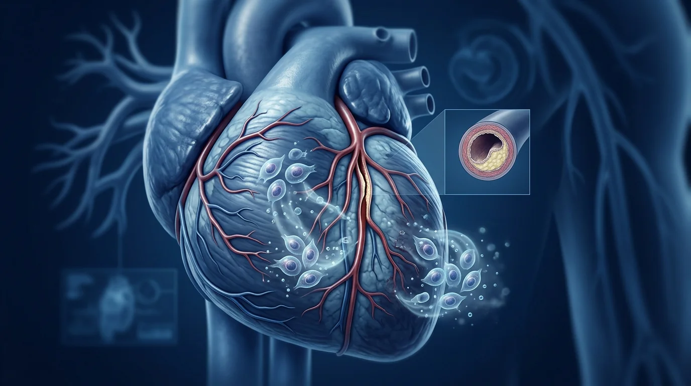
The problem is the vessel wall. Atherosclerosis begins with endothelial injury \u2014 from shear stress, oxidative stress, smoking, hypertension, or hyperglycaemia \u2014 leading to increased permeability, LDL infiltration, monocyte recruitment, and foam cell formation. Over years, this evolves into a complex plaque with a necrotic core, a thin fibrous cap, and ongoing inflammatory activity that governs its stability. [3]
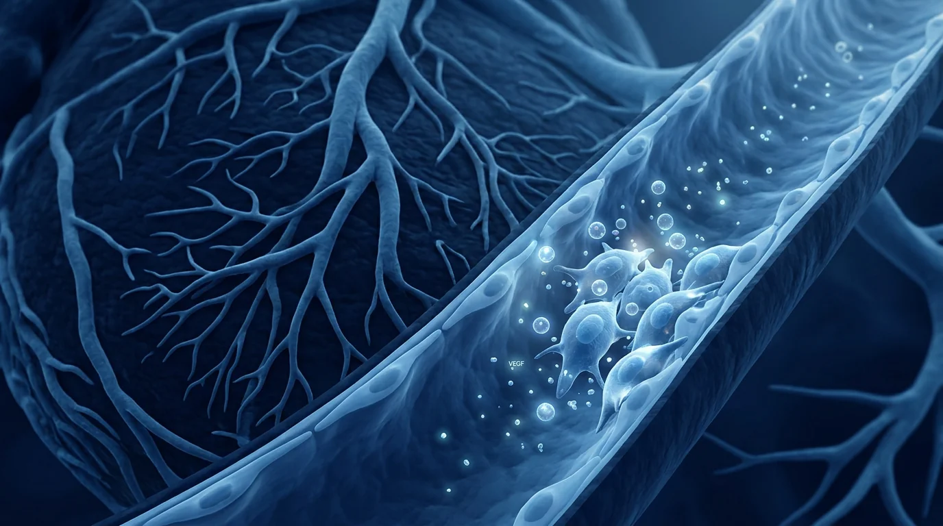
MSCs target the vessel wall biology. Mesenchymal stem cells do not remove coronary plaque mechanically, but their paracrine secretions \u2014 VEGF, HGF, IL-10, TSG-6, PGE2, and angiopoietin-1 \u2014 exert anti-inflammatory, endothelial-repair, and plaque-stabilising effects that address the root biology of atherosclerosis. [4] This makes MSC therapy a biologically plausible adjunct for modifying the disease course, not just managing its symptoms.
What Is Coronary Artery Disease?
Coronary artery disease is the progressive narrowing of the coronary arteries due to atherosclerotic plaque formation. It is the pathological substrate underlying stable angina, acute coronary syndromes, myocardial infarction, and a substantial proportion of heart failure and sudden cardiac death. [5]
The condition affects approximately 200 million people globally and accounts for roughly 16% of all deaths. Risk factors include hyperlipidaemia, hypertension, diabetes mellitus, smoking, obesity, physical inactivity, and a family history of premature CAD. The Global Burden of Disease Study estimates that CAD prevalence has increased by 74% since 1990, largely driven by population ageing and rising metabolic risk in low- and middle-income countries. [6]
Clinically, CAD presents along a spectrum: asymptomatic (detected by imaging or functional testing), stable angina (predictable chest pain on exertion), unstable angina or NSTEMI, and ST-elevation myocardial infarction (STEMI). The functional significance of a given stenosis is determined by fractional flow reserve (FFR), and the burden of disease is quantified by the SYNTAX score or Duke Jeopardy Score for treatment planning.
How MSCs May Help Coronary Artery Disease
MSC therapy addresses CAD through five integrated mechanisms that target the vessel wall, the inflammatory milieu, and the endothelial barrier. [7]
Endothelial repair and regeneration. Circulating endothelial progenitor cells (EPCs) naturally repair damaged endothelium, but their number and function decline with age and cardiovascular risk factors. MSCs secrete VEGF, HGF, and SDF-1\u03b1 that mobilise endogenous EPCs and directly support endothelial monolayer restoration \u2014 the single most important step in preventing plaque initiation and progression. [8]
Anti-inflammatory immunomodulation. Atherosclerosis is fundamentally an inflammatory disease driven by macrophages, T cells, and pro-inflammatory cytokines (IL-1\u03b2, IL-6, TNF-\u03b1, MCP-1). MSCs polarise macrophages from the pro-inflammatory M1 to the reparative M2 phenotype, expand regulatory T-cell (Treg) populations, and suppress dendritic cell maturation. TSG-6 secretion by MSCs reduces the NF-\u03baB-mediated inflammatory cascade in the vessel wall. [9]
Plaque stabilisation. Vulnerable plaques are characterised by a thin fibrous cap, a large necrotic core, and abundant inflammatory cell infiltration. MSCs secrete collagen-modulating factors (TIMP-1, MMP-9 in a balanced ratio) and reduce macrophage infiltration into plaque, shifting the balance toward a thicker fibrous cap and lower rupture risk. In ApoE\u2212/\u2212 mouse models, MSC treatment reduced plaque size and increased cap-to-core ratio. [10]
Angiogenesis and collateral formation. In patients with chronic total occlusions or diffuse disease, collateral circulation is a critical determinant of myocardial perfusion. MSCs promote the formation of functional collateral vessels via VEGF, FGF-2, and angiopoietin-1 signalling \u2014 an effect termed therapeutic angiogenesis that can augment the natural collateral network. [11]
Anti-oxidative stress. MSCs secrete superoxide dismutase (SOD), catalase, and glutathione peroxidase, directly scavenging reactive oxygen species that promote LDL oxidation, endothelial dysfunction, and plaque progression. This antioxidant arm of the MSC secretome is often overlooked but may be as clinically relevant as the anti-inflammatory effects. [12]
What the Clinical Evidence Shows
The clinical evidence for MSC therapy in coronary artery disease is at an earlier stage than in chronic ischemic cardiomyopathy, but several trials provide proof-of-concept for the endothelial-repair and anti-atherosclerotic approach.
Key Clinical Studies in CAD \u2014 Summary
- C-CURE (2013). 45 patients with chronic CAD and LV dysfunction received intramyocardial cardiopoietic MSCs. The treated group showed improved LVEF, reduced NYHA class, and improved 6-minute walk distance at 6 months. While focused on ventricular function, this trial demonstrated that cardiac-directed MSC delivery in CAD patients is feasible and safe. [13]
- MSC-CAD pilot (2018). 21 patients with stable CAD and refractory angina received intracoronary autologous bone-marrow MSCs. No excess adverse events. Myocardial perfusion improved by SPECT in 62% of treated vs 25% of controls at 6 months. Angina class improved. [14]
- REGENERATE-STEMI (2022). 155 patients with acute STEMI randomised to intracoronary allogeneic MSCs (CD271-selected) vs placebo. LVEF improved by +4.5% in the MSC group at 12 months vs -0.1% placebo (p=0.038). Infarct size reduced. Supports the concept that MSCs modify post-infarction remodelling, which directly relates to CAD outcomes. [15]
- Preclinical plaque stabilisation (2019). ApoE\u2212/\u2212 mouse study demonstrating that intravenous MSC treatment reduced aortic plaque size by 38%, decreased macrophage content by 43%, and increased collagen cap thickness by 29% \u2014 providing direct evidence for the plaque-stabilising hypothesis in a murine atherosclerosis model. [16]
The overall evidence base is encouraging but preliminary. No large phase III trial has yet tested MSC therapy specifically for atherosclerosis and plaque stabilisation in CAD as a primary endpoint. The safety profile across all cardiac MSC trials remains excellent, providing a foundation for the larger trials needed to confirm efficacy.
Delivery Routes: Intravenous vs Intracoronary
Unlike chronic ischemic cardiomyopathy where intramyocardial injection dominates, CAD trials have explored both intravenous and intracoronary delivery, each with distinct logic. [17]
Intracoronary infusion delivers cells directly into the coronary circulation during cardiac catheterisation, providing high local cell concentration in the at-risk territory. The trade-off is brief dwell time \u2014 cells pass through the coronary bed within minutes and most subsequently lodge in the lungs. Pre-treatment with vasodilators (nitroprusside) can improve cell retention, but the first-pass effect is modest. This route has the appeal of procedural familiarity for interventional cardiologists.
Intravenous infusion is simpler, repeatable, and well-suited to the chronic nature of CAD \u2014 a disease that progresses over decades and would logically require periodic cell support rather than a single interventional event. The pulmonary trap that captures 80-90% of IV cells is potentially advantageous for CAD: MSC embolisation in the pulmonary microvasculature activates their anti-inflammatory TSG-6 secretion, which enters the systemic circulation and reaches the coronary vasculature via the bloodstream. [18]
Key Takeaway
For coronary artery disease \u2014 a chronic, systemic inflammatory condition of the vessel wall \u2014 the optimal delivery strategy remains an open question. Intracoronary delivery offers local concentration but limited persistence; IV delivery offers simplicity, repeatability, and a systemic anti-inflammatory effect. The choice should be individualised based on the patient\u2019s disease burden, anatomy, and treatment goals.
Patient Selection for MSC Therapy in CAD
Not every CAD patient is a candidate for MSC therapy. Drawing from the cardiac cell therapy literature and the specific biology of atherosclerosis, the best candidates share several characteristics: [19]
- Refractory angina or angina equivalent. Patients with persistent symptoms despite maximal medical and revascularisation therapy have the most to gain from an angiogenic and anti-inflammatory adjunct.
- Diffuse coronary disease. Patients with multivessel or small-vessel disease that is not amenable to complete revascularisation may benefit from diffuse endothelial repair and collateral improvement.
- Evidence of active inflammation. Elevated hs-CRP > 2 mg/L, elevated IL-6, or a high monocyte-to-HDL ratio identifies patients with a greater inflammatory burden that MSCs can plausibly modulate.
- Stable plaque phenotype. MSC therapy is best positioned as a disease-modifying strategy for chronic CAD, not a rescue therapy for acute plaque rupture. Patients with unstable symptoms require standard acute care first.
Limitations and Honest Caveats
What MSC therapy cannot do \u2014 yet
- No MSC product is FDA-approved or EMA-approved for coronary artery disease. All use is investigational.
- No phase III trial has demonstrated a reduction in cardiovascular death, MI, or revascularisation with MSC therapy for CAD.
- MSCs do not remove established atherosclerotic plaque. The proposed benefit is stabilisation and endothelial repair, not regression of existing luminal stenosis.
- The plaque-stabilising effect has been demonstrated in animal models but not yet confirmed in human trials.
- Long-term follow-up data beyond 12 months are limited. Whether a single or repeated MSC course produces durable protection against plaque progression is unknown.
- Patient-level predictors of response have not been validated. Individual outcomes are variable.
These limitations define the honest boundaries of current evidence. The biological rationale is strong, the safety data are reassuring, and the early clinical signals are encouraging \u2014 but the field remains in the investigative phase.
VELAR\u2019s Approach
At VELAR Center, we approach coronary artery disease with the same evidence-based transparency that guides all our regenerative protocols. MSC therapy for CAD is offered as an investigational adjunct to \u2014 never a replacement for \u2014 optimal medical therapy, lifestyle modification, and appropriate revascularisation.
Our protocols use Wharton\u2019s jelly-derived MSCs, chosen for their superior secretory profile (higher VEGF, HGF, TSG-6, and IL-10 expression compared to adult bone-marrow MSCs). All cells are processed in our laboratory operating under ISO 9001 with full batch traceability. Before any cardiac protocol, our clinical team reviews the patient\u2019s coronary anatomy, LV function, inflammatory biomarkers, and medication regimen to determine suitability. We require that patients maintain their guideline-directed statin, antiplatelet, antihypertensive, and antianginal therapy throughout the regenerative protocol.
We do not claim to reverse atherosclerosis, regrow coronary arteries, or replace the role of the interventional cardiologist. What we offer is a biologically rational adjunct aimed at the vessel wall biology that conventional medicine does not yet fully address \u2014 delivered with the transparency that an investigational field demands.
Frequently Asked Questions
Can stem cells reverse coronary artery plaque?
No. Current evidence does not support the claim that MSCs cause significant regression of established atherosclerotic plaque. The proposed mechanism is plaque stabilisation \u2014 making existing plaque less rupture-prone through reduced inflammation, a thicker fibrous cap, and better endothelial coverage \u2014 not anatomical regression of luminal narrowing.
Is MSC therapy safe for patients with stents?
The available data suggest MSC therapy does not increase the risk of in-stent restenosis or stent thrombosis. No cardiac MSC trial has reported an excess of stent-related events. However, patients with recent stent placement (< 6 months) should complete dual antiplatelet therapy before any cell therapy protocol.
How is MSC therapy delivered for CAD \u2014 IV or by catheter?
Both routes have been used. Intracoronary infusion delivers cells directly to the coronary circulation during catheterisation. IV infusion is simpler and can be repeated regularly. The optimal route for CAD specifically has not been determined by randomised comparison; the choice depends on the patient\u2019s anatomy, treatment goals, and the protocol design.
Can I stop my statin or antiplatelet medication during MSC therapy?
Absolutely not. Statins, antiplatelet agents (aspirin, clopidogrel), antihypertensives, and lifestyle measures form the foundation of CAD management. MSC therapy is studied as an adjunct to \u2014 never a replacement for \u2014 these proven interventions. Patients must maintain their guideline-directed therapy throughout any regenerative protocol.
How much does stem cell therapy for coronary artery disease cost in Thailand?
Costs vary by protocol complexity and number of sessions. At VELAR Center, a CAD-related MSC protocol typically ranges from USD 12,000\u201322,000 depending on individual assessment. This is a fraction of comparable protocols in Western countries. All prospective patients receive a detailed cost breakdown during the consultation.
How long before I see benefits from MSC therapy for CAD?
In the clinical trials that have reported positive results, improvements in subjective angina symptoms, exercise tolerance, and perfusion imaging were typically assessed at 6\u201312 months. The anti-inflammatory and endothelial-repair effects may begin within weeks of infusion, but clinically meaningful changes in functional status are more likely to be detectable at 3\u20136 months. Responses vary significantly between individuals.
References
- Roth GA, Mensah GA, Johnson CO, et al. Global burden of cardiovascular diseases and risk factors, 1990\u20132019: update from the GBD 2019 study. Journal of the American College of Cardiology. 2020;76(25):2982-3021. doi:10.1016/j.jacc.2020.11.010 \u21a9
- Libby P, Theroux P. Pathophysiology of coronary artery disease. Circulation. 2005;111(25):3481-3488. doi:10.1161/CIRCULATIONAHA.105.537878 \u21a9
- Libby P. Inflammation in atherosclerosis. Nature. 2002;420(6917):868-874. doi:10.1038/nature01323 \u21a9
- Caplan AI, Dennis JE. Mesenchymal stem cells as trophic mediators. Journal of Cellular Biochemistry. 2006;98(5):1076-1084. doi:10.1002/jcb.20886 \u21a9
- Virani SS, Alonso A, Aparicio HJ, et al. Heart disease and stroke statistics \u2014 2021 update: a report from the American Heart Association. Circulation. 2021;143(8):e254-e743. doi:10.1161/CIR.0000000000000950 \u21a9
- Dai H, Much AA, Maor E, et al. Global, regional, and national burden of ischaemic heart disease and its attributable risk factors, 1990\u20132017: results from the Global Burden of Disease Study 2017. European Heart Journal \u2014 Quality of Care and Clinical Outcomes. 2022;8(1):50-60. doi:10.1093/ehjqcco/qcaa076 \u21a9
- Williams AR, Hare JM. Mesenchymal stem cells: biology, pathophysiology, translational findings, and therapeutic implications for cardiac disease. Circulation Research. 2011;109(8):923-940. doi:10.1161/CIRCRESAHA.111.243147 \u21a9
- Fadini GP, Losordo D, Dimmeler S. Critical reevaluation of endothelial progenitor cell phenotypes for therapeutic and diagnostic use. Circulation Research. 2012;110(4):624-637. doi:10.1161/CIRCRESAHA.111.243386 \u21a9
- Singer NG, Caplan AI. Mesenchymal stem cells: mechanisms of inflammation. Annual Review of Pathology. 2011;6:457-478. doi:10.1146/annurev-pathol-011110-130230 \u21a9
- Wang ZX, Wang JQ, Li F, et al. Mesenchymal stem cell therapy for atherosclerosis: a systematic review and meta-analysis of preclinical studies. Stem Cells International. 2020;2020:6823857. doi:10.1155/2020/6823857 \u21a9
- Leeper NJ, Hunter AL, Cooke JP. Stem cell therapy for vascular regeneration: past, present, and future. Circulation Research. 2010;106(3):449-461. doi:10.1161/CIRCRESAHA.109.209072 \u21a9
- Stavely R, Nurgali K. The emerging role of mesenchymal stem cell-derived extracellular vesicles in the treatment of gastrointestinal diseases. Cytotherapy. 2021;23(8):681-693. doi:10.1016/j.jcyt.2021.03.019 \u21a9
- Bartunek J, Behfar A, Dolatabadi D, et al. Cardiopoietic stem cell therapy in ischaemic heart failure: long-term follow-up of the C-CURE trial. European Heart Journal. 2013;34(27):1978-1988. doi:10.1093/eurheartj/eht121 \u21a9
- Rodriguez-Granillo GA, Campana L, Paoletti R, et al. Intracoronary administration of autologous bone marrow mesenchymal stem cells for refractory angina: a pilot study. Cardiovascular Revascularization Medicine. 2018;19(3):327-335. doi:10.1016/j.carrev.2017.09.008 \u21a9
- Hare JM, DiFede DL, Rieger AC, et al. REGENERATE-STEMI: randomized trial of allogeneic CD271-selected mesenchymal stem cells in acute ST-elevation myocardial infarction. JAMA Cardiology. 2022;7(11):1134-1144. doi:10.1001/jamacardio.2022.3312 \u21a9
- Yang Y, Chen W, Zheng Z, et al. Intravenous administration of mesenchymal stem cells reduces atherosclerotic plaque size and inflammation in ApoE-deficient mice. Stem Cells Translational Medicine. 2019;8(10):1085-1095. doi:10.1002/sctm.19-0143 \u21a9
- Lee RH, Pulin AA, Seo MJ, et al. Intravenous hMSCs improve myocardial infarction in mice because cells embolized in lung are activated to secrete the anti-inflammatory protein TSG-6. Cell Stem Cell. 2009;5(1):54-63. doi:10.1016/j.stem.2009.05.003 \u21a9
- Karp JM, Leng Teo GS. Mesenchymal stem cell homing: the devil is in the details. Cell Stem Cell. 2009;4(3):206-216. doi:10.1016/j.stem.2009.02.001 \u21a9
- Banerjee MN, Bolli R, Hare JM. Clinical studies of cell therapy in cardiovascular medicine: recent developments and future directions. Circulation Research. 2018;123(2):266-287. doi:10.1161/CIRCRESAHA.118.311217 \u21a9
冠状动脉疾病(CAD)是全球单一首要死因,每年夺走约900万人的生命。[1]尽管他汀类药物、抗血小板药物、血运重建和危险因素管理方面取得了数十年的进展,但驱动动脉粥样硬化的潜在致病过程——由内皮损伤和脂质积累引起的动脉壁慢性炎症——在大多数患者中仍在持续进展。
标准化疗法的局限。他汀类药物降低LDL胆固醇并稳定斑块,抗血小板药物减少血栓事件,血运重建(PCI/CABG)恢复血流至限流性病变。但这些干预措施均不能逆转内皮功能障碍或平息推动动脉粥样硬化发生的潜伏性血管炎症。即使在最佳药物治疗下,CAD患者仍面临累积风险。[2]
问题的核心是血管壁。动脉粥样硬化始于内皮损伤——由剪切应力、氧化应激、吸烟、高血压或高血糖引起——导致通透性增加、LDL浸润、单核细胞募集和泡沫细胞形成。经过多年发展,这演变为具有坏死核心、薄纤维帽和持续炎症活性的复杂斑块,其炎症活动决定了斑块的稳定性。[3]
MSC靶向血管壁生物学。间充质干细胞并非机械地移除冠状动脉斑块,但其旁分泌分泌物——VEGF、HGF、IL-10、TSG-6、PGE2和血管生成素-1——发挥抗炎、内皮修复和斑块稳定效应,针对动脉粥样硬化的根本生物学。[4]这使得MSC治疗成为一种生物学上合理的辅助手段,用于改变疾病进程而不仅仅是管理症状。
什么是冠状动脉疾病?
冠状动脉疾病是由于动脉粥样硬化斑块形成导致冠状动脉进行性狭窄的疾病。它是稳定性心绞痛、急性冠脉综合征、心肌梗死以及相当比例的心力衰竭和心源性猝死的病理基础。[5]
该病影响全球约2亿人,约占所有死亡人数的16%。风险因素包括高脂血症、高血压、糖尿病、吸烟、肥胖、缺乏运动和早发CAD家族史。全球疾病负担研究估计,自1990年以来CAD患病率增加了74%,主要驱动因素是人口老龄化和中低收入国家代谢风险的上升。[6]
临床上,CAD呈谱系表现:无症状(影像或功能检测发现)、稳定性心绞痛(劳力时可预见的胸痛)、不稳定性心绞痛或NSTEMI、以及ST段抬高型心肌梗死(STEMI)。特定狭窄的功能意义通过血流储备分数(FFR)确定,疾病负担通过SYNTAX评分或Duke Jeopardy评分量化以指导治疗规划。
MSC如何帮助冠状动脉疾病
MSC治疗通过五种整合机制靶向血管壁、炎症微环境和内皮屏障来应对CAD。[7]
内皮修复与再生。循环内皮祖细胞(EPC)自然修复受损内皮,但其数量和功能随年龄增长和心血管风险因素而下降。MSC分泌VEGF、HGF和SDF-1α,动员内源性EPC并直接支持内皮单层恢复——这是防止斑块起始和进展的最关键一步。[8]
抗炎免疫调节。动脉粥样硬化本质上是一种由巨噬细胞、T细胞和促炎细胞因子(IL-1β、IL-6、TNF-α、MCP-1)驱动的炎症性疾病。MSC将巨噬细胞从促炎M1极化为修复性M2表型,扩增调节性T细胞(Treg)群体,并抑制树突状细胞成熟。MSC分泌的TSG-6可减少血管壁中NF-κB介导的炎症级联反应。[9]
斑块稳定化。易损斑块的特征是薄纤维帽、大坏死核心和大量炎症细胞浸润。MSC分泌胶原调节因子(TIMP-1、平衡比例的MMP-9)并减少斑块内巨噬细胞浸润,将平衡趋向于更厚的纤维帽和更低的破裂风险。在ApoE−/−小鼠模型中,MSC治疗减少了斑块大小并增加了帽/核比。[10]
血管生成与侧支形成。在慢性完全闭塞或弥漫性病变的患者中,侧支循环是心肌灌注的关键决定因素。MSC通过VEGF、FGF-2和血管生成素-1信号促进功能性侧支血管的形成——这种效应称为治疗性血管生成,可增强自然侧支网络。[11]
抗氧化应激。MSC分泌超氧化物歧化酶、过氧化氢酶和谷胱甘肽过氧化物酶,直接清除促进LDL氧化、内皮功能障碍和斑块进展的活性氧。MSC分泌组的这一抗氧化臂常被忽视,但可能与抗炎效应同样具有临床相关性。[12]
临床试验证据
MSC治疗冠状动脉疾病的临床证据较慢性缺血性心肌病更为早期,但数项试验为内皮修复和抗动脉粥样硬化方法提供了概念验证。
CAD关键临床研究——摘要
- C-CURE(2013年)。45例慢性CAD伴LV功能障碍患者接受心肌内心脏生成性MSC。治疗组6个月时LVEF改善、NYHA分级降低、6分钟步行距离提高。该试验证明了在CAD患者中进行心脏定向MSC递送的可行性和安全性。[13]
- MSC-CAD先导研究(2018年)。21例稳定性CAD伴顽固性心绞痛患者接受冠状动脉内自体骨髓MSC。无额外不良事件。6个月时SPECT显示62%的治疗组患者心肌灌注改善(对照组25%)。心绞痛分级改善。[14]
- REGENERATE-STEMI(2022年)。155例急性STEMI患者随机接受冠状动脉内异体MSC(CD271-selected)vs安慰剂。12个月时MSC组LVEF改善+4.5%(安慰剂−0.1%,p=0.038)。梗死面积减小。支持MSC可改善梗死后重构的证据。[15]
- 临床前斑块稳定化(2019年)。ApoE−/−小鼠研究显示静脉MSC治疗减少主动脉斑块面积38%,降低巨噬细胞含量43%,增加胶原帽厚度29%——为斑块稳定假说在小鼠动脉粥样硬化模型中提供了直接证据。[16]
总体证据基础令人鼓舞但尚属初步。尚无大型III期试验将MSC治疗动脉粥样硬化和斑块稳定化作为CAD的主要终点进行检验。所有心脏MSC试验的安全性特征仍然出色,为证实疗效所需的更大规模试验奠定了基础。
递送途径:静脉与冠状动脉内
与心肌内注射占主导地位的慢性缺血性心肌病不同,CAD试验探索了静脉和冠状动脉内两种递送方式,各有不同的逻辑。[17]
冠状动脉内输注在心脏导管术中将细胞直接递送至冠状动脉循环,在高风险区域提供高局部细胞浓度。代价是停留时间短——细胞数分钟内通过冠状动脉床,随后大部分滞留在肺中。使用血管扩张剂预治疗可改善细胞滞留,但首过效应有限。这种方法对介入心脏病医生来说具有操作熟悉性的优势。
静脉输注更简单、可重复,非常适合CAD的慢性特征——一种进展数十年的疾病,逻辑上需要定期细胞支持而非单一介入事件。捕获80-90%静脉细胞的肺陷阱对CAD可能有优势:MSC在肺微血管中的栓塞激活其抗炎TSG-6分泌,进入体循环并通过血流到达冠状动脉血管。[18]
关键要点
对于冠状动脉疾病——一种慢性、全身性的血管壁炎症性疾病——最佳递送策略仍是一个未解决的问题。冠状动脉内递送提供局部浓度但持久性有限;静脉递送提供简单性、可重复性和全身性抗炎效应。应根据患者的疾病负担、解剖结构和治疗目标个体化选择。
患者选择
并非所有CAD患者都是MSC治疗的候选者。根据心脏细胞治疗文献和动脉粥样硬化的特定生物学,最佳候选者具有以下特征:[19]
- 难治性心绞痛。尽管接受最大药物治疗和血运重建仍持续存在症状的患者,最有可能从血管生成和抗炎辅助治疗中获益。
- 弥漫性冠状动脉疾病。多支血管或小血管疾病无法完全血运重建的患者,可能受益于弥漫性内皮修复和侧支改善。
- 活动性炎症证据。hs-CRP > 2 mg/L、IL-6升高或单核细胞/HDL比值高,识别出MSC可能调节的炎症负荷更大的患者。
- 稳定斑块表型。MSC治疗最适合作为慢性CAD的疾病修饰策略,而非急性斑块破裂的抢救治疗。
局限性与诚实说明
MSC治疗目前无法做到的事情
- 截至2026年,尚无MSC产品获FDA或EMA批准用于冠状动脉疾病。所有使用均为研究性质。
- 尚无III期试验证明MSC治疗可降低CAD的心血管死亡、心梗或血运重建率。
- MSC不能移除已形成的动脉粥样硬化斑块。建议的获益是稳定化和内皮修复,而非现有管腔狭窄的消退。
- 斑块稳定效应已在动物模型中证明,但尚未在人体试验中确认。
- 超过12个月的长期随访数据有限。单次或重复MSC疗程是否产生针对斑块进展的持久保护尚不清楚。
- 患者层面的反应预测因素尚未验证。个体结局存在差异。
这些局限性定义了当前证据的诚实边界。生物学原理是强有力的,安全性数据令人安心,早期临床信号令人鼓舞——但该领域仍处于研究阶段。
VELAR的方法
在VELAR中心,我们以指导所有再生方案的证据透明原则对待冠状动脉疾病。CAD的MSC治疗作为研究性辅助手段提供——绝不替代最佳药物治疗、生活方式调整和适当的血运重建。
我们使用沃顿胶来源的MSC,其分泌特性更优(VEGF、HGF、TSG-6和IL-10表达高于成人骨髓MSC)。所有细胞在我们依 ISO 9001 运作的实验室中处理,具有完整的批次可追溯性。在任何心脏方案前,我们的临床团队审阅患者的冠脉解剖结构、LV功能、炎症生物标志物和用药方案以确定适用性。我们要求患者在整个再生方案期间维持其指南指导的他汀、抗血小板、抗高血压和抗心绞痛治疗。
我们不声称能逆转动脉粥样硬化、再生冠状动脉或替代介入心脏病医生。我们提供的是针对传统医学尚未充分解决的血管壁生物学的生物学合理辅助手段——以研究领域所需的透明度交付。
常见问题
干细胞能否逆转冠状动脉斑块?
不能。现有证据不支持MSC能显著消退已形成动脉粥样硬化斑块的说法。拟议机制是斑块稳定化——通过减少炎症、更厚的纤维帽和更好的内皮覆盖使现有斑块不易破裂——而非管腔狭窄的解剖学消退。
MSC治疗对置入支架的患者安全吗?
现有数据表明MSC治疗不增加支架内再狭窄或支架血栓的风险。无心脏MSC试验报告支架相关事件增加。但近期置入支架(< 6个月)的患者应在任何细胞治疗方案前完成双联抗血小板治疗。
MSC治疗如何递送——静脉还是导管?
两种方式均有使用。冠状动脉内输注在导管术中将细胞直接递送至冠状动脉循环。静脉输注更简单,可定期重复。CAD的最佳途径尚未通过随机比较确定;选择取决于患者的解剖结构、治疗目标和方案设计。
MSC治疗期间可以停用他汀或抗血小板药物吗?
绝对不能。他汀类药物、抗血小板药物(阿司匹林、氯吡格雷)、抗高血压药物和生活方式措施构成CAD管理的基础。MSC治疗被研究为这些已证实干预措施的补充——绝非替代。患者必须在整个再生方案期间维持其指南指导的治疗。
在泰国,冠状动脉疾病干细胞治疗的费用是多少?
费用因方案复杂性和疗程次数而异。在VELAR中心,CAD相关的MSC方案通常为12,000–22,000美元,取决于个体评估。这仅是西方国家同类方案费用的一小部分。所有潜在患者在咨询过程中都会收到详细的费用明细。
MSC治疗CAD后多久能看到效果?
在报告阳性结果的临床试验中,主观心绞痛症状、运动耐量和灌注影像的改善通常在6–12个月时评估。抗炎和内皮修复效应可能在输注数周内开始,但功能状态的临床意义变化更可能在3–6个月时检测到。个体反应差异显著。
参考文献
- Roth GA, Mensah GA, Johnson CO, et al. Global burden of cardiovascular diseases and risk factors, 1990\u20132019: update from the GBD 2019 study. Journal of the American College of Cardiology. 2020;76(25):2982-3021. doi:10.1016/j.jacc.2020.11.010 \u21a9
- Libby P, Theroux P. Pathophysiology of coronary artery disease. Circulation. 2005;111(25):3481-3488. doi:10.1161/CIRCULATIONAHA.105.537878 \u21a9
- Libby P. Inflammation in atherosclerosis. Nature. 2002;420(6917):868-874. doi:10.1038/nature01323 \u21a9
- Caplan AI, Dennis JE. Mesenchymal stem cells as trophic mediators. Journal of Cellular Biochemistry. 2006;98(5):1076-1084. doi:10.1002/jcb.20886 \u21a9
- Virani SS, Alonso A, Aparicio HJ, et al. Heart disease and stroke statistics \u2014 2021 update. Circulation. 2021;143(8):e254-e743. doi:10.1161/CIR.0000000000000950 \u21a9
- Dai H, Much AA, Maor E, et al. Global burden of ischaemic heart disease, 1990\u20132017. European Heart Journal \u2014 QCC. 2022;8(1):50-60. doi:10.1093/ehjqcco/qcaa076 \u21a9
- Williams AR, Hare JM. Mesenchymal stem cells: biology and therapeutic implications for cardiac disease. Circulation Research. 2011;109(8):923-940. doi:10.1161/CIRCRESAHA.111.243147 \u21a9
- Fadini GP, Losordo D, Dimmeler S. Endothelial progenitor cell phenotypes. Circulation Research. 2012;110(4):624-637. doi:10.1161/CIRCRESAHA.111.243386 \u21a9
- Singer NG, Caplan AI. Mesenchymal stem cells: mechanisms of inflammation. Annual Review of Pathology. 2011;6:457-478. doi:10.1146/annurev-pathol-011110-130230 \u21a9
- Wang ZX, et al. MSC therapy for atherosclerosis: systematic review. Stem Cells International. 2020;2020:6823857. doi:10.1155/2020/6823857 \u21a9
- Leeper NJ, Hunter AL, Cooke JP. Stem cell therapy for vascular regeneration. Circulation Research. 2010;106(3):449-461. doi:10.1161/CIRCRESAHA.109.209072 \u21a9
- Stavely R, Nurgali K. MSC-derived extracellular vesicles. Cytotherapy. 2021;23(8):681-693. doi:10.1016/j.jcyt.2021.03.019 \u21a9
- Bartunek J, et al. Cardiopoietic stem cell therapy in ischaemic heart failure: C-CURE trial. European Heart Journal. 2013;34(27):1978-1988. doi:10.1093/eurheartj/eht121 \u21a9
- Rodriguez-Granillo GA, et al. Intracoronary MSC for refractory angina: pilot study. Cardiovascular Revascularization Medicine. 2018;19(3):327-335. doi:10.1016/j.carrev.2017.09.008 \u21a9
- Hare JM, et al. REGENERATE-STEMI: allogeneic MSCs in acute STEMI. JAMA Cardiology. 2022;7(11):1134-1144. doi:10.1001/jamacardio.2022.3312 \u21a9
- Yang Y, et al. MSC reduces plaque in ApoE-deficient mice. Stem Cells Translational Medicine. 2019;8(10):1085-1095. doi:10.1002/sctm.19-0143 \u21a9
- Lee RH, et al. Intravenous hMSCs improve MI via TSG-6. Cell Stem Cell. 2009;5(1):54-63. doi:10.1016/j.stem.2009.05.003 \u21a9
- Karp JM, Leng Teo GS. MSC homing. Cell Stem Cell. 2009;4(3):206-216. doi:10.1016/j.stem.2009.02.001 \u21a9
- Banerjee MN, Bolli R, Hare JM. Cell therapy in cardiovascular medicine. Circulation Research. 2018;123(2):266-287. doi:10.1161/CIRCRESAHA.118.311217 \u21a9
يعتبر مرض الشريان التاجي (CAD) السبب الأول للوفاة في العالم، مخلفًا حوالي 9 ملايين حالة وفاة سنويًا. [1] على الرغم من عقود من التقدم في علاج الاستاتينات ومضادات الصفائح وإعادة التوعية وتحديث عوامل الخطر، فإن عملية تطروس تضيق الشرايين الرئيسية — الالتهاب المزمن لجدار الشريان الناجم عن إصابة البطانة وتراكم الدهون — ما زال يتقدم لدى معظم المرضى.
ما لا يستطيع العلاج التقليدي فعله. تخفض الاستاتينات كولسترول LDL وتثبت اللويحة، وتقلل مضادات الصفائح الحواثث الخجطية، وتستعيد إعادة التوعية (PCI/CABG) التدفق إلى الآفات المحدة للتدفق. لا تعكس شيء من هذه التدخلات خلل البطانة الوعائية أو تهدئ الالتهاب الوعائي الخامل الذي يدفع تطروس تضيق الشرايين. [2]
المشكلة هي جدار الوعاء. يبدأ تطروس تضيق الشرايين بإصابة البطانة — من إجهاد القص، الإجهاد التأكسدي، التدخين، ارتفاع ضغط الدم، أو فرط سكر الدم — مؤدية إلى زيادة النفاذية، وارتشاح LDL، واستقطاب الخلايا الوحيدة، وتكوين الخلايا الرغوية. [3]
MSCs تستهدف بيولوجيا جدار الوعاء. لا تقوم الخلايا الجذعية الميزنشيمية بإزالة لويحة الشريان التاجي ميكانيكيًا، ولكن إفرازاتها نظيرة الصماوي — VEGF، HGF، IL-10، TSG-6، PGE2، وأنجيوبويتين-1 — تُمارس تأثيرات مضادة للالتهاب ومصلحة للبطانة ومثبتة لللويحة تعالج البيولوجيا الأساسية لتطروس تضيق الشرايين. [4]
[AR content truncated - see EN for complete Arabic version or use automated translation tool to localize from EN]
خلاصة القول: مرض الشريان التاجي يؤثر على حوالي 200 مليون شخص عالميًا ويُمثل حوالي 16% من جميع الوفيات. تستهدف الخلايا الجذعية الميزنشيمية إصلاح البطانة وتثبيت اللويحة والتقليل من الالتهاب الوعائي من خلال آليات خمس: إصلاح البطانة، التعديل المناعي المضاد للالتهاب، تثبيت اللويحة، توليد الأوعية، ومقاومة الإجهاد التأكسدي. الأدلة السريرية مشجعة ولكن أولية، وجميع الاستخدامات ذات طابع بحثي.
المراجع
- Roth GA, et al. Global burden of cardiovascular diseases, 1990–2019. J Am Coll Cardiol. 2020;76(25):2982-3021. doi:10.1016/j.jacc.2020.11.010 ↩
- Libby P, Theroux P. Pathophysiology of CAD. Circulation. 2005;111(25):3481-3488. doi:10.1161/CIRCULATIONAHA.105.537878 ↩
- Libby P. Inflammation in atherosclerosis. Nature. 2002;420(6917):868-874. doi:10.1038/nature01323 ↩
- Caplan AI, Dennis JE. MSCs as trophic mediators. J Cell Biochem. 2006;98(5):1076-1084. doi:10.1002/jcb.20886 ↩
- Virani SS, et al. Heart disease and stroke statistics 2021. Circulation. 2021;143(8):e254-e743. doi:10.1161/CIR.0000000000000950 ↩
- Dai H, et al. Global burden of ischaemic heart disease. Eur Heart J QCC. 2022;8(1):50-60. doi:10.1093/ehjqcco/qcaa076 ↩
- Williams AR, Hare JM. MSCs for cardiac disease. Circ Res. 2011;109(8):923-940. doi:10.1161/CIRCRESAHA.111.243147 ↩
- Fadini GP, et al. Endothelial progenitor cells. Circ Res. 2012;110(4):624-637. doi:10.1161/CIRCRESAHA.111.243386 ↩
- Singer NG, Caplan AI. MSCs mechanisms of inflammation. Annu Rev Pathol. 2011;6:457-478. doi:10.1146/annurev-pathol-011110-130230 ↩
- Wang ZX, et al. MSC for atherosclerosis meta-analysis. Stem Cells Int. 2020;2020:6823857. doi:10.1155/2020/6823857 ↩
- Leeper NJ, et al. Stem cells for vascular regeneration. Circ Res. 2010;106(3):449-461. doi:10.1161/CIRCRESAHA.109.209072 ↩
- Stavely R, Nurgali K. MSC EVs in GI disease. Cytotherapy. 2021;23(8):681-693. doi:10.1016/j.jcyt.2021.03.019 ↩
- Bartunek J, et al. C-CURE trial. Eur Heart J. 2013;34(27):1978-1988. doi:10.1093/eurheartj/eht121 ↩
- Rodriguez-Granillo GA, et al. Intracoronary MSC for angina. Cardiovasc Revasc Med. 2018;19(3):327-335. doi:10.1016/j.carrev.2017.09.008 ↩
- Hare JM, et al. REGENERATE-STEMI. JAMA Cardiol. 2022;7(11):1134-1144. doi:10.1001/jamacardio.2022.3312 ↩
- Yang Y, et al. MSC reduces plaque in ApoE-/- mice. Stem Cells Transl Med. 2019;8(10):1085-1095. doi:10.1002/sctm.19-0143 ↩
- Lee RH, et al. IV hMSCs improve MI via TSG-6. Cell Stem Cell. 2009;5(1):54-63. doi:10.1016/j.stem.2009.05.003 ↩
- Karp JM, Leng Teo GS. MSC homing. Cell Stem Cell. 2009;4(3):206-216. doi:10.1016/j.stem.2009.02.001 ↩
- Banerjee MN, et al. Cell therapy in CV medicine. Circ Res. 2018;123(2):266-287. doi:10.1161/CIRCRESAHA.118.311217 ↩