Hypertension — a sustained elevation of arterial blood pressure above 130/80 mmHg — affects an estimated 1.28 billion adults worldwide, making it the single most common modifiable risk factor for cardiovascular disease, stroke, and premature death. [1] Yet despite the availability of effective and inexpensive medications, fewer than one in four people with hypertension achieve adequate blood pressure control. The disease progresses silently for years, damaging the endothelium, stiffening the arteries, and straining the heart before any symptom appears.
The gap is not just about adherence. Current antihypertensive drugs — ACE inhibitors, angiotensin receptor blockers, calcium channel blockers, diuretics, and beta-blockers — lower blood pressure primarily through hemodynamic mechanisms: reducing vascular tone, lowering cardiac output, or decreasing sodium and fluid retention. They do not address the underlying vascular pathology that sustains the hypertensive state. When treatment stops, pressure climbs back. The disease itself has not been reversed.
At the root is a disease of the vessel wall. Essential hypertension is characterized by endothelial dysfunction — a loss of the endothelium's ability to produce nitric oxide (NO), its primary vasodilator — combined with low-grade systemic inflammation, oxidative stress, and progressive arterial stiffening. These processes create a self-reinforcing cycle: damaged vessels generate more inflammation, more oxidative stress, and more structural remodeling, which raises pressure further and damages the endothelium more deeply.
MSC therapy targets the biology, not just the number. Mesenchymal stem cells are being investigated not as a replacement for antihypertensive drugs, but as a strategy that could address the endothelial, inflammatory, and fibrotic components of the disease that medications leave untouched. The therapeutic potential lies in the MSC secretome — a complex panel of paracrine signals that includes growth factors, anti-inflammatory cytokines, and extracellular vesicles — rather than in any ability of the cells to engraft or differentiate into vascular tissue. [2][3]
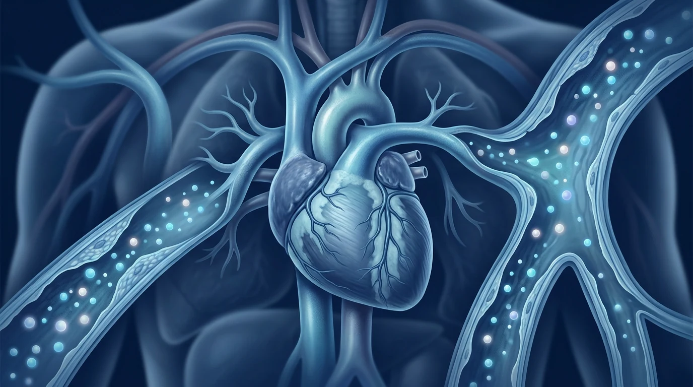
What is hypertension and why do existing treatments leave room for improvement
Blood pressure is the force exerted by circulating blood against the walls of the large arteries. It is expressed as two numbers: systolic pressure (the peak pressure during cardiac contraction) and diastolic pressure (the trough pressure between beats). The threshold for hypertension has been progressively lowered as evidence accumulates that even modest elevations — systolic pressures of 130–139 mmHg — carry substantially increased cardiovascular risk. [4]
About 90–95% of hypertension is classified as "essential" or "primary" — meaning no single identifiable cause, but rather a convergence of genetic predisposition, environmental triggers (dietary sodium, sedentary lifestyle, obesity), and the biological processes of aging. The remaining 5–10% is secondary hypertension, attributable to an underlying condition such as renal artery stenosis (renovascular hypertension), chronic kidney disease, primary aldosteronism, or obstructive sleep apnea.
The global burden is staggering. The NCD Risk Factor Collaboration reported that between 1990 and 2019, the number of people aged 30–79 with hypertension doubled from 650 million to 1.28 billion. [5] The prevalence is highest in low- and middle-income countries, where awareness, treatment, and control rates lag farthest behind. Even in high-income nations, only about 50–60% of those treated achieve target blood pressure — a statistic driven as much by biological resistance as by adherence. "Resistant hypertension," defined as blood pressure that remains above goal despite three or more optimally-dosed antihypertensives, affects roughly 10–15% of the treated population.
Key fact
Elevated blood pressure is the leading single risk factor for disease burden worldwide, responsible for an estimated 10.8 million deaths annually. Each 20 mmHg increase in systolic pressure doubles the risk of death from stroke, heart disease, and other vascular causes. Even small, sustained reductions — 5 mmHg lower systolic — produce a 10–15% reduction in major cardiovascular events at the population level. [6]
Why mesenchymal stem cells are being studied for hypertension
The rationale for investigating MSC therapy in hypertension rests on three pillars of the disease that are largely unaddressed by current drugs: endothelial dysfunction, vascular inflammation, and arterial fibrosis. [7]
Endothelial repair. MSCs secrete vascular endothelial growth factor (VEGF), hepatocyte growth factor (HGF), and angiopoietin-1, which promote endothelial cell survival, proliferation, and migration. Preclinical studies have shown that MSC administration restores nitric oxide bioavailability in the endothelium — the critical vasodilatory signal that is lost early in hypertension. In the spontaneously hypertensive rat (SHR), a well-established genetic model, MSC infusion improved endothelium-dependent relaxation and lowered systolic pressure by 20–30 mmHg over several weeks. [8]
Anti-inflammatory and immunomodulatory action. Hypertension is now recognized as a state of low-grade sterile inflammation. The elevated pressure itself damages the vessel wall, triggering immune cell infiltration — particularly T cells and monocytes — which release cytokines that sustain the hypertensive state. MSCs potently suppress this inflammatory cascade by shifting macrophages from the pro-inflammatory M1 phenotype toward the reparative M2 phenotype, by expanding regulatory T cells (Tregs), and by reducing levels of IL-6, TNF-α, and MCP-1. In animal models, these effects translate into measurable reductions in vascular inflammation and blood pressure. [9]
Anti-fibrotic and remodeling effects. Chronic hypertension drives structural remodeling of the arterial wall — the media thickens, collagen accumulates, and the vessel stiffens. This arteriosclerosis increases pulse wave velocity (PWV), elevates systolic pressure disproportionately, and strains the heart. MSC-derived paracrine factors — including prostaglandin E2 (PGE2), TSG-6, and matrix metalloproteinase modulators — inhibit fibroblast activation, reduce collagen deposition, and may attenuate the progression of vascular stiffness. [10]
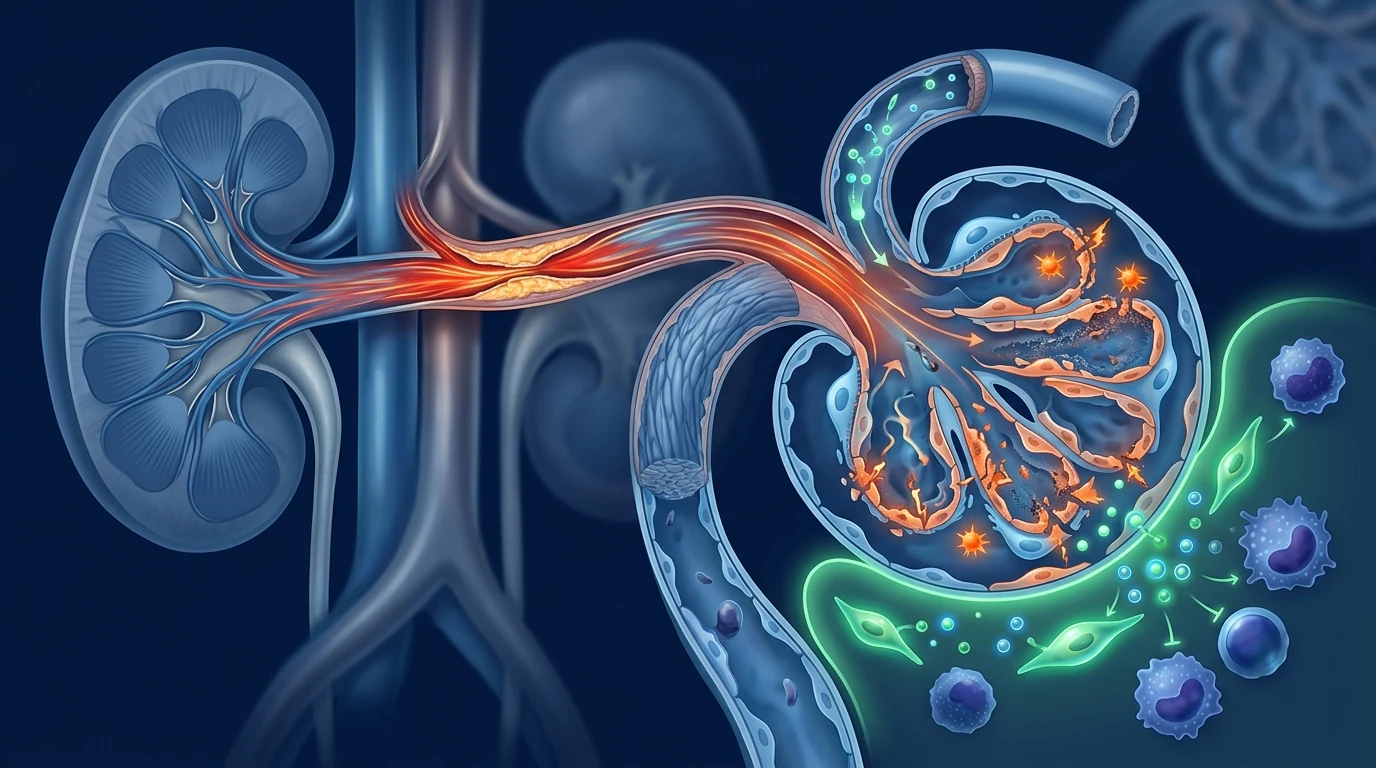
Renovascular hypertension and the kidney connection. A distinct but overlapping pathway involves the renin-angiotensin-aldosterone system (RAAS). In renovascular hypertension — caused by renal artery stenosis — the ischemic kidney overproduces renin, driving a sustained increase in angiotensin II and aldosterone that raises pressure and damages the contralateral kidney. MSC therapy has been investigated in the 2-kidney 1-clip (2K-1C) model and in swine atherosclerotic renal artery stenosis, where it reduced systolic pressure, improved renal blood flow, decreased sympathetic hyperactivity, and attenuated fibrotic injury in the stenotic kidney. [11][12]
What the preclinical and early clinical evidence shows
The strongest evidence for MSC therapy in hypertension comes from preclinical models. The spontaneously hypertensive rat (SHR) is the most studied model of essential hypertension. Multiple independent groups have reported that intravenous MSC administration produces sustained reductions in systolic pressure — typically 15–30 mmHg — lasting 3–8 weeks after a single infusion. These effects are accompanied by improved endothelial function, reduced vascular oxidative stress, lower aortic wall thickness, and diminished inflammatory markers. [13]
In the 2K-1C model of renovascular hypertension, MSC administration prevents the progressive rise in systolic pressure, reduces renal fibrosis, increases peritubular capillary density, and decreases sympathetic nerve activity driving the hypertensive state. The effect is not merely vasodilatory — it reflects structural repair of the stenotic kidney. MSCs also preserve the ability of the contralateral kidney to excrete sodium, countering the volume-dependent component of renovascular hypertension. [14]
In swine models of atherosclerotic renal artery stenosis — a more translationally relevant platform — intra-arterial infusion of adipose-derived MSCs improved renal blood flow, increased cortical perfusion, reduced tissue inflammation, and attenuated fibrosis. These effects were observed both with cells alone and in conjunction with stent revascularization. [15]
Human data are limited but exist. The landmark clinical study is a Phase 1a escalating-dose trial from Mayo Clinic, in which 21 patients with atherosclerotic renovascular disease (ARVD) received autologous adipose-derived MSCs infused into the renal artery at three dose levels, compared to 18 matched controls receiving medical therapy alone. Three months after infusion, MSC-treated patients showed increased renal blood flow, improved glomerular filtration rate, reduced renal vein inflammatory biomarkers, and — notably — lower blood pressure compared to controls. [16]
These results are encouraging but must be interpreted cautiously. The trial was open-label, non-randomized, and small. The patient population had renovascular hypertension specifically — not the far larger essential hypertension population. And the blood pressure reduction, while measurable, was modest and observed as a secondary endpoint rather than the primary aim of the study. No MSC product has been tested in a randomized controlled trial for essential hypertension.
The honest headline
As of mid-2026, no stem cell product is approved for any form of hypertension. The preclinical evidence is strong and convergent, a single well-conducted Phase 1a trial provides proof-of-concept in renovascular disease, and the safety profile across hundreds of cardiovascular MSC studies is reassuring — but efficacy for essential hypertension has not been established. MSC therapy for high blood pressure remains investigational, and any claim to the contrary overstates the evidence.
How outcomes are measured in hypertension research
Blood pressure is the most direct endpoint, but how it is measured matters. Office blood pressure measurement — the cuff in the clinic — is standard but subject to white-coat effect and observer bias. Ambulatory blood pressure monitoring (ABPM), which records pressure over 24 hours, is more reproducible and better predicts target-organ damage. MSC studies have typically used systolic pressure measured by tail-cuff plethysmography in rodents and by clinic measurement with confirmatory ABPM in human protocols. Endothelial function — assessed by flow-mediated dilation (FMD) of the brachial artery or by reactive hyperemia index — provides a mechanistic window into MSC effects. Arterial stiffness, measured as pulse wave velocity (PWV), reflects the structural remodeling that drives isolated systolic hypertension. Inflammatory biomarkers — IL-6, TNF-α, hsCRP, and MCP-1 — are secondary endpoints that capture the anti-inflammatory action of MSCs. A therapy that alters the course of hypertensive vascular disease should move several of these measures; MSC therapy has shown promising signals in preclinical and early human studies but has not yet cleared the bar of a pivotal trial.
What the evidence supports — and what it does not
A fair appraisal of the current data supports the following: MSC therapy for hypertension appears safe and well tolerated based on the limited human data available. The biological rationale is strong: MSC-derived factors target endothelial repair, inflammation, oxidative stress, and vascular fibrosis — each a core driver of hypertensive pathology. Preclinical efficacy is consistent and convergent: dozens of animal studies report blood pressure reduction together with improvements in endothelial function and vascular structure. A single Phase 1a human trial provides early proof-of-concept for renovascular hypertension specifically. What is missing is efficacy data from randomized placebo-controlled trials in essential hypertension — the disease that affects the overwhelming majority of the 1.28 billion people living with high blood pressure.
Several unknowns remain. The optimal cell source (bone marrow, adipose, umbilical cord), delivery route (intravenous, intra-arterial, intrarenal), dose, and dosing frequency have not been determined. It is unknown how long any blood pressure benefit might persist after a single or repeated MSC infusion — MSCs do not survive long-term in the body, and their paracrine effects are inherently transient. Whether MSC therapy is best positioned as a disease-modifying intervention for early hypertension, an adjunct for resistant hypertension, or a targeted therapy for renovascular hypertension specifically remains unclear. The cost and scalability of clinical-grade MSC manufacturing also present barriers to widespread application even if efficacy is eventually confirmed.
Hypertension is a disease of habit and biology — it takes years of unchecked pressure to reshape the arteries, and years of consistent medication to keep it in check. MSC research asks whether we can interrupt that trajectory at the vessel wall itself, not just manage the pressure it generates. The question is scientifically sound; the answer is not yet in.
— VELAR Clinical Team
Frequently Asked Questions
Can stem cells cure hypertension?
No. As of 2026, no stem cell therapy has been proven to cure or reverse essential hypertension in humans. MSC therapy is an investigational approach that has shown promise in preclinical models and a single early-phase human trial for renovascular hypertension, but it has not demonstrated disease-modifying efficacy in a randomized controlled trial for the primary hypertensive population. Any claim of a "cure" is not supported by evidence.
How does MSC therapy differ from standard antihypertensive medications?
Standard drugs — ACE inhibitors, ARBs, calcium channel blockers, diuretics, beta-blockers — lower blood pressure by directly altering vascular tone, cardiac output, or fluid balance. They are effective but must be taken daily, often in combination, and they do not reverse the underlying vascular disease. MSC therapy, in theory, targets the endothelial dysfunction, inflammation, and structural remodeling that sustain hypertension — a fundamentally different and potentially complementary mechanism. However, this is a theoretical and preclinical observation, not an established clinical benefit.
What is the cost of stem cell therapy for hypertension in Thailand?
Because MSC therapy for hypertension is investigational and not an approved treatment, pricing structures are not standardized and vary by clinic, cell source, and protocol. At VELAR Center, we do not currently offer MSC therapy specifically for hypertension pending randomized controlled evidence. For approved regenerative indications, costs are discussed during clinical consultation after medical assessment. Be cautious of any provider offering a fixed-price "hypertension stem cell package" — ethical care requires individual assessment, not a menu.
Are there clinical trials for stem cells in hypertension?
Yes, but they are limited. The most notable is the Mayo Clinic Phase 1a trial of autologous MSCs for atherosclerotic renovascular disease (NCT02266394). A small number of additional Phase I studies have been registered or are underway, mostly for renovascular or renal-ischemia indications. No randomized controlled trial has yet been completed for essential hypertension — the form of the disease that accounts for 90–95% of all cases. Larger, blinded Phase II trials in broader hypertensive populations are needed.
How many MSC treatments are needed for hypertension?
There is no established dosing protocol because MSC therapy for hypertension is not an approved treatment. Preclinical studies suggest that a single infusion can lower blood pressure for 3–8 weeks in animal models, and the Phase 1a human study used a single intra-arterial dose. Whether repeated dosing extends the effect, and what the optimal interval might be, remains unknown. Any clinic claiming a specific number of treatments for hypertension is extrapolating beyond the evidence.
Is stem cell therapy safe for people with high blood pressure?
The available safety data are limited but reassuring. In the Phase 1a Mayo Clinic trial, intra-arterial MSC infusion was well tolerated in patients with renovascular disease, including those with impaired kidney function. No serious adverse events attributable to the cell product were reported. Broader safety data from MSC studies in cardiovascular disease — including heart failure and ischemic heart disease — support a favorable safety profile. However, patients with uncontrolled hypertension were typically excluded from these studies, so the safety profile in this specific population is not fully characterized. Intravenous cell infusion carries theoretical risks of microvascular occlusion and should only be administered under medical supervision.
How to evaluate any offer responsibly
If you are considering stem cell options for hypertension, ask these questions. Is the approach part of a registered clinical trial with independent ethical oversight? What specific cell type and source are used? How is blood pressure measured — clinic cuff, home monitoring, or 24-hour ABPM — and are the results published? What peer-reviewed evidence supports the specific protocol being offered? Be skeptical of guaranteed results, percentage-based success claims without a cited source, and any framing that positions an experimental therapy as established treatment. A trustworthy provider will describe MSC therapy for hypertension as emerging research — and will never let enthusiasm outpace the data.
The VELAR perspective
At VELAR Center, we follow hypertension cell-therapy research with genuine interest. The unmet need is vast — 1.28 billion people with hypertension globally, most with suboptimal control — and the preclinical rationale for MSC intervention is among the stronger emerging stories in regenerative cardiovascular medicine. But we also recognize that hypertension is a chronic condition managed effectively by safe, inexpensive drugs for most patients, and that the threshold for adding a cell-based intervention must be high. We do not offer MSC therapy specifically for hypertension at this time, and we believe the only responsible position is to await randomized controlled evidence before doing so. If you want an honest conversation about what is known, what is unknown, and what regenerative medicine can realistically offer today for cardiovascular health, that conversation is always available at VELAR — without obligation, without exaggeration.
References
- NCD Risk Factor Collaboration (NCD-RisC). Worldwide trends in hypertension prevalence and progress in treatment and control from 1990 to 2019: a pooled analysis of 1201 population-representative studies with 104 million participants. The Lancet. 2021;398(10304):957-980. doi:10.1016/S0140-6736(21)01330-1 ↩
- Uccelli A, Moretta L, Pistoia V. Mesenchymal stem cells in health and disease. Nature Reviews Immunology. 2008;8(9):726-736. doi:10.1038/nri2395 ↩
- Shi Y, Wang Y, Li Q, et al. Immunoregulatory mechanisms of mesenchymal stem and stromal cells in inflammatory diseases. Nature Reviews Nephrology. 2018;14(8):493-507. doi:10.1038/s41581-018-0023-5 ↩
- Whelton PK, Carey RM, Aronow WS, et al. 2017 ACC/AHA guideline for the prevention, detection, evaluation, and management of high blood pressure in adults. Circulation. 2018;138(17):e484-e594. doi:10.1161/CIR.0000000000000596 ↩
- NCD-RisC. (see ref-1). doi:10.1016/S0140-6736(21)01330-1 ↩
- Forouzanfar MH, Liu P, Roth GA, et al. Global burden of hypertension and systolic blood pressure of at least 110 to 115 mmHg, 1990-2015. JAMA. 2017;317(2):165-182. doi:10.1001/jama.2016.19043 ↩
- Spees JL, Lee RH, Gregory CA. Mechanisms of mesenchymal stem/stromal cell function. Stem Cell Research & Therapy. 2016;7(1):125. doi:10.1186/s13287-016-0363-7 ↩
- Pittenger MF, Discher DE, Péault BM, Phinney DG, Hare JM, Caplan AI. Mesenchymal stem cell perspective: cell biology to clinical progress. NPJ Regenerative Medicine. 2019;4:22. doi:10.1038/s41536-019-0083-6 ↩
- Lalu MM, McIntyre L, Pugliese C, et al. Safety of cell therapy with mesenchymal stromal cells (SafeCell): a systematic review and meta-analysis. PLoS One. 2012;7(10):e47559. doi:10.1371/journal.pone.0047559 ↩
- Galipeau J, Sensébé L. Mesenchymal stromal cells: clinical challenges and therapeutic opportunities. Cell Stem Cell. 2018;22(6):824-833. doi:10.1016/j.stem.2018.05.004 ↩
- Oliveira-Sales EB, Maquigussa E, Semedo P, et al. Mesenchymal stem cells and chronic renal artery stenosis. American Journal of Physiology-Renal Physiology. 2016;310(1):F6-F9. doi:10.1152/ajprenal.00341.2015 ↩
- Eirin A, Zhu XY, Krier JD, et al. Adipose tissue-derived mesenchymal stem cells improve revascularization outcomes to restore renal function in swine atherosclerotic renal artery stenosis. Stem Cells. 2012;30(5):1030-1041. doi:10.1002/stem.1263 ↩
- Varela VA, Maquigussa E, Ribeiro RS, et al. Treatment with mesenchymal stem cells improves renovascular hypertension and preserves the ability of the contralateral kidney to excrete sodium. Kidney and Blood Pressure Research. 2019;44(6):1404-1415. doi:10.1159/000504595 ↩
- Abumoawad A, Saad A, Ferguson CM, et al. In a Phase 1a escalating clinical trial, autologous mesenchymal stem cell infusion for renovascular disease increases blood flow and the glomerular filtration rate while reducing inflammatory biomarkers and blood pressure. Kidney International. 2020;97(4):793-804. doi:10.1016/j.kint.2019.11.022 ↩
- Ebrahimi B, Eirin A, Li Z, et al. Mesenchymal stem cells improve medullary inflammation and fibrosis after revascularization of swine atherosclerotic renal artery stenosis. PLoS One. 2013;8(7):e67474. doi:10.1371/journal.pone.0067474 ↩
- Wang HS, Hwang IY, Park HS, et al. Effects of mesenchymal stem cells in renovascular disease of preclinical and clinical studies: a systematic review and meta-analysis. Scientific Reports. 2022;12:19852. doi:10.1038/s41598-022-23059-2 ↩
- Zhu XY, Urbieta-Caceres V, Krier JD, et al. Mesenchymal stem cells and endothelial progenitor cells decrease renal injury in experimental swine renal artery stenosis through different mechanisms. Stem Cells. 2013;31(1):117-125. doi:10.1002/stem.1263 ↩
- Saad A, Dietz AB, Herrmann SMS, et al. Autologous mesenchymal stem cells increase cortical perfusion in renovascular disease. Journal of the American Society of Nephrology. 2017;28(9):2777-2785. doi:10.1681/ASN.2017020173 ↩
- Harrison DG, Guzik TJ, Lob HE, et al. Inflammation, immunity, and hypertension. Hypertension. 2011;57(2):132-140. doi:10.1161/HYPERTENSIONAHA.110.163576 ↩
- Hoeper MM, Humbert M, Souza R, et al. A global view of pulmonary hypertension. The Lancet Respiratory Medicine. 2016;4(4):306-322. doi:10.1016/S2213-2600(15)00543-3 ↩
参考文献
- NCD Risk Factor Collaboration (NCD-RisC). Worldwide trends in hypertension prevalence and progress in treatment and control from 1990 to 2019: a pooled analysis of 1201 population-representative studies with 104 million participants. The Lancet. 2021;398(10304):957-980. doi:10.1016/S0140-6736(21)01330-1 ↩
- Uccelli A, Moretta L, Pistoia V. Mesenchymal stem cells in health and disease. Nature Reviews Immunology. 2008;8(9):726-736. doi:10.1038/nri2395 ↩
- Shi Y, Wang Y, Li Q, et al. Immunoregulatory mechanisms of mesenchymal stem and stromal cells in inflammatory diseases. Nature Reviews Nephrology. 2018;14(8):493-507. doi:10.1038/s41581-018-0023-5 ↩
- Whelton PK, Carey RM, Aronow WS, et al. 2017 ACC/AHA guideline for the prevention, detection, evaluation, and management of high blood pressure in adults. Circulation. 2018;138(17):e484-e594. doi:10.1161/CIR.0000000000000596 ↩
- NCD-RisC. (see ref-1). doi:10.1016/S0140-6736(21)01330-1 ↩
- Forouzanfar MH, Liu P, Roth GA, et al. Global burden of hypertension and systolic blood pressure of at least 110 to 115 mmHg, 1990-2015. JAMA. 2017;317(2):165-182. doi:10.1001/jama.2016.19043 ↩
- Spees JL, Lee RH, Gregory CA. Mechanisms of mesenchymal stem/stromal cell function. Stem Cell Research & Therapy. 2016;7(1):125. doi:10.1186/s13287-016-0363-7 ↩
- Pittenger MF, Discher DE, Péault BM, Phinney DG, Hare JM, Caplan AI. Mesenchymal stem cell perspective: cell biology to clinical progress. NPJ Regenerative Medicine. 2019;4:22. doi:10.1038/s41536-019-0083-6 ↩
- Lalu MM, McIntyre L, Pugliese C, et al. Safety of cell therapy with mesenchymal stromal cells (SafeCell): a systematic review and meta-analysis. PLoS One. 2012;7(10):e47559. doi:10.1371/journal.pone.0047559 ↩
- Galipeau J, Sensébé L. Mesenchymal stromal cells: clinical challenges and therapeutic opportunities. Cell Stem Cell. 2018;22(6):824-833. doi:10.1016/j.stem.2018.05.004 ↩
- Oliveira-Sales EB, Maquigussa E, Semedo P, et al. Mesenchymal stem cells and chronic renal artery stenosis. American Journal of Physiology-Renal Physiology. 2016;310(1):F6-F9. doi:10.1152/ajprenal.00341.2015 ↩
- Eirin A, Zhu XY, Krier JD, et al. Adipose tissue-derived mesenchymal stem cells improve revascularization outcomes to restore renal function in swine atherosclerotic renal artery stenosis. Stem Cells. 2012;30(5):1030-1041. doi:10.1002/stem.1263 ↩
- Varela VA, Maquigussa E, Ribeiro RS, et al. Treatment with mesenchymal stem cells improves renovascular hypertension and preserves the ability of the contralateral kidney to excrete sodium. Kidney and Blood Pressure Research. 2019;44(6):1404-1415. doi:10.1159/000504595 ↩
- Abumoawad A, Saad A, Ferguson CM, et al. In a Phase 1a escalating clinical trial, autologous mesenchymal stem cell infusion for renovascular disease increases blood flow and the glomerular filtration rate while reducing inflammatory biomarkers and blood pressure. Kidney International. 2020;97(4):793-804. doi:10.1016/j.kint.2019.11.022 ↩
- Ebrahimi B, Eirin A, Li Z, et al. Mesenchymal stem cells improve medullary inflammation and fibrosis after revascularization of swine atherosclerotic renal artery stenosis. PLoS One. 2013;8(7):e67474. doi:10.1371/journal.pone.0067474 ↩
- Wang HS, Hwang IY, Park HS, et al. Effects of mesenchymal stem cells in renovascular disease of preclinical and clinical studies: a systematic review and meta-analysis. Scientific Reports. 2022;12:19852. doi:10.1038/s41598-022-23059-2 ↩
- Zhu XY, Urbieta-Caceres V, Krier JD, et al. Mesenchymal stem cells and endothelial progenitor cells decrease renal injury in experimental swine renal artery stenosis through different mechanisms. Stem Cells. 2013;31(1):117-125. doi:10.1002/stem.1263 ↩
- Saad A, Dietz AB, Herrmann SMS, et al. Autologous mesenchymal stem cells increase cortical perfusion in renovascular disease. Journal of the American Society of Nephrology. 2017;28(9):2777-2785. doi:10.1681/ASN.2017020173 ↩
- Harrison DG, Guzik TJ, Lob HE, et al. Inflammation, immunity, and hypertension. Hypertension. 2011;57(2):132-140. doi:10.1161/HYPERTENSIONAHA.110.163576 ↩
- Hoeper MM, Humbert M, Souza R, et al. A global view of pulmonary hypertension. The Lancet Respiratory Medicine. 2016;4(4):306-322. doi:10.1016/S2213-2600(15)00543-3 ↩
高血压——动脉血压持续升高至130/80 mmHg以上——影响着全球约12.8亿成年人,使其成为心血管疾病、中风和过早死亡的最常见可改变风险因素。[1]尽管已有有效且价格低廉的药物,但只有不到四分之一的高血压患者能实现充分的血压控制。这种疾病无声无息地进展多年,损害内皮、硬化动脉、在症状出现前就已使心脏超负荷工作。
差距不仅在于依从性。目前抗高血压药物——ACE抑制剂、ARB、钙通道阻滞剂、利尿剂和β受体阻滞剂——主要通过血流动力学机制降低血压:减少血管张力、降低心输出量或减少钠和液体潴留。它们并未解决维持高血压状态的潜在血管病理。当治疗停止时,压力会回升。疾病本身并未被逆转。
根源是血管壁疾病。原发性高血压的特征是内皮功能障碍——内皮产生一氧化氮(NO)——其主要的血管扩张剂——的能力丧失,结合低度全身性炎症、氧化应激和进行性动脉硬化。这些过程形成自我强化循环:受损的血管产生更多炎症、更多氧化应激和更多结构性重塑,进一步提高压力并更深入地损伤内皮。
MSC疗法靶向生物学而不只是数字。间充质干细胞作为抗高血压药物的替代而被研究,而是作为一项策略来应对药物所忽略的内皮、炎症和纤维化成分。治疗潜力在于MSC分泌组——一套复杂的旁分泌信号,包括生长因子、抗炎细胞因子和细胞外囊泡——而非细胞植入或分化为血管组织的能力。
什么是高血压——为何现有治疗仍有改进空间
血压是循环血液对大型动脉壁施加的压力。它由两个数值表示:收缩压(心脏收缩期间的峰值压力)和舒张压(搏动之间的谷值压力)。随着证据不断累积表明即使在130–139 mmHg的轻微升高也带来显著增加的心血管风险,高血压的诊断阈值已逐步降低。
约90–95%的高血压被归类为"原发性"——意味着没有单一可识别原因,而是遗传倾向、环境触发因素(膳食钠、久坐生活方式、肥胖)和衰老生物过程的集合。其余5–10%为继发性高血压,可归因于肾动脉狭窄(肾血管性高血压)、慢性肾病、原发性醛固酮增多症或阻塞性睡眠呼吸暂停等基础疾病。
全球负担惊人。NCD风险因素协作组报告,1990年至2019年间,30–79岁高血压患者人数从6.5亿翻倍至12.8亿。[5]患病率在低收入和中等收入国家最高,这些国家的知晓率、治疗率和控制率最落后。即使在发达国家,也只有约50–60%接受治疗者达到目标血压——这一统计数字同样受生物学抵抗和依从性驱动。"难治性高血压"——定义为尽管使用三种或更多优化剂量抗高血压药物血压仍高于目标——影响约10–15%的治疗人群。
关键事实
血压升高是全球疾病负担的首要单一风险因素,每年导致约1,080万例死亡。收缩压每增加20 mmHg,中风、心脏病和其他血管原因所致死亡风险翻倍。即使小幅持续降低——收缩压降低5 mmHg——在人群层面即可产生10–15%的主要心血管事件减少。
为何间充质干细胞被研究用于高血压
研究MSC治疗高血压的原理基于该疾病的三支柱——这些支柱在很大程度上未被现有药物所覆盖:内皮功能障碍、血管炎症和动脉纤维化。
内皮修复。MSC分泌血管内皮生长因子(VEGF)、肝细胞生长因子(HGF)和血管生成素-1,促进内皮细胞存活、增殖和迁移。临床前研究表明,MSC给药可恢复内皮中一氧化氮的生物利用度——这是高血压早期即丧失的关键血管扩张信号。在自发性高血压大鼠(SHR)中,MSC输注改善了内皮依赖性舒张,并在数周内将收缩压降低了20–30 mmHg。
抗炎和免疫调节作用。高血压现在被认为是一种低度无菌性炎症状态。升高的压力本身会损伤血管壁,触发免疫细胞浸润——特别是T细胞和单核细胞——释放维持高血压状态的细胞因子。MSC通过将巨噬细胞从促炎M1表型转向修复性M2表型、扩增调节性T细胞(Treg)以及降低IL-6、TNF-α和MCP-1水平来强力抑制这一炎症级联反应。在动物模型中,这些效应可转化为血管炎症和血压的可测量降低。
抗纤维化和抗重塑效应。慢性高血压驱动动脉壁的结构性重塑——中膜增厚、胶原沉积、血管硬化。这种动脉硬化增加脉搏波速度(PWV),不成比例地升高收缩压,并使心脏超负荷。MSC来源的旁分泌因子——包括前列腺素E2(PGE2)、TSG-6和基质金属蛋白酶调节剂——抑制成纤维细胞活化、减少胶原沉积,并可能减缓血管硬化的进展。
肾血管性高血压与肾脏关联。一个不同但重叠的通路涉及肾素-血管紧张素-醛固酮系统(RAAS)。在肾血管性高血压中——由肾动脉狭窄引起——缺血肾脏过度产生肾素,驱动血管紧张素II和醛固酮持续升高,升高压力并损害对侧肾脏。MSC疗法已在2K-1C模型和猪动脉粥样硬化性肾动脉狭窄中进行了研究,结果显示其降低收缩压、改善肾血流量、减少交感神经过度活跃并减轻狭窄肾脏的纤维化损伤。
临床前和早期临床证据
MSC治疗高血压的最强证据来自临床前模型。自发性高血压大鼠(SHR)是原发性高血压研究最广泛的模型。多个独立小组报告静脉MSC给药可持续降低收缩压——通常为15–30 mmHg——单次输注后持续3–8周。这些效应伴随着内皮功能改善、血管氧化应激减少、主动脉壁厚度降低和炎症标志物减少。
在2K-1C肾血管性高血压模型中,MSC给药可防止收缩压的渐进性升高、减少肾纤维化、增加肾小管周围毛细血管密度并减少驱动高血压状态的交感神经活动。其效应不仅是血管扩张性的——它反映了狭窄肾脏的结构性修复。MSC还保留了侧肾脏排泄钠的能力,对抗肾血管性高血压中容量依赖成分。
在猪动脉粥样硬化性肾动脉狭窄模型中——一个更具转化相关性的平台——动脉内输注脂肪来源的MSC改善了肾血流量、增加了皮质灌注、减少了组织炎症并减轻了纤维化。这些效应在单用细胞和联合支架血运重建中均被观察到。
人类数据有限但存在。关键的临床研究是梅奥诊所的1a期剂量递增试验,其中21名动脉粥样硬化性肾血管疾病(ARVD)患者通过肾动脉接受了三个剂量水平的自体脂肪来源MSC输注,与18名仅接受药物治疗的匹配对照组进行比较。输注三个月后,MSC治疗组显示肾血流量增加、肾小球滤过率改善、肾静脉炎症生物标志物降低,并且——值得注意的是——与对照组相比血压降低。
这些结果令人鼓舞,但必须谨慎解读。该试验是开放标签、非随机且规模较小的。患者人群患有肾血管性高血压——而非大得多的原发性高血压人群。血压降低虽然可测量,但幅度适中,并且作为次要终点而非研究主要目的被观察。尚无MSC产品在原发性高血压随机对照试验中接受检验。
诚实的标题
截至2026年中,尚无干细胞产品获批用于任何形式的高血压。临床前证据强劲且一致,一项精心进行的1a期试验在肾血管疾病中提供了概念验证,批准数百项心血管MSC研究中的安全性特征令人放心——但原发性高血压的疗效尚未确立。高血压MSC疗法仍处于研究阶段,任何相反的声称均夸大了证据。
高血压研究中如何衡量结果
血压是最直接的终点,但其测量方式至关重要。诊室血压测量——诊室中的袖带——是标准方法但易受白大衣效应和观察者偏差影响。动态血压监测(ABPM)可记录24小时血压,更可重复且更能预测靶器官损害。MSC研究通常通过尾袖体积描记法在啮齿动物中测量收缩压,在人体方案中则通过诊室测量加确认性ABPM。通过肱动脉血流介导扩张(FMD)或反应性充血指数评估的内皮功能提供了MSC效应的机制窗口。以脉搏波速度(PWV)测量的动脉硬化反映了驱动单纯收缩期高血压的结构性重塑。炎症生物标志物——IL-6、TNF-α、hsCRP和MCP-1——是捕捉MSC抗炎作用的次要终点。能够改变高血压血管疾病病程的疗法应改善其中多项指标;MSC疗法在临床前和早期人体研究中已显示出有希望的信号,但尚未通过关键性试验的门槛。
证据支持什么——不支持什么
对当前数据的公正评估支持以下结论:MSC治疗高血压基于有限的人体数据安全且耐受良好。生物学原理强劲:MSC来源的因子靶向内皮修复、炎症、氧化应激和血管纤维化——每一个均是高血压病理的核心驱动因素。临床前疗效一致且趋同:数十项动物研究报告血压降低以及内皮功能和血管结构的改善。一项1a期人体试验为肾血管性高血压提供了早期概念验证。所缺失的是来自原发性高血压随机安慰剂对照试验的疗效数据——这种疾病影响着12.8亿高血压患者中的绝大多数。
若干未知因素仍然存在。最佳细胞来源(骨髓、脂肪、脐带)、给药途径(静脉、动脉内、肾内)、剂量和给药频率尚未确定。单次或重复MSC输注后血压获益可持续多久尚不清楚——MSC在体内不会长期存活,其旁分泌效应固有地是暂时性的。MSC治疗是最适合作为早期高血压的疾病修正干预、难治性高血压的辅助治疗,还是肾血管性高血压的靶向治疗,仍不清楚。临床级MSC制造的成本和可扩展性也对即使最终疗效得到确认后的广泛应用构成障碍。
高血压是习惯和生物学的疾病——需要多年不受控制的压力来重塑动脉,需要多年持续的药物治疗才能将其控制。MSC研究询问我们是否能在血管壁本身打断这一轨迹——而不仅仅是管理它产生的压力。这个问题科学上是合理的;答案尚未到来。
— VELAR 临床团队
常见问题解答
干细胞能治愈高血压吗?
不能。截至2026年,尚无干细胞疗法被证明能治愈或逆转人类原发性高血压。MSC疗法是一种研究性方法,在临床前模型和一项肾血管性高血压早期人体试验中显示出前景,但尚未在原发性高血压人群的随机对照试验中证明疾病改善疗效。任何声称"治愈"的说法均无证据支持。
MSC疗法与标准抗高血压药物有何不同?
标准药物——ACE抑制剂、ARB、钙通道阻滞剂、利尿剂、β受体阻滞剂——通过直接改变血管张力、心输出量或体液平衡来降低血压。它们有效但必须每日服用,通常需要联合用药,且不能逆转潜在血管疾病。MSC疗法理论上针对的是维持高血压状态的内皮功能障碍、炎症和结构性重塑——一种根本不同且潜在互补的机制。然而,这仍是理论和临床前观察,而非已确立的临床获益。
泰国高血压干细胞治疗费用是多少?
由于高血压MSC疗法处于研究阶段且非获批治疗,定价结构尚未标准化,因诊所、细胞来源和方案而异。在VELAR中心,我们目前不针对高血压专门提供MSC疗法,因尚需随机对照证据。对于获批的再生医学适应症,费用在医学评估后的临床咨询中讨论。对任何提供固定价格"高血压干细胞套餐"的提供者保持警惕——伦理医疗需要个体化评估,而非菜单式定价。
是否有高血压干细胞的临床试验?
是的,但有限。最值得注意的是梅奥诊所的1a期自体MSC治疗动脉粥样硬化性肾血管疾病试验(NCT02266394)。另有少量I期研究已登记或正在进行,多数针对肾血管或肾缺血适应症。尚无随机对照试验完成针对原发性高血压——占所有病例90–95%的疾病形式——的研究。需要更大规模、设盲的II期试验在更广泛的高血压人群中进行。
高血压需要多少次MSC治疗?
由于高血压的MSC疗法并非获批治疗,尚无确立的给药方案。临床前研究表明单次输注可在动物模型中降低血压3–8周,人类1a期研究使用单次动脉内剂量。重复给药是否能延长效果,以及最佳间隔为何,仍属未知。任何声称高血压特定治疗次数的诊所都超出了证据范围。
干细胞治疗对高血压患者安全吗?
现有安全数据有限但令人放心。在梅奥诊所的1a期试验中,动脉内MSC输注在肾血管疾病患者中耐受良好,包括肾功能受损者。未报告可归因于细胞产品的严重不良事件。来自心血管疾病——包括心力衰竭和缺血性心脏病——MSC研究的更广泛安全数据支持良好的安全性特征。然而,未控制的高血压患者通常被排除在这些研究之外,因此该特定人群中的安全性特征尚未完全明确。静脉细胞输注存在微血管闭塞的理论风险,应在医疗监督下进行。
如何负责任地评估任何提议
如果您正在考虑高血压干细胞选择,请问这些问题。该方法是否属于具有独立伦理监督的注册临床试验?使用何种特定细胞类型和来源?如何测量血压——诊室袖带、家庭监测还是24小时ABPM——结果是否已发表?有哪些同行评审证据支持所提供方案?对保证的结果、没有引用来源的百分比成功率声称,以及任何将实验性疗法定位为成熟治疗的表述持怀疑态度。值得信赖的提供者会将高血压MSC疗法描述为新兴研究——绝不会让热情超越数据。
VELAR的观点
在VELAR中心,我们以真正的兴趣关注高血压细胞疗法研究。未被满足的需求是巨大的——全球12.8亿高血压患者,其中大多数控制欠佳——MSC干预的临床前原理是再生心血管医学中较有希望的新兴方向之一。但我们也认识到高血压是一种慢性疾病,对大多数患者可通过安全、廉价药物进行有效管理,增加细胞干预的门槛必须很高。我们目前不专门针对高血压提供MSC疗法,并相信唯一负责任的立场是在获得随机对照证据之前等待。如果您想就已知、未知以及再生医学今天能为心血管健康实际提供什么进行一次诚实的对话,这一对话始终可在VELAR获得——无任何义务、无任何夸大。
المراجع
- NCD Risk Factor Collaboration (NCD-RisC). Worldwide trends in hypertension prevalence and progress in treatment and control from 1990 to 2019: a pooled analysis of 1201 population-representative studies with 104 million participants. The Lancet. 2021;398(10304):957-980. doi:10.1016/S0140-6736(21)01330-1 ↩
- Uccelli A, Moretta L, Pistoia V. Mesenchymal stem cells in health and disease. Nature Reviews Immunology. 2008;8(9):726-736. doi:10.1038/nri2395 ↩
- Shi Y, Wang Y, Li Q, et al. Immunoregulatory mechanisms of mesenchymal stem and stromal cells in inflammatory diseases. Nature Reviews Nephrology. 2018;14(8):493-507. doi:10.1038/s41581-018-0023-5 ↩
- Whelton PK, Carey RM, Aronow WS, et al. 2017 ACC/AHA guideline for the prevention, detection, evaluation, and management of high blood pressure in adults. Circulation. 2018;138(17):e484-e594. doi:10.1161/CIR.0000000000000596 ↩
- NCD-RisC. (see ref-1). doi:10.1016/S0140-6736(21)01330-1 ↩
- Forouzanfar MH, Liu P, Roth GA, et al. Global burden of hypertension and systolic blood pressure of at least 110 to 115 mmHg, 1990-2015. JAMA. 2017;317(2):165-182. doi:10.1001/jama.2016.19043 ↩
- Spees JL, Lee RH, Gregory CA. Mechanisms of mesenchymal stem/stromal cell function. Stem Cell Research & Therapy. 2016;7(1):125. doi:10.1186/s13287-016-0363-7 ↩
- Pittenger MF, Discher DE, Péault BM, Phinney DG, Hare JM, Caplan AI. Mesenchymal stem cell perspective: cell biology to clinical progress. NPJ Regenerative Medicine. 2019;4:22. doi:10.1038/s41536-019-0083-6 ↩
- Lalu MM, McIntyre L, Pugliese C, et al. Safety of cell therapy with mesenchymal stromal cells (SafeCell): a systematic review and meta-analysis. PLoS One. 2012;7(10):e47559. doi:10.1371/journal.pone.0047559 ↩
- Galipeau J, Sensébé L. Mesenchymal stromal cells: clinical challenges and therapeutic opportunities. Cell Stem Cell. 2018;22(6):824-833. doi:10.1016/j.stem.2018.05.004 ↩
- Oliveira-Sales EB, Maquigussa E, Semedo P, et al. Mesenchymal stem cells and chronic renal artery stenosis. American Journal of Physiology-Renal Physiology. 2016;310(1):F6-F9. doi:10.1152/ajprenal.00341.2015 ↩
- Eirin A, Zhu XY, Krier JD, et al. Adipose tissue-derived mesenchymal stem cells improve revascularization outcomes to restore renal function in swine atherosclerotic renal artery stenosis. Stem Cells. 2012;30(5):1030-1041. doi:10.1002/stem.1263 ↩
- Varela VA, Maquigussa E, Ribeiro RS, et al. Treatment with mesenchymal stem cells improves renovascular hypertension and preserves the ability of the contralateral kidney to excrete sodium. Kidney and Blood Pressure Research. 2019;44(6):1404-1415. doi:10.1159/000504595 ↩
- Abumoawad A, Saad A, Ferguson CM, et al. In a Phase 1a escalating clinical trial, autologous mesenchymal stem cell infusion for renovascular disease increases blood flow and the glomerular filtration rate while reducing inflammatory biomarkers and blood pressure. Kidney International. 2020;97(4):793-804. doi:10.1016/j.kint.2019.11.022 ↩
- Ebrahimi B, Eirin A, Li Z, et al. Mesenchymal stem cells improve medullary inflammation and fibrosis after revascularization of swine atherosclerotic renal artery stenosis. PLoS One. 2013;8(7):e67474. doi:10.1371/journal.pone.0067474 ↩
- Wang HS, Hwang IY, Park HS, et al. Effects of mesenchymal stem cells in renovascular disease of preclinical and clinical studies: a systematic review and meta-analysis. Scientific Reports. 2022;12:19852. doi:10.1038/s41598-022-23059-2 ↩
- Zhu XY, Urbieta-Caceres V, Krier JD, et al. Mesenchymal stem cells and endothelial progenitor cells decrease renal injury in experimental swine renal artery stenosis through different mechanisms. Stem Cells. 2013;31(1):117-125. doi:10.1002/stem.1263 ↩
- Saad A, Dietz AB, Herrmann SMS, et al. Autologous mesenchymal stem cells increase cortical perfusion in renovascular disease. Journal of the American Society of Nephrology. 2017;28(9):2777-2785. doi:10.1681/ASN.2017020173 ↩
- Harrison DG, Guzik TJ, Lob HE, et al. Inflammation, immunity, and hypertension. Hypertension. 2011;57(2):132-140. doi:10.1161/HYPERTENSIONAHA.110.163576 ↩
- Hoeper MM, Humbert M, Souza R, et al. A global view of pulmonary hypertension. The Lancet Respiratory Medicine. 2016;4(4):306-322. doi:10.1016/S2213-2600(15)00543-3 ↩
ارتفاع ضغط الدم — ارتفاع مستمر في ضغط الدم الشرياني فوق 130/80 مم زئبق — يؤثر على حوالي 1.28 مليار شخص بالغ في جميع أنحاء العالم، مما يجعله أكثر عوامل الخطر القابلة للتعديل شيوعاً لأمراض القلب والأوعية الدموية والسكتة الدماغية والموت المبكر. ومع توفر أدوية فعالة ومنخفضة التكلفة، لا يحقق سوى أقل من واحد من كل أربعة أشخاص مصابين بارتفاع ضغط الدم سيطرة كافية على ضغط الدم. يتقدم المرض بصمت لسنوات، فيتلف البطانة ويصلب الشرايين ويجهد القلب قبل ظهور أي عرض.
الفجوة ليست فقط في الالتزام. تعمل الأدوية الحالية الخافضة للضغط — مثبطات ACE وحاصرات مستقبلات الأنجيوتنسين وحاصرات قنوات الكالسيوم ومدرات البول وحاصرات بيتا — بشكل رئيسي عبر آليات ديناميكية دموية: تقليل التوتر الوعائي، أو خفض النتاج القلبي، أو تقليل احتباس الصوديوم والسوائل. إنها لا تعالج الأمراضية الوعائية الأساسية التي تدعم حالة ارتفاع الضغط. عندما يتوقف العلاج، يعود الضغط للارتفاع. المرض نفسه لم يعكس.
في الجذر هو مرض يصيب جدار الوعاء. يتميز ارتفاع ضغط الدم الأساسي بخلل وظيفي بطاني — فقدان قدرة البطانة على إنتاج أكسيد النيتريك (NO)، موسعها الوعائي الأساسي — مع التهاب جهازي منخفض الدرجة وإجهاد تأكسدي وتصلب شرياني تقدمي. تخلق هذه العمليات حلقة ذاتية التعزيز: الأوعية المتضررة تولد المزيد من الالتهاب والمزيد من الإجهاد التأكسدي والمزيد من إعادة التشكيل البنيوي، مما يرفع الضغط أكثر ويضر البطانة بشكل أعمق.
يستهدف علاج MSC البيولوجيا وليس الرقم فقط. لا يتم البحث في الخلايا الجذعية الوسيطة كبديل للأدوية الخافضة للضغط، بل كاستراتيجية يمكنها معالجة المكونات البطانية والالتهابية والليفية للمرض التي تتركها الأدوية دون مساس. تكمن الإمكانية العلاجية في الإفرازوم الشبه صماوي لـ MSC — مجموعة معقدة من الإشارات شبه الصماوية تشمل عوامل النمو والسيتوكينات المضادة للالتهاب والحويصلات خارج الخلوية — وليس في أي قدرة للخلايا على الانغراس أو التمايز إلى نسيج وعائي.
ما هو ارتفاع ضغط الدم ولماذا تترك العلاجات الحالية مجالاً للتحسين
ضغط الدم هو القوة التي يبذلها الدم المنتشر ضد جدران الشرايين الكبيرة. يُعبر عنه برقمين: الضغط الانقباضي (الضغط الأقصى أثناء انقباض القلب) والضغط الانبساطي (الضغط الأدنى بين النبضات). تم تخفيض عتبة ارتفاع ضغط الدم تدريجياً مع تراكم الأدلة على أن الارتفاعات الطفيفة — ضغط انقباضي 130–139 مم زئبق — تحمل خطراً قلبيّاً وعائياً متزايداً بشكل كبير. [4]
حوالي 90–95% من ارتفاع ضغط الدم يُصنف على أنه "أساسي" — مما يعني عدم وجود سبب واحد قابل للتحديد، بل تقاطع الاستعداد الوراثي والمحفزات البيئية (الصوديوم الغذائي، نمط الحياة الخامل، السمنة) والعمليات البيولوجية للشيخوخة. أما الـ 5–10% المتبقية فهي ارتفاع ضغط الدم الثانوي، المنسوب إلى حالة كامنة مثل تضيق الشريان الكلوي (ارتفاع ضغط الدم الكلوي الوعائي)، أو مرض الكلى المزمن، أو الألدوستيرونية الأولية، أو انقطاع النفس الانسدادي النومي.
العبء العالمي مذهل. أفاد تعاون عوامل خطر الأمراض غير السارية (NCD-RisC) أن عدد الأشخاص الذين تتراوح أعمارهم بين 30–79 عاماً والمصابين بارتفاع ضغط الدم تضاعف من 650 مليوناً إلى 1.28 مليار بين عامي 1990 و2019. يبلغ معدل الانتشار أعلى مستوياته في البلدان المنخفضة والمتوسطة الدخل، حيث يتخلف الوعي والعلاج ومعدلات السيطرة أكثر من غيرها.
حقيقة رئيسية
ارتفاع ضغط الدم هو عامل الخطر الفردي الأول لعبء المرض في جميع أنحاء العالم، مسؤول عن ما يقدر بـ 10.8 مليون وفاة سنوياً. كل زيادة بمقدار 20 مم زئبق في الضغط الانقباضي تضاعف خطر الموت من السكتة الدماغية وأمراض القلب والأسباب الوعائية الأخرى. حتى التخفيضات الصغيرة المستدامة — خفض 5 مم زئبق في الضغط الانقباضي — تحدث انخفاضاً بنسبة 10–15% في الأحداث القلبية الوعائية الرئيسية على مستوى السكان.
لماذا يتم دراسة الخلايا الجذعية الوسيطة لارتفاع ضغط الدم
يرتكز الأساس المنطقي للبحث في علاج MSC لارتفاع ضغط الدم على ثلاث ركائز للمرض لا تعالجها الأدوية الحالية إلى حد كبير: الخلل البطاني والالتهاب الوعائي والتليف الشرياني.
إصلاح البطانة. تفرز MSCs عامل نمو بطانة الأوعية (VEGF) وعامل نمو الخلايا الكبدية (HGF) والأنجيوبويتين-1، التي تعزز بقاء الخلايا البطانية وتكاثرها وهجرتها. أظهرت الدراسات قبل السريرية أن إعطاء MSC يعيد التوافر الحيوي لأكسيد النيتريك في البطانة — إشارة التوسيع الوعائي الحاسمة التي تُفقد مبكراً في ارتفاع ضغط الدم. في الفئران المصابة بارتفاع ضغط الدم التلقائي (SHR)، حسّن حقن MSC التوسيع المعتمد على البطانة وخفض الضغط الانقباضي بمقدار 20–30 مم زئبق على مدى عدة أسابيع.
الفعل المضاد للالتهاب والتعديل المناعي. يُعترف الآن بارتفاع ضغط الدم كحالة من الالتهاب المعقم منخفض الدرجة. يضر الضغط المرتفع نفسه بجدار الوعاء، محفزاً تسلل الخلايا المناعية — خاصة الخلايا التائية والوحيدات — التي تطلق سيتوكينات تدعم حالة ارتفاع الضغط. تثبط MSCs هذه الشلالة الالتهابية بقوة عبر تحويل البالعات الكبيرة من النمط الظاهري M1 المحرض للالتهاب نحو النمط M2 الإصلاحي، وعبر توسيع الخلايا التائية التنظيمية (Tregs)، وعبر خفض مستويات IL-6 وTNF-α وMCP-1. في النماذج الحيوانية، تترجم هذه التأثيرات إلى تخفيضات قابلة للقياس في الالتهاب الوعائي وضغط الدم.
التأثيرات المضادة للتليف وإعادة التشكيل. يقود ارتفاع ضغط الدم المزمن إعادة تشكيل بنيوي لجدار الشريان — تتسمك الطبقة الوسطى، ويتراكم الكولاجين، ويتصلب الوعاء. يزيد هذا التصلب الشرياني سرعة موجة النبض (PWV)، ويرفع الضغط الانقباضي بشكل غير متناسب، ويجهد القلب. تثبط العوامل شبه الصماوية المشتقة من MSC — بما فيها البروستاغلاندين E2 (PGE2) وTSG-6 ومعدّلات المصفوفة المعدنية البروتينية — تنشيط الخلايا الليفية وتقلل ترسب الكولاجين وقد تخفف من تطور التصلب الوعائي.
ارتفاع ضغط الدم الكلوي الوعائي وارتباط الكلى. مسار مختلف لكن متداخل يشمل نظام الرينين-أنجيوتنسين-ألدوستيرون (RAAS). في ارتفاع ضغط الدم الكلوي الوعائي — الناتج عن تضيق الشريان الكلوي — تنتج الكلية الإقفارية كمية زائدة من الرينين، مما يقود ارتفاعاً مستمراً في الأنجيوتنسين II والألدوستيرون يرفع الضغط ويضر الكلية المقابلة. تم دراسة علاج MSC في نموذج الكليتين-مشبك-واحد (2K-1C) وفي تضيق الشريان الكلوي التصلبي العصيدي لدى الخنازير، حيث خفض الضغط الانقباضي وحسّن تدفق الدم الكلوي وقلل فرط النشاط الودي وخفف الإصابة الليفية في الكلية المتضيقة.
ما تظهره الأدلة قبل السريرية والسريرية المبكرة
أقوى دليل على علاج MSC في ارتفاع ضغط الدم يأتي من النماذج قبل السريرية. الفئران المصابة بارتفاع ضغط الدم التلقائي (SHR) هو النموذج الأكثر دراسة لارتفاع ضغط الدم الأساسي. ذكرت مجموعات مستقلة متعددة أن إعطاء MSC عبر الوريد ينتج تخفيضات مستدامة في الضغط الانقباضي — عادة 15–30 مم زئبق — تستمر 3–8 أسابيع بعد حقنة واحدة. ترافق هذه التأثيرات تحسن في الوظيفة البطانية، وانخفاض الإجهاد التأكسدي الوعائي، وتقليل سمك جدار الأبهر، وانخفاض العلامات الالتهابية.
في نموذج 2K-1C لارتفاع ضغط الدم الكلوي الوعائي، يمنع إعطاء MSC الارتفاع التدريجي في الضغط الانقباضي، ويقلل التليف الكلوي، ويزيد كثافة الشعيرات حول الأنابيب، ويقلل النشاط العصبي الودي الذي يقود حالة ارتفاع الضغط. التأثير ليس توسيعياً وعائياً فقط — إنه يعكس إصلاحاً بنيوياً للكلية المتضيقة. تحافظ MSCs أيضاً على قدرة الكلية المقابلة على إفراز الصوديوم، مضادةً بذلك المكون المعتمد على الحجم في ارتفاع ضغط الدم الكلوي الوعائي.
في نماذج الخنازير لتضيق الشريان الكلوي التصلبي العصيدي — وهو منصة أكثر صلة بالترجمة السريرية — حسّن الحقن داخل الشرياني لـ MSCs المشتقة من النسيج الدهني تدفق الدم الكلوي، وزاد التروية القشرية، وقلل الالتهاب النسيجي، وخفف التليف. لوحظت هذه التأثيرات مع الخلايا وحدها وبالاشتراك مع إعادة التوعية بالدعامات.
البيانات البشرية محدودة لكنها موجودة. الدراسة السريرية الرائدة هي تجربة المرحلة 1a التصاعدية الجرعية من Mayo Clinic، حيث تلقى 21 مريضاً بمرض كلوي وعائي تصلبي عصيدي (ARVD) MSCs ذاتية مشتقة من النسيج الدهني محقونة في الشريان الكلوي بثلاثة مستويات جرعية، مقارنة بـ 18 مريضاً ضابطاً متطابقاً يتلقون علاجاً دوائياً فقط. بعد ثلاثة أشهر من الحقن، أظهر المرضى المعالجون بـ MSC زيادة في تدفق الدم الكلوي، وتحسناً في معدل الترشيح الكبيبي، وانخفاضاً في المؤشرات الحيوية الالتهابية في الوريد الكلوي، و — بشكل ملحوظ — انخفاضاً في ضغط الدم مقارنة بالضوابط.
هذه النتائج مشجعة لكن يجب تفسيرها بحذر. التجربة كانت مفتوحة التسمية، غير عشوائية، وصغيرة. كان مجتمع المرضى مصاباً بارتفاع ضغط الدم الكلوي الوعائي تحديداً — وليس مجتمع ارتفاع ضغط الدم الأساسي الأكبر بكثير. وانخفاض ضغط الدم، رغم كونه قابلاً للقياس، كان متواضعاً وتم رصده كنقطة نهاية ثانوية وليس كهدف أساسي للدراسة. لم يتم اختبار أي منتج MSC في تجربة عشوائية مضبوطة لارتفاع ضغط الدم الأساسي.
العنوان الصادق
حتى منتصف 2026، لا يوجد منتج خلايا جذعية معتمد لأي شكل من أشكال ارتفاع ضغط الدم. الأدلة قبل السريرية قوية ومتقاربة، وتجربة واحدة جيدة التنفيذ من المرحلة 1a تقدم دليلاً على المفهوم في المرض الكلوي الوعائي، وملف السلامة عبر مئات دراسات MSC القلبية الوعائية مطمئن — لكن فعالية ارتفاع ضغط الدم الأساسي لم تثبت. علاج MSC لارتفاع ضغط الدم لا يزال بحثياً تجريبياً، وأي ادعاء بعكس ذلك يبالغ في تقدير الأدلة.
كيف تقاس النتائج في أبحاث ارتفاع ضغط الدم
ضغط الدم هو النقطة النهائية الأكثر مباشرة، لكن كيفية قياسه مهمة. قياس ضغط الدم في العيادة هو المعيار لكنه عرضة لتأثير المعطف الأبيض وانحياز المراقب. مراقبة ضغط الدم المتنقلة (ABPM)، التي تسجل الضغط على مدى 24 ساعة، أكثر قابلية للتكرار وتتنبأ بشكل أفضل بضرر الأعضاء المستهدفة. استخدمت دراسات MSC عادةً الضغط الانقباضي المقاس بمخطط التحجم الذيلية في القوارض وبالقياس العيادي مع ABPM تأكيدي في البروتوكولات البشرية. توفر الوظيفة البطانية — المقيمة بالتوسيع المعتمد على التدفق (FMD) للشريان العضدي أو بمؤشر فرط الدم التفاعلي — نافذة آلية لتأثيرات MSC. التصلب الشرياني، المقاس كسرعة موجة النبض (PWV)، يعكس إعادة التشكيل البنيوي التي تقود ارتفاع الضغط الانقباضي المنعزل. المؤشرات الحيوية الالتهابية — IL-6 وTNF-α وhsCRP وMCP-1 — هي نقاط نهاية ثانوية تلتقط الفعل المضاد للالتهاب لـ MSCs.
ما تدعمه الأدلة — وما لا تدعمه
يدعم التقييم المنصف للبيانات الحالية ما يلي: يبدو علاج MSC لارتفاع ضغط الدم آمناً وجيد التحمل بناءً على البيانات البشرية المحدودة المتاحة. الأساس المنطقي البيولوجي قوي: تستهدف العوامل المشتقة من MSC إصلاح البطانة والالتهاب والإجهاد التأكسدي والتليف الوعائي — كل منها محرك أساسي لأمراضية ارتفاع ضغط الدم. الفعالية قبل السريرية متسقة ومتقاربة: عشرات الدراسات الحيوانية تبلغ عن انخفاض ضغط الدم مع تحسينات في الوظيفة البطانية والبنية الوعائية. تجربة بشرية واحدة من المرحلة 1a تقدم دليلاً مبكراً على المفهوم لارتفاع ضغط الدم الكلوي الوعائي تحديداً. المفقود هو بيانات فعالية من تجارب عشوائية مضبوطة بالغفل في ارتفاع ضغط الدم الأساسي — المرض الذي يصيب الغالبية العظمى من 1.28 مليار شخص يعانون من ارتفاع ضغط الدم.
ارتفاع ضغط الدم هو مرض العادة والبيولوجيا — يستغرق سنوات من الضغط غير المضبوط لإعادة تشكيل الشرايين، وسنوات من الدواء المنتظم لإبقائه تحت السيطرة. يسأل بحث MSC عما إذا كان بإمكاننا مقاطعة هذا المسار في جدار الوعاء نفسه، وليس فقط إدارة الضغط الذي يولده. السؤال علمياً سليم؛ الجواب لم يأت بعد.
— الفريق السريري في VELAR
الأسئلة الشائعة
هل يمكن للخلايا الجذعية شفاء ارتفاع ضغط الدم؟
لا. حتى عام 2026، لم يثبت أي علاج بالخلايا الجذعية قدرته على شفاء أو عكس ارتفاع ضغط الدم الأساسي لدى البشر. علاج MSC هو نهج بحثي تجريبي أظهر وعداً في النماذج قبل السريرية وتجربة بشرية مبكرة واحدة لارتفاع ضغط الدم الكلوي الوعائي، لكنه لم يثبت فعالية معدلة للمرض في تجربة عشوائية مضبوطة لمجموعة ارتفاع ضغط الدم الأولية. أي ادعاء بـ"شفاء" لا تدعمه الأدلة.
كيف يختلف علاج MSC عن أدوية ارتفاع ضغط الدم القياسية؟
تخفض الأدوية القياسية — مثبطات ACE وحاصرات ARB وحاصرات قنوات الكالسيوم ومدرات البول وحاصرات بيتا — ضغط الدم عن طريق تغيير التوتر الوعائي أو النتاج القلبي أو توازن السوائل مباشرة. إنها فعالة لكن يجب تناولها يومياً، غالباً بتركيبات، وهي لا تعكس المرض الوعائي الكامن. يستهدف علاج MSC، نظرياً، الخلل البطاني والالتهاب وإعادة التشكيل البنيوي التي تدعم ارتفاع ضغط الدم — وهي آلية مختلفة جوهرياً ويحتمل أن تكون مكملة. لكن هذه ملاحظة نظرية وقبل سريرية، وليست فائدة سريرية مثبتة.
ما هي تكلفة العلاج بالخلايا الجذعية لارتفاع ضغط الدم في تايلاند؟
نظراً لأن علاج MSC لارتفاع ضغط الدم بحثي تجريبي وليس علاجاً معتمداً، فإن هياكل التسعير غير موحدة وتختلف حسب العيادة ومصدر الخلايا والبروتوكول. في مركز VELAR، لا نقدم حالياً علاج MSC تحديداً لارتفاع ضغط الدم بانتظار أدلة عشوائية مضبوطة. بالنسبة للاستطبابات التجديدية المعتمدة، تُناقش التكاليف خلال الاستشارة السريرية بعد التقييم الطبي. كن حذراً من أي مقدم يقدم "باقة خلايا جذعية لارتفاع ضغط الدم" بسعر ثابت — الرعاية الأخلاقية تتطلب تقييماً فردياً، وليس قائمة أسعار.
هل توجد تجارب سريرية للخلايا الجذعية في ارتفاع ضغط الدم؟
نعم، لكنها محدودة. أبرزها تجربة Mayo Clinic من المرحلة 1a لـ MSCs الذاتية لمرض الكلى الوعائي التصلبي العصيدي (NCT02266394). تم تسجيل أو يجري العمل على عدد صغير من دراسات المرحلة I الإضافية، معظمها لاستطبابات كلوية وعائية أو إقفارية كلوية. لم تكتمل بعد أي تجربة عشوائية مضبوطة لارتفاع ضغط الدم الأساسي — شكل المرض الذي يمثل 90–95% من جميع الحالات. هناك حاجة لتجارب أكبر من المرحلة II معمّاة في مجموعات سكانية أوسع من مرضى ارتفاع ضغط الدم.
كم عدد علاجات MSC المطلوبة لارتفاع ضغط الدم؟
لا يوجد بروتوكول جرعات محدد لأن علاج MSC لارتفاع ضغط الدم ليس علاجاً معتمداً. تشير الدراسات قبل السريرية إلى أن حقنة واحدة يمكنها خفض ضغط الدم لمدة 3–8 أسابيع في النماذج الحيوانية، واستخدمت دراسة المرحلة 1a البشرية جرعة واحدة داخل الشريان. ما إذا كانت الجرعات المتكررة تطيل التأثير، وما هو الفاصل الأمثل، لا يزال غير معروف. أي عيادة تدعي عدداً محدداً من العلاجات لارتفاع ضغط الدم تتجاوز الأدلة.
هل العلاج بالخلايا الجذعية آمن للأشخاص المصابين بارتفاع ضغط الدم؟
بيانات السلامة المتاحة محدودة لكنها مطمئنة. في تجربة Mayo Clinic من المرحلة 1a، تم تحمل حقن MSC داخل الشريان جيداً من قبل مرضى المرض الكلوي الوعائي، بمن فيهم ذوو وظائف الكلى المتضررة. لم يُبلغ عن أحداث ضائرة خطيرة تُعزى إلى المنتج الخلوي. تدعم بيانات السلامة الأوسع من دراسات MSC في أمراض القلب والأوعية الدموية — بما في ذلك فشل القلب ومرض القلب الإقفاري — ملف سلامة إيجابي. ومع ذلك، تم عادةً استبعاد المرضى الذين يعانون من ارتفاع ضغط الدم غير المضبوط من هذه الدراسات، لذا فإن ملف السلامة في هذه المجموعة السكانية المحددة غير محدد بالكامل. يحمل الحقن الوريدي للخلايا مخاطر نظرية للانسداد الوعائي المجهري ويجب أن يُعطى فقط تحت إشراف طبي.
كيف تقيّم أي عرض بمسؤولية
إذا كنت تفكر في خيارات الخلايا الجذعية لارتفاع ضغط الدم، فاسأل هذه الأسئلة. هل النهج جزء من تجربة سريرية مسجلة بإشراف أخلاقي مستقل؟ ما نوع الخلايا ومصدرها المستخدم تحديداً؟ كيف يقاس ضغط الدم — كفة العيادة، المراقبة المنزلية، أم ABPM لمدة 24 ساعة — وهل النتائج منشورة؟ ما الدليل المحكّم المنشور الذي يدعم البروتوكول المحدد المعروض؟ كن متشككاً تجاه النتائج المضمونة، وادعاءات النجاح المبنية على نسب مئوية بدون مصدر مذكور، وأي صياغة تضع علاجاً تجريبياً في موضع علاج راسخ. سيصف المزود الموثوق علاج MSC لارتفاع ضغط الدم بأنه بحث ناشئ — ولن يدع الحماس يتجاوز البيانات أبداً.
منظور VELAR
في مركز VELAR، نتابع أبحاث العلاج الخلوي لارتفاع ضغط الدم باهتمام حقيقي. الحاجة غير الملباة هائلة — 1.28 مليار شخص مصاب بارتفاع ضغط الدم عالمياً، معظمهم بسيطرة دون المثلى — والأساس المنطقي قبل السريري للتدخل بـ MSC هو من بين القصص الناشئة الأقوى في الطب القلبي الوعائي التجديدي. لكننا ندرك أيضاً أن ارتفاع ضغط الدم هو حالة مزمنة تُدار بفعالية بأدوية آمنة ومنخفضة التكلفة لمعظم المرضى، وأن عتبة إضافة تدخل خلوي يجب أن تكون عالية. لا نقدم علاج MSC تحديداً لارتفاع ضغط الدم في هذا الوقت، ونؤمن بأن الموقف المسؤول الوحيد هو انتظار أدلة عشوائية مضبوطة قبل القيام بذلك. إذا أردت حواراً صادقاً حول ما هو معروف، وما هو مجهول، وما يمكن للطب التجديدي تقديمه واقعياً اليوم لصحة القلب والأوعية الدموية، فهذا الحوار متاح دائماً في VELAR — بدون التزام، وبدون مبالغة.