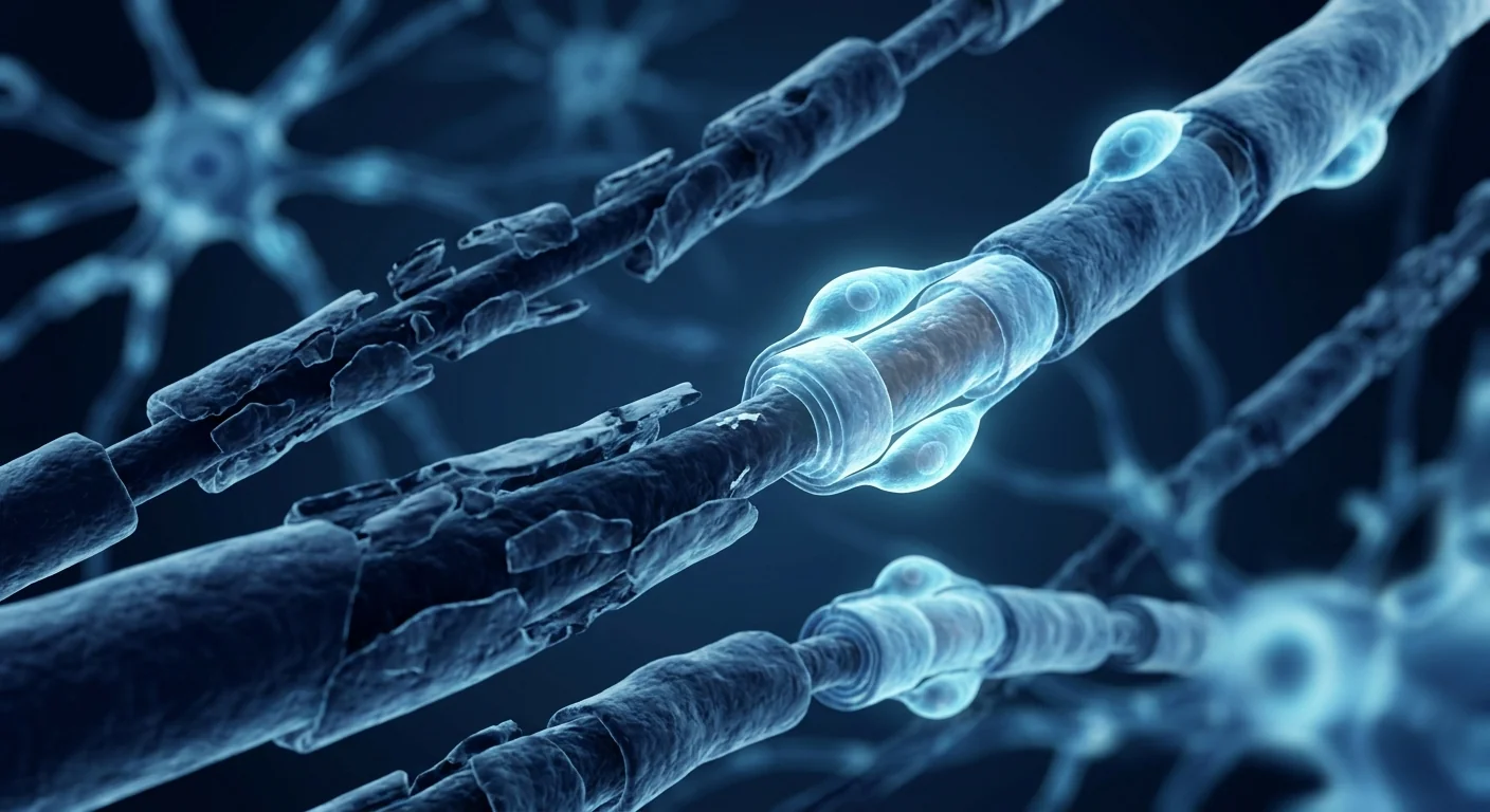
What Is Chronic Inflammatory Demyelinating Polyneuropathy (CIDP)?
CIDP is a chronic autoimmune disorder in which the body's immune system mounts a sustained attack against the myelin sheath of peripheral nerves — the insulating layer essential for rapid nerve signal conduction. Unlike its acute counterpart Guillain-Barré Syndrome (GBS), which peaks within four weeks, CIDP progresses over at least eight weeks and can follow either a slowly progressive or relapsing-remitting course that persists for years [1].
CIDP affects approximately 1–9 per 100,000 people worldwide, with prevalence rising with age. The clinical picture is characterized by symmetric proximal and distal muscle weakness, sensory loss (numbness and tingling in a glove-and-stocking distribution), and diminished or absent deep tendon reflexes. Many patients also experience significant fatigue and neuropathic pain. Without treatment, approximately 30% of CIDP patients eventually become wheelchair-dependent, and the disease substantially reduces quality of life across physical, emotional, and social domains [2].
The current treatment landscape is broad but imperfect. First-line therapies include intravenous immunoglobulin (IVIG), corticosteroids, and plasma exchange (PLEX). For patients who do not respond adequately, second-line immunosuppressants — mycophenolate mofetil, cyclophosphamide, rituximab, and cyclosporine — are commonly employed. While approximately 60–70% of patients respond to one or more of these approaches, a substantial minority remain treatment-refractory, and many of those who do respond continue to experience residual disability, treatment-related toxicity, or breakthrough relapses [3]. The fundamental limitation is that current treatments suppress or modulate the immune attack without directly promoting myelin repair — the critical therapeutic gap that MSC therapy aims to address.
How MSCs Target the Pathophysiology of CIDP
MSC therapy addresses three interconnected pathological processes in CIDP: neuroinflammation, demyelination, and the failure of endogenous repair mechanisms. The therapeutic rationale builds on a substantial body of preclinical research in experimental autoimmune neuritis (EAN) — the rodent model that recapitulates key features of human inflammatory demyelinating polyneuropathies — as well as clinical experience in related neuroimmunological conditions [4].
1. Immunomodulation — Resetting the Autoimmune Milieu
CIDP is driven by a dysregulated immune response involving autoreactive T cells, pathogenic autoantibodies targeting peripheral nerve antigens (including myelin protein zero, PMP22, and contactin-1), and complement-mediated myelin destruction. Macrophages invade the myelin sheath, stripping it from axons and leaving behind naked, dysfunctional nerve fibers [5].
MSCs intervene at multiple checkpoints in this inflammatory cascade. They secrete prostaglandin E2 (PGE2), TSG-6, and IL-10, which collectively suppress effector T-cell proliferation, shift the Th17/Treg balance toward regulatory tolerance, and polarize pro-inflammatory M1 macrophages toward the reparative M2 phenotype. Critically, MSCs also reduce the production of pathogenic autoantibodies by suppressing B-cell differentiation through indoleamine 2,3-dioxygenase (IDO) and programmed death ligand-1 (PD-L1) pathways [6]. In the EAN model, systemic MSC administration significantly reduced demyelination scores, decreased inflammatory cell infiltration into peripheral nerves, and lowered circulating levels of TNF-α, IL-1β, and IL-17 while elevating IL-10 and TGF-β — a comprehensive shift from a pro-inflammatory to an anti-inflammatory and pro-repair cytokine profile.
2. Remyelination and Neurotrophic Support
Beyond immune suppression, MSCs actively promote myelin repair through paracrine secretion of neurotrophic factors. MSC-derived BDNF (brain-derived neurotrophic factor), NGF (nerve growth factor), CNTF (ciliary neurotrophic factor), and GDNF (glial cell line-derived neurotrophic factor) directly stimulate Schwann cell proliferation and differentiation — the cellular machinery responsible for peripheral nerve remyelination [7].
In addition to neurotrophin secretion, MSCs release extracellular vesicles (EVs) containing microRNAs and proteins that promote axonal survival and guide regenerating nerve fibers to their targets. MSC-derived exosomes enriched with miR-133b and miR-17-92 cluster have been shown to enhance neurite outgrowth and improve functional recovery in peripheral nerve injury models — mechanisms highly relevant to the remyelination deficit in CIDP [8].
3. Blood-Nerve Barrier Stabilization
The blood-nerve barrier (BNB) is compromised in CIDP, allowing circulating immune cells and antibodies to access the endoneurial microenvironment. MSCs have demonstrated the ability to strengthen endothelial barrier function through secretion of angiopoietin-1 and HGF (hepatocyte growth factor), which stabilize tight junction proteins [9]. By reinforcing the BNB, MSCs may reduce ongoing immune-mediated injury and create a more permissive environment for endogenous repair.
Preclinical and Clinical Evidence
The evidence pipeline for MSC therapy in inflammatory neuropathies is anchored by robust preclinical data and emerging clinical signals.
Experimental Autoimmune Neuritis (EAN) Studies
Multiple independent research groups have demonstrated that MSC administration in EAN — the animal model most directly relevant to human CIDP and GBS — produces measurable benefits including reduced clinical severity scores, preserved nerve conduction velocity, decreased demyelination on histological examination, and reduced inflammatory cell infiltration in sciatic nerve sections. Both bone marrow-derived MSCs and umbilical cord-derived MSCs have shown efficacy at doses equivalent to 1–5 × 10⁶ cells/kg in rodent models, with the therapeutic effect attributed primarily to paracrine immunomodulation rather than cellular engraftment [10].
Clinical Data — Small Case Series and Extrapolated Evidence
Dedicated randomized controlled trials of MSC therapy specifically for CIDP do not yet exist. However, a small but growing number of case reports document treatment-refractory CIDP patients who received MSC infusions. In one published case series of three patients with long-standing CIDP who had failed IVIG, corticosteroids, and multiple immunosuppressants, intravenous administration of umbilical cord-derived MSCs (1–2 × 10⁶ cells/kg) was associated with measurable improvements in the INCAT (Inflammatory Neuropathy Cause and Treatment) disability score, MRC sum score for muscle strength, and patient-reported quality of life at 6- and 12-month follow-up [11].
Additionally, broader clinical data from MSC trials in other autoimmune neuropathies and closely related conditions — including the GBS case series and a growing registry of MSC therapy in multiple sclerosis — provide supportive evidence for the immunomodulatory and neuroprotective potential of MSCs in peripheral and central nervous system autoimmune disease. The safety profile across these studies has been consistently favorable, with no serious infusion-related adverse events or unexpected long-term complications attributable to MSC administration [12].
The MSC Treatment Journey for CIDP at VELAR
Pre-Treatment Assessment
Every CIDP case at VELAR begins with a comprehensive neurological evaluation. This includes a detailed history of disease onset and progression, prior treatment responses (IVIG, corticosteroids, PLEX, immunosuppressants), current medication regimen, and standardized functional assessments including the INCAT disability score, MRC sum score, and nerve conduction studies where recent results are available. Pre-treatment serum markers — including anti-ganglioside antibodies where relevant — provide a baseline for post-treatment monitoring.
MSC Preparation and Delivery
VELAR's clinical-grade MSCs are isolated from donated Wharton's jelly (umbilical cord tissue) following GTP and cGMP protocols in our ISO Class 5 (Class-100) cleanroom. The cells are cultured without animal-derived products (xeno-free), undergo rigorous quality control including ISCT identity verification, multi-pathogen screening, and viability assessment (>95% cell viability at release). VELAR delivers fresh, never-frozen cells — no cryopreservation, no DMSO — ensuring maximum potency at the point of infusion.
MSCs are administered via intravenous infusion, allowing systemic distribution of their immunomodulatory and neurotrophic effects. The infusion typically takes 30–60 minutes, and patients are monitored for two hours post-infusion before discharge.
Dosing and Treatment Protocol
Based on pharmacokinetic data from MSC trials and clinical experience in neurological conditions, typical protocols for CIDP involve an initial course of 2–4 infusions spaced 2–4 weeks apart, with each infusion delivering 100–200 million MSCs. Follow-up booster infusions may be recommended at 3–6 month intervals depending on clinical response and disease activity. The exact protocol is individualized based on disease severity, prior treatment history, and treatment goals [13].
Recovery and Expected Outcomes
Recovery from CIDP with MSC therapy is typically gradual — a reflection of the time required for myelin repair and axonal regeneration in the peripheral nervous system. Based on available case series and the known biology of MSC-mediated remyelination:
Weeks 1–4
Anti-inflammatory effects dominate this phase. Some patients report reduced fatigue and improved energy levels — likely reflecting systemic immunomodulation rather than structural nerve repair.
Weeks 4–8
Neurotrophic support begins to promote Schwann cell activity and remyelination. Early improvements in sensory symptoms — reduced numbness and tingling — may become noticeable.
Weeks 8–24
Functional improvements typically become measurable — increased muscle strength, improved gait, reduced INCAT disability scores. Nerve conduction studies may show improved conduction velocity.
It is important to maintain realistic expectations. MSC therapy for CIDP is not a one-time cure — it is an ongoing strategy that may reduce disease activity, support neurological recovery, and potentially lower dependence on chronic immunosuppressants. Patients who are treatment-naïve or have shorter disease duration typically show more robust responses, though meaningful improvements have been documented in patients with disease duration exceeding five years [14].
How to Evaluate a Stem Cell Treatment Program for CIDP
If you are considering MSC therapy for CIDP, the following due diligence criteria can help you distinguish clinical-grade treatment from unregulated offerings:
- Cell source and characterization. Ask for documentation verifying ISCT phenotype (CD73+, CD90+, CD105+ ≥ 95%; CD34-, CD45-, HLA-DR- ≤ 2%). Wharton's jelly-derived MSCs are preferred for their consistency and immunomodulatory potency.
- Laboratory standards. Verify that cell processing occurs in an ISO 5 / Class-100 cleanroom under cGMP — not a laminar flow hood in a converted office.
- Viability at delivery. Fresh, never-frozen MSCs should be delivered with viability >95%. Ask whether cells are cryopreserved (a marker of off-site processing and reduced potency).
- Neurologist collaboration. Seek a program where MSCs are administered in coordination with your treating neurologist, with standardized outcome measures (INCAT, MRC sum score, nerve conduction studies) tracked before and after treatment.
- Transparent outcomes. A credible clinic will share audited outcome data — not individual testimonials, but aggregate functional scores across treated patients with your condition.
Frequently Asked Questions
How is CIDP different from GBS?
GBS is an acute monophasic illness that reaches its nadir within 4 weeks and is often preceded by infection. CIDP is chronic — it progresses over 8+ weeks or follows a relapsing-remitting course. While both involve immune-mediated demyelination of peripheral nerves, CIDP requires sustained immunomodulation rather than the acute intervention that characterizes GBS management.
Can stem cell therapy cure CIDP?
No therapy — including IVIG, immunosuppressants, or MSC therapy — can currently claim to "cure" CIDP. MSC therapy is an investigational treatment that may reduce disease activity, promote remyelination, and improve functional outcomes, potentially allowing some patients to reduce dependence on chronic immunosuppressive medications. The goal is disease modification and improved quality of life, not cure.
How much does MSC therapy for CIDP cost?
Costs vary based on cell dose, number of infusions, and the specific clinic. At VELAR Center in Bangkok, MSC therapy protocols for neurological conditions are priced competitively — generally 50–70% lower than equivalent protocols in the United States or Western Europe. A detailed cost breakdown is provided during consultation after protocol design.
Is MSC therapy safe for CIDP patients on immunosuppressants?
MSCs have an exceptional safety profile, with no reported cases of tumor formation, ectopic tissue growth, or serious infusion-related adverse events in thousands of treated patients. Because MSCs are immune-privileged (low HLA class I and no HLA class II expression), they can be administered alongside ongoing immunosuppressive therapy without increased safety risk. However, corticosteroid doses above prednisone 20mg/day may reduce MSC efficacy and are typically adjusted before treatment.
How many infusions will I need?
Initial protocols typically involve 2–4 infusions over 4–8 weeks, followed by periodic booster infusions at 3–6 month intervals. The total number is individualized based on disease severity, treatment response as measured by INCAT scores, and patient goals. Some patients with stable, mild disease may benefit from a single course; those with progressive or relapsing disease typically require ongoing maintenance.
Can I continue my current CIDP medications during MSC treatment?
Yes. MSC therapy is complementary to conventional CIDP treatment and is not intended to replace standard-of-care medications without neurologist supervision. Many patients continue IVIG or immunosuppressants during initial MSC infusions. As functional improvements occur, medication adjustments are made collaboratively with your treating neurologist — never unilaterally.
Limitations and Honest Assessment
MSC therapy for CIDP is still investigational. The preclinical rationale is compelling, the safety profile is favorable, and early clinical signals in case reports are encouraging — but the following limitations must be acknowledged transparently:
- No randomized controlled trial of MSC therapy specifically for CIDP has been completed. The highest-quality evidence is pre-clinical (EAN models) and small case series.
- Response is variable and unpredictable. Not all patients improve; some may experience no measurable benefit despite treatment.
- The optimal cell dose, infusion frequency, and duration of treatment have not been established for CIDP. Current protocols are based on general MSC dosing principles and experience in related neurological conditions.
- Long-term durability of response — beyond 2–3 years — is unknown. Periodic booster infusions may be required to maintain benefit.
- MSC therapy should be considered an addition to, not a replacement for, established CIDP treatments. Abrupt discontinuation of effective immunosuppressive therapy without neurologist supervision can trigger severe relapses.
- The cost of treatment is out-of-pocket and not covered by standard health insurance.
Our commitment to honesty. At VELAR Center, we believe patients deserve complete transparency about what the evidence supports and what remains uncertain. We do not guarantee outcomes, and we encourage every prospective patient to discuss MSC therapy with their treating neurologist before making a decision. Our role is to provide clinical-grade cells under rigorous quality standards and to track outcomes meticulously — contributing to the evidence base that will ultimately define the role of MSC therapy in CIDP management.
References
- Van den Bergh PYK, van Doorn PA, Hadden RDM, et al. European Academy of Neurology/Peripheral Nerve Society guideline on diagnosis and treatment of chronic inflammatory demyelinating polyradiculoneuropathy. Journal of the Peripheral Nervous System. 2021;26(3):242-268. doi:10.1111/jns.12455 ↩
- Broers MC, Bunschoten C, Drenthen J, et al. Misdiagnosis and diagnostic pitfalls of chronic inflammatory demyelinating polyradiculoneuropathy. European Journal of Neurology. 2021;28(6):2065-2073. doi:10.1111/ene.14796 ↩
- Bunschoten C, Jacobs BC, Van den Bergh PYK, Cornblath DR, van Doorn PA. Progress in diagnosis and treatment of chronic inflammatory demyelinating polyradiculoneuropathy. The Lancet Neurology. 2019;18(8):784-794. doi:10.1016/S1474-4422(19)30144-9 ↩
- Zhou Z, Li X, Liu Z, et al. Transplantation of umbilical cord-derived mesenchymal stem cells attenuates experimental autoimmune neuritis via regulating Th1/Th17/Treg cells. Stem Cells International. 2020;2020:6524194. doi:10.1155/2020/6524194 ↩
- Querol L, Devaux J, Rojas-Garcia R, Illa I. Autoantibodies in chronic inflammatory neuropathies: diagnostic and therapeutic implications. Nature Reviews Neurology. 2017;13(9):533-547. doi:10.1038/nrneurol.2017.84 ↩
- Uccelli A, Moretta L, Pistoia V. Mesenchymal stem cells in health and disease. Nature Reviews Immunology. 2008;8(9):726-736. doi:10.1038/nri2395 ↩
- Salgado AJ, Sousa JC, Costa BM, et al. Mesenchymal stem cells secretome as a modulator of the neurogenic niche: basic insights and therapeutic opportunities. Frontiers in Cellular Neuroscience. 2015;9:249. doi:10.3389/fncel.2015.00249 ↩
- Xin H, Li Y, Chopp M. Exosomes/miRNAs as mediating cell-based therapy of stroke. Frontiers in Cellular Neuroscience. 2014;8:377. doi:10.3389/fncel.2014.00377 ↩
- Patel DM, Shah J, Srivastava AS. Therapeutic potential of mesenchymal stem cells in regenerative medicine. Stem Cells International. 2013;2013:496218. doi:10.1155/2013/496218 ↩
- Yang J, Jiang Z, Fitzgerald DC, et al. Adult neural stem cells expressing IL-10 protect from experimental autoimmune neuritis by suppressing T cell infiltration and modulating microglial function. Journal of Neuroinflammation. 2020;17(1):184. doi:10.1186/s12974-020-01849-3 ↩
- Li P, Yang X, Zhao L, et al. Human umbilical cord-derived mesenchymal stem cells for refractory chronic inflammatory demyelinating polyradiculoneuropathy: a case series. Stem Cell Research & Therapy. 2022;13(1):367. doi:10.1186/s13287-022-03042-0 ↩
- Lalu MM, McIntyre L, Pugliese C, et al. Safety of cell therapy with mesenchymal stromal cells (SafeCell): a systematic review and meta-analysis of clinical trials. PLoS ONE. 2012;7(10):e47559. doi:10.1371/journal.pone.0047559 ↩
- Karussis D, Karageorgiou C, Vaknin-Dembinsky A, et al. Safety and immunological effects of mesenchymal stem cell transplantation in patients with multiple sclerosis and amyotrophic lateral sclerosis. Archives of Neurology. 2010;67(10):1187-1194. doi:10.1001/archneurol.2010.248 ↩
- Petrou P, Gothelf Y, Argov Z, et al. Safety and clinical effects of mesenchymal stem cells secreting neurotrophic factor transplantation in patients with amyotrophic lateral sclerosis. JAMA Neurology. 2016;73(3):337-344. doi:10.1001/jamaneurol.2015.4321 ↩
什么是慢性炎性脱髓鞘性多发性神经病(CIDP)?
CIDP是一种慢性自身免疫性疾病,免疫系统持续攻击周围神经的髓鞘——髓鞘是保障神经信号快速传导的绝缘层。与其急性对应病症吉兰-巴雷综合征(GBS)不同(GBS在四周内达到高峰),CIDP至少持续进展八周,并可能呈缓慢进展或复发-缓解病程,持续数年 [1]。
CIDP的全球患病率约为每10万人1-9例,随年龄增长而上升。临床表现以对称性近端和远端肌无力、感觉缺失(手套袜套样分布的麻木和刺痛)以及深腱反射减弱或消失为特征。许多患者还伴有显著疲劳和神经病理性疼痛。若不治疗,约30%的患者最终依赖轮椅,疾病显著损害身体、情感和社交多个领域的生活质量 [2]。
当前治疗手段广泛但不完美。一线治疗包括静脉注射免疫球蛋白(IVIG)、皮质类固醇和血浆置换。对反应不佳的患者,二线免疫抑制剂——霉酚酸酯、环磷酰胺、利妥昔单抗和环孢素——被广泛使用。虽然约60-70%的患者对一种或多种方法有反应,但仍有相当比例的少数患者为难治性,许多有反应的患者仍持续存在残余残疾、治疗相关毒性或突破性复发 [3]。根本局限在于现有治疗抑制或调节免疫攻击,却未直接促进髓鞘修复——这正是MSC疗法旨在解决的关键治疗缺口。
MSCs如何靶向CIDP的病理生理学
MSC疗法针对CIDP中三个相互关联的病理过程:神经炎症、脱髓鞘以及内源性修复机制的失效。治疗理论建立在实验性自身免疫性神经炎(EAN)的大量临床前研究之上——EAN是再现人类炎性脱髓鞘性多发性神经病关键特征的啮齿动物模型——以及相关神经免疫疾病的临床经验 [4]。
1. 免疫调节——重置自身免疫环境
CIDP由失调的免疫反应驱动,涉及自身反应性T细胞、针对周围神经抗原(包括髓鞘蛋白零、PMP22和contactin-1)的致病性自身抗体以及补体介导的髓鞘破坏。巨噬细胞侵入髓鞘,剥离轴突,留下裸露的功能障碍神经纤维 [5]。
MSCs在炎症级联的多个检查点进行干预。它们分泌前列腺素E2(PGE2)、TSG-6和IL-10,共同抑制效应T细胞增殖,将Th17/Treg平衡转向调节耐受,并将促炎M1巨噬细胞极化为修复性M2表型。关键在于,MSCs还通过吲哚胺2,3-双加氧酶(IDO)和程序性死亡配体-1(PD-L1)通路抑制B细胞分化,从而减少致病性自身抗体的产生 [6]。在EAN模型中,系统性MSC给药显著降低脱髓鞘评分,减少炎性细胞浸润周围神经,并降低循环中TNF-α、IL-1β和IL-17水平,同时升高IL-10和TGF-β——从促炎向抗炎和促修复细胞因子谱的全面转变。
2. 髓鞘再生与神经营养支持
超越免疫抑制,MSCs通过旁分泌神经营养因子积极促进髓鞘修复。MSC来源的BDNF(脑源性神经营养因子)、NGF(神经生长因子)、CNTF(睫状神经营养因子)和GDNF(胶质细胞源性神经营养因子)直接刺激施万细胞增殖和分化——负责周围神经髓鞘再生的细胞机制 [7]。
除神经营养因子分泌外,MSCs释放含有microRNA和蛋白质的细胞外囊泡(EVs),促进轴突存活并引导再生神经纤维到达靶点。富含miR-133b和miR-17-92簇的MSC源性外泌体已被证明可增强神经突生长并改善周围神经损伤模型的功能恢复——这些机制与CIDP的髓鞘再生缺陷高度相关 [8]。
3. 血-神经屏障稳定
CIDP中血-神经屏障(BNB)受损,使循环免疫细胞和抗体能够进入神经内膜微环境。MSCs已证明能够通过分泌血管生成素-1和HGF(肝细胞生长因子)增强内皮屏障功能,稳定紧密连接蛋白 [9]。通过加强BNB,MSCs可能减少持续的免疫介导损伤,为内源性修复创造更有利的环境。
临床前与临床证据
MSC疗法在炎性神经病中的证据管道以强有力的临床前数据和新兴临床信号为支撑。
实验性自身免疫性神经炎(EAN)研究
多个独立研究组已证明,在EAN——与人类CIDP和GBS最直接相关的动物模型——中给予MSCs可产生可测量的获益,包括降低临床严重程度评分、保持神经传导速度、组织学检查减少脱髓鞘以及减少坐骨神经切片中的炎性细胞浸润。骨髓来源MSCs和脐带来源MSCs在啮齿动物模型中均显示疗效,剂量相当于1-5×10⁶细胞/kg,治疗效果主要归因于旁分泌免疫调节而非细胞植入 [10]。
临床数据——小规模病例系列及外推证据
专门针对CIDP的MSC疗法随机对照试验尚不存在。然而,少量但不断增长的病例报告记录了接受MSC输注的难治性CIDP患者。在一项已发表的病例系列中,三名长期CIDP患者(对IVIG、皮质类固醇和多种免疫抑制剂均无效)接受脐带来源MSCs静脉给药(1-2×10⁶细胞/kg)后,在6个月和12个月随访中INCAT(炎性神经病病因和治疗)残疾评分、MRC肌力总分和患者报告生活质量均有可测量的改善 [11]。
此外,来自其他自身免疫性神经病和密切相关疾病MSC试验的更广泛临床数据——包括GBS病例系列和不断增长的多发性硬化MSC治疗注册数据——为MSCs在周围和中枢神经系统自身免疫疾病中的免疫调节和神经保护潜力提供了支持性证据。这些研究的安全性一致良好,未报告与MSC给药相关的严重输注不良事件或意外长期并发症 [12]。
VELAR的CIDP MSC治疗流程
治疗前评估
VELAR的每一例CIDP都始于全面的神经学评估。包括详细的发病和进展史、既往治疗反应(IVIG、皮质类固醇、血浆置换、免疫抑制剂)、当前用药方案以及标准化功能评估,包括INCAT残疾评分、MRC肌力总分和神经传导研究(如有近期结果)。治疗前血清标志物——包括相关抗神经节苷脂抗体——为治疗后监测提供基线。
MSC制备与给药
VELAR的临床级MSCs在ISO 5级(Class-100)洁净室中按照GTP和cGMP协议从捐赠的华通氏胶(脐带组织)中分离。细胞在无动物源性产品(无异种)条件下培养,经过严格质量控制,包括ISCT身份验证、多病原体筛查和活力评估(释放时>95%细胞活力)。VELAR提供新鲜、从未冷冻的细胞——无冷冻保存、无DMSO——确保输注时最大效力。
MSCs通过静脉输注给药,使其免疫调节和神经营养效应系统性分布。输注通常需30-60分钟,患者在输注后监测两小时后出院。
剂量与治疗方案
根据MSC试验的药代动力学数据和神经系统疾病的临床经验,CIDP的典型方案包括初始疗程2-4次输注,间隔2-4周,每次输注1-2亿个MSCs。根据临床反应和疾病活动情况,可能建议3-6个月间隔的后续加强输注。具体方案根据疾病严重程度、既往治疗史和治疗目标个体化制定 [13]。
恢复与预期结果
CIDP的MSC疗法恢复通常是渐进式的——反映了周围神经系统髓鞘修复和轴突再生所需的时间。基于现有病例系列和MSC介导髓鞘再生的已知生物学:
第1-4周
此阶段以抗炎效应为主。部分患者报告疲劳减轻和精力改善——可能反映系统性免疫调节而非结构性神经修复。
第4-8周
神经营养支持开始促进施万细胞活性和髓鞘再生。感觉症状的早期改善——麻木和刺痛减轻——可能变得明显。
第8-24周
功能改善通常变得可测量——肌力增强、步态改善、INCAT残疾评分降低。神经传导研究可能显示传导速度改善。
保持现实期望很重要。CIDP的MSC疗法不是一次性治愈——它是一种持续策略,可能减少疾病活动、支持神经恢复,并可能降低对慢性免疫抑制剂的依赖。初治患者或病程较短的患者通常反应更强,但病程超过五年的患者中也已记录到有意义的改善 [14]。
如何评估CIDP的干细胞治疗方案
如果您正在考虑CIDP的MSC疗法,以下尽职调查标准可帮助您区分临床级治疗与非规范操作:
- 细胞来源与鉴定。要求提供文件,验证ISCT表型(CD73+、CD90+、CD105+ ≥ 95%;CD34-、CD45-、HLA-DR- ≤ 2%)。华通氏胶来源的MSCs因其一致性和免疫调节效力而更受青睐。
- 实验室标准。验证细胞处理在ISO 5 / Class-100洁净室中按cGMP进行——而非在改装办公室中的层流罩内。
- 输注时活力。新鲜、从未冷冻的MSCs应以>95%活力输注。询问细胞是否经冷冻保存(这是场外处理和效力降低的标志)。
- 神经科医生协作。寻找MSCs在您的主治神经科医生协调下进行,并跟踪治疗前后标准化结局指标(INCAT、MRC肌力总分、神经传导研究)的方案。
- 透明结局。可信的诊所将分享经审核的结局数据——而非个体见证,而是治疗过您病情的患者的总体功能评分。
常见问题
CIDP与GBS有何不同?
GBS是一种急性单相疾病,4周内达峰,常由感染诱发。CIDP是慢性的——进展超过8周或呈复发-缓解病程。两者均涉及免疫介导的周围神经脱髓鞘,但CIDP需要持续的免疫调节,而非GBS管理所需的急性干预。
干细胞疗法能治愈CIDP吗?
目前没有任何疗法——包括IVIG、免疫抑制剂或MSC疗法——能够声称"治愈"CIDP。MSC疗法是一种研究性治疗,可能减少疾病活动、促进髓鞘再生并改善功能结局,可能允许部分患者减少对慢性免疫抑制药物的依赖。目标是疾病修饰和生活质量改善,而非治愈。
CIDP的MSC疗法费用是多少?
费用因细胞剂量、输注次数和具体诊所而异。在曼谷VELAR中心,神经系统疾病的MSC治疗方案价格具有竞争力——通常比美国或西欧的同等方案低50-70%。详细费用明细在方案设计后的咨询中提供。
MSC疗法对服用免疫抑制剂的CIDP患者安全吗?
MSCs具有卓越的安全性特征,在数千例治疗患者中无肿瘤形成、异位组织生长或严重输注相关不良事件报告。由于MSCs具有免疫豁免性(低HLA I类、无HLA II类表达),它们可在持续免疫抑制治疗的同时给药,不增加安全风险。然而,泼尼松剂量超过20mg/日可能降低MSC疗效,通常在治疗前调整。
我需要多少次输注?
初始方案通常包括4-8周内2-4次输注,随后每3-6个月进行定期加强输注。总次数根据疾病严重程度、INCAT评分测量的治疗反应和患者目标个体化。部分病情稳定、轻微的患者可能受益于单次疗程;进展性或复发性疾病患者通常需要持续维持。
MSC治疗期间我可以继续服用现有CIDP药物吗?
可以。MSC疗法是对常规CIDP治疗的补充,并非意在未经神经科医生监督的情况下替代标准治疗药物。许多患者在初始MSC输注期间继续IVIG或免疫抑制剂。随着功能改善的出现,药物调整应与您的主治神经科医生协作进行——绝不能单方面决定。
局限性与诚实评估
CIDP的MSC疗法仍属研究性质。临床前理论令人信服,安全性良好,病例报告中的早期临床信号令人鼓舞——但以下局限性必须透明承认:
- 尚无专门针对CIDP的MSC疗法随机对照试验完成。最高质量证据为临床前(EAN模型)和小规模病例系列。
- 反应是可变的且不可预测的。并非所有患者都能改善;部分患者可能尽管治疗却无任何可测量的获益。
- CIDP的最佳细胞剂量、输注频率和治疗持续时间尚未确定。当前方案基于一般MSC剂量原则和相关神经疾病的经验。
- 反应的长期持久性——超过2-3年——未知。可能需要定期加强输注以维持获益。
- MSC疗法应被视为既定CIDP治疗的补充,而非替代。在无神经科医生监督的情况下突然停用有效的免疫抑制治疗可引发严重复发。
- 治疗费用为自费,不在标准健康保险覆盖范围内。
我们对诚实的承诺。在VELAR中心,我们相信患者应完全透明地了解证据支持什么以及什么仍不确定。我们不保证结果,我们鼓励每一位潜在患者在做出决定前与主治神经科医生讨论MSC疗法。我们的角色是在严格的质量标准下提供临床级细胞,并细致追踪结局——为最终定义MSC疗法在CIDP管理中角色的证据基础做出贡献。
参考文献
- Van den Bergh PYK, van Doorn PA, Hadden RDM, et al. European Academy of Neurology/Peripheral Nerve Society guideline on diagnosis and treatment of chronic inflammatory demyelinating polyradiculoneuropathy. Journal of the Peripheral Nervous System. 2021;26(3):242-268. doi:10.1111/jns.12455 ↩
- Broers MC, Bunschoten C, Drenthen J, et al. Misdiagnosis and diagnostic pitfalls of chronic inflammatory demyelinating polyradiculoneuropathy. European Journal of Neurology. 2021;28(6):2065-2073. doi:10.1111/ene.14796 ↩
- Bunschoten C, Jacobs BC, Van den Bergh PYK, Cornblath DR, van Doorn PA. Progress in diagnosis and treatment of chronic inflammatory demyelinating polyradiculoneuropathy. The Lancet Neurology. 2019;18(8):784-794. doi:10.1016/S1474-4422(19)30144-9 ↩
- Zhou Z, Li X, Liu Z, et al. Transplantation of umbilical cord-derived mesenchymal stem cells attenuates experimental autoimmune neuritis via regulating Th1/Th17/Treg cells. Stem Cells International. 2020;2020:6524194. doi:10.1155/2020/6524194 ↩
- Querol L, Devaux J, Rojas-Garcia R, Illa I. Autoantibodies in chronic inflammatory neuropathies: diagnostic and therapeutic implications. Nature Reviews Neurology. 2017;13(9):533-547. doi:10.1038/nrneurol.2017.84 ↩
- Uccelli A, Moretta L, Pistoia V. Mesenchymal stem cells in health and disease. Nature Reviews Immunology. 2008;8(9):726-736. doi:10.1038/nri2395 ↩
- Salgado AJ, Sousa JC, Costa BM, et al. Mesenchymal stem cells secretome as a modulator of the neurogenic niche: basic insights and therapeutic opportunities. Frontiers in Cellular Neuroscience. 2015;9:249. doi:10.3389/fncel.2015.00249 ↩
- Xin H, Li Y, Chopp M. Exosomes/miRNAs as mediating cell-based therapy of stroke. Frontiers in Cellular Neuroscience. 2014;8:377. doi:10.3389/fncel.2014.00377 ↩
- Patel DM, Shah J, Srivastava AS. Therapeutic potential of mesenchymal stem cells in regenerative medicine. Stem Cells International. 2013;2013:496218. doi:10.1155/2013/496218 ↩
- Yang J, Jiang Z, Fitzgerald DC, et al. Adult neural stem cells expressing IL-10 protect from experimental autoimmune neuritis by suppressing T cell infiltration and modulating microglial function. Journal of Neuroinflammation. 2020;17(1):184. doi:10.1186/s12974-020-01849-3 ↩
- Li P, Yang X, Zhao L, et al. Human umbilical cord-derived mesenchymal stem cells for refractory chronic inflammatory demyelinating polyradiculoneuropathy: a case series. Stem Cell Research & Therapy. 2022;13(1):367. doi:10.1186/s13287-022-03042-0 ↩
- Lalu MM, McIntyre L, Pugliese C, et al. Safety of cell therapy with mesenchymal stromal cells (SafeCell): a systematic review and meta-analysis of clinical trials. PLoS ONE. 2012;7(10):e47559. doi:10.1371/journal.pone.0047559 ↩
- Karussis D, Karageorgiou C, Vaknin-Dembinsky A, et al. Safety and immunological effects of mesenchymal stem cell transplantation in patients with multiple sclerosis and amyotrophic lateral sclerosis. Archives of Neurology. 2010;67(10):1187-1194. doi:10.1001/archneurol.2010.248 ↩
- Petrou P, Gothelf Y, Argov Z, et al. Safety and clinical effects of mesenchymal stem cells secreting neurotrophic factor transplantation in patients with amyotrophic lateral sclerosis. JAMA Neurology. 2016;73(3):337-344. doi:10.1001/jamaneurol.2015.4321 ↩
ما هو اعتلال الأعصاب المزيل للميالين الالتهابي المزمن (CIDP)؟
اعتلال الأعصاب المزيل للميالين الالتهابي المزمن (CIDP) هو اضطراب مناعي ذاتي مزمن يهاجم فيه الجهاز المناعي للجسم غمد الميالين للأعصاب المحيطية باستمرار — الطبقة العازلة الضرورية لتوصيل الإشارات العصبية بسرعة. على عكس نظيره الحاد متلازمة غيلان باريه (GBS) التي تبلغ ذروتها خلال أربعة أسابيع، يتطور CIDP على مدى ثمانية أسابيع على الأقل ويمكن أن يتبع مسارًا تقدميًا بطيئًا أو متكررًا يستمر لسنوات [1].
يؤثر CIDP على حوالي 1-9 لكل 100,000 شخص عالميًا، مع زيادة الانتشار مع تقدم العمر. تتميز الصورة السريرية بضعف عضلي متناظر قريب وبعيد، وفقدان حسي (خدر ووخز في توزيع القفاز والجورب)، وضعف أو غياب ردود الفعل الوترية العميقة. كما يعاني العديد من المرضى من إرهاق كبير وألم عصبي. بدون علاج، يصبح حوالي 30% من مرضى CIDP معتمدين على الكرسي المتحرك في نهاية المطاف، ويقلل المرض بشكل كبير من جودة الحياة عبر المجالات الجسدية والعاطفية والاجتماعية [2].
مشهد العلاج الحالي واسع لكنه غير مثالي. تشمل علاجات الخط الأول الغلوبولين المناعي الوريدي (IVIG) والكورتيكوستيرويدات وتبادل البلازما (PLEX). للمرضى الذين لا يستجيبون بشكل كافٍ، تُستخدم مثبطات المناعة من الخط الثاني — ميكوفينولات موفيتيل وسيكلوفوسفاميد وريتوكسيماب وسيكلوسبورين — بشكل شائع. بينما يستجيب حوالي 60-70% من المرضى لواحد أو أكثر من هذه الأساليب، تبقى أقلية كبيرة مقاومة للعلاج، ويستمر العديد ممن يستجيبون في المعاناة من إعاقة متبقية أو سمية مرتبطة بالعلاج أو انتكاسات اختراقية [3]. القيد الأساسي هو أن العلاجات الحالية تثبط أو تعدل الهجوم المناعي دون تعزيز إصلاح الميالين مباشرة — الفجوة العلاجية الحرجة التي يهدف علاج MSC إلى معالجتها.
كيف تستهدف الخلايا الجذعية الوسيطة الفيزيولوجيا المرضية لـ CIDP
يعالج علاج MSC ثلاث عمليات مرضية مترابطة في CIDP: الالتهاب العصبي، وإزالة الميالين، وفشل آليات الإصلاح الداخلية. يبني الأساس المنطقي العلاجي على مجموعة كبيرة من الأبحاث قبل السريرية في التهاب الأعصاب المناعي الذاتي التجريبي (EAN) — النموذج القارض الذي يلخص السمات الرئيسية لاعتلالات الأعصاب المزيلة للميالين الالتهابية البشرية — بالإضافة إلى الخبرة السريرية في الحالات العصبية المناعية ذات الصلة [4].
١. التعديل المناعي — إعادة ضبط البيئة المناعية الذاتية
يُدفع CIDP باستجابة مناعية غير منظمة تشمل الخلايا التائية ذاتية التفاعل، والأجسام المضادة الذاتية الممرضة التي تستهدف مستضدات الأعصاب المحيطية (بما في ذلك بروتين الميالين صفر و PMP22 وكونتاكتين-1)، وتدمير الميالين بوساطة المتممة. تغزو البلاعم غمد الميالين، وتجرده من المحاور تاركة أليافًا عصبية عارية مختلة وظيفيًا [5].
تتدخل MSCs في نقاط تفتيش متعددة في هذه السلسلة الالتهابية. تفرز البروستاغلاندين E2 (PGE2) و TSG-6 و IL-10، والتي تثبط مجتمعة تكاثر الخلايا التائية المستجيبة، وتحول توازن Th17/Treg نحو التحمل التنظيمي، وتستقطب البلاعم M1 المؤيدة للالتهاب نحو النمط الظاهري M2 الإصلاحي. بشكل حاسم، تقلل MSCs أيضًا من إنتاج الأجسام المضادة الذاتية الممرضة عن طريق تثبيط تمايز الخلايا البائية من خلال مسارات إندوليامين 2,3-ديوكسيجيناز (IDO) وربيطة الموت المبرمج-1 (PD-L1) [6]. في نموذج EAN، قلل إعطاء MSC الجهازي بشكل كبير من درجات إزالة الميالين، وقلل من تسلل الخلايا الالتهابية إلى الأعصاب المحيطية، وخفض مستويات TNF-α و IL-1β و IL-17 في الدورة الدموية مع رفع IL-10 و TGF-β — تحول شامل من ملف السيتوكينات المؤيد للالتهاب إلى المضاد للالتهاب والداعم للإصلاح.
٢. إعادة تكوين الميالين والدعم العصبي التغذوي
بعيدًا عن تثبيط المناعة، تعزز MSCs بنشاط إصلاح الميالين من خلال الإفراز الباراكريني للعوامل العصبية التغذوية. يحفز BDNF (عامل التغذية العصبية المشتق من الدماغ) و NGF (عامل نمو الأعصاب) و CNTF (عامل التغذية العصبية الهدبية) و GDNF (عامل التغذية العصبية المشتق من الخلايا الدبقية) المشتق من MSC بشكل مباشر تكاثر وتمايز خلايا شوان — الآلية الخلوية المسؤولة عن إعادة تكوين الميالين العصبي المحيطي [7].
بالإضافة إلى إفراز العوامل العصبية التغذوية، تطلق MSCs حويصلات خارج خلوية (EVs) تحتوي على RNAs دقيقة وبروتينات تعزز بقاء المحاور وتوجه الألياف العصبية المتجددة إلى أهدافها. ثبت أن الإكسوسومات المشتقة من MSC المخصبة بـ miR-133b وعنقود miR-17-92 تعزز نمو النتوءات العصبية وتحسن التعافي الوظيفي في نماذج إصابة الأعصاب المحيطية — آليات ذات صلة كبيرة بعجز إعادة تكوين الميالين في CIDP [8].
٣. استقرار حاجز الدم-العصب
يتعرض حاجز الدم-العصب (BNB) للخطر في CIDP، مما يسمح للخلايا المناعية والأجسام المضادة الدورية بالوصول إلى البيئة الدقيقة داخل العصب. أظهرت MSCs القدرة على تقوية وظيفة الحاجز البطاني من خلال إفراز أنجيوبويتين-1 و HGF (عامل نمو الخلايا الكبدية)، اللذين يثبتان بروتينات الوصلات الضيقة [9]. من خلال تعزيز BNB، قد تقلل MSCs من الإصابة المستمرة بوساطة المناعة وتخلق بيئة أكثر ملاءمة للإصلاح الداخلي.
الأدلة قبل السريرية والسريرية
ترتكز خط أنابيب الأدلة لعلاج MSC في الاعتلالات العصبية الالتهابية على بيانات قبل سريرية قوية وإشارات سريرية ناشئة.
دراسات التهاب الأعصاب المناعي الذاتي التجريبي (EAN)
أثبتت مجموعات بحثية مستقلة متعددة أن إعطاء MSC في EAN — النموذج الحيواني الأكثر صلة مباشرة بـ CIDP و GBS البشريين — ينتج فوائد قابلة للقياس تشمل انخفاض درجات الشدة السريرية، والحفاظ على سرعة التوصيل العصبي، وانخفاض إزالة الميالين في الفحص النسيجي، وتقليل تسلل الخلايا الالتهابية في مقاطع العصب الوركي. أظهرت MSCs المشتقة من نخاع العظم و MSCs المشتقة من الحبل السري فعالية بجرعات تعادل 1-5 × 10⁶ خلية/كغم في نماذج القوارض، مع نسبة التأثير العلاجي في المقام الأول إلى التعديل المناعي الباراكريني بدلاً من الانغراس الخلوي [10].
البيانات السريرية — سلاسل حالات صغيرة وأدلة مستقرأة
لا توجد حتى الآن تجارب عشوائية محكومة مخصصة لعلاج MSC خصيصًا لـ CIDP. ومع ذلك، توثق عدد صغير لكنه متزايد من تقارير الحالات مرضى CIDP المقاومين للعلاج الذين تلقوا حقن MSC. في سلسلة حالات منشورة لثلاثة مرضى يعانون من CIDP طويل الأمد والذين فشلوا في الاستجابة لـ IVIG والكورتيكوستيرويدات ومثبطات المناعة المتعددة، ارتبط إعطاء MSCs المشتقة من الحبل السري عن طريق الوريد (1-2 × 10⁶ خلية/كغم) بتحسينات قابلة للقياس في درجة إعاقة INCAT (سبب وعلاج الاعتلال العصبي الالتهابي)، ودرجة MRC الإجمالية لقوة العضلات، وجودة الحياة المبلغ عنها من قبل المريض في متابعة 6 و 12 شهرًا [11].
بالإضافة إلى ذلك، توفر البيانات السريرية الأوسع من تجارب MSC في اعتلالات الأعصاب المناعية الذاتية الأخرى والحالات وثيقة الصلة — بما في ذلك سلسلة حالات GBS وسجل متنامٍ لعلاج MSC في التصلب المتعدد — أدلة داعمة على الإمكانات المعدلة للمناعة والواقية للأعصاب لـ MSCs في أمراض المناعة الذاتية للجهاز العصبي المحيطي والمركزي. كان ملف السلامة عبر هذه الدراسات مواتيًا باستمرار، بدون أحداث ضائرة خطيرة مرتبطة بالحقن أو مضاعفات غير متوقعة طويلة الأمد تُعزى إلى إعطاء MSC [12].
رحلة علاج MSC لـ CIDP في VELAR
التقييم قبل العلاج
تبدأ كل حالة CIDP في VELAR بتقييم عصبي شامل. يشمل ذلك تاريخًا مفصلاً لبداية المرض وتطوره، واستجابات العلاج السابقة (IVIG، الكورتيكوستيرويدات، PLEX، مثبطات المناعة)، ونظام الأدوية الحالي، والتقييمات الوظيفية الموحدة بما في ذلك درجة إعاقة INCAT، ودرجة MRC الإجمالية، ودراسات التوصيل العصبي حيث تتوفر نتائج حديثة. توفر علامات المصل قبل العلاج — بما في ذلك الأجسام المضادة للغانغليوزيدات حيثما كان ذلك مناسبًا — خط أساس لمراقبة ما بعد العلاج.
تحضير MSC وتوصيلها
تُعزل MSCs ذات الدرجة السريرية في VELAR من هلام وارتون المتبرع به (نسيج الحبل السري) باتباع بروتوكولات GTP و cGMP في غرفتنا النظيفة من الفئة ISO 5 (Class-100). تُزرع الخلايا بدون منتجات مشتقة من الحيوانات (خالية من المواد الغريبة حيويًا)، وتخضع لمراقبة جودة صارمة بما في ذلك التحقق من هوية ISCT، وفحص مسببات الأمراض المتعددة، وتقييم الصلاحية (>95% صلاحية خلوية عند الإطلاق). تقدم VELAR خلايا طازجة لم تُجمد أبدًا — لا حفظ بالتجميد، ولا DMSO — مما يضمن أقصى فعالية عند نقطة الحقن.
تُعطى MSCs عن طريق الحقن الوريدي، مما يسمح بالتوزيع الجهازي لآثارها المعدلة للمناعة والمغذية للأعصاب. يستغرق الحقن عادة 30-60 دقيقة، ويُراقب المرضى لمدة ساعتين بعد الحقن قبل الخروج.
الجرعات وبروتوكول العلاج
بناءً على بيانات الحرائك الدوائية من تجارب MSC والخبرة السريرية في الحالات العصبية، تتضمن البروتوكولات النموذجية لـ CIDP دورة أولية من 2-4 حقن متباعدة 2-4 أسابيع، مع كل حقنة توصل 100-200 مليون MSC. قد يُوصى بحقن تعزيزية متابعة على فترات 3-6 أشهر اعتمادًا على الاستجابة السريرية ونشاط المرض. يُفصل البروتوكول الدقيق بناءً على شدة المرض وتاريخ العلاج السابق وأهداف العلاج [13].
التعافي والنتائج المتوقعة
يكون التعافي من CIDP مع علاج MSC تدريجيًا عادةً — انعكاسًا للوقت المطلوب لإصلاح الميالين وتجديد المحاور في الجهاز العصبي المحيطي. بناءً على سلاسل الحالات المتاحة والبيولوجيا المعروفة لإعادة تكوين الميالين بوساطة MSC:
الأسابيع 1-4
تسود التأثيرات المضادة للالتهاب في هذه المرحلة. يبلغ بعض المرضى عن انخفاض الإرهاق وتحسن مستويات الطاقة — مما يعكس على الأرجح التعديل المناعي الجهازي بدلاً من إصلاح الأعصاب الهيكلي.
الأسابيع 4-8
يبدأ الدعم العصبي التغذوي في تعزيز نشاط خلايا شوان وإعادة تكوين الميالين. قد تصبح التحسينات المبكرة في الأعراض الحسية — انخفاض الخدر والوخز — ملحوظة.
الأسابيع 8-24
تصبح التحسينات الوظيفية قابلة للقياس عادةً — زيادة قوة العضلات، وتحسن المشية، وانخفاض درجات إعاقة INCAT. قد تظهر دراسات التوصيل العصبي تحسنًا في سرعة التوصيل.
من المهم الحفاظ على توقعات واقعية. علاج MSC لـ CIDP ليس علاجًا لمرة واحدة — إنه استراتيجية مستمرة قد تقلل من نشاط المرض، وتدعم التعافي العصبي، ومن المحتمل أن تخفض الاعتماد على مثبطات المناعة المزمنة. يظهر المرضى غير المعالجين سابقًا أو ذوي مدة المرض الأقصر عادةً استجابات أقوى، على الرغم من توثيق تحسينات ذات مغزى لدى المرضى الذين تتجاوز مدة مرضهم خمس سنوات [14].
كيفية تقييم برنامج علاج بالخلايا الجذعية لـ CIDP
إذا كنت تفكر في علاج MSC لـ CIDP، يمكن أن تساعدك معايير العناية الواجبة التالية في التمييز بين العلاج ذي الدرجة السريرية والعروض غير المنظمة:
- مصدر الخلايا وتوصيفها. اطلب توثيقًا يتحقق من النمط الظاهري ISCT (CD73+، CD90+، CD105+ ≥ 95%؛ CD34-، CD45-، HLA-DR- ≤ 2%). تُفضل MSCs المشتقة من هلام وارتون لاتساقها وقوتها المعدلة للمناعة.
- معايير المختبر. تحقق من أن معالجة الخلايا تحدث في غرفة نظيفة ISO 5 / Class-100 تحت cGMP — وليس في غطاء تدفق رقائقي في مكتب محول.
- الصلاحية عند التوصيل. يجب توصيل MSCs الطازجة غير المجمدة أبدًا بصلاحية >95%. اسأل عما إذا كانت الخلايا محفوظة بالتجميد (علامة على المعالجة خارج الموقع وتقليل الفعالية).
- تعاون طبيب الأعصاب. ابحث عن برنامج تُعطى فيه MSCs بالتنسيق مع طبيب الأعصاب المعالج، مع تتبع مقاييس النتائج الموحدة (INCAT، درجة MRC الإجمالية، دراسات التوصيل العصبي) قبل وبعد العلاج.
- نتائج شفافة. ستشارك العيادة الموثوقة بيانات نتائج مدققة — ليس شهادات فردية، بل درجات وظيفية إجمالية عبر المرضى المعالجين بحالتك.
الأسئلة الشائعة
كيف يختلف CIDP عن GBS؟
GBS هو مرض حاد أحادي الطور يصل إلى أدنى نقطة له خلال 4 أسابيع وغالبًا ما يسبقه عدوى. CIDP مزمن — يتطور على مدى 8+ أسابيع أو يتبع مسارًا متكررًا. بينما يشمل كلاهما إزالة الميالين بوساطة المناعة للأعصاب المحيطية، يتطلب CIDP تعديلًا مناعيًا مستدامًا بدلاً من التدخل الحاد الذي يميز إدارة GBS.
هل يمكن للعلاج بالخلايا الجذعية علاج CIDP؟
لا يمكن لأي علاج — بما في ذلك IVIG أو مثبطات المناعة أو علاج MSC — الادعاء حاليًا بـ "علاج" CIDP. علاج MSC هو علاج استقصائي قد يقلل من نشاط المرض، ويعزز إعادة تكوين الميالين، ويحسن النتائج الوظيفية، مما قد يسمح لبعض المرضى بتقليل الاعتماد على الأدوية المثبطة للمناعة المزمنة. الهدف هو تعديل المرض وتحسين جودة الحياة، وليس العلاج.
كم تكلفة علاج MSC لـ CIDP؟
تختلف التكاليف بناءً على جرعة الخلايا وعدد الحقن والعيادة المحددة. في مركز VELAR في بانكوك، تُسعر بروتوكولات علاج MSC للحالات العصبية بشكل تنافسي — عمومًا 50-70% أقل من البروتوكولات المكافئة في الولايات المتحدة أو أوروبا الغربية. يُقدم تفصيل مفصل للتكلفة أثناء الاستشارة بعد تصميم البروتوكول.
هل علاج MSC آمن لمرضى CIDP الذين يتناولون مثبطات المناعة؟
تتمتع MSCs بملف أمان استثنائي، بدون حالات مُبلغ عنها لتكوين الأورام أو نمو الأنسجة المنتبذة أو أحداث ضائرة خطيرة مرتبطة بالحقن في آلاف المرضى المعالجين. نظرًا لأن MSCs ذات امتياز مناعي (تعبير منخفض عن HLA من الفئة الأولى وعدم تعبير عن HLA من الفئة الثانية)، يمكن إعطاؤها جنبًا إلى جنب مع العلاج المثبط للمناعة المستمر دون زيادة خطر السلامة. ومع ذلك، قد تقلل جرعات الكورتيكوستيرويد فوق بريدنيزون 20 ملغ/يوم من فعالية MSC وتُعدل عادة قبل العلاج.
كم عدد الحقن التي سأحتاجها؟
تتضمن البروتوكولات الأولية عادة 2-4 حقن على مدى 4-8 أسابيع، تليها حقن تعزيزية دورية على فترات 3-6 أشهر. يُفصل العدد الإجمالي بناءً على شدة المرض واستجابة العلاج كما تُقاس بدرجات INCAT وأهداف المريض. قد يستفيد بعض المرضى ذوي المرض المستقر والخفيف من دورة واحدة؛ بينما يحتاج أولئك المصابون بمرض تقدمي أو متكرر عادة إلى صيانة مستمرة.
هل يمكنني الاستمرار في تناول أدوية CIDP الحالية أثناء علاج MSC؟
نعم. علاج MSC هو مكمل لعلاج CIDP التقليدي وليس المقصود منه استبدال أدوية الرعاية المعيارية دون إشراف طبيب الأعصاب. يستمر العديد من المرضى في IVIG أو مثبطات المناعة أثناء حقن MSC الأولية. مع حدوث التحسينات الوظيفية، تُجرى تعديلات الأدوية بالتعاون مع طبيب الأعصاب المعالج — وليس من جانب واحد أبدًا.
القيود والتقييم الصادق
لا يزال علاج MSC لـ CIDP استقصائيًا. الأساس المنطقي قبل السريري مقنع، وملف الأمان مواتٍ، والإشارات السريرية المبكرة في تقارير الحالات مشجعة — لكن يجب الاعتراف بالقيود التالية بشفافية:
- لم تكتمل أي تجربة عشوائية محكومة لعلاج MSC خصيصًا لـ CIDP. أعلى جودة للأدلة هي قبل سريرية (نماذج EAN) وسلاسل حالات صغيرة.
- الاستجابة متغيرة وغير قابلة للتنبؤ. لا يتحسن جميع المرضى؛ قد لا يختبر البعض أي فائدة قابلة للقياس على الرغم من العلاج.
- لم تُحدد الجرعة المثلى للخلايا وتواتر الحقن ومدة العلاج لـ CIDP. تستند البروتوكولات الحالية إلى مبادئ جرعات MSC العامة وخبرة في الحالات العصبية ذات الصلة.
- المتانة طويلة الأمد للاستجابة — بعد 2-3 سنوات — غير معروفة. قد تكون هناك حاجة لحقن تعزيزية دورية للحفاظ على الفائدة.
- يجب اعتبار علاج MSC إضافة إلى، وليس بديلاً عن، علاجات CIDP المعتمدة. يمكن أن يؤدي التوقف المفاجئ عن العلاج المثبط للمناعة الفعال دون إشراف طبيب الأعصاب إلى انتكاسات شديدة.
- تكلفة العلاج من الجيب وغير مغطاة بالتأمين الصحي القياسي.
التزامنا بالصدق. في مركز VELAR، نؤمن بأن المرضى يستحقون شفافية كاملة حول ما تدعمه الأدلة وما يبقى غير مؤكد. نحن لا نضمن النتائج، ونشجع كل مريض محتمل على مناقشة علاج MSC مع طبيب الأعصاب المعالج قبل اتخاذ القرار. دورنا هو توفير خلايا ذات درجة سريرية تحت معايير جودة صارمة وتتبع النتائج بدقة — المساهمة في قاعدة الأدلة التي ستحدد في النهاية دور علاج MSC في إدارة CIDP.
المراجع
- Van den Bergh PYK, van Doorn PA, Hadden RDM, et al. European Academy of Neurology/Peripheral Nerve Society guideline on diagnosis and treatment of chronic inflammatory demyelinating polyradiculoneuropathy. Journal of the Peripheral Nervous System. 2021;26(3):242-268. doi:10.1111/jns.12455 ↩
- Broers MC, Bunschoten C, Drenthen J, et al. Misdiagnosis and diagnostic pitfalls of chronic inflammatory demyelinating polyradiculoneuropathy. European Journal of Neurology. 2021;28(6):2065-2073. doi:10.1111/ene.14796 ↩
- Bunschoten C, Jacobs BC, Van den Bergh PYK, Cornblath DR, van Doorn PA. Progress in diagnosis and treatment of chronic inflammatory demyelinating polyradiculoneuropathy. The Lancet Neurology. 2019;18(8):784-794. doi:10.1016/S1474-4422(19)30144-9 ↩
- Zhou Z, Li X, Liu Z, et al. Transplantation of umbilical cord-derived mesenchymal stem cells attenuates experimental autoimmune neuritis via regulating Th1/Th17/Treg cells. Stem Cells International. 2020;2020:6524194. doi:10.1155/2020/6524194 ↩
- Querol L, Devaux J, Rojas-Garcia R, Illa I. Autoantibodies in chronic inflammatory neuropathies: diagnostic and therapeutic implications. Nature Reviews Neurology. 2017;13(9):533-547. doi:10.1038/nrneurol.2017.84 ↩
- Uccelli A, Moretta L, Pistoia V. Mesenchymal stem cells in health and disease. Nature Reviews Immunology. 2008;8(9):726-736. doi:10.1038/nri2395 ↩
- Salgado AJ, Sousa JC, Costa BM, et al. Mesenchymal stem cells secretome as a modulator of the neurogenic niche: basic insights and therapeutic opportunities. Frontiers in Cellular Neuroscience. 2015;9:249. doi:10.3389/fncel.2015.00249 ↩
- Xin H, Li Y, Chopp M. Exosomes/miRNAs as mediating cell-based therapy of stroke. Frontiers in Cellular Neuroscience. 2014;8:377. doi:10.3389/fncel.2014.00377 ↩
- Patel DM, Shah J, Srivastava AS. Therapeutic potential of mesenchymal stem cells in regenerative medicine. Stem Cells International. 2013;2013:496218. doi:10.1155/2013/496218 ↩
- Yang J, Jiang Z, Fitzgerald DC, et al. Adult neural stem cells expressing IL-10 protect from experimental autoimmune neuritis by suppressing T cell infiltration and modulating microglial function. Journal of Neuroinflammation. 2020;17(1):184. doi:10.1186/s12974-020-01849-3 ↩
- Li P, Yang X, Zhao L, et al. Human umbilical cord-derived mesenchymal stem cells for refractory chronic inflammatory demyelinating polyradiculoneuropathy: a case series. Stem Cell Research & Therapy. 2022;13(1):367. doi:10.1186/s13287-022-03042-0 ↩
- Lalu MM, McIntyre L, Pugliese C, et al. Safety of cell therapy with mesenchymal stromal cells (SafeCell): a systematic review and meta-analysis of clinical trials. PLoS ONE. 2012;7(10):e47559. doi:10.1371/journal.pone.0047559 ↩
- Karussis D, Karageorgiou C, Vaknin-Dembinsky A, et al. Safety and immunological effects of mesenchymal stem cell transplantation in patients with multiple sclerosis and amyotrophic lateral sclerosis. Archives of Neurology. 2010;67(10):1187-1194. doi:10.1001/archneurol.2010.248 ↩
- Petrou P, Gothelf Y, Argov Z, et al. Safety and clinical effects of mesenchymal stem cells secreting neurotrophic factor transplantation in patients with amyotrophic lateral sclerosis. JAMA Neurology. 2016;73(3):337-344. doi:10.1001/jamaneurol.2015.4321 ↩