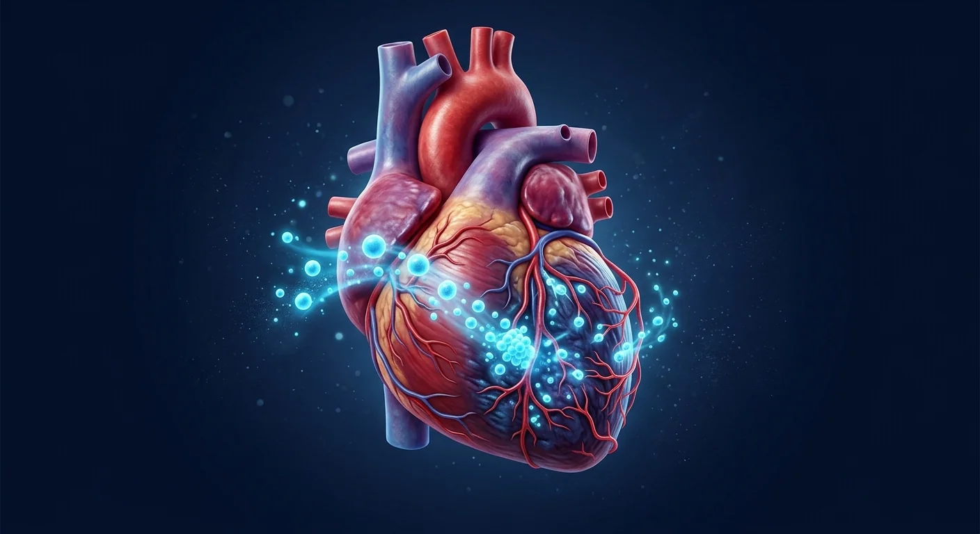
Chronic ischemic cardiomyopathy affects an estimated 2–5 million people in the United States alone, making it one of the most prevalent causes of heart failure with reduced ejection fraction (HFrEF). [1] The scarred, thinned ventricular wall that follows one or more myocardial infarctions progressively loses its ability to pump, and guideline-directed medical therapy — while life-extending — does not reverse the structural damage.
Where conventional therapy reaches its ceiling. Beta-blockers, ACE inhibitors, mineralocorticoid receptor antagonists, and device therapy (ICDs, CRT) slow the downward trajectory, but patients continue to accumulate myocardial scar, and a substantial proportion progress to end-stage disease despite optimal management. The gap is structural: no drug regrows contractile tissue.
The problem is the scar. After myocardial infarction, necrotic cardiomyocytes are replaced by a collagen-rich extracellular matrix that neither contracts nor conducts. This fibrotic replacement stabilises the ventricular wall acutely but becomes pathological when it expands — a process termed adverse ventricular remodelling — leading to progressive chamber dilation, worsened wall stress, and functional mitral regurgitation. [2]
MSCs target the scar microenvironment. Rather than attempting to replace dead cardiomyocytes directly, mesenchymal stem cells secrete a broad repertoire of paracrine factors — including VEGF, HGF, IGF-1, and TGF-β modulators — that collectively reduce fibrosis, stimulate resident cardiac progenitor activity, promote angiogenesis in the peri-infarct border zone, and suppress maladaptive inflammation. [3] This multi-target mechanism is well-suited to the complex pathophysiology of ischemic cardiomyopathy.
What Is Chronic Ischemic Cardiomyopathy?
Chronic ischemic cardiomyopathy is a distinct form of heart failure caused by coronary artery disease that has produced irreversible myocardial damage. It is defined by left ventricular systolic dysfunction (LVEF ≤ 40%) attributable to ischemia, with evidence of prior myocardial infarction, hibernating myocardium, or both. [4]
The condition differs from non-ischemic dilated cardiomyopathy in both pathophysiology and prognosis. Because the underlying driver is atherosclerotic coronary disease, the myocardial injury tends to be regional and patchy rather than global, and interventions that improve coronary perfusion (revascularisation) can recover some function in hibernating segments. However, once fibrotic scar has formed, that tissue is permanently non-contractile.
Clinically, patients present with the classic heart failure syndrome — exertional dyspnoea, orthopnoea, paroxysmal nocturnal dyspnoea, fatigue, and peripheral oedema — superimposed on a history of angina, prior MI, or coronary revascularisation. Diagnosis rests on echocardiography demonstrating regional wall motion abnormalities in a coronary distribution, supported by stress testing, cardiac MRI with late gadolinium enhancement confirming transmural or subendocardial scar, and coronary angiography documenting the culprit lesions.
How MSCs May Help Ischemic Cardiomyopathy
MSC therapy for chronic ischemic cardiomyopathy operates through four interconnected mechanisms, none of which requires the transplanted cells to become new cardiomyocytes. [5]
Paracrine signalling. MSCs secrete a rich cocktail of growth factors and cytokines — VEGF, HGF, IGF-1, SDF-1, and angiopoietin-1 — that act on the host myocardium. VEGF stimulates angiogenesis in the ischaemic border zone, recruiting new capillary networks that improve oxygen delivery to hibernating but viable tissue. HGF and IGF-1 activate resident cardiac stem cells and promote cardiomyocyte survival. This "bystander effect" is now considered the dominant mechanism of MSC benefit in the heart. [6]
Anti-fibrotic remodelling. MSCs modulate the TGF-β/Smad signalling axis and secrete matrix metalloproteinases (MMPs) and their tissue inhibitors (TIMPs) in a balanced ratio that favours scar resorption over expansion. Preclinical models consistently show reduced collagen volume fraction in MSC-treated hearts, with improved ventricular compliance and reduced end-diastolic pressure. [7]
Immunomodulation. The post-infarction myocardium is a pro-inflammatory environment driven by M1 macrophages, IL-1β, IL-6, and TNF-α. MSCs polarise macrophages toward the reparative M2 phenotype, expand regulatory T-cell populations, and suppress neutrophil infiltration — shifting the milieu from chronic destructive inflammation toward tissue repair. [8]
Angiogenesis and perfusion. Beyond VEGF secretion, MSCs differentiate into pericyte-like cells that stabilise nascent vessels, and their exosomes carry microRNAs (miR-21, miR-210) that promote endothelial cell proliferation and tube formation. Together, these effects improve microvascular density in peri-infarct regions — a parameter that correlates with functional recovery on serial echocardiography. [9]
What the Clinical Trials Show
The clinical evidence for MSC therapy in ischemic cardiomyopathy comes from a series of phase I/II trials. While results are mixed and modest, they establish a safety foundation and signal of potential benefit.
Key Clinical Trials in Ischemic Cardiomyopathy — Summary
- POSEIDON (2012). 30 patients with ischemic cardiomyopathy randomised to autologous vs allogeneic bone-marrow MSCs via transendocardial injection. Both groups showed improved 6-minute walk distance and reduced infarct size on CT at 13 months. Allogeneic MSCs were not immunogenic. [10]
- TAC-HFT (2014). 65 patients comparing MSC vs bone marrow mononuclear cells. MSC group showed significantly greater reduction in infarct size (−18.9% vs −7.0%, p=0.05) and improved regional contractility at 12 months. [11]
- MSC-HF (2015). 60 patients with severe ischemic HF (LVEF <45%) randomised to intramyocardial autologous bone-marrow MSCs vs placebo. MSC group showed mean LVEF improvement of +6.2% (placebo: −1.4%, p<0.001) and reduced end-systolic volume at 6 months. [12]
- DREAM-HF (2023). 537 patients with HFrEF (60% ischemic) randomised to transendocardial allogeneic MPCs (rexlemestrocel-L) vs sham control. Did not meet primary endpoint of recurrent HF hospitalisations, but pre-specified analysis showed 24% reduction in cardiac death/MI/stroke composite in the treated group. Safe across the cohort. [13]
- CHART-1 (2016). 315 patients with ischemic HF receiving cardiopoietic bone-marrow cells vs sham. Negative for primary endpoint, but post hoc analysis suggested benefit in patients with baseline LVEDV 200–370 mL. Underscores the importance of patient selection. [14]
The consistent pattern across these trials is reassuring safety — no excess arrhythmias, no tumour formation — with modest but statistically significant improvements in surrogate endpoints (LVEF, infarct size, 6MWD) that have not yet translated into a definitive mortality or hospitalisation benefit. The field is in a phase of refining cell product, dose, delivery route, and patient selection to amplify the signal.
How Outcomes Are Measured
Understanding the cardiac imaging and biomarker endpoints used in MSC trials is essential for evaluating claims. [15]
LVEF (Left Ventricular Ejection Fraction). The percentage of blood ejected from the left ventricle with each contraction. Measured by echocardiography or cardiac MRI. Normal ≥ 52% (male) or ≥ 54% (female). In the MSC-HF trial, treated patients gained a mean of 6.2 LVEF percentage points over 6 months — a modest but clinically meaningful change.
Infarct Size / Scar Burden. Quantified by late gadolinium enhancement (LGE) on cardiac MRI. This is arguably the most direct measure of MSC anti-fibrotic effect. TAC-HFT reported an 18.9% relative reduction in infarct size in the MSC arm.
LV Volumes (LVEDV, LVESV). Left ventricular end-diastolic and end-systolic volumes track adverse remodelling. A decrease in LVESV (reverse remodelling) is a positive prognostic sign and has been observed across multiple MSC trials.
6-Minute Walk Distance (6MWD). A simple, reproducible functional endpoint. Improvements of 30–50 metres reached statistical significance in POSEIDON and other early-phase trials.
NT-proBNP. A natriuretic peptide biomarker that reflects ventricular wall stress. Reductions in NT-proBNP correlate with improved haemodynamics and are an established surrogate in heart failure trials. [16]
Delivery Routes: Intravenous vs Intramyocardial
How MSCs reach the heart matters profoundly. The two principal routes each carry distinct advantages and limitations.
Intramyocardial injection — typically via transendocardial catheter (NOGA mapping) or direct surgical injection during CABG — deposits cells directly into the peri-infarct zone, bypassing the pulmonary first-pass trap that captures 80–90% of intravenously delivered cells. This is the preferred route in most major trials (POSEIDON, TAC-HFT, MSC-HF, DREAM-HF). The trade-off is invasiveness and the requirement for specialised mapping equipment.
Intravenous infusion is simpler, repeatable, and suitable for ambulatory care, but the lung trapping means only a small fraction of cells reach the coronary circulation. Some investigators argue this is acceptable because the paracrine effect may be systemic rather than local, and trapped MSCs in the pulmonary microvasculature still secrete anti-inflammatory factors that enter the systemic circulation. This hypothesis remains unproven. [17]
Key Takeaway
The largest and most rigorously controlled trials in ischemic cardiomyopathy have used direct intramyocardial delivery. IV delivery for cardiac indications lacks comparable phase III evidence. When evaluating a treatment proposal, ask which delivery route is planned and what published evidence supports that specific route for the patient's condition.
Patient Selection: Who Stands to Benefit Most?
Not all patients with ischemic cardiomyopathy are equally likely to respond to MSC therapy. Post hoc analyses from the larger trials point to several predictors of response. [18]
- Moderate LV dysfunction (LVEF 25–40%). Patients with very advanced disease (LVEF <20%) have extensive scar with little viable myocardium to rescue. Those with moderate dysfunction retain a border zone of hibernating tissue — the substrate MSCs are most likely to benefit.
- Middle-range LV volumes (LVEDV 200–370 mL). CHART-1 identified this window as the responsive subgroup. Severely dilated ventricles may have wall stress exceeding what paracrine repair can overcome.
- Absence of large, transmural scar in all territories. Late gadolinium enhancement >50% wall thickness in the target zone limits the viable substrate available for functional recovery.
- Biomarker profile. Elevated baseline CRP and IL-6 predict greater anti-inflammatory benefit, as these patients have the most active inflammatory drive to suppress.
Limitations and Honest Caveats
What MSC therapy cannot do — yet
- No MSC product is FDA-approved or EMA-approved for ischemic cardiomyopathy as of 2026. All use is investigational.
- The largest phase III trial (DREAM-HF, n=537) did not meet its primary endpoint. The signals are in secondary and subgroup analyses.
- LVEF improvements in the positive trials are modest (mean +3–6 percentage points) — not "reversal" of heart failure.
- MSCs do not regenerate substantial volumes of new myocardium. The benefit is paracrine, not cardiomyogenic.
- Long-term durability beyond 12–24 months is under-studied. Most trials report at 6–12 months.
- Patient-level predictors of response are statistical associations, not validated biomarkers. Individual response is variable.
These limitations do not mean the approach is invalid — many therapies for chronic diseases produce modest, incremental benefits that are nonetheless clinically meaningful. But they define the honest boundaries of current knowledge.
VELAR's Approach
At VELAR Center, we approach ischemic cardiomyopathy with the same principles that guide all our regenerative protocols: transparency about the evidence, conservative claims, and integration with — never replacement of — conventional cardiology care.
Our MSC protocols for cardiac patients are grounded in the paracrine biology described above. We use Wharton's jelly-derived MSCs, which have a more potent secretory profile than adult bone-marrow MSCs, with higher expression of VEGF, HGF, and anti-inflammatory cytokines. All cells are processed in our laboratory operating under ISO 9001 with full batch traceability. For patients with ischemic cardiomyopathy, our clinical team reviews cardiac imaging, ejection fraction, NYHA class, and medication regimen before determining suitability — and we require that patients maintain their guideline-directed medical therapy throughout the regenerative protocol.
We do not claim to reverse heart failure, regrow dead muscle, or replace cardiology care. What we offer is a biologically rational adjunct grounded in acknowledged paracrine mechanisms, delivered with the transparency that an investigational field demands.
Frequently Asked Questions
Can stem cells regrow dead heart muscle after a heart attack?
No. Current evidence does not support the claim that MSCs become large numbers of new cardiomyocytes. The benefit — when it occurs — is paracrine: MSCs secrete factors that reduce scar, promote angiogenesis, and suppress inflammation in the surviving border zone. This is tissue support, not tissue replacement.
How much LVEF improvement can I realistically expect from MSC therapy?
In the clinical trials that showed benefit (MSC-HF, TAC-HFT), the mean LVEF improvement was 3–7 percentage points over 6–12 months. This is a modest but clinically meaningful change for patients with moderate dysfunction. Responses vary; some patients experience no measurable improvement.
Is MSC therapy safe for patients with ICDs or pacemakers?
Yes. No trial has reported an excess of arrhythmic events or device interactions with MSC therapy. The arrhythmia rate in treated groups has been comparable to placebo across all major trials — one of the strongest safety signals in the field.
How is MSC therapy delivered to the heart — IV or injection?
In the major clinical trials (POSEIDON, DREAM-HF, MSC-HF), cells were delivered by transendocardial catheter injection under electromechanical mapping — a catheter-based procedure that deposits cells directly into the heart wall. IV delivery is simpler but results in pulmonary trapping of most cells and has substantially less clinical evidence for cardiac indications.
How much does stem cell therapy for heart failure cost in Thailand?
Costs vary by protocol complexity, cell dose, and number of sessions. At VELAR Center, a cardiac MSC protocol typically ranges from USD 15,000–25,000 depending on individual assessment. This is a fraction of comparable protocols in the United States or Europe, where costs frequently exceed USD 40,000. All prospective patients receive a detailed cost breakdown during the consultation process.
Will I still need my heart failure medications during MSC therapy?
Absolutely. MSC therapy is an adjunct, not a replacement, for guideline-directed medical therapy. Beta-blockers, ACE inhibitors/ARBs, MRAs, and SGLT2 inhibitors remain the foundation of heart failure management. MSCs are studied as a supplement to — never a substitute for — these proven treatments.
References
- Benjamin EJ, Muntner P, Alonso A, et al. Heart disease and stroke statistics — 2019 update: a report from the American Heart Association. Circulation. 2019;139(10):e56-e528. doi:10.1161/CIR.0000000000000659 ↩
- Prabhu SD, Frangogiannis NG. The biological basis for cardiac repair after myocardial infarction: from inflammation to fibrosis. Circulation Research. 2016;119(1):91-112. doi:10.1161/CIRCRESAHA.116.303577 ↩
- Gnecchi M, Zhang Z, Ni A, Dzau VJ. Paracrine mechanisms in adult stem cell signaling and therapy. Circulation Research. 2008;103(11):1204-1219. doi:10.1161/CIRCRESAHA.108.176826 ↩
- Felker GM, Shaw LK, O'Connor CM. A standardized definition of ischemic cardiomyopathy for use in clinical research. Journal of the American College of Cardiology. 2002;39(2):210-218. doi:10.1016/S0735-1097(01)01738-7 ↩
- Williams AR, Hare JM. Mesenchymal stem cells: biology, pathophysiology, translational findings, and therapeutic implications for cardiac disease. Circulation Research. 2011;109(8):923-940. doi:10.1161/CIRCRESAHA.111.243147 ↩
- Ranganath SH, Levy O, Inamdar MS, Karp JM. Harnessing the mesenchymal stem cell secretome for the treatment of cardiovascular disease. Cell Stem Cell. 2012;10(3):244-258. doi:10.1016/j.stem.2012.02.005 ↩
- Mias C, Lairez O, Trouche E, et al. Mesenchymal stem cells promote matrix metalloproteinase secretion by cardiac fibroblasts and reduce cardiac ventricular fibrosis after myocardial infarction. Stem Cells. 2009;27(11):2734-2743. doi:10.1002/stem.169 ↩
- Ben-Mordechai T, Holbova R, Landa-Rouben N, et al. Macrophage subpopulations are essential for infarct repair with and without stem cell therapy. Journal of the American College of Cardiology. 2013;62(20):1890-1901. doi:10.1016/j.jacc.2013.07.057 ↩
- Bian S, Zhang L, Duan L, Wang X, Min Y, Yu H. Extracellular vesicles derived from human bone marrow mesenchymal stem cells promote angiogenesis in a rat myocardial infarction model. Journal of Molecular Medicine. 2014;92(4):387-397. doi:10.1007/s00109-013-1110-5 ↩
- Hare JM, Fishman JE, Gerstenblith G, et al. Comparison of allogeneic vs autologous bone marrow–derived mesenchymal stem cells delivered by transendocardial injection in patients with ischemic cardiomyopathy: the POSEIDON randomized trial. JAMA. 2012;308(22):2369-2379. doi:10.1001/jama.2012.25321 ↩
- Heldman AW, DiFede DL, Fishman JE, et al. Transendocardial mesenchymal stem cells and mononuclear bone marrow cells for ischemic cardiomyopathy: the TAC-HFT randomized trial. JAMA. 2014;311(1):62-73. doi:10.1001/jama.2013.282909 ↩
- Mathiasen AB, Qayyum AA, Jørgensen E, et al. Bone marrow–derived mesenchymal stromal cell treatment in patients with severe ischaemic heart failure: a randomized placebo-controlled trial (MSC-HF trial). European Heart Journal. 2015;36(27):1744-1753. doi:10.1093/eurheartj/ehv136 ↩
- Perin EC, Borow KM, Henry TD, et al. Randomized trial of targeted transendocardial mesenchymal precursor cell therapy in patients with heart failure. Journal of the American College of Cardiology. 2023;81(9):849-863. doi:10.1016/j.jacc.2022.11.061 ↩
- Bartunek J, Terzic A, Davison BA, et al. Cardiopoietic cell therapy for advanced ischaemic heart failure: results at 39 weeks of the prospective, randomized, double blind, sham-controlled CHART-1 clinical trial. European Heart Journal. 2017;38(9):648-660. doi:10.1093/eurheartj/ehw543 ↩
- Sanganalmath SK, Bolli R. Cell therapy for heart failure: a comprehensive overview of experimental and clinical studies, current challenges, and future directions. Circulation Research. 2013;113(6):810-834. doi:10.1161/CIRCRESAHA.113.300219 ↩
- Januzzi JL, Troughton R. Are serial BNP measurements useful in heart failure management? Serial natriuretic peptide measurements are useful in heart failure management. Circulation. 2013;127(4):500-508. doi:10.1161/CIRCULATIONAHA.112.120485 ↩
- Lee RH, Pulin AA, Seo MJ, et al. Intravenous hMSCs improve myocardial infarction in mice because cells embolized in lung are activated to secrete the anti-inflammatory protein TSG-6. Cell Stem Cell. 2009;5(1):54-63. doi:10.1016/j.stem.2009.05.003 ↩
- Banerjee MN, Bolli R, Hare JM. Clinical studies of cell therapy in cardiovascular medicine: recent developments and future directions. Circulation Research. 2018;123(2):266-287. doi:10.1161/CIRCRESAHA.118.311217 ↩
慢性缺血性心肌病仅在美国就影响着约200万至500万人,是射血分数降低型心力衰竭(HFrEF)最常见的原因之一。[1]一次或多次心肌梗死后留下的瘢痕化、变薄的心室壁逐渐丧失泵血能力,而指南指导的药物治疗虽然能延长生命,却无法逆转结构性损伤。
常规治疗的局限性。β受体阻滞剂、ACE抑制剂、盐皮质激素受体拮抗剂和器械治疗(ICD、CRT)能够减缓病情恶化,但患者仍会持续积累心肌瘢痕,相当一部分人即使在最佳管理下仍会进展至终末期疾病。这个缺口是结构性的:没有任何药物能再生收缩组织。
问题的核心是瘢痕。心肌梗死后,坏死的心肌细胞被富含胶原的细胞外基质替代,这种基质既不收缩也不传导。这种纤维化替代在急性期稳定了心室壁,但一旦扩展——即不良心室重构——就变成病理性的,导致进行性心室扩张、室壁应力加剧和功能性二尖瓣反流。[2]
MSC靶向瘢痕微环境。间充质干细胞并非直接替代死亡的心肌细胞,而是分泌一系列旁分泌因子——包括VEGF、HGF、IGF-1和TGF-β调节剂——共同减少纤维化、激活常驻心脏祖细胞、促进梗死边缘区血管新生,并抑制异常的炎症反应。[3]这一多靶点机制非常适合缺血性心肌病复杂的病理生理学。
什么是慢性缺血性心肌病?
慢性缺血性心肌病是由冠状动脉疾病引起的特殊类型心力衰竭,已造成不可逆的心肌损伤。其定义为归因于缺血的左心室收缩功能障碍(LVEF ≤ 40%),并伴有既往心肌梗死、冬眠心肌或两者兼具的证据。[4]
该病在病理生理学和预后上都与非缺血性扩张型心肌病不同。由于其潜在驱动因素是动脉粥样硬化性冠状动脉疾病,心肌损伤往往是区域性、斑片状的而非全心的,改善冠脉灌注的干预措施(血运重建)可以恢复冬眠节段的部分功能。然而,一旦纤维化瘢痕形成,该组织便永久失去收缩能力。
临床上,患者表现为典型的心力衰竭综合征——劳力性呼吸困难、端坐呼吸、阵发性夜间呼吸困难、疲劳和外周水肿——叠加于心绞痛、既往心梗或冠脉血运重建病史之上。诊断依赖于超声心动图显示冠脉分布区域的节段性室壁运动异常,辅以负荷试验、钆延迟增强心脏MRI确认透壁或心内膜下瘢痕,以及冠脉造影记录罪犯病变。
MSC如何帮助缺血性心肌病
MSC治疗慢性缺血性心肌病通过四个相互关联的机制进行,其中没有一项要求移植细胞成为新的心肌细胞。[5]
旁分泌信号。MSC分泌丰富的生长因子和细胞因子——VEGF、HGF、IGF-1、SDF-1和血管生成素-1——作用于宿主心肌。VEGF刺激缺血边缘区的血管新生,募集新的毛细血管网络以改善对冬眠但存活组织的氧供。HGF和IGF-1激活常驻心脏干细胞并促进心肌细胞存活。这种"旁观者效应"现被认为是MSC心脏获益的主要机制。[6]
抗纤维化重构。MSC调节TGF-β/Smad信号轴,并以有利于瘢痕吸收而非扩展的平衡比例分泌基质金属蛋白酶(MMP)及其组织抑制剂(TIMP)。临床前模型一致显示MSC处理的心脏中胶原体积分数降低,心室顺应性改善,舒张末期压力下降。[7]
免疫调节。梗死后的心肌是一个促炎环境,由M1巨噬细胞、IL-1β、IL-6和TNF-α驱动。MSC将巨噬细胞极化为修复性M2表型,扩增调节性T细胞群体,抑制中性粒细胞浸润——将微环境从慢性破坏性炎症转向组织修复。[8]
血管新生与灌注。除VEGF分泌外,MSC分化为周细胞样细胞稳定新生血管,其外泌体携带促进内皮细胞增殖和管腔形成的microRNA(miR-21、miR-210)。这些效应共同改善梗死周围区域的微血管密度——这一参数与超声心动图随访中的功能恢复相关。[9]
临床试验证据
MSC治疗缺血性心肌病的临床证据来自一系列I/II期试验。虽然结果参差不齐且效应幅度有限,但它们建立了安全性基础并提示了潜在获益信号。
缺血性心肌病关键临床试验——摘要
- POSEIDON(2012年)。30例缺血性心肌病患者随机分配至自体vs异体骨髓MSC经心内膜注射。两组在13个月时均显示6分钟步行距离改善和CT梗死面积缩小。异体MSC不具免疫原性。[10]
- TAC-HFT(2014年)。65例患者比较MSC与骨髓单个核细胞。MSC组梗死面积减小显著更大(−18.9% vs −7.0%,p=0.05),12个月时局部收缩功能改善。[11]
- MSC-HF(2015年)。60例重度缺血性心衰患者(LVEF <45%)随机分配至心肌内自体骨髓MSC vs安慰剂。MSC组LVEF平均改善+6.2%(安慰剂:−1.4%,p<0.001),6个月时收缩末期容积减小。[12]
- DREAM-HF(2023年)。537例HFrEF患者(60%缺血性)随机分配至经心内膜异体MPC(rexlemestrocel-L)vs假手术对照。未达到心衰住院复发的首要终点,但预设分析显示治疗组心脏死亡/心梗/卒中复合终点减少24%。全组安全性良好。[13]
- CHART-1(2016年)。315例缺血性心衰患者接受心脏生成性骨髓细胞vs假手术。主要终点阴性,但事后分析提示基线LVEDV 200–370 mL的患者可能获益。凸显了患者选择的重要性。[14]
这些试验的一致模式是令人安心的安全性——无过度心律失常、无肿瘤形成——以及在替代终点(LVEF、梗死面积、6MWD)上的适度但统计学显著的改善,但这些尚未转化为明确的死亡率或住院获益。该领域正处于优化细胞产品、剂量、递送途径和患者选择以放大信号的阶段。
如何测量结局
理解MSC试验中使用的心脏影像和生物标志物终点对于评估疗效主张至关重要。[15]
LVEF(左心室射血分数)。每次收缩时左心室搏出血量的百分比。通过超声心动图或心脏MRI测量。正常值 ≥ 52%(男性)或 ≥ 54%(女性)。在MSC-HF试验中,治疗组患者6个月内LVEF平均增加6.2个百分点——虽温和但具有临床意义。
梗死面积/瘢痕负荷。通过心脏MRI钆延迟增强定量。这可以说是MSC抗纤维化效果最直接的测量指标。TAC-HFT报告MSC组梗死面积相对减少18.9%。
LV容积(LVEDV、LVESV)。左心室舒张末期和收缩末期容积追踪不良重构。LVESV减小(逆重构)是积极的预后信号,已在多个MSC试验中观察到。
6分钟步行距离(6MWD)。一个简单、可重复的功能终点。POSEIDON及其他早期试验中,30–50米的改善达到了统计学显著性。
NT-proBNP。反映心室壁应力的利钠肽生物标志物。NT-proBNP降低与血流动力学改善相关,是心力衰竭试验中已确立的替代指标。[16]
递送途径:静脉vs心肌内
MSC如何到达心脏至关重要。两种主要途径各有独特的优势和局限。
心肌内注射——通常通过经心内膜导管(NOGA标测)或CABG术中直接手术注射——将细胞直接沉积到梗死周围区域,避开了肺首过捕获80-90%静脉输注细胞的陷阱。这是大多数主要试验(POSEIDON、TAC-HFT、MSC-HF、DREAM-HF)的首选途径。代价是侵袭性和需要专门的标测设备。
静脉输注更简单、可重复,适合门诊治疗,但肺捕获意味着只有一小部分细胞到达冠脉循环。一些研究者认为这是可接受的,因为旁分泌效应可能是全身性的而非局部的,被捕获在肺微血管中的MSC仍可分泌进入体循环的抗炎因子。这一假说尚未得到证实。[17]
关键要点
缺血性心肌病中规模最大、对照最严格的试验均采用直接心肌内递送。针对心脏适应证的静脉递送缺乏可比的III期证据。在评估治疗方案时,应询问计划使用哪种递送途径,以及有何已发表证据支持该特定途径用于患者的具体病情。
患者选择:谁最可能获益?
并非所有缺血性心肌病患者对MSC治疗的反应概率相同。较大试验的事后分析指出了若干反应预测因素。[18]
- 中度LV功能障碍(LVEF 25–40%)。病情极晚期患者(LVEF <20%)有广泛的瘢痕,可挽救的存活心肌很少。中度功能障碍者保留了一个冬眠组织边缘区——MSC最可能获益的基质。
- 中等范围LV容积(LVEDV 200–370 mL)。CHART-1确定这一窗口为反应性亚组。严重扩张的心室可能具有超出旁分泌修复能力的室壁应力。
- 所有区域均无大型透壁瘢痕。目标区域钆延迟增强 >50%室壁厚度会限制功能恢复所需的存活基质。
- 生物标志物谱。基线CRP和IL-6升高预示更大的抗炎获益,因为这些患者具有最活跃的炎症驱动力可供抑制。
局限性与诚实说明
MSC治疗目前无法做到的事情
- 截至2026年,尚无MSC产品获得FDA或EMA批准用于缺血性心肌病。所有使用均为研究性质。
- 最大的III期试验(DREAM-HF,n=537)未达到其主要终点。信号存在于次要分析和亚组分析中。
- 阳性试验中的LVEF改善幅度有限(平均+3–6个百分点)——不是心衰的"逆转"。
- MSC不会再生大量新心肌。获益是旁分泌性的,而非心肌生成性的。
- 超过12–24个月的长期持久性研究不足。大多数试验在6–12个月时报告结果。
- 患者层面的反应预测因素是统计关联,而非验证的生物标志物。个体反应存在差异。
这些局限性并不意味着这种方法无效——许多慢性疾病疗法产生温和、渐进的获益,却具有临床意义。但它们定义了当前知识的诚实边界。
VELAR的方法
在VELAR中心,我们以指导所有再生方案的原则对待缺血性心肌病:透明说明证据、保守表述主张、与常规心脏病治疗整合——而非替代。
我们针对心脏患者的MSC方案基于上述旁分泌生物学。我们使用沃顿胶来源的MSC,其分泌特征比成人骨髓MSC更强效,VEGF、HGF和抗炎细胞因子表达更高。所有细胞在我们依 ISO 9001 运作的实验室中处理,具有完整的批次可追溯性。对于缺血性心肌病患者,我们的临床团队在确定适用性之前审阅心脏影像、射血分数、NYHA分级和用药方案——并且我们要求患者在整个再生方案期间维持其指南指导的药物治疗。
我们不声称能逆转心衰、再生死亡肌肉或替代心脏病治疗。我们提供的是基于公认旁分泌机制的生物学合理补充疗法,以研究领域所要求的透明方式提供。
常见问题
干细胞能否在心脏病发作后再生死亡的心肌?
不能。现有证据不支持MSC成为大量新心肌细胞的说法。获益——当发生时——是旁分泌性的:MSC分泌减少瘢痕、促进血管新生和抑制存活边缘区炎症的因子。这是组织支持,而非组织替代。
通过MSC治疗可以现实地期望多少LVEF改善?
在显示获益的临床试验中(MSC-HF、TAC-HFT),平均LVEF改善在6–12个月内为3–7个百分点。这对于中度功能障碍患者是温和但有临床意义的变化。反应存在差异;部分患者未出现可测量的改善。
MSC治疗对装有ICD或起搏器的患者安全吗?
安全。没有试验报告MSC治疗导致心律失常事件或器械相互作用增加。在所有主要试验中,治疗组的心律失常发生率与安慰剂相当——这是该领域最强的安全性信号之一。
MSC治疗如何递送到心脏——静脉还是注射?
在主要临床试验(POSEIDON、DREAM-HF、MSC-HF)中,细胞通过经心内膜导管注射在心电机械标测引导下递送——一种基于导管的操作,将细胞直接沉积到心脏壁中。静脉递送更简单,但导致大多数细胞被肺捕获,且对心脏适应证具有较少的临床证据。
在泰国,干细胞治疗心力衰竭的费用是多少?
费用因方案复杂性、细胞剂量和疗程次数而异。在VELAR中心,心脏MSC方案通常为15,000–25,000美元,具体取决于个体评估。这仅是美国或欧洲同类方案费用(通常超过40,000美元)的一小部分。所有潜在患者在咨询过程中都会收到详细的费用明细。
在MSC治疗期间,我是否仍需服用心力衰竭药物?
绝对需要。MSC治疗是指南指导药物治疗的补充,而非替代。β受体阻滞剂、ACE抑制剂/ARB、MRA和SGLT2抑制剂仍然是心力衰竭管理的基础。MSC被研究作为这些已证实治疗的补充——绝非替代。
参考文献
- Benjamin EJ, Muntner P, Alonso A, et al. Heart disease and stroke statistics — 2019 update: a report from the American Heart Association. Circulation. 2019;139(10):e56-e528. doi:10.1161/CIR.0000000000000659 ↩
- Prabhu SD, Frangogiannis NG. The biological basis for cardiac repair after myocardial infarction: from inflammation to fibrosis. Circulation Research. 2016;119(1):91-112. doi:10.1161/CIRCRESAHA.116.303577 ↩
- Gnecchi M, Zhang Z, Ni A, Dzau VJ. Paracrine mechanisms in adult stem cell signaling and therapy. Circulation Research. 2008;103(11):1204-1219. doi:10.1161/CIRCRESAHA.108.176826 ↩
- Felker GM, Shaw LK, O'Connor CM. A standardized definition of ischemic cardiomyopathy for use in clinical research. Journal of the American College of Cardiology. 2002;39(2):210-218. doi:10.1016/S0735-1097(01)01738-7 ↩
- Williams AR, Hare JM. Mesenchymal stem cells: biology, pathophysiology, translational findings, and therapeutic implications for cardiac disease. Circulation Research. 2011;109(8):923-940. doi:10.1161/CIRCRESAHA.111.243147 ↩
- Ranganath SH, Levy O, Inamdar MS, Karp JM. Harnessing the mesenchymal stem cell secretome for the treatment of cardiovascular disease. Cell Stem Cell. 2012;10(3):244-258. doi:10.1016/j.stem.2012.02.005 ↩
- Mias C, Lairez O, Trouche E, et al. Mesenchymal stem cells promote matrix metalloproteinase secretion by cardiac fibroblasts and reduce cardiac ventricular fibrosis after myocardial infarction. Stem Cells. 2009;27(11):2734-2743. doi:10.1002/stem.169 ↩
- Ben-Mordechai T, Holbova R, Landa-Rouben N, et al. Macrophage subpopulations are essential for infarct repair with and without stem cell therapy. Journal of the American College of Cardiology. 2013;62(20):1890-1901. doi:10.1016/j.jacc.2013.07.057 ↩
- Bian S, Zhang L, Duan L, Wang X, Min Y, Yu H. Extracellular vesicles derived from human bone marrow mesenchymal stem cells promote angiogenesis in a rat myocardial infarction model. Journal of Molecular Medicine. 2014;92(4):387-397. doi:10.1007/s00109-013-1110-5 ↩
- Hare JM, Fishman JE, Gerstenblith G, et al. Comparison of allogeneic vs autologous bone marrow–derived mesenchymal stem cells delivered by transendocardial injection in patients with ischemic cardiomyopathy: the POSEIDON randomized trial. JAMA. 2012;308(22):2369-2379. doi:10.1001/jama.2012.25321 ↩
- Heldman AW, DiFede DL, Fishman JE, et al. Transendocardial mesenchymal stem cells and mononuclear bone marrow cells for ischemic cardiomyopathy: the TAC-HFT randomized trial. JAMA. 2014;311(1):62-73. doi:10.1001/jama.2013.282909 ↩
- Mathiasen AB, Qayyum AA, Jørgensen E, et al. Bone marrow–derived mesenchymal stromal cell treatment in patients with severe ischaemic heart failure: a randomized placebo-controlled trial (MSC-HF trial). European Heart Journal. 2015;36(27):1744-1753. doi:10.1093/eurheartj/ehv136 ↩
- Perin EC, Borow KM, Henry TD, et al. Randomized trial of targeted transendocardial mesenchymal precursor cell therapy in patients with heart failure. Journal of the American College of Cardiology. 2023;81(9):849-863. doi:10.1016/j.jacc.2022.11.061 ↩
- Bartunek J, Terzic A, Davison BA, et al. Cardiopoietic cell therapy for advanced ischaemic heart failure: results at 39 weeks of the prospective, randomized, double blind, sham-controlled CHART-1 clinical trial. European Heart Journal. 2017;38(9):648-660. doi:10.1093/eurheartj/ehw543 ↩
- Sanganalmath SK, Bolli R. Cell therapy for heart failure: a comprehensive overview of experimental and clinical studies, current challenges, and future directions. Circulation Research. 2013;113(6):810-834. doi:10.1161/CIRCRESAHA.113.300219 ↩
- Januzzi JL, Troughton R. Are serial BNP measurements useful in heart failure management? Serial natriuretic peptide measurements are useful in heart failure management. Circulation. 2013;127(4):500-508. doi:10.1161/CIRCULATIONAHA.112.120485 ↩
- Lee RH, Pulin AA, Seo MJ, et al. Intravenous hMSCs improve myocardial infarction in mice because cells embolized in lung are activated to secrete the anti-inflammatory protein TSG-6. Cell Stem Cell. 2009;5(1):54-63. doi:10.1016/j.stem.2009.05.003 ↩
- Banerjee MN, Bolli R, Hare JM. Clinical studies of cell therapy in cardiovascular medicine: recent developments and future directions. Circulation Research. 2018;123(2):266-287. doi:10.1161/CIRCRESAHA.118.311217 ↩
يؤثر اعتلال عضلة القلب الإقفاري المزمن على ما يقدر بـ 2–5 مليون شخص في الولايات المتحدة وحدها، مما يجعله أحد أكثر أسباب فشل القلب مع انخفاض الكسر القذفي (HFrEF) شيوعًا. [1] يفقد الجدار البطيني المتندب والرقيق الذي يتبع احتشاءً واحدًا أو أكثر من عضلة القلب قدرته على الضخ تدريجيًا، والعلاج الدوائي الموجه بالمبادئ التوجيهية — رغم أنه يطيل العمر — لا يعكس الضرر البنيوي.
حيث يصل العلاج التقليدي إلى سقفه. تعمل حاصرات بيتا ومثبطات الإنزيم المحول للأنجيوتنسين ومضادات مستقبلات القشرانيات المعدنية والعلاج بالأجهزة (ICD، CRT) على إبطاء المسار التنازلي، لكن المرضى يستمرون في تراكم الندبة القلبية، وتتقدم نسبة كبيرة منهم إلى مرحلة المرض النهائي على الرغم من الإدارة المثلى. الفجوة بنيوية: لا يوجد دواء يعيد نمو النسيج الانقباضي.
المشكلة هي الندبة. بعد احتشاء عضلة القلب، تُستبدل الخلايا العضلية القلبية النخرية بمطرس خارج خلوي غني بالكولاجين لا ينقبض ولا ينقل الإشارات. هذا الاستبدال الليفي يثبت الجدار البطيني في المرحلة الحادة لكنه يصبح مرضيًا عندما يتوسع — وهي عملية تسمى إعادة التشكيل البطيني العكسي — مما يؤدي إلى توسع تدريجي للحجرات وتفاقم إجهاد الجدار وقلس تاجي وظيفي. [2]
تستهدف الخلايا الجذعية الميزنشيمية البيئة الدقيقة للندبة. بدلاً من محاولة استبدال الخلايا العضلية القلبية الميتة مباشرة، تفرز الخلايا الجذعية الميزنشيمية مجموعة واسعة من العوامل نظيرة الصماوي — بما في ذلك VEGF وHGF وIGF-1 ومعدلات TGF-β — التي تقلل التليف بشكل جماعي وتحفز نشاط الخلايا السلفية القلبية المقيمة وتعزز تولد الأوعية في المنطقة الحدودية حول الاحتشاء وتثبط الالتهاب غير التكيفي. [3] هذه الآلية متعددة الأهداف مناسبة تمامًا للفسيولوجيا المرضية المعقدة لاعتلال عضلة القلب الإقفاري.
ما هو اعتلال عضلة القلب الإقفاري المزمن؟
اعتلال عضلة القلب الإقفاري المزمن هو شكل مميز من فشل القلب ناجم عن مرض الشريان التاجي الذي أنتج ضررًا قلبيًا غير قابل للعكس. يُعرّف بخلل وظيفي انقباضي في البطين الأيسر (LVEF ≤ 40%) يُعزى إلى الإقفار، مع دليل على احتشاء عضلة قلبية سابق أو عضلة قلبية في حالة سبات أو كليهما. [4]
تختلف هذه الحالة عن اعتلال عضلة القلب التوسعي غير الإقفاري في كل من الفسيولوجيا المرضية والإنذار. نظرًا لأن المحرك الأساسي هو مرض الشريان التاجي التصلبي العصيدي، تميل إصابة عضلة القلب إلى أن تكون إقليمية ومتقطعة بدلاً من شاملة، ويمكن للتدخلات التي تحسن التروية التاجية (إعادة التوعية) استعادة بعض الوظيفة في الأجزاء السباتية. ومع ذلك، بمجرد تشكل الندبة الليفية، يصبح هذا النسيج غير قابل للانقباض بشكل دائم.
سريريًا، يظهر المرضى بمتلازمة فشل القلب الكلاسيكية — ضيق التنفس الجهدي وضيق التنفس الاضطجاعي وضيق التنفس الليلي الانتيابي والإرهاق والوذمة المحيطية — متراكبة على تاريخ من الذبحة الصدرية أو احتشاء عضلة قلبية سابق أو إعادة توعية تاجية. يعتمد التشخيص على تخطيط صدى القلب الذي يظهر اضطرابات حركة الجدار الإقليمية في توزيع تاجي، مدعومًا باختبار الإجهاد والتصوير بالرنين المغناطيسي القلبي مع تعزيز الجادولينيوم المتأخر الذي يؤكد الندبة عبر الجدارية أو تحت الشغافية، وتصوير الأوعية التاجية الذي يوثق الآفات المسببة.
كيف يمكن للخلايا الجذعية الميزنشيمية مساعدة اعتلال عضلة القلب الإقفاري
يعمل علاج MSC لاعتلال عضلة القلب الإقفاري المزمن من خلال أربع آليات مترابطة، لا تتطلب أي منها أن تصبح الخلايا المزروعة خلايا عضلية قلبية جديدة. [5]
الإشارات نظيرة الصماوي. تفرز MSCs مزيجًا غنيًا من عوامل النمو والسيتوكينات — VEGF وHGF وIGF-1 وSDF-1 وأنجيوبويتين-1 — التي تعمل على عضلة القلب المضيفة. يحفز VEGF تولد الأوعية في المنطقة الحدودية الإقفارية، مجندًا شبكات شعرية جديدة تحسن توصيل الأكسجين إلى الأنسجة السباتية ولكن القابلة للحياة. ينشط HGF وIGF-1 الخلايا الجذعية القلبية المقيمة ويعززان بقاء الخلايا العضلية القلبية. يعتبر "تأثير المتفرج" هذا الآن الآلية السائدة لفائدة MSC في القلب. [6]
إعادة التشكيل المضادة للتليف. تعدل MSCs محور إشارات TGF-β/Smad وتفرز ميتالوبروتينازات المطرس (MMPs) ومثبطاتها النسيجية (TIMPs) بنسبة متوازنة تفضل ارتشاف الندبة على التوسع. تظهر النماذج قبل السريرية باستمرار انخفاض حجم الكولاجين في القلوب المعالجة بـ MSC، مع تحسن الامتثال البطيني وانخفاض الضغط الانبساطي النهائي. [7]
التعديل المناعي. عضلة القلب بعد الاحتشاء هي بيئة مؤيدة للالتهاب تدفعها البلعميات M1 وIL-1β وIL-6 وTNF-α. تستقطب MSCs البلعميات نحو النمط الظاهري M2 الإصلاحي وتوسع مجموعات الخلايا التائية التنظيمية وتثبط ارتشاح العدلات — محولة البيئة الدقيقة من التهاب مزمن مدمر نحو إصلاح الأنسجة. [8]
تولد الأوعية والتروية. بالإضافة إلى إفراز VEGF، تتمايز MSCs إلى خلايا شبيهة بالخلايا الحولية تثبت الأوعية الناشئة، وتحمل حويصلاتها خارج الخلوية RNAs دقيقة (miR-21، miR-210) تعزز تكاثر الخلايا البطانية وتشكيل الأنابيب. معًا، تحسن هذه التأثيرات كثافة الأوعية الدقيقة في المناطق حول الاحتشاء — وهو معيار يرتبط بالتعافي الوظيفي في تخطيط صدى القلب المتسلسل. [9]
ما تظهره التجارب السريرية
تأتي الأدلة السريرية لعلاج MSC في اعتلال عضلة القلب الإقفاري من سلسلة من تجارب المرحلة الأولى/الثانية. بينما النتائج مختلطة ومتواضعة، إلا أنها تؤسس أساسًا للسلامة وإشارة للفائدة المحتملة.
التجارب السريرية الرئيسية في اعتلال عضلة القلب الإقفاري — ملخص
- POSEIDON (2012). 30 مريضًا باعتلال عضلة القلب الإقفاري عشوائيًا إلى MSCs ذاتية مقابل خيفية من نخاع العظم عبر الحقن عبر الشغاف. أظهرت كلا المجموعتين تحسنًا في مسافة المشي لمدة 6 دقائق وانخفاض حجم الاحتشاء على التصوير المقطعي عند 13 شهرًا. لم تكن MSCs الخيفية مولدة للمناعة. [10]
- TAC-HFT (2014). 65 مريضًا يقارنون MSC مقابل خلايا نخاع العظم وحيدة النواة. أظهرت مجموعة MSC انخفاضًا أكبر بكثير في حجم الاحتشاء (−18.9% مقابل −7.0%، p=0.05) وتحسن الانقباض الإقليمي عند 12 شهرًا. [11]
- MSC-HF (2015). 60 مريضًا بفشل قلب إقفاري شديد (LVEF <45%) عشوائيًا إلى MSCs ذاتية من نخاع العظم داخل عضلة القلب مقابل الدواء الوهمي. أظهرت مجموعة MSC تحسنًا متوسطًا في LVEF بنسبة +6.2% (الدواء الوهمي: −1.4%، p<0.001) وانخفاض الحجم الانقباضي النهائي عند 6 أشهر. [12]
- DREAM-HF (2023). 537 مريضًا بـ HFrEF (60% إقفاري) عشوائيًا إلى MPCs خيفية عبر الشغاف (rexlemestrocel-L) مقابل تحكم وهمي. لم يحقق نقطة النهاية الأولية لدخول المستشفى المتكرر لفشل القلب، لكن التحليل المحدد مسبقًا أظهر انخفاضًا بنسبة 24% في نقطة النهاية المركبة للوفاة القلبية/احتشاء عضلة القلب/السكتة الدماغية في المجموعة المعالجة. آمن عبر المجموعة. [13]
- CHART-1 (2016). 315 مريضًا بفشل قلب إقفاري يتلقون خلايا نخاع العظم المولدة للقلب مقابل علاج وهمي. سلبي لنقطة النهاية الأولية، لكن التحليل البعدي اقترح فائدة في المرضى الذين لديهم LVEDV أساسي 200–370 مل. يؤكد أهمية اختيار المريض. [14]
النمط الثابت عبر هذه التجارب هو سلامة مطمئنة — لا اضطرابات نظم زائدة، لا تشكل أورام — مع تحسينات متواضعة لكنها ذات دلالة إحصائية في نقاط النهاية البديلة (LVEF، حجم الاحتشاء، 6MWD) التي لم تترجم بعد إلى فائدة محددة في الوفيات أو دخول المستشفى. المجال في مرحلة تحسين المنتج الخلوي والجرعة وطريق التوصيل واختيار المريض لتضخيم الإشارة.
كيف تُقاس النتائج
فهم نقاط نهاية تصوير القلب والواسمات الحيوية المستخدمة في تجارب MSC ضروري لتقييم الادعاءات. [15]
LVEF (الكسر القذفي للبطين الأيسر). النسبة المئوية للدم المقذوف من البطين الأيسر مع كل انقباض. يقاس بتخطيط صدى القلب أو التصوير بالرنين المغناطيسي القلبي. الطبيعي ≥ 52% (ذكور) أو ≥ 54% (إناث). في تجربة MSC-HF، اكتسب المرضى المعالجون متوسط 6.2 نقطة مئوية من LVEF على مدى 6 أشهر — تغيير متواضع لكنه ذو مغزى سريري.
حجم الاحتشاء / عبء الندبة. يُقاس كميًا بتعزيز الجادولينيوم المتأخر (LGE) على التصوير بالرنين المغناطيسي القلبي. هذا يمكن القول إنه المقياس الأكثر مباشرة لتأثير MSC المضاد للتليف. أبلغت TAC-HFT عن انخفاض نسبي بنسبة 18.9% في حجم الاحتشاء في ذراع MSC.
أحجام LV (LVEDV، LVESV). تتبع أحجام البطين الأيسر الانبساطية والانقباضية النهائية إعادة التشكيل العكسي. انخفاض LVESV (إعادة التشكيل العكسي) هو علامة إنذارية إيجابية وقد لوحظ عبر تجارب MSC متعددة.
مسافة المشي لمدة 6 دقائق (6MWD). نقطة نهاية وظيفية بسيطة وقابلة للتكرار. وصلت تحسينات 30–50 مترًا إلى دلالة إحصائية في POSEIDON وتجارب المرحلة المبكرة الأخرى.
NT-proBNP. واسم حيوي ببتيد مدر للصوديوم يعكس إجهاد جدار البطين. ترتبط انخفاضات NT-proBNP بتحسن الديناميكا الدموية وهي بديل مثبت في تجارب فشل القلب. [16]
طرق التوصيل: الوريدي مقابل داخل عضلة القلب
كيفية وصول MSCs إلى القلب مهمة للغاية. يحمل كل من الطريقين الرئيسيين مزايا وقيودًا مميزة.
الحقن داخل عضلة القلب — عادة عبر قسطرة عبر الشغاف (تخطيط NOGA) أو الحقن الجراحي المباشر أثناء CABG — يودع الخلايا مباشرة في المنطقة حول الاحتشاء، متجاوزًا مصيدة المرور الرئوي الأول التي تلتقط 80–90% من الخلايا الموصولة وريديًا. هذا هو الطريق المفضل في معظم التجارب الرئيسية (POSEIDON، TAC-HFT، MSC-HF، DREAM-HF). المقايضة هي الغزو والحاجة إلى معدات تخطيط متخصصة.
التسريب الوريدي أبسط وقابل للتكرار ومناسب للرعاية الإسعافية، لكن احتباس الرئة يعني أن جزءًا صغيرًا فقط من الخلايا يصل إلى الدورة التاجية. يجادل بعض الباحثين بأن هذا مقبول لأن التأثير نظير الصماوي قد يكون جهازيًا وليس موضعيًا، وأن MSCs المحتجزة في الأوعية الدقيقة الرئوية لا تزال تفرز عوامل مضادة للالتهاب تدخل الدورة الجهازية. تبقى هذه الفرضية غير مثبتة. [17]
نقطة رئيسية
أكبر وأكثر التجارب تحكمًا في اعتلال عضلة القلب الإقفاري استخدمت التوصيل المباشر داخل عضلة القلب. يفتقر التوصيل الوريدي لمؤشرات قلبية إلى أدلة المرحلة الثالثة المماثلة. عند تقييم اقتراح علاجي، اسأل عن طريق التوصيل المخطط له وما هي الأدلة المنشورة التي تدعم هذا الطريق المحدد لحالة المريض.
اختيار المريض: من الأكثر احتمالاً للاستفادة؟
ليس كل مرضى اعتلال عضلة القلب الإقفاري متساوين في احتمالية الاستجابة لعلاج MSC. تشير التحليلات البعدية من التجارب الأكبر إلى عدة مؤشرات للاستجابة. [18]
- خلل LV متوسط (LVEF 25–40%). المرضى الذين يعانون من مرض متقدم جدًا (LVEF <20%) لديهم ندبة واسعة مع القليل من عضلة القلب القابلة للحياة للإنقاذ. أولئك الذين يعانون من خلل متوسط يحتفظون بمنطقة حدودية من الأنسجة السباتية — الركيزة التي من المرجح أن تستفيد منها MSCs.
- أحجام LV متوسطة المدى (LVEDV 200–370 مل). حددت CHART-1 هذه النافذة كمجموعة فرعية مستجيبة. قد يكون للبطينين المتوسعين بشدة إجهاد جدار يتجاوز ما يمكن للإصلاح نظير الصماوي التغلب عليه.
- غياب ندبة كبيرة عبر الجدارية في جميع المناطق. تعزيز الجادولينيوم المتأخر >50% من سمك الجدار في المنطقة المستهدفة يحد من الركيزة القابلة للحياة المتاحة للتعافي الوظيفي.
- ملف الواسمات الحيوية. CRP وIL-6 الأساسيان المرتفعان يتنبآن بفائدة أكبر مضادة للالتهاب، حيث أن هؤلاء المرضى لديهم الدافع الالتهابي الأكثر نشاطًا للقمع.
القيود والتحفظات الصادقة
ما لا يمكن لعلاج MSC فعله — حتى الآن
- لا يوجد منتج MSC معتمد من FDA أو EMA لاعتلال عضلة القلب الإقفاري حتى عام 2026. جميع الاستخدامات استقصائية.
- أكبر تجربة من المرحلة الثالثة (DREAM-HF، n=537) لم تحقق نقطة نهايتها الأولية. الإشارات في التحليلات الثانوية والمجموعات الفرعية.
- تحسينات LVEF في التجارب الإيجابية متواضعة (متوسط +3–6 نقاط مئوية) — وليس "عكس" فشل القلب.
- لا تجدد MSCs كميات كبيرة من عضلة القلب الجديدة. الفائدة نظيرة صماوية، وليست مولدة لعضلة القلب.
- المتانة طويلة المدى بعد 12–24 شهرًا غير مدروسة بشكل كافٍ. معظم التجارب تبلغ عند 6–12 شهرًا.
- مؤشرات الاستجابة على مستوى المريض هي ارتباطات إحصائية، وليست واسمات حيوية مثبتة. الاستجابة الفردية متغيرة.
هذه القيود لا تعني أن النهج غير صالح — العديد من علاجات الأمراض المزمنة تنتج فوائد متواضعة وتدريجية لكنها ذات مغزى سريري. لكنها تحدد الحدود الصادقة للمعرفة الحالية.
نهج VELAR
في مركز VELAR، نتعامل مع اعتلال عضلة القلب الإقفاري بنفس المبادئ التي توجه جميع بروتوكولاتنا التجديدية: الشفافية حول الأدلة، والادعاءات المتحفظة، والتكامل مع — وليس استبدال — رعاية القلب التقليدية.
تستند بروتوكولات MSC الخاصة بنا لمرضى القلب إلى البيولوجيا نظيرة الصماوي الموصوفة أعلاه. نستخدم MSCs المشتقة من هلام وارتون، التي تمتلك ملفًا إفرازيًا أقوى من MSCs نخاع العظم البالغة، مع تعبير أعلى عن VEGF وHGF والسيتوكينات المضادة للالتهاب. تتم معالجة جميع الخلايا في مختبرنا العامل وفق ISO 9001 مع قابلية تتبع كاملة للدفعات. بالنسبة لمرضى اعتلال عضلة القلب الإقفاري، يراجع فريقنا السريري تصوير القلب والكسر القذفي وتصنيف NYHA ونظام الأدوية قبل تحديد الملاءمة — ونطلب من المرضى الحفاظ على علاجهم الدوائي الموجه بالمبادئ التوجيهية طوال البروتوكول التجديدي.
نحن لا ندعي عكس فشل القلب أو إعادة نمو العضلات الميتة أو استبدال رعاية القلب. ما نقدمه هو مكمل عقلاني بيولوجيًا قائم على آليات نظيرة صماوي معترف بها، مقدم بالشفافية التي يتطلبها مجال استقصائي.
الأسئلة الشائعة
هل يمكن للخلايا الجذعية إعادة نمو عضلة القلب الميتة بعد النوبة القلبية؟
لا. الأدلة الحالية لا تدعم الادعاء بأن MSCs تصبح أعدادًا كبيرة من الخلايا العضلية القلبية الجديدة. الفائدة — عندما تحدث — نظيرة صماوية: تفرز MSCs عوامل تقلل الندبة وتعزز تولد الأوعية وتثبط الالتهاب في المنطقة الحدودية الباقية. هذا دعم نسجي، وليس استبدالاً نسجيًا.
ما مقدار تحسن LVEF الذي يمكنني توقعه بشكل واقعي من علاج MSC؟
في التجارب السريرية التي أظهرت فائدة (MSC-HF، TAC-HFT)، كان متوسط تحسن LVEF 3–7 نقاط مئوية على مدى 6–12 شهرًا. هذا تغيير متواضع لكنه ذو مغزى سريري للمرضى الذين يعانون من خلل متوسط. تختلف الاستجابات؛ بعض المرضى لا يختبرون تحسنًا قابلًا للقياس.
هل علاج MSC آمن للمرضى الذين لديهم ICD أو منظم ضربات القلب؟
نعم. لم تبلغ أي تجربة عن زيادة في الأحداث النظمية أو تفاعلات الأجهزة مع علاج MSC. كان معدل اضطراب النظم في المجموعات المعالجة مشابهًا للدواء الوهمي عبر جميع التجارب الرئيسية — واحدة من أقوى إشارات السلامة في المجال.
كيف يتم توصيل علاج MSC إلى القلب — وريدي أم حقن؟
في التجارب السريرية الرئيسية (POSEIDON، DREAM-HF، MSC-HF)، تم توصيل الخلايا عن طريق حقن قسطرة عبر الشغاف تحت التخطيط الكهروميكانيكي — إجراء قائم على القسطرة يودع الخلايا مباشرة في جدار القلب. التوصيل الوريدي أبسط لكنه يؤدي إلى احتباس رئوي لمعظم الخلايا وله أدلة سريرية أقل بكثير للمؤشرات القلبية.
كم تكلفة العلاج بالخلايا الجذعية لفشل القلب في تايلاند؟
تختلف التكاليف حسب تعقيد البروتوكول وجرعة الخلايا وعدد الجلسات. في مركز VELAR، يتراوح بروتوكول MSC القلبي عادة من 15,000–25,000 دولار أمريكي حسب التقييم الفردي. هذا جزء بسيط من البروتوكولات المماثلة في الولايات المتحدة أو أوروبا، حيث تتجاوز التكاليف غالبًا 40,000 دولار أمريكي. يتلقى جميع المرضى المحتملين تفصيلاً مفصلاً للتكاليف أثناء عملية الاستشارة.
هل سأظل بحاجة إلى أدوية فشل القلب أثناء علاج MSC؟
بالتأكيد. علاج MSC هو مكمل، وليس بديلاً، للعلاج الدوائي الموجه بالمبادئ التوجيهية. تبقى حاصرات بيتا ومثبطات الإنزيم المحول للأنجيوتنسين/ARBs وMRAs ومثبطات SGLT2 أساس إدارة فشل القلب. تُدرس MSCs كمكمل لهذه العلاجات المثبتة — وليس بديلاً عنها أبدًا.
المراجع
- Benjamin EJ, Muntner P, Alonso A, et al. Heart disease and stroke statistics — 2019 update: a report from the American Heart Association. Circulation. 2019;139(10):e56-e528. doi:10.1161/CIR.0000000000000659 ↩
- Prabhu SD, Frangogiannis NG. The biological basis for cardiac repair after myocardial infarction: from inflammation to fibrosis. Circulation Research. 2016;119(1):91-112. doi:10.1161/CIRCRESAHA.116.303577 ↩
- Gnecchi M, Zhang Z, Ni A, Dzau VJ. Paracrine mechanisms in adult stem cell signaling and therapy. Circulation Research. 2008;103(11):1204-1219. doi:10.1161/CIRCRESAHA.108.176826 ↩
- Felker GM, Shaw LK, O'Connor CM. A standardized definition of ischemic cardiomyopathy for use in clinical research. Journal of the American College of Cardiology. 2002;39(2):210-218. doi:10.1016/S0735-1097(01)01738-7 ↩
- Williams AR, Hare JM. Mesenchymal stem cells: biology, pathophysiology, translational findings, and therapeutic implications for cardiac disease. Circulation Research. 2011;109(8):923-940. doi:10.1161/CIRCRESAHA.111.243147 ↩
- Ranganath SH, Levy O, Inamdar MS, Karp JM. Harnessing the mesenchymal stem cell secretome for the treatment of cardiovascular disease. Cell Stem Cell. 2012;10(3):244-258. doi:10.1016/j.stem.2012.02.005 ↩
- Mias C, Lairez O, Trouche E, et al. Mesenchymal stem cells promote matrix metalloproteinase secretion by cardiac fibroblasts and reduce cardiac ventricular fibrosis after myocardial infarction. Stem Cells. 2009;27(11):2734-2743. doi:10.1002/stem.169 ↩
- Ben-Mordechai T, Holbova R, Landa-Rouben N, et al. Macrophage subpopulations are essential for infarct repair with and without stem cell therapy. Journal of the American College of Cardiology. 2013;62(20):1890-1901. doi:10.1016/j.jacc.2013.07.057 ↩
- Bian S, Zhang L, Duan L, Wang X, Min Y, Yu H. Extracellular vesicles derived from human bone marrow mesenchymal stem cells promote angiogenesis in a rat myocardial infarction model. Journal of Molecular Medicine. 2014;92(4):387-397. doi:10.1007/s00109-013-1110-5 ↩
- Hare JM, Fishman JE, Gerstenblith G, et al. Comparison of allogeneic vs autologous bone marrow–derived mesenchymal stem cells delivered by transendocardial injection in patients with ischemic cardiomyopathy: the POSEIDON randomized trial. JAMA. 2012;308(22):2369-2379. doi:10.1001/jama.2012.25321 ↩
- Heldman AW, DiFede DL, Fishman JE, et al. Transendocardial mesenchymal stem cells and mononuclear bone marrow cells for ischemic cardiomyopathy: the TAC-HFT randomized trial. JAMA. 2014;311(1):62-73. doi:10.1001/jama.2013.282909 ↩
- Mathiasen AB, Qayyum AA, Jørgensen E, et al. Bone marrow–derived mesenchymal stromal cell treatment in patients with severe ischaemic heart failure: a randomized placebo-controlled trial (MSC-HF trial). European Heart Journal. 2015;36(27):1744-1753. doi:10.1093/eurheartj/ehv136 ↩
- Perin EC, Borow KM, Henry TD, et al. Randomized trial of targeted transendocardial mesenchymal precursor cell therapy in patients with heart failure. Journal of the American College of Cardiology. 2023;81(9):849-863. doi:10.1016/j.jacc.2022.11.061 ↩
- Bartunek J, Terzic A, Davison BA, et al. Cardiopoietic cell therapy for advanced ischaemic heart failure: results at 39 weeks of the prospective, randomized, double blind, sham-controlled CHART-1 clinical trial. European Heart Journal. 2017;38(9):648-660. doi:10.1093/eurheartj/ehw543 ↩
- Sanganalmath SK, Bolli R. Cell therapy for heart failure: a comprehensive overview of experimental and clinical studies, current challenges, and future directions. Circulation Research. 2013;113(6):810-834. doi:10.1161/CIRCRESAHA.113.300219 ↩
- Januzzi JL, Troughton R. Are serial BNP measurements useful in heart failure management? Serial natriuretic peptide measurements are useful in heart failure management. Circulation. 2013;127(4):500-508. doi:10.1161/CIRCULATIONAHA.112.120485 ↩
- Lee RH, Pulin AA, Seo MJ, et al. Intravenous hMSCs improve myocardial infarction in mice because cells embolized in lung are activated to secrete the anti-inflammatory protein TSG-6. Cell Stem Cell. 2009;5(1):54-63. doi:10.1016/j.stem.2009.05.003 ↩
- Banerjee MN, Bolli R, Hare JM. Clinical studies of cell therapy in cardiovascular medicine: recent developments and future directions. Circulation Research. 2018;123(2):266-287. doi:10.1161/CIRCRESAHA.118.311217 ↩